A giant keratoacanthoma of the cheek
Edoardo Pica Alfieri, Andrea Sisti, Giuseppe Nisi, Cesare Brandi, Luca Grimaldi,
Carlo D’Aniello
Division of Plastic and Reconstructive Surgery, Department of Medicine, Surgery and Neuroscience, University Hospital “San-ta Maria alle Scotte”, University of Siena, Siena, I“San-taly
Summary. Keratoacanthoma (KA) is a cutaneous tumor arising on sun-exposed skin and characterized by
self-limiting growth and involution. We reported a case of a 92-year-old man presented a 4.5x3.5 cm nodular lesion with a central keratin-filled crater on his left cheek. We performed surgical excision and histopatho-logical examination revealed a keratoacanthoma with perineural invasion. A close follow-up was carried out. (www.actabiomedica.it)
Key words: keratoacanthoma, skin cancer, squamous cell carcinoma
Acta Biomed 2019; Vol. 90, N. 4: 580-582 DOI: 10.23750/abm.v90i4.7409 © Mattioli 1885
C a s e r e p o r t
Introduction
The nonmelanoma skin cancers (NMSCs) most frequently diagnosed are basal cell carcinoma (BCC) and squamous cell carcinoma (SCC) (1).
Cutaneous SCC represents an uncontrolled growth of abnormal keratinocytes, arising on sun-ex-posed skin (2). The precancerous lesion is actinic kera-tosis (AK), a keratinocyte-derived precursor found predominantly in fair-skinned people, which can advance to SCC in situ, invasive SCC and finally to metastatic SCC (3).
Keratoacanthoma (KA) is a cutaneous neoplasia arising on sun-exposed surfaces and characterized by self-limiting growth and involution. The life cycle con-sists of three distinct stages (proliferative, stabilization and involutional) and takes about 4 to 6 months (4). The differential diagnosis between this neoplasia and SCC is a challenge due to their similar appearance.
Case Report
A 92-year-old man presented a nodular lesion with a central keratin-filled crater on his left cheek.
His daughter reported it appeared about 2 months be-fore as a small nodule and increased in size with fast growth. He was previously visited by a dermatologist who suggested surgical excision.
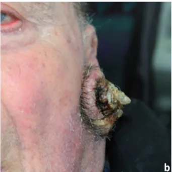
Physical examination revealed a 4.5x3.5 cm skin horn lesion: crateriform architecture filled with a kera-tin plug, which gave conical projection, no signs of in-flammation in peripheral skin and painless (Figure 1a and 1b). There were no palpable regional limph nodes.
The lesion was excised under local anesthesia and the skin loss was repaired with local advancement flaps. Histopathological examination revealed a “ker-atoacanthoma-type squamous cell carcinoma” (or only “keratoacanthoma”) with perineural invasion.
During postoperative follow-up of 6 months, no recurrence was noted (Figure 1c).
Discussion
Exophytic lesions with a central keratin-filled crater are difficult to diagnose clinically (5). Kerato-canthoma and squamous cell carcinoma shares some features so they cannot be confidently differentiated by dermoscopy (6).
A giant keratoacanthoma of the cheek 581
Histopathological diagnosis allows to classify epi-thelial crateriform tumors into seven types: crateriform verruca, crateriform seborrheic keratosis, keratoacan-thoma (KA), KA with a conventional squamous cell carcinoma (SCC) component (KA-like SCC and KA with malignant transformation), crateriform Bowen’s disease, crateriform SCC arising from solar keratosis and crater form of infundibular SCC (7).
Whether KA is benign or malignant is contro-versial: it can be classified as a benign self-limited squamous proliferation (8), or as a type of well dif-ferentiated squamous cell carcinoma (SCC) capable of spontaneous regression (9).
KA is usually solitary but can be multiple (10). It is assumed to originate from the hair follicle: KA ex-hibits markers corresponding with those found in the follicular isthmus and infundibulum. Its life cycle with proliferative, mature and involutional phases mimics the hair cycle (11).
Perineural invasion of keratoacanthoma is rare (12). Keratoacanthomas of head and neck with peri-neural invasion have a greater potential for aggres-siveness: their spread into the mimic muscles, cranial nerves or sinus cavernosus, local recurrences, metas-tases in the parotis gland and regional lymph nodes have been reported in literature (13). When perineural invasion is extended, the prognosis can correspond to that of squamous cell carcinoma with perineural infil-tration, so a closer follow-up of the patient is recom-mended (12-14).
Figure 1. Preoperative and postoperative pictures. a) and b):
Keratoacanthoma: a 4.5x3.5 cm skin horn lesion with crateri-form architecture filled with a keratin plug and no signs of in-flammation in peripheral skin; c: Postoperative image
E. Pica Alfieri, A. Sisti, G. Nisi, et al. 582
Conflict of interest: Each author declares that he or she has no commercial associations (e.g. consultancies, stock ownership, equity interest, patent/licensing arrangement etc.) that might pose a con-flict of interest in connection with the submitted article
References
1. Dye K, Saucedo M, Raju D, Aydin N. A common cancer in an uncommon location: A case report of squamous cell carcinoma of the nipple. International journal of surgery case reports 2017; 36: 94-7.
2. Yesantharao P, Wang W, Ioannidis NM, Demehri S, Whit-temore AS, Asgari MM. Cutaneous squamous cell cancer (cSCC) risk and the human leukocyte antigen (HLA) sys-tem. Human immunology 2017; 78(4): 327-35.
3. Ratushny V, Gober MD, Hick R, Ridky TW, Seykora JT. From keratinocyte to cancer: the pathogenesis and modeling of cutaneous squamous cell carcinoma. The Journal of clinical investigation 2012; 122(2): 464-72.
4. Watanabe IC, Magalhaes RF, de Moraes AM, et al. Kera-toacanthoma and KeraKera-toacanthoma-Like Squamous Cell Carcinoma: Similar Morphology but Different Pathogenesis. Medicine 2015; 94(23): e934.
5. Misago N, Inoue T, Koba S, Narisawa Y. Keratoacanthoma and other types of squamous cell carcinoma with crateriform architecture: classification and identification. The Journal of dermatology 2013; 40(6): 443-52.
6. Rosendahl C, Cameron A, Argenziano G, Zalaudek I, Tschandl P, Kittler H. Dermoscopy of squamous cell carci-noma and keratoacanthoma. Archives of dermatology 2012; 148(12): 1386-92.
7. Ogita A, Ansai SI, Misago N, Anan T, Fukumoto T, Saeki H. Histopathological diagnosis of epithelial crateriform tumors: Keratoacanthoma and other epithelial crateriform tumors. The Journal of dermatology 2016; 43(11): 1321-31.
8. Mandrell JC, Santa Cruz D. Keratoacanthoma: hyperpla-sia, benign neoplasm, or a type of squamous cell carcinoma? Seminars in diagnostic pathology 2009; 26(3): 150-63.
9. Beham A, Regauer S, Soyer HP, Beham-Schmid C. Kerato-acanthoma: a clinically distinct variant of well differentiated squamous cell carcinoma. Advances in anatomic pathology 1998; 5(5): 269-80.
10. Wu TP, Miller K, Cohen DE, Stein JA. Keratoacanthomas arising in association with prurigo nodules in pruritic, ac-tinically damaged skin. Journal of the American Academy of Dermatology 2013; 69(3): 426-30.
11. Kwiek B, Schwartz RA. Keratoacanthoma (KA): An update and review. Journal of the American Academy of Dermatol-ogy 2016; 74(6): 1220-33.
12. Schuurmann M, Ponitzsch I, Simon JC, Ziemer M. Don’t miss the base - keratoacanthoma-type squamous cell car-cinoma with perineural invasion during BRAF inhibitor therapy for melanoma. Journal der Deutschen Dermatolo-gischen Gesellschaft = Journal of the German Society of Dermatology: JDDG 2015; 13(12): 1279-81.
13. Basoglu Y, Metze D, Nashan D, Stander S. Keratoacan-thoma with perineural invasion: an indicator for aggressive behavior? Journal der Deutschen Dermatologischen Ge-sellschaft = Journal of the German Society of Dermatology: JDDG 2008; 6(11): 952-5.
14. Petrie M, Eliezri Y, Campanelli C. Keratoacanthoma of the head and neck with perineural invasion: incidental finding or cause for concern? Dermatologic surgery : official publi-cation for American Society for Dermatologic Surgery [et al] 2010; 36(7): 1209-13.
Received: 9 June 2018 Accepted: 11 July 2018 Correspondence: Edoardo Pica Alfieri, MD
Division of Plastic and Reconstructive Surgery Department of Medicine, Surgery and Neuroscience University Hospital “Santa Maria alle Scotte” University of Siena, Siena, Italy