UNIVERSITÀ DEGLI STUDI DI MACERATA
Dipartimento di Giurisprudenza
CORSO DI DOTTORATO DI RICERCA IN SCIENZE GIURICHE
__________________________________________________________________________
CICLO XXXII
Development of third molar and Third molar
index: I
3M
Global observations
DOTTORANDO
Dott. Luz Andrea Velandia Palacio
RELATORE
Chiar.mo Prof. Roberto Cameriere
TERMINOLOGY ACRONYMS INTRODUCTION
CHAPTER 1 - AGE ESTIMATION IN THE LIVING (JUVENILES) 1.1. HISTORY AND CONTEXT
1.2. ASSESSMENT OF SECONDARY SEXUAL DEVELOPMENT 1.3. PSYCHOLOGICAL DEVELOPMENT
1.4. SKELETAL MATURITY
1.4.1 HAND-WRIST : Greulich and Pyle (GP)/ Tanner-Whitehouse (TW) 1.4.2 CLAVICLE
1.5 DENTAL ASSESSMENT: 1.5.1 General anatomy
1.5.2. Dental methods for estimation of adult age. -Third molar
-Staging Methods (Demirjian-Moorres) 1.5.3. Third molar maturity Index
-Imaging in age estimation
CHAPTER 2. LEGAL REQUIREMENTS FOR AGE ESTIMATION IN JUVENILES 2..1 IRREGULAR MIGRATION AND ASYLUM SEEKERS
2.2 HUMAN TRAFFICKING 2.3 CRIMINAL RESPONSIBILITY 2.4 CHILD BRIDES
CHAPTER 3. THE 18 YEARS OLD CHALLENGE- DOES POPULATION MATTER FOR I3M? 3.1 Population group differences for dental age estimation
3.2 Third Molar Index: Observations around the world, data
TERMINOLOGY
Age assessment: Age assessment is an individual concept. It is the estimation of the
most probable age of an individual considering their skeletal and dental age and their
specific demographic characteristics (sex, health, physical activity, intake, etc.).
Accuracy: also called validity is the degree of conformity of a measured or calculated
quantity to its actual true value
Bone age -general degree of maturation of bone that subjects of a population reach at
a certain average age.
Chronological age: The age of a person as measured from birth to a given date Dental age -A measure of age based on the general degree of development of teeth Precision: also called reliability is the degree to which further measurements or
calculations give the same or similar results
Repeatability: the efforts made to keep conditions constant by using the same observer
( intra-observer reliability)
Reproducibility: The same measurement process is successfully carried out by
different observers (inter-observer reliability
Skeletal age -general degree of maturation of the skeleton that subjects of a population
reach at a certain average age. This term may be used when talking about the entire
ACRONYMS
CT: Computed Tomography
GP: Greulich& Pyle
OPG: Orthopantomograms
MACR: Minimum Age of Criminal Responsibility
MRI: Magnetic Resonance Imaging
TW: Tanner-Whitehouse
I3M: Third molar maturity Index
IOM: International Organization for Migration
UNHCR:United Nations High Commissioner for Refugees
UNODC: United Nations Office on Drugs and Crime
US: Ultrasound
INTRODUCTION
In general, Forensic science is defined as the use of science and its application to assist the law, forensic dentistry is a specialized branch of dentistry that apply specialized knowledge in order to evaluate and analyze dental evidence to help in legal proceedings. The contributions of forensic dentistry to the field have involved dental identification, sex estimation, analysis of bite marks, as well as expert opinion in cases of dental malpractice and negligence, and especially in cases of age estimation.[1]
Age estimation is one of the most complex parts of identification both in the living and in human remains. It has become an important and relevant subject in the forensic field as part of the identification process in cases of victims of different crimes or mass disasters. In the living, it is required in diverse context, for example, in criminal and civil procedures for determining the legal course of action for a subject according to his age and depending on the minimum age of criminal responsibility (MACR). In civil law it is required for determining the legal age to work to claim social benefits or to get married. [2][3] .
In the last few decades, there has been a constant increase in cross border migration around the world due in part to the economic globalization.[4] This phenomenon has posed new challenges specially in cases of irregular migration where people’s identification document or birth certificates are not reliable or are missing. In Europe, forensic age estimation is mainly done on unaccompanied minor immigrants who enter Europe lacking an adult reference figure. An adequate method for age estimation in such circumstance is vital and it is not only in the best interest of the country of arrival but is a way to protect the asylum seekers from sexual abuse, exploitation and to be certain that they receive the benefits they are entitled to according to their age[5].
Age assessment of living individuals who presumably claim to be minors or give inaccurate information regarding their age, has become an inherent part of the daily forensic work. Courts demand appropriate medical tests aimed at age estimation in these cases and thanks to this increased requirement there has been several papers interested in studying methods from skeletal to dental specifically targeting 18 years old. Since in most countries the age of majority is fixed at this important age [6]
In order to estimate chronological age, scientist have studied physiological age defined as the age at which a developing system or an organ reaches a specific stage [7]. For an organ or system to be used as a predictor of chronological age it must comply with certain criteria. It must develop over a long period of time, have a recognizable and measurable developmental stage and must be possible to assess both in the living and the dead. However, the stages must happen over a relative short period of time and ideally should be less affected by racial and environmental factors as well as survive inhumation[8].
In finding the best way to estimate chronological age the first features to be studied were height, weight and secondary sex characteristics and then with the discovery of the x rays the interest shifted to bone and dental development. The main idea was to identify the changes that happened to all people at similar ages to then to correlate them with chronological age.
Considering the requirements mentioned for a system or organ to be a good predictor of chronological age, the teeth meet more closely these criteria since they are less affected by factors such as malnutrition and hormonal disorders than bones. The hard structure of the teeth also makes them more resistant to time, mechanical, chemical and physical influences and therefore well preserved for long periods of time. The developmental stages are also clear, recognizable and each is morphologically unique from the other, adding to that they are also less influenced by elements such as environment, living conditions and nutrition [8].
However when it comes to the specific threshold of adult age, it becomes more complex to study since as an individual approaches adulthood there are less morphological predictors of age to study, the subject reaches the end of skeletal and dental development and the only remaining teeth undergoing developmental changes in late adolescence and early adulthood are the third molars since at the age of 15 years, all permanent teeth have reached maturation.
Despite being one of the most variable teeth regarding development, the degree of mineralization of the third molar through radiological examination is the most prevalent method of assessing the likelihood that age 18 has been achieved, is therefore considered that when it comes to forensic age estimation, apical closure of the third molar is the smallest morphological change in dental development that has a huge impact legally since it occur around the threshold of 18 years. [9] However, it presents several challenges, there are individual variations in time of formation of third molar as well as in time of eruption and the effects of factors such as regional differences are not fully known.
To study the third molar mineralization, it has been used different approaches, one of the most known is the classification system in stages based on scores given to teeth according to its maturation and eruption. This has been the methodology followed by Mincer[10], Demirjian[11] et al and Moorrees et al [12] they have shown a higher degree of intra- and inter-observer error because of the subjective component involved in assigning an stage and as a consequence an increase in prediction error [13]
To overcome this drawback another method was introduced for assessing adult age by Cameriere et al.[14] this method requires the measurement of apical pulp width and tooth length as observed in dental orthopantomograph. It has the advantage of using these measurements to calculate a ratio, the derived index is scale invariant and the magnification of the measurements inherent to x- rays is no longer an issue. Since the resultant index does not require choosing a stage it also avoids the subjectivity involved in previous methods.
Thanks to the observations done with the application of this methodology, the group of AgeEstimation project was created to validate Cameriere’s results in several populations foreign to the original reference sample, with positive results. [15] [16][17] [18]
However, in the study of the third molar for age estimation of legal age, there has been several challenges mainly the individual variations in time of formation of the third molar as well as the effects of factors such as regional differences that are not fully known. Having studied in the group of age estimation project many samples for the study of the third molar coming from different countries this thesis respond to the need first of summarizing the results found during this three years and second to establish if the third molar index is not only reliable to discriminating minor from adults but if is also applicable to any country regardless of the ethnicity of the individual studied.
CHAPTER 1 – AGE ESTIMATION IN THE LIVING (JUVENILES)
1.1. HISTORY AND CONTEXT
In the field of age estimation there are several points in history that have been important in determining how age is determined. In particular, the onset of the Industrial Revolution in England set the first precedent. It was very common during this time to have very young children, sometimes as young as seven, employed in factories and specially in the coal mines that were the source of power to keep these newly industries going. They worked for 12 -14 hours a day and sometimes not even had a day off during the week. In 1815 the age of employment was limited to no younger than nine years and the hours of employment to 10 per day. The poor economic situation of most families in Britain and the financial concerns of the industrial owners that did not want to change their advantageous employment policies led to this legislation to be ignored. There were not organized birth registry at the time that could produce reliable documents to prove someone’s age, and the way to estimate it then was by a subjective estimation or using the age given by a parent.
Edward Saunders in 1837 considered the use of dental eruption as the best mean for assessing age in children instead of the conventional ones that among others used parameters like height, weight and robustness [19]. Since at the time there were not civil registers Saunders demonstrated the inapplicability of the law and suggested the use of dental eruption as a better and more reliable alternative, he developed toot eruption charts based on his observations on 1046 children. Although tooth eruption is still considered a general guideline for estimating age, it is not considered valid due to the variability of the eruption process.
The second moment that marked history was the discovery in 1895 of X-rays by Röntgen, the multiple applications that were derived from this knowledge affected several fields in medical research. In the case of age estimation, the use of x rays expanded the possibilities to different levels since it was then possible for researchers to directly observe the formation of the dentition in living people.
The publication of the Atlas of Skeletal Maturation by Todd in 1937 used the radiographic technique by collecting a series of radiographs of the hand of the upper middle-class American children and adolescents. The radiographs were used to observe the relationship between the
changes in hand and wrist bones in both genders and their relationship with chronological age[20] . Despite the bias in the origin of the sample, it remained as gold standard until Greulich &Pyle reviewed the work in 1950 and 1959. Their research continue to be of relevance both in clinical practice and the forensic field [21]. Tanner and Whitehouse (TW1) also developed a bone age assessment protocol and used several indicators that were later revised into what is called TW2 and TW3 [22][23].
Dental age studies in the modern era have been concentrated in two main subjects, general dental development and the development of the third molar. As mentioned in the introduction of this work the third molar is the only tooth still in development during adolescence and therefore relevant for the assessment of legal age of adulthood.
The fact that the third molar has bring all the attention of researchers demonstrate the need to finding a method that helps in assessing legal age for the law. The main reason for this to happen are the cases of undocumented immigrants that usually are unaccompanied minors.
According to the European legislation and different international treaties, the minors must be protected on arrival at any country. However, there is lack of consensus regarding the techniques that should be used for estimating age since each country chooses its own methodology. Knowing the conflicting situations, different international groups have created some guidelines and recommendations in an effort to establish standardization of methods to be applied when estimating age[24].
There are numerous researches that have studied the correlations between development and chronological age as well as methods from skeletal to dental for age assessment. This has been driven by the increasing demand in the society to determine an individuals’ age and the need to ensure high accuracy since errors particularly in juveniles, may cause unwarranted consequences and violation of human rights.
In the next sections will be described the current methods used as part of the methodology for age estimation in juveniles
1.2 ASESSMENT OF SECONDARY SEXUAL DEVELOPMENT
Puberty is a period of biological maturation marked by the appearance of secondary sexual characteristics, growth spurt, and changes in body composition. Throughout puberty it is possible to assess subjectively the presence and prevalence of secondary sexual characteristics that appear as a result of sexual maturity. The assessment of sexual maturation is based on secondary sex characteristics: breast development and age at menarche in girls, genital (penis and testes) development in boys, and pubic hair in both sexes. Development of the breasts, genitals, and pubic hair is most often rated on five-point scales, Stage 1 of each characteristic indicates the prepubertal state (absence of development) and stage 2 the initial, overt development of each characteristic that marks the transition into puberty. Stages 3 and 4 mark progress in maturation, and stage five 5 indicates the adult (mature) as described by Tanner[25]
Nevertheless, their use as a method for evaluating age is not reliable, as stated by Chipkevitch 2001 adolescents with the same age are frequently in different stages of puberty considering that its onset and progression are highly variable and the influence of several elements such as geographic location, ethnicity and individual body shape/size affects the development of secondary sexual characteristics. [26]
This influence has been observed in different researches, Wu et al studied the development of secondary sexual characteristic for African, Caucasian, and Hispanic American girls aged between 8 and 16 years. They observed that girls enter puberty at slightly different chronological ages according to their race and ethnicity, and when considering pubic hair, the mean ages of development varied also according to ethnicity between 9.5 years for African American, 10.5 years for Caucasian and 10.3 years for Hispanic girls. Their observations concluded that black and Mexican American girls had pubic hair and breast development and had achieved menarche at younger ages than white girls and suggested to look for external factors that may influence the differences observed.
The resulting variations in timing of sexual maturity showed the lack of correlation between chronological age and secondary sexual characteristics indicating that this method was not suitable for assessing age in living individuals. [27]
This observation was later supported in a research that studied ethnically diverse US boys to evaluate the ages of onset of secondary sexual characteristics. Using Tanner stages they
observed that African American boys were advanced in their degree of genital and pubic hair development by an average of 1–2 years compared to Hispanic and non-Hispanic white boys and further observed that mean ages of beginning genital and pubic hair growth and early testicular volumes were 6 months to 2 years earlier than in past studies, depending on the characteristic and race/ethnicity, showing that there was also a significative influence from external factors [28].
In line with the previous researches in 2005 it was carried out a study assessing the developmental age in a cross- sectional analysis of secondary sexual characteristics in a sample of 8675 German boys and 8689 girls between 8 and 17 years. They observed a significant difference in the developmental tempo not only between the two sexes but also within the same sex, and additionally early developers looked older than their chronological age and late developers instead seemed younger than their chronological age. There was also a variation in breast development within the sample that was attributed to body type [29].
The results of the previous studies evidenced that developmental age of a growing person does not necessarily relate to his or her chronological age which imply that the evaluation of secondary sexual characteristics is not the appropriate method to use for ascertaining chronological age in juveniles.
.
1.3 PSYCHOLOGICAL DEVELOPMENT
Age estimation has been mainly studied in the framework of asylum seekers due to the exposition by the media of the migration crisis in Europe and United States, is then of no surprise that the United Nations High Commissioner for Refugees (UNHCRS) is the institution that has suggested a guideline in this subject. They have proposed a multidisciplinary evaluation that additionally to the physical and developmental evaluation also considers a psychological assessment[30]
As part of a holistic approach for age estimation also the Royal College of Physicians (RCP) recognize the necessity of considering the social history of an individual child in addition to the other physical methods. The elements that make part of the Social history include among others lifestyle, familial role in country of origin and education received. [31]
Psychosocial maturity has been defined as the general level of an individual’s socioemotional competence and adaptive functioning[32] it is recognized that during adolescence there are important developments happening such as perspective taking, emotion regulation, identity formation, independence, affiliation and striving for achievement, which make complex to define psychosocial maturity by just one criteria . A more complete definition encompass the ability of the individual to adapt to changing circumstances and that includes the capacities of intelligence, learning , memory , skills, feelings as well as motivations and emotions to apply behavioral control and self-regulation [33] However changes that occur during that period of life can be expressed in many different ways and at varying times.
There are several factors then that influence the psychological development of age in a child and as suggested both by the UNHCRS and the RCP it is paramount to consider it as part of the holistic estimation of age in living individuals.
Estimating age of psychological maturity it has been implemented by several countries as part of the assessment performed to evaluate if a child has reached or not the age of adulthood, unfortunately, there is a large variation according to the country’s jurisdiction in the way they choose to assess the psychological aspect in age estimation
The social and psychosocial evaluation is performed by countries such as Austria, Sweden, the UK and the. The social aspect looks for information that involves age of siblings, relationship with the family and family composition, education history and social exposure.
In a UN report in only few cases the analysis is conducted by a licensed psychologist and the interviews and assessments are then performed by immigration officers or social workers and consisted of a medical evaluation and a short interview. For a psychosocial evaluation to present a reliable outcome there are a lot of issues to overcome. It is stressed the importance of skilled professionals to have experience and training in aspects such as how to address to children that come from different cultural backgrounds and how they can cope with prolonged questioning. It has been demonstrated the influence of risk factors during early childhood in several developmental characteristics which makes important for interviewers to have the skills required to recognize child development issues that results from the challenging locations that most of them are originally from. [34]
The ideal method for assessment of psychological development in the context of age estimation require an approach that involves not just an evaluation of the psychological aspect but also that take into account the socio-cultural history and even further to include issues concerning the assessment of children with developmental disability or delay.[30]
1.4 SKELETAL MATURITY
Osteogenesis also known as ossification is the process of bone tissue formation from which the skeleton is derived from. The conversion of other types of connective tissue into bone can be classified according to the precursor tissue type, the intramembranous or periosteal ossification has as precursor the mesenchyme that is a primitive connective tissue template, the Endochondral ossification instead has as a precursor a cartilaginous tissue template. Each skeletal element in the body start from an initial locus of bone formation named primary center of ossification. That will continue to form either the entirety or the main component of an adult bone. Once a primary center is stablished some bones will develop secondary bone formation loci defined as secondary ossification centers or epiphyses. The longitudinal growth of bones will occur in the area between the adjacent primary and secondary centers named growth plate. By the time the secondary ossification center will fuse with the primary center at the position of the growth plate the longitudinal growth of the bone will stop, establishing the attainment of skeletal maturity. These processes of ossification and fusion in the skeleton happen during certain specific developmental period that in consequence can be used for the purposes of age estimation.[34]
When evaluating biological age what is being observed is the positive correlation between growth and age although growth does progress with age it has been proved that this does not happen in a linear manner and hence the lack of pattern is observed as variations among individuals not to mention that the influence of external environment also contribute to the variation
For the assessment of age, the observation of skeletal development associated to maturity through the application of clinical imaging has been widely used. The most common type of
imaging include x- ray radiography, computed tomography (CT), magnetic resonance imaging (MRI) and ultrasound (US) with different degree of image quality.
The maturation of the skeleton is observed and then maturity is evaluated according to the changes that take place from initial ossification to adult morphology of individual bones. The study of skeletal maturation has been developed as a diagnostic measure to assess clinical issues related to growth abnormalities, or to stablish the response to a medical treatment and to determine the growth potential of a children.
When it comes to its use in forensics as a tool for estimating age in living individuals it is important to remember that these standards have been developed to respond to a clinical necessity rather than to being used to assess age in forensic context. It is important to exert some caution in the application of these methods and are always confronted with any other findings done by experienced practitioners[35].
The concept of estimating skeletal age is based in the possibility of associating biological age with chronological age, to be considered as a useful age indicators it is necessary that the changes observed in the process or traits should change unidirectionally with age, then correlate with chronological age and change in a consistent manner across individuals . Currently the approaches used to assess skeletal maturity are mainly Greulich&Pyle , and Tanner-Whitehouse methods.
3.4.2.
HAND–WRISTThe anatomy of the hand and wrist is of importance when estimating skeletal age through the development of its bones, is composed of 19 short bones ( 5 metacarpals and 14 phalanges) , 7 carpals and the radius and ulna (Fig. 1). There is a variation among individuals in the maturation rate of the carpals and compared to the long and short bones the carpals complete their maturation earlier.
For estimation of subadult age using skeletal maturation in the hand-wrist complex: Greulich and Pyle and Tanner et al are the most used methods.
Figure 1. Hand and wrist anatomy [36]
Greulich And Pyle (GP)
Greulich & Pyle (GP) atlas is the work developed by Todd and the method was described on “ The Radiographic Atlas of Skeletal Development of the Hand and Wrist’, by Dr William Walter Greulich and Dr Sarah Idell Pyle [21], is one of the most commonly used atlases for evaluation of skeletal age. The atlas is a series of sex-specific radiographs of the left hand/wrist that are used as reference of the maturity status at a given chronological age from birth till 18 years of age for females and 19 years of age for males. It also has comments explaining the gradual age-related changes observed in the bone structure besides each standard image. The atlas was done using the radiograph founded to be the most typical from about 100 of each sex at each age level to be the reference plate. The radiographs correspond to a large sample of children from the Brush Foundation Study
Each bone on the standard plates represents its median appearance at a given chronological age (however, in some plates of the atlas, there is considerably variation for a given chronological age). The method is based on the radiographs of a large sample of
children coming of high socioeconomic status in Cleveland, Ohio, USA from the Brush Foundation Study developed to chart the growth of healthy children.
The calculation of bone age is done by comparison of the hand radiograph of the subject in question and that of the atlas looking for the nearest matching reference. The skeletal age is provided by the one founded to be in the closest standard.
Frequently the skeletal age assigned is based on the plate that most closely matches that of the child ignoring the differences among bones in the hand-wrist and among the standard plates. Some consider that the ideal application of the method should be to match each individual bone to the atlas plate and assigned the skeletal age of the one with which the individual bone most closely coincides and the final skeletal age should be the median value of the skeletal ages of all bones, however other studies have suggested that this does not necessarily improves the accuracy of the statement.[37]
Although is useful as a guide for age estimation this atlas was not designed for this purpose but as a guide of skeletal growth for clinicians such as pediatricians interested to follow the skeletal status of their patients with chronological known age and to assess growth and development compared with that of other children of the same sex and age
Recognizing the original purpose of this atlases helps to question how effective it is to use it as a mean to estimate the legal age of 18 years old. The limitations of this method are more clearly stated in the work of the Australian human rights commission site,
When a hand-writs x ray is used to assess chronological age, it will only be useful until the moment in which the skeletal maturity is achieved, once arrived to that point it won’t be possible to see any modification of the characteristics of the hand and wrist. The challenge then is if is possible to say that skeletal maturity is equivalent to achieving 18 years old when on average hand writs achieve the traits of skeletal maturity when under the age of 18 years or at an age close to 18 years.
Assessing 18 years old with the hand wrist method implies two main issues one is that most of the studies regarding chronological age estimation of adult age have shown that hand wrist is a useful method when evaluating subjects younger than 16 and therefore is not suitable for 18 years old. Actually an study from Second issue is the high influence of external factors on the attaining of skeletal maturity , is well study how nutrition,
environment, social and general health conditions affects the development of bones and in consequence have an impact on when skeletal maturity is reached and therefore the association with chronological age is less clear.
Greulich and Pyle method has been studied in several samples specifically for the ages of 14 and 18 years and the general results concluded that this method tend to overestimate age in samples of females aged up to 17 years and for males it tends to underestimate it in samples aged up to 15 years.[38], [39]
The legal weight that age estimation carries for the life of a person is well stated by Black et al [40] when said that underestimating age when solicited by a court most likely have smaller impact on the human rights of a person instead overestimating age can have serious consequences and will vastly influence the future of the concerned individual.
Based on this statement it is important to understand the type of errors described by Garamendi et al [41] that pointed out that technically unacceptable errors are the one that happened when a subject that is 18 years or older is deemed a minor and ethically unacceptable error when a minor is classified as an adult. The risk of incurring in an ethically unacceptable error when estimating age in criminal procedures with Greulich and Pyle method tends to be high when evaluating ages 14 and 18 and is recommended to be used carefully and in addition to other methods to secure a better reliability.
.
Tanner-Whitehouse (TW)
Is a scoring method that was initially developed in 1962 in which they analyzed the anterior-posterior hand-wrist radiographs of a sample of British origin between 1 and 21 years of age. They evaluated 20 bones: radius, ulna, metacarpals 1, 3 and 5 and all carpals except the pisiform.
The revision named TW2 method was based on the level of maturity for 20 selected regions of interest (ROI) in specific bones of the wrist and added an age estimation method using the radius ulna and short bones. The developmental level of each region is then assigned into specific stages (A,B,C,D) and a score is given to each stage for each bone. Summing all these scores a total maturity final score is calculated and then correlated with skeletal age individually for males and females. Later this method was updated and named TW3, is considered to be more accurate than TW2 because is suggested that currently children
mature more rapidly and any skeletal maturity score is reached faster and earlier in chronological age then in the past when compared with GP method is considered more reliable specially when assessing cases of asylum seekers due to the tendency of GP to overestimate age, in general is recommended not to use just one method but a combination of TW3 with GP together with the additional evaluations done to the subject to improve the accuracy of the observation.
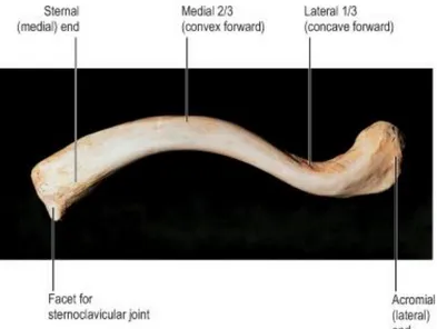
1.4.2. CLAVICLE
The clavicle bone is an S-shaped bone located anterior to the base of the neck. It is of importance in age estimation for several particularities , in the period of adolescence at the medial end of the clavicle a secondary epiphyseal ossification center arise which extend the growth and remodeling of the bone till fusion at approximately 22 years resulting in the clavicle to be the last bone to ossify from the whole skeleton. While at age 18 the hand bones have already finished ossification as well as the third molar has completed mineralization, the medial end of clavicle can still be used to estimate bone age from 18 to 22 years.[36]
Figure 2: Right clavicle viewed from above S.Jacob (Human Anatomy 2008)
Due to these characteristics is relevant to evaluate the progress of ossification of the cartilage at the sternal end of the clavicle to help in the estimation of 18 years old.
The first research suggesting that the growth of the medial clavicle could be of use in age estimation was done by Buikstra and Ubelaker [42]in 1994 observing the developing epiphyseal in medial clavicle and the union of clavicular shaft. The study of Todd and D’Errico and McKerna and Stewart observed the potential of evaluating clavicular development to provide age assessment. They focused on the use of anatomical specimens to evaluate associations between age and epiphyseal fusion in the medial clavicle. Thanks to these early studies the methodologies developed for the observation of epiphyseal fusion in subcategorizing stages are still the basis used in present research.
Most methods were developed as scoring methods, Webb and Suchey categorized the clavicle fusion in a 4 phase scoring method. In phase 1 was non-union with no epiphysis, phase 2 was for no union with separate epiphysis, phase 3 defined as partial union and phase 4 was for complete union. The ages assigned to phase 1 were 25 for males and 23 for females, phase 2 was between age 16 to 22 in males and between age 16 to 22 years in females, phase 3 was observed from 17 to 30 years in males and from 16 to 33 in females and the last phase from age 21 to 21 in males and in females from age 20 to 34 years for complete fusion. In the study of Black and Scheurer they used a 5-phase scoring method observing the morphological changes at the medial end of the clavicle using a sample of skeletons of known age.[43]
For age estimation in the living the stage systems used in direct observation of the bones is still a useful methodology, however the assessment of the medial clavicular bone is not an easy task when done by traditional radiography, due to the anatomical position of this bone there are a lot of overlapping shadows resulting from the vertebrae and the ribs. The trend is to move to more reliable systems that allow a better observation such as computed tomography and more recently magnetic resonance.
Regardless of the device used the studies looks to grade the ossification in the medial clavicle and then assign an stage with a corresponding age range. One of the most recognized studies was the one developed by Schmeling[44] that used 873 chest radiographs of subjects aged between 16 and 30 years and assigned a stage as follows:
Stage 1: the ossification center has not yet ossified, Stage 2: the ossification center has ossified, the epiphyseal cartilage has not ossified, Stage 3: the epiphyseal cartilage is partially ossified , Stage 4: the epiphyseal cartilage is fully ossified, Stage 5: the epiphyseal cartilage has fused
completely and the epiphyseal scar is no longer visible. This result suggested that in terms of criminal responsibility a subject classified under Stage 4 have not yet reached 18 years old. Since the visualization in conventional radiography is not accurate, the previous results serve to the authors to study the clavicle in age estimation with thin-layer computed tomography[45], their results led to a sub-staging of their original 2 and 3 stages as observed in (Fig 3)
Figure 4. Schematic drawings and pictures of stages 2a-3c of clavicular ossification[45]
Their observations with these sub stages allowed them to narrow the interval of the estimated age and to evidence that the late stage 3 first appeared at age 19 and in consequence it was possible to infer that if it is found a late stage 3 the subject it is most likely to have already reached the legal age of 18 years old. Still it is always noteworthy to remember that although the ethnic origin has demonstrated not to strongly influence the skeletal development, a low socio-economic status does, causing usually a delay in development. The evaluation in such cases would most likely result in an underestimation of age which would cause then a technical error but not an ethical one. [41] Some other techniques of evaluating the development of the clavicle are the ones using CT scans in order to improve the possibility of having a better quality of image and some using the same sub- staging method than the ones used with previous studies (Fig 4)
Most recently with the ethical debate regarding the exposure to ionizing radiation without a medical reason for it , like in the case of estimating age in undocumented allegedly minors, Magnetic Resonance Imaging (MRI) offer a choice that is radiation free and would decrease the amount of radiation caused by other techniques. There are several studies focusing on the evaluation of the clavicle for age estimation with a relatively same degree of precision than conventional radiographs [47][46] , The main pitfall of this technique is the variability in the protocols according to the specific device used added to the different staging methods applied to assess clavicular ossification that makes difficult to run a systematic review to establish its efficacy [48]
1.5 DENTAL ASSESSMENT
Dental age, as mentioned briefly in the introduction, is the most used anatomical element to evaluate physiological age. To understand the reasons why teeth have become paramount for age estimation it is necessary to know some basic information.
1.5.1.General anatomy
Teeth are anatomically comprised by three main parts: the crown the neck and the root. The crown is the top portion that is composed by enamel in the external part, is the one that is seen above the gum level and is more specifically known as clinical crown. The neck is the narrowing part t located between the crown and root. The root is the lower portion that is in contact with the alveolus, the bone portion of the maxilla or mandible where roots articulate. (Fig 5)
Figure 5. Anatomy of a tooth and its surrounding tissue.[49]
The configuration of the root from the inside to the outside is the pulp cavity, dentin and cementum. The inner part is the pulp chamber that host the terminal nerves and blood vessels that arrive from the opening of the root. The pulp cavity narrows towards the apex of the toot root to form the root canal, that opens at the apex as the apical foramen. The crown is covered by the hardest tissue of the human body, the enamel, the next layer inside is dentin a softer tissue that supports the function of the crown and the pulp chamber inside.
In human’s dentition is what is called diphyodont, meaning that we develop two sets of teeth. The deciduous teeth that begin their formation in the 16th week of intra-uterine life and the
second set that would be the permanent dentition starting to develop a month before birth. The sequential process of teeth formation is what facilitates the study of dental age estimation through the formative stages of dental development [50]
To observe the developmental stages of teeth for age estimation, X rays are the most used method, and allows to see also the unerupted teeth in formation (Fig 6). The development of each tooth starts within the alveolar bone of the jaw from a germ that is later mineralized. The crown and part of the root are formed before the beginning of eruption, by the time a tooth emerged in the oral cavity, around one third to three quarters of the root is formed. In a
radiographic image the enamel is seen as the most radiopaque part of the tooth and so the crown or developing crown of a tooth can be observed as the whitest part.
Figure 6.OPG from a 9-year-old subject showing mixed dentition[51]
Dentin and cementum are composted by less radiopaque material than enamel and it appear greyer but is not always easily distinguished from one another in an x-ray. The pulp chamber and the periodontal ligament are radiolucent and appear dark in radiographs, the parts of the alveolar bone observed in radiographs are the lamina dura, the layer of compact bone that covers the tooth socket and the alveolar crest, which is the gingival margin of the alveolus. In a healthy erupted tooth, the alveolar crest is located just below the level of the cervical margin of the tooth.
1.5.2.Dental Methods for estimation of adult age
Age assessment by teeth can be done through dental radiographs or by clinically observing the eruption of teeth. Eruption is defined as the emergence of teeth in the mouth through the movement of the developing tooth from the depth of the dental crypt to the edge of the alveolar bone and finally to the occlusal level. It has the advantage of not requiring special equipment or training to be studied and during the early days of age estimation research by teeth, the study
of eruption became a standard method. Although it was recognized an individual variability in the appearance of teeth from subject to subject researchers turned to developing ranges in order to overcome this shortcoming. Nevertheless as the studies advanced it was noticeable that eruption was affected by different events such as dental decay, systemic illness, tooth malposition and premature loss of deciduous teeth[12][52]
The exception to the rule of observing only the emergence of teeth was the study by Legros and Magitot in 1880 in which they examined corpses from children to adults to observe the development of the dental structures and came up with a tooth formation chart that remained standard up until the 1930s. Later on the studies on dental formation became more and more frequent, and were observed through dental x rays as the process of laying down matrix and mineralization of the dental tissues, the methods that follows the development of teeth observing the morphological changes as a continuous process in an individual have been more accurate for estimating age than eruption or other methods. The advantages of teeth mineralization for age estimation over methods that study skeletal maturation is that tooth calcification is less variable compared to ossification and it has been observed to be less affected by extrinsic environmental influences.
The age of majority is fixed at 18 years in most countries REF, its importance is reflected by the rights and duties that imply being an adult. Different situations around the world require the estimation of adult age, the irregular allegedly minor migrants without reliable documents , the foreigner minor children claiming to be adults to get married , the athletes in youth games that require monitoring of compliance with the age limits and so on, the context variates but the need remain the same, to assess if they are minors or adults.
Third molar
When it comes to dental assessment of age the teeth that has received the most attention is the third molar , as stated in the introduction the period of mineralization of the third molar that spans from 14 to 21 years combined to a small environmental influence in its development have fuel the multiple studies of the third molar in relation to age determination particularly of 18 years. .
Mineralization of third molar is radiographically visible starting as early as age 7 to 9 years for upper third molars and 8 to 10 years for lower third molars and the las evidence of apical closure occurs generally post onset of puberty and can be from 18 to 25 allowing a long developmental time extent to be studied as biological indicator of adult age. Despite of the positive traits of third molars as an adult age indicator is important to know there are differences among upper and lower third molars in time of mineralization and some studies have found variances between sexes. Maxillary third molars mature first than mandibular third molars, but discrepancies between left and right symmetry have not been significant. (15-21)
Staging Methods
In the literature there are multiple staging systems developed to evaluate the dental maturation process of the third molar. Examples of staging systems are Demirjian, Goldstein and Tanner from 1973 (8 stages denoted A to H) [53] and Gleiser Hunt and from 1955 (15 stages) [54]. There are several variations of the latter, such as Moorrees et al., 1963 (14 stages)[12], Haaviko et al., 1970 (12 stages)[55] Kullmann et al., 1992 (7 stages) [56] and Köhler et al., 1994 (10 stages) [57].
For the specific staging of third molar In 2005 Olze[58] et all studied a group of five most common classification systems of staging the lower third molar to assess their validation, the methods included: Gleiser and Hunt, Demirjian et al[53], Gustafson and Koch, Harris and Nortje and Kullman et al. They study the correlation between true age and estimated age using the results obtained from the several methods and the method that provided the maximum correlation was considered the most appropriate. Their results concluded that Demirjian et al classification was the staging system that achieved the highest value in observer agreement and correlation between the stages defined by the method and true age.
Demirjian original method used subjects of French-Canadian origin. The number of stages was reduced to eigh with a detailed description radiograph and line drawing of each stage, each toot was assigned a score depending on its state of development. Weighted scores were added to produce a total maturity score that was plotted against age.
Demirjian et al uses an 8 stage system based on the graphic representations of each stage and a written description (Fig 7).
Figure 7. Diagrammatic representation of the stage classification by Demirjian et al.[11]
According to the authors, the reasons why Demirjian system stand out from other staging classification methods and performed better in this study was because Demirjian is based on a sufficient number of stages and the fact that they do not rely on an arbitrary length measurement to determine the developmental stage, but utilize anatomically recognizable features, such as root length equal to crown length.
However some other studies have found a lack of accuracy of the method reported in different populations[59][60] and since then several researches have tried to modify it to improve the results. The most popular modification is the scoring system developed by Willems et al[61] A later study by Thevissen evaluated nine staging including the same five Olze evaluated and he found that there were similar results with all nine classification systems and stated that the small benefit from the information provided by the technique with the highest amount of stages should not compromise the correctly classification of the described stages, this observation suggested that the number of stages had no influence in decreasing the accuracy of the age estimation[57].
In 2015 a research developed by Roberts et al concluded that having less stages to choose from led to fewer error in assigning the appropriate one and increased the inter an intraobserver agreement, they stated that having stages clearly defined and with fewer intermediate stages allowed for a better reproducibility . Nevertheless, the differences observed between a 12 stages system such as the modification of Demirjian by Kasper and draft (Fig 8) and an 8 staged system in correctly estimating the dental age of the subjects seemed to be not significant. It is necessary to understand that sometimes tooth formation stages are not equally spaced in time
and sometimes the first half of molar crown height viewed from a radiograph does not take the same amount of time to develop as the second half. Sometimes the first quarter of molar root length takes considerably longer to develop than later fractions[54].
In the staging system of Demirjian the H end stage of development represent a complete apical closure. This stage has not an interval staging but represent the finished state of tooth development and has been argued that this create difficulties in producing an accurate assessment of chronological age of the third molar and then when using stage H there is a high probability of overestimating true chronological age.
Figure 8. Demirjian molar staging chart modified by kasper and draft[62]
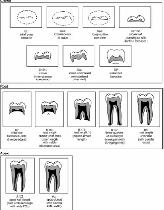
Another well-known staging system is that from Moorrees et al. [12](1963) divided dental maturation into 14 stages, The Moorrees technique includes diagrammatic and written descriptors for the morphology of the developing dentition and provides normative data for dental development for the specific purpose of assessing growth acceleration or retardation. He studied a population of 380 predominantly healthy white children ages birth to 21 years from the Ohio and Boston areas of the United States. They developed a morphologic staging chart with graphic representations of progressive dental crown and root maturation and a corresponding chart with associated written descriptors. These charts consist of 13 stages for
single rooted teeth and 14 stages for multirooted teeth. Multirooted teeth have the additional cleft stage.
Subsequently, this methodological approach has been employed to develop age estimation standards suitable for forensic application and has been validated in other populations. AlQahtani et al. [63]modified these to 13 stages. These stages start with “initial cusp” formation and then proceed to eventual “apical closure complete. (Fig 9)
Figure 9: Stages of formation for the crown, root and apex of multirooted teeth (modified from Moorrees et al. 1963, Buikstra & Ubelaker 1994, Schaefer et al. 2009 and Al Qahtani et al. (2010). The additions in parentheses, e.g., (with defined pulp roof) are modifications added by AlQahtani et al. (2010). *not included in the AlQahtani system. PDL = periodontal ligament space.[49]
Previous research has indicated that the application of the Moorrees standards potentially results in the consistent underestimation of age, for example: -0.91 years in a mixed
ethnicity population from South Africa;75 and -0.10 years (standard deviation [SD] 0.97 years) in a British population of mixed ethnicity and temporal origin. Further, a technical difficulty in using the Moorrees standards is that the results are presented in a cumbersome format and the graphs require manual interpretation, unlike the Demirjian standards, which can be interpreted using a contemporary computational approach.[64]
What can be extracted from these different studies is that the positive performance of a staging system has little to do with the number of stages used or the written descriptions but is an inherent shortcoming from the methods which is their qualitative nature rather than quantitative. The stage classification assigned by the observer add a component of subjectivity that can diminish the performance of an age estimating method.
I3M THIRD MOLAR MATURITY INDEX
As part of the work of the Age estimation project Cameriere’s et al. in 2008 published a study that had a twofold aims, first to observe the ability of open apices of third molars in discriminating between individuals who are or are not 18 years of age or older and fixing a cutoff value for evaluation of the age of 18 years and second to compare the sensitivity and specificity of this method with stages G and H of Demirjian.[65]
The method described in the article analyzed the apical ends of the roots of the left mandibular third molar of each individual the third molar maturity index I3M as follows: if the third molar
has root development complete meaning apical ends of the roots fully closed, then I3; =0, if not I3M is evaluated as observed in (Fig 10) the sum of the distances between the inner sides of the two open apices divided by the tooth length. The maturity index I3M is evaluated as a ratio .
Individual age was taken as a dichotomous response variable (E=1 if an individual is at least 18 years of age, E=0 otherwise) and gender and the third molar maturity index I3M as
predictor variables, we derived a generalized linear model to predict whether an individual is older (E=1) or younger (E=0) than 18 years of age using a logistic model.
In statistics a characteristic receiver operating curve (ROC) (Fig11) is used to compare diagnostic tests. It is a plot of the true positive rate against the false positive rate and in this case asses the predictive accuracy of the model. First all the most significant variables were considered to test the question if an individual is older or younger than 18 years of age. The test was developed to identify a cutoff or threshold that could be useful to assign an individual to one of two populations groups, those younger (T=0) or those older (T=1) than 18. It was evaluated both sensitivity p1 of the test defined as the proportion of children for or older than
18 years of age that would be verified by event T=1 and specificity p2 that is defined as the
proportion of individuals younger than 18 that would be verified by event T=0. The open apices in teeth would assist in discriminating between those individuals that are aged 18 years or more from those that are not by the posttest probability of being 18 years or more ( meaning the proportion of individuals aged 18 or over in whom the event E=1 is verified)
Figure 11: Receiver operating characteristic curve for “18 years of age or
older” status[65] Se: Sensitivity Sp: Specificity
In this case it is important that the test shows a low proportion of individuals that were younger than 18 whose test is positive (T=1) , meaning minors that were classified as adults by the test so it was appropriate to be more attentive of the chances of a false positive that of a false
negative. Keeping in mind this it was established then that an individual is considered to be 18 years of age or older (the test is positive, T=1) if I3M is lower than the cutoff value of 0.08. The
sensitivity of the test which means the proportion of individuals being 18 years of age or older whose test is positive was 70% and its specificity the proportion of individuals younger than18 whose test is negative was 95%. The proportion of correctly classified individuals was 83%. This study led to multiple validation studies that we will summarize further; the main accomplishment of this methodology was to propose a different type of approach to the ones already in use that were based in variations of the Demirjian stage approach, that as it was already stated earlier had the shortcoming of having a subjectivity component when the examiner must to choose between one stage or the other. The third molar index is a quantitative approach which means that is not affected by subjectivity because it is a measure that retrieve a numerical result. The test presents a value that can be simply interpreted as positive: 18 years or older or negative: younger than 18 avoiding the bias of choosing a stage.
Imaging in age estimation
The conflict arising due to the use of x-rays as a means of assessing adult age, has made it more difficult to tackle the issue of age estimation using radiographs. Ethical values have been introduced in this discussion stating that use of x-rays must be confined strictly to diagnostic or therapeutic purposes. Even though the amount of radiation that is used to make x rays of the hands or teeth are low, it falls under the classification of invasive examination which forbids its use other than for medical reasons [67],[68] .
Age estimation methods developed for application in the living are required to be noninvasive, accurate, and medically ethical (eg, minimal radiation exposure. The radiographic examination of dental development is relatively noninvasive and only requires exposure to small radiation doses (0.026 mSv) to acquire a suitable orthopantomogram (OPG). This level of radiation is equivalent to 4.5 days of naturally occurring radiation exposure and is thus ethically permissible. Additionally the risks associated with radiation related to panoramic radiographs less when compared to other risks in life namely assault, mass disaster, homicide etc. [69]
This does not mean that it is advisable to proceed with X-Rays from forensic age-estimation procedures under all circumstances but is necessary to reexamine the conditions of harm-benefit ratio. The calculation of of risk-harm-benefit ratio in procedures of age estimation is more complex than those done for medical care or therapeutic indication, but it should be consider that the main interest in assessing age is for requirement from the court in criminal proceedings because of the age of criminal responsibility.
The Australian Radiation Protection and Nuclear Safety Agency and the Australian Society of Forensic Odontology confirm that the radiation associated with dental X-rays is minimal. However, there is a consensus among these agencies that all deliberate exposures to radiation should be justified, and that it must be consider the potential damages arising due to the vulnerability and age of the individual.
Most of methods to evaluate third molar development for age assessment are done using conventional radiographs, but the best imaging technique for reading radiographs specially for staging purposes is the computed tomography (CT) but a study from 33ased et al [70]compared the panoramic radiographs with computed tomography and concluded that for the observation of developmental stages there were no statistically significant differences between the two types of images.
The limitations posed in the use of x-rays have led the research community in the direction of looking for alternatives that would diminish the radiation exposure. For instance, the use of magnetic resonance imaging (MRI) or ultrasound that will produce images equally valuable to those obtained by the x-rays without interfering with the rights of the subjects.
However MRI presents some limitations when studying the third molar, as observed by---- in a study comparing MRI and OPG for age assessment using Demirjian stages and Olze eruption method studying the molars in the four quadrants, they observed that the different stages could be identified equally well both for OPG and MRI and that It was easier to evaluate the mineralization stages on MRI than OPG because the roots on OPG are often superimposed (OPG is a projection method). The results showed that MRI tends to assign lower stages compared to OPG and that the mineralization stages can be affected by the imaging procedures. For instance when observing the dental follicle that covers the dental tissue, the interface is surrounded by watery content which creates enough contrast to be observed by
MRI in early stages of tooth development but in later stages it becomes more difficult since the two main features to study are the remnant of the dental follicle and closing of the apex [71] and In these stages due to the lack of contrast between dental tissue and bone the measurements can be difficult to observe and therefore most likely inaccurate. They concluded that MRI imaging is highly dependent on the protocol used to optimize the contrast in later stages of molar mineralization and although they deemed comparable to OPG in third molars, the difficulty in standardization of protocols and the high cost compared to regular x rays do not make it the first choice.
CHAPTER 2. LEGAL REQUIREMENTS FOR AGE ESTIMATION IN
JUVENILES
2.1 IRREGULAR MIGRATION AND ASYLUM SEEKERS
It is a complex situation for countries of origin, transit and destination of irregular migrants to manage properly the several issues that arise with this phenomenon. The reasons for migration are multiple but more recently the displacement cause by civil and transnational conflicts such as acts of violent extremism that are not confined to the actual war zones are considered one of the increasing ones.
The magnitude of the problem remains difficult to clearly estimate since information regarding migrants in irregular situation is not easy to obtain and therefore the figures variates according to the source. The international Organization for Migration (IOM) has an estimate of 10-15 per cent of the world’s 214 million international migrants in 2010 were undocumented [72] . But the actual figure is unknown United States have a relatively accurate estimate of undocumented migrants, and is estimated at 11.7 million by 2012 according to Pew Research Center [73].
The issues irregular migrants must confront due to their vulnerable situation are multiple, they are exposed to abuse and discrimination and often are victims of exploitation by crime organizations that are linked to human trafficking and migrant smuggling. Based on figures from the United Nations High Commissioner for Refugees (UNHCR) almost half of people forcibly displaced from their homes are children, which double the possibilities of being victims of abuse, violence and in some cases forced military recruitment. Most of the countries members of the United Nations have signed the Convention on the Rights of the Child to ensure the rights of each child within their jurisdiction.
The issue arises when an alleged child does not have a legitimate document to support his/her condition as a minor. To confront this case the UNHCR Guidelines on Policies and Procedures in Dealing with Unaccompanied Children Seeking Asylum [30] suggest that in order to protect children rights when age is uncertain it should be given the benefit of doubt, and that age assessments should be performed considering always whether the individual display a vulnerability that call for a more sensitive treatment. These considerations look to protect children rights and to do so they proposed that age evaluation should be done using a combination of criteria such as psychological maturity and physical appearance. The scientific
procedures applied to assess age estimation should include the margins of error and should be minimally invasive and respectful of human dignity to comply with the ethical treatment of children and human dignity. [74]
The UNHCR Detention Guidelines recommend that children should not be detained in the process of seeking asylum and therefore they have the right to remain with their parents and not to be separated from them against their will. The UNHCR new Issue Brief published in 2017 go further and stated that “ children should never be detained for immigration related purposes, irrespective of their legal/migratory status or that of their parents, and that detention is never in their best interests”[75] and additionally confirms that in all cases there must be appropriate care arrangements and that to ensure a suitable reception of children and their families the implementation of community-based alternatives should be in place.
Not all countries comply with these guidelines, and contrary to the principles stated there, the detention of children is the first action of the Australian Government that require a mandatory detention of all non-citizen children in Australia without a valid visa. A recent article in 2019 exposed the case of the immigration detention facility in Nauru the South pacific Island where asylum seekers arriving in Australia by boat are detained. The article confirmed that although the children are – since the introduction of the ‘open’ center arrangements – no longer technically detained, they are regarded as remaining in a detention-like environment. The article concluded that Australia’s treatment of asylum seeker and refugee children violates key obligations under the Convention on the Rights of the Child (CRC) and stated that under the principles laid down by this Convention, they should remove these children from Nauru and settle them in Australia [76]
This case exemplifies the challenges that children under the conditions of asylum seekers face; the deleterious consequences on their mental and emotional health have been well documented not to mention the high possibility to end up being victim of human trafficking and abuse. When the detention and separation of minors from their families occurs because a child is wrongfully deemed as an adult the effects on the children are equally harmful.
2.2 HUMAN TRAFFICKING
The UN Protocol to Prevent, Suppress and Punish Trafficking in Persons, Especially Women and Children” is the main global legal tool that provides a commonly approved definition on trafficking in persons. [77] Also the US federal law defined child sex trafficking as the
engaging of a person younger than 18 years in a commercial sex act including prostitution , sexual exploitation in the context of travel and tourism, the so called mail-order bride trade and early forced marriage, production of child pornography, live online sexual abuse and performing in sexual venues[78].
However despite of the strong legislation against human trafficking the figures continue to increase and victims can come from any country, based on data of the United Nations from the identified victims between 2010 and 2012, their citizenship included 152 different
countries in cases across 124 countries and it is estimated that from the minors victims the average child’s age is 12 to 14 years. [79]
The many branches for people trafficking can include sexual exploitation, forced labor such as catering and domestic servitude, begging and organ harvesting. In armed conflicts, situations such as child soldiers are another form of exploitation that the more recent report of the United Nations Office on Drugs and Crime UNODC stressed about. Despite the laws still there is a huge level of impunity as exposed by data that shows the number of prosecutions and convictions from 2007 to 2014 (Tab 1.). The number of prosecutions is shockingly low for an industry that victimizes an estimated 21 million people around the world[80]
Latin America is considered a primary source region for people trafficked to the United States, frequently the victims are first exploited in their countries of origin and later trafficked to different destinations managed by organized crime networks.
The report regarding South America showed that the majority of victims were trafficked for sexual exploitation (Fig 12) specially in countries such as Colombia and Venezuela. Child victims are more frequently girls than boys and there were more child victims than adults in Bolivia and Peru. Exploitation for illegal adoption and forced begging are the other forms of trafficking more common in south America with Bolivia reporting a number of 170 detected victims for illegal adoption from 2014 to 2017. The data concerning the victims age and gender in South America showed that most of the victims are women including when separating the data by age, from the 37% of the minor victims, 31% are girls.
Figure 12. Share of detected victims of trafficking in South America, by age group and sex,
2016 [79]
Another form of trafficking is domestic labor, that receive massive attention in the news on October 2019 because of the large use of application such as Instagram to lure particularly women, into the slave market.[81] This case shed light on the magnitude of the problem and how the different forms of trafficking are evolving, the use of virtual platforms and applications, can act as an amplifier of the problem and pose more difficulties in tracking and stopping this type of exploitation.
Human trafficking is a problem that goes beyond a humanitarian and social problem, it is also a matter of health interested, a study from 2016[82] addressed to emergency clinicians exposed the health issues related to exploitation not only to inform about the consequences
but so that emergency staff would be able to recognize the possible injuries and spot a victim, in (Tab.2) are listed the potential health consequences from the different forms of abuse.
Table 2. Physical, sexual, and psychological abuse and substance misuse, and potential health
consequences associated with human trafficking.* TOP: Termination of pregnancy[82]
The profile of the exploited victims around the world is usually the same, a low social and economic background. Trafficking take advantage of the most vulnerable population and that is the reason for targeting also children, that once absorbed into the trafficked industry are usually used in criminal activities and more prone to be provided with false identity documents.
![Figure 1. Hand and wrist anatomy [36]](https://thumb-eu.123doks.com/thumbv2/123dokorg/2960896.25843/15.918.116.722.105.522/figure-hand-and-wrist-anatomy.webp)
![Figure 3. Sub-staging of clavicle original 2 and 3 Stages [46]](https://thumb-eu.123doks.com/thumbv2/123dokorg/2960896.25843/20.918.95.610.281.593/figure-sub-staging-clavicle-original-stages.webp)
![Figure 4. Schematic drawings and pictures of stages 2a-3c of clavicular ossification[45]](https://thumb-eu.123doks.com/thumbv2/123dokorg/2960896.25843/21.918.117.484.102.634/figure-schematic-drawings-pictures-stages-a-clavicular-ossification.webp)
![Figure 5. Anatomy of a tooth and its surrounding tissue.[49]](https://thumb-eu.123doks.com/thumbv2/123dokorg/2960896.25843/23.918.298.549.152.471/figure-anatomy-tooth-surrounding-tissue.webp)
![Figure 6.OPG from a 9-year-old subject showing mixed dentition[51]](https://thumb-eu.123doks.com/thumbv2/123dokorg/2960896.25843/24.918.187.731.180.471/figure-opg-year-old-subject-showing-mixed-dentition.webp)
![Figure 7. Diagrammatic representation of the stage classification by Demirjian et a l.[11]](https://thumb-eu.123doks.com/thumbv2/123dokorg/2960896.25843/27.918.95.483.185.385/figure-diagrammatic-representation-stage-classification-demirjian-et-l.webp)
![Figure 8. Demirjian molar staging chart modified by kasper and draft[62]](https://thumb-eu.123doks.com/thumbv2/123dokorg/2960896.25843/28.918.87.775.433.691/figure-demirjian-molar-staging-chart-modified-kasper-draft.webp)
![Figure 10: Example of measurement of a third molar for I 3 M[66]](https://thumb-eu.123doks.com/thumbv2/123dokorg/2960896.25843/30.918.88.344.726.1023/figure-example-measurement-molar-i-m.webp)
