DOI 10.1007/s11547-011-0660-2
Abstract
Purpose. This study was undertaken to explore the capabilities of an open-confi guration, low-fi eld, tilting, magnetic resonance (MR) system for investigating pelvic fl oor disorders and to compare the results obtained with the patient in the semiorthostatic and supine positions.
Materials and methods. Eighteen female patients with a diagnosis of pelvic fl oor disorder (physical examination and conventional defecography) underwent dynamic MR defecography (MRD) with a 0.25-T tilting MR system (G-scan, Esaote). Images were obtained after administration of contrast agent into the rectum, bladder and vagina in both the orthostatic and supine positions. Three-dimensional T2-weighted hybrid contrast-enhanced (HYCE) sequences and dynamic T1-weighted gradient echo (GE) sequences were acquired at rest, during maximal contraction of the anal sphincter, straining and defecation.
Results. Good image quality was obtained in 15/18 patients; three presented severe artefacts due to motion, and three had incontinence, which hampered the functional studies. Better anatomical detail was obtained with MRD compared with conventional defecography. Three prolapses were observed in the semiorthostatic position only, and seven were found to be more severe in the orthostatic than in the supine position.
Conclusions. Dynamic MRD with an open-confi guration, low-fi eld, tilting MR system is a feasible and promising tool for studying the pelvic fl oor. Larger series are necessary to assess its real diagnostic value.
Riassunto
Obiettivo. Abbiamo voluto valutare la capacità di un sistema di risonanza magnetica (RM) aperto a basso campo a decubito variabile di esaminare i disordini del pavimento pelvico e di confrontare i risultati ottenuti con paziente in posizione supina e in ortostatismo.
Materiali e metodi. Diciotto pazienti donne con diagnosi di patologia del pavimento pelvico (esame clinico e defecografi a convenzionale) hanno eseguito una defecografi a con risonanza magnetica dinamica (MRD), utilizzando un sistema di RM da 0,25 T ribaltabile (G-scan, Esaote). Le immagini sono state acquisite dopo sommministrazione di mezzo di contrasto nel retto, nella vescica e nella vagina, sia nella posizione supina che in ortostatismo. Sequenze 3D T2 hybrid contrast enhanced (HYCE) e sequenze dinamiche gradient echo (GE) T1-pesate sono state acquisite rispettivamente a riposo, durante la massima contrazine dello sfi ntere anale, ponzamento ed evacuazione.
Risultati. Una buona qualità delle immagini è stata ottenuta in 15/18 pazienti; 3 pazienti hanno presentato gravi artefatti dovuti al movimento, 3 pazienti
presentavano incontinenza che ha impedito gli studi funzionali. Migliori dettagli anatomici sono stati ottenuti dalla MRD in confronto alla defecografi a convenzionale. Tre prolassi sono stati osservati soltanto nella posizione semi-ortostatica, 7 prolassi sono risultati più grandi nella posizione ortostatica rispetto a quella supina.
Conclusioni. La MRD dinamica con un sistema di RM aperto a basso campo ribaltabile è uno strumento ABDOMINAL RADIOLOGY
RADIOLOGIA ADDOMINALE
Dynamic MR defecography with an open-confi guration, low-fi eld, tilting
MR system in patients with pelvic fl oor disorders
Defecografi a-RM con un sistema di risonanza magnetica aperto a basso
campo a decubito variabile. Tecnica e risultati clinici preliminari
V. Fiaschetti • E. Squillaci • D. Pastorelli • M. Rascioni • V. Funel • C. Salimbeni • E. Fanucci
G. Simonetti
Dipartimento di Diagnostica per Immagini ed Imaging Molecolare, Radioterapia e Radiologia Interventistica, Fondazione Policlinico Universitario Tor Vergata (PTV), Viale Oxford 81, 00133 Rome, Italy
Correspondence to: E. Squillaci, Tel.: +39-062-0902374, Fax: +39-062-0902404, e-mail: [email protected] Received: 1 April 2010 / Accepted: 9 July 2010 / Published online: 19 March 2011
attuabile e promettente per lo studio del pavimento pelvico. Casistiche più ampie sono necessarie per stabilire il reale valore diagnostico.
Parole chiave Defecografi a RM · Patologia del pavimento pelvico · RM a basso campo · RM a decubito variabile
Introduzione
Le disfunzioni del pavimento pelvico rappresentano uno spettro di patologie largamente diffuse nella popolazione, in particolare nel sesso femminile e nelle fasce di età più avanzate [1, 2], e infl uenzano in maniera determinante la qualità di vita dei soggetti che ne soffrono [3]. Tali disturbi comprendono una serie di alterazioni anatomiche e funzio-nali a carico dei tre compartimenti anatomici della pelvi: il compartimento anteriore, che comprende vescica e uretra; il compartimento medio, costituito dall’utero, dagli annessi e del fornice vaginale; il compartimento posteriore, formato dal retto e dal canale anale. In Italia uno studio effettuato su un campione di 1.200.000 donne d’età compresa tra i 55 e 75 anni ha documentato una prevalenza delle disfunzioni del pavimento pelvico pari a circa il 22% [4]. La defecogra-fi a convenzionale ha migliorato notevolmente la diagnosi dei disturbi della defecazione, ma risulta spesso inadeguata alla valutazione delle anomalie degli altri compartimenti [5, 6]. La defeco-risonanza magnetica (RM) (MRD) seppur rivesta ancora un ruolo sperimentale, consente lo studio del pavimento pelvico e presenta il duplice vantaggio di evitare l’esposizione dei pazienti a radiazioni ionizzanti e quello di una ottimale valutazione dei tessuti molli e delle strutture di supporto degli organi pelvici.
Scopo di questo studio è quello di esaminare la possibili-tà di utilizzare, nella valutazione dei disturbi del pavimento pelvico, un sistema di RM aperto, a basso campo (0,25 T) e a decubito variabile, dedicato usualmente alle studio del rachide lombare e del ginocchio sotto carico, confrontando i risultati ottenuti nello stesso soggetto in decubito supino ed in ortostasi.
Materiali e metodi
Pazienti
Nel periodo compreso fra ottobre 2009 e aprile 2010 sono stati arruolati nello studio 18 pazienti che riferivano sin-tomi da disfunzioni del pavimento pelvico. Tutti i soggetti dello studio erano di sesso femminile e di età compresa fra 24 e 67 anni (età media 45,5 anni). Tutti i soggetti riferiva-no sintomi clinici (Tabella 1) e da ciascuriferiva-no di loro è stato
Keywords MR defecography · Pelvic fl oor disorders ·
Low-fi eld MR · Tilt system MR
Introduction
Pelvic fl oor disorders encompass a spectrum of conditions that are very common in the general population, especially among women and in the older age groups [1, 2], and which greatly affect quality of life [3]. Pelvic fl oor disorders in-clude a series of anatomical and functional changes affect-ing the three anatomical compartments of the pelvis: the anterior compartment, which comprises the bladder and the urethra; the middle compartment, consisting of the uterus, the adnexa and the vaginal fornix; and the posterior com-partment, composed of the rectum and anal canal. A survey conducted on a sample of 1,200,000 Italian women aged 55–75 years found a prevalence of pelvic fl oor disorders of approximately 22% [4]. Although conventional defecogra-phy has considerably improved the diagnosis of evacuation disorders, the examination often proves inadequate in evalu-ating abnormalities affecting the other compartments [5, 6]. Magnetic resonance defecography (MRD), although still an experimental technique, allows the study of the pelvic fl oor with the double advantage of sparing the patient exposure to ionising radiation and permitting optimal evaluation of soft tissues and support structures of the pelvic organs.
The aim of this study was to investigate the possibility of using an open-confi guration, low-fi eld (0.25 T), tilting MR system normally used for imaging the lumbar spine and knee under weight-bearing conditions in evaluating pelvic fl oor disorders and to compare results obtained in the same individual but imaged from the supine and orthostatic posi-tions.
Materials and methods
Patients
Eighteen patients presenting with symptoms due to pelvic fl oor disorders were enrolled between October 2009 and April 2010. All patients were women aged between 24 and 67 (mean 45.5) years. All were symptomatic (Table 1) and provided informed consent for the MRD examination. One patient had previously undergone transanal rectal resection. Only two women were nulliparous, and one woman had had a hysterectomy.
ottenuto un consenso informato all’esecuzione dell’esame MRD. Uno dei soggetti era stato precedentemente sottopo-sto ad intervento di resezione rettale transanale. Solo due soggetti erano nullipare. Uno dei soggetti in esame aveva subito un’isterectomia.
Tecniche di imaging Colpo-cisto-MRD
Gli esami RM sono stati eseguiti su magnete permanente aperto a decubito variabile con campo magnetico staziona-rio di 0,25 T gradienti dinamici con potenza di 20 mT/m e slew rate di 25 mT/m/s (G-SCAN, Esaote, Italia) e shimming passivo. Il tavolo del magnete è provvisto di un meccanismo di ribaltamento da 0° a 90° con passi di 2° che ha consen-tito lo studio del paziente in decubito supino ed ortostatico. Come bobina di ricezione è stata utilizzata una bobina di superfi cie Lombar Spine DPA, costituita da una base rigida (Lunghezza [L]×profondità [P]×altezza [H]=320 mm×280 mm×45 mm) e una fascia fl essibile anteriore di dimen-sioni variabili (fascia grande 89×18,5 cm; fascia piccola 69×18,5 cm) in funzione della taglia del soggetto in esa-me. Prima dell’esame il retto dei soggetti in esame è stato riempito con circa 200 ml di mezzo di sospensione unito ad 1 ml di mezzo di contrasto paramagnetico gadobutrolo (Gadovist 1,0 mol/l, Bayer Schering AG, Berlin, Germania). Sono state utilizzate 3 diverse sospensioni: gel ecografi ci (n=2), composto di fi bre (Psyllogel) (n=2), purea di patate (sono stati utilizati vari prodotti commerciali preparati in sede di esame mediante aggiunta di acqua) (n=4). È stata opacizzata anche la vescica mediante 180 ml di soluzione fi siologica unita a 3 ml di mezzo di contrasto paramagnetico gadobutrolo previo posizionamento di catetere Foley a dop-pia via da 16 french, lasciato in sede durante l’esame. Infi ne è stato opacizzato il canale vaginale mediante sospensione di 10 ml di gel ecografi co e 0,5 ml di mezzo di contrasto pa-ramagnetico gadobutrolo. Il tempo medio di preparazione del paziente è stato di 20 minuti (range 14–27 min).
L’esame è stato inizialmente acquisito in decubito orto-statico con tavolo del magnete angolato ad 80°. Lo studio statico ha previsto l’utilizzo sui tre piani ortogonali dello spazio di sequenze 3D hybrid contrast enhanced (HYCE), gradient echo (GE) bilanciata con le seguenti caratteristi-che: tempo di ripetizione (TR) 10 ms; tempo di eco (TE) 5 ms; fl ip angle (FA) 90°; strati 20; spessore di strato 2,5 mm; campo di vista (FOV) 280×280; matrice 200×160. Sono state inoltre acquisite immagini statiche sul piano sagittale durante rilassamento, contrazione sfi nteriale e ponzamento, mediante sequenze GE T1-pesate con le seguenti caratte-ristiche: TR 35 ms; TE 10 ms; FA 90°; atrati 1; spessore di strato 5,5 mm; FOV 300×300; matrice 192×128. Infi -ne è stato condotto lo studio dinamico durante evacuazio-Imaging techniques
MR colpocystodefecography
The MR examinations were performed on an open-confi g-uration, tilting-gantry, permanent magnet with a stationary magnetic fi eld of 0.25 T, a dynamic gradient strength of 20 mT/m and a slew rate of 25 mT/m/s (G-SCAN, Esaote, Italy) and passive shimming. The system’s table is provided with a tilting mechanism (0–90° with 2° increments), al-lowing the patient to be studied in the supine and orthos-tatic position. The receiver coil was a lumbar spine DPA surface coil consisting of a rigid base (width 320 mm, depth 280 mm, height 45 mm) and a fl exible belt available in two sizes (large, 89×18.5 cm; small, 69×18.5 cm) to fi t the di-mensions of the individual to be studied. Prior to the study, the patient’s rectum was fi lled with approximately 200 ml of suspension medium containing 1 ml of gadobutrol paramagnetic contrast medium (Gadovist 1.0 mol/L, Bayer Schering AG, Berlin, Germany). Three different suspen-sions were used: sonographic gel (n=2), a psyllium fi bre compound (Psyllogel) (n=2) and mashed potatoes (prepared by adding water to different commercial products in the ex-amination room) (n=4). The bladder was opacifi ed with 180 ml of saline solution added to 3 ml of gadobutrol contrast medium (Gadovist 1.0 mol/L, Bayer Schering) after hav-ing inserted a 16-French, two-way Foley catheter durhav-ing the examination. Finally, the vaginal canal was opacifi ed with a suspension of 10 ml of sonographic gel and 0.5 ml of gadobutrol. Mean patient preparation time was 20 (range 14–27) min.
The examination was initially acquired with the patient in the orthostatic position and the table tilted to 80°. The
Table 1 Clinical symptoms in our patients
Symptom Patients, n
Sense of incomplete evacuation 10/18 Pain during defecation 9/18 Faecal incontinence 3/18 Chronic constipation 12/18
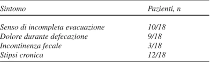
Tabella 1 Sintomatologia clinica dei pazienti sottoposti allo studio
Sintomo Pazienti, n Senso di incompleta evacuazione 10/18 Dolore durante defecazione 9/18 Incontinenza fecale 3/18 Stipsi cronica 12/18
ne dell’ampolla rettale mediante sequenza GE T1-pesata orientata sul piano sagittale con le seguenti caratteristiche: TR 30 ms; TE 6 ms; FA 90°; strati 1; spessore di strato 5,5 mm; FOV 300×300; matrice 192×128; tempo di acquisi-zione 3 s/immagine. Previo secondo riempimento dell’am-polla rettale, è stato condotto analogo protocollo d’esame in decubito supino con tavolo del magnete a 0°. Il tempo complessivo di occupazione della macchina per la completa acquisizione dell’esame nei due decubiti, compreso il se-condo riempimento del retto, è stato in media di 68 minuti (range 42–93 minuti).
Analisi delle immagini
Le immagini RM acquisite sul piano sagittale sono state ana-lizzate da due radiologi che hanno effettuato separatamente le rispettive misurazioni. Sono state prese in considerazione tutte le immagini acquisite, sia in decubito ortostatico sia in decubito prono. Per consentire una standardizzazione delle misure e per determinare la gravità delle disfunzioni del pa-vimento pelvico è stata identifi cata la linea pubo-coccigea (LPC), delimitante la base del pavimento pelvico, tesa tra il margine inferiore della sinfi si pubica e l’ultima articolazione del coccige. Al fi ne di valutare la lassità del pavimento pelvi-co abbiamo misurato la linea H e la linea M: la linea H è misurata tracciando una linea dal margine inferiore della sinfi -si pubica alla parete posteriore del retto a livello della giun-zione ano-rettale e rappresenta la porgiun-zione più caudale del gruppo dei muscoli elevatori dell’ano (muscolo pubo-rettale); inoltre, essa corrisponde allo iato pubo-rettale, che consente la massima apertura in senso antero-posteriore dell’anello pelvico durante il ponzamento. In accordo con i dati in lette-ratura abbiamo considerato l’ampliamento dello iato pubo-rettale patologico quando la linea H supera la lunghezza di 6 cm [7]; la linea M si estende perpendicolarmente dal piano della LPC alla fi ne della linea H. Abbiamo considerato pato-logica una lunghezza della linea H superiore ai 2 cm.
Abbiamo considerato il prolasso come la protrusione di qualsiasi organo (vescica, uretra, vagina, piccolo intesti-no) attraverso lo iato pubo-rettale (linea H). L’analisi della gravità del prolasso si è ottenuta considerando la più breve distanza intercorrente tra il margine più caudale dell’orga-no prolassato e la linea H. La quantifi cazione del rettocele anteriore è avvenuta prolungando superiormente a livello del retto la tangente alla parete anteriore del canale anale e misurando la distanza tra tale linea e la parete anteriore del bulging nel punto di maggiore ampiezza. Abbiamo conside-rato patologico un rettocele di almeno 2 cm [7].
Qualità delle immagini
Abbiamo valutato la qualità delle immagini analizzando il rapporto segnale/rumore ed il livello di artefatti mediante static study included acquisition of 3D hybrid
contrast-en-hanced (HYCE) gradient echo (GE) sequences and three or-thogonal spatial planes with the following parameters: rep-etition time 10 ms; echo-train length 5 ms; fl ip angle 90°; slices 20; slice thickness 2.5 mm; fi eld of view 280×280; matrix, 200×160. Static images were also acquired in the sagittal plane at rest and during sphincter contraction and straining using T1-weighted GE sequences with the follow-ing parameters: repetition time 35 ms; echo-train length 10 ms; fl ip angle 90°; slices 1; slice thickness 5.5 mm; fi eld of view 300×300; matrix 192×128. Finally, a dynamic study was performed during rectal evacuation using an axial T1-weighted GE sequence with the following parameters: rep-etition time 30 ms; echo-train length 6 ms; fl ip angle 90°; slices 1; slice thickness 5.5 mm; fi eld of view 300×300; matrix 192×128; acquisition time 3 s/image. After refi lling the rectal ampulla, the scan protocol was repeated with the patient in the supine position and table tilt set at 0°. Overall MR room time for the examinations in the two patient posi-tions, and including refi lling of the rectum, was approxi-mately 68 (range 42–93) min.
Image analysis
MR images acquired in the sagittal plane were analysed by two radiologists who took the measurements separately. All images acquired in both supine and orthostatic posi-tions were taken into consideration. To allow measurement standardisation and determine severity of the pelvic fl oor disorder, we considered the pubococcygeal line (PCL), which delimits the base of the pelvic fl oor and extends from the inferior border of the pubic symphysis and the last coc-cygeal joint. To evaluate pelvic fl oor laxity, measurements of the H and M lines were obtained: the H line is measured by tracing a line from the inferior border of the pubic sym-physis to the posterior wall of the rectum at the anorectal junction level. This line represents the most caudal portion of the levator ani complex (puborectalis muscle) and cor-responds to the puborectal hiatus, which allows grading of the maximal widening in the anteroposterior direction of the pelvic sling during straining. In agreement with the literature, we considered puborectal hiatus widening to be pathological when the H line exceeded 6 cm [7]. The M line extends perpendicularly from the PCL to the end of the H line. The M line exceeding 2 cm was considered patho-logical.
Organ prolapse was defi ned as protrusion of any organ (bladder, urethra, vagina, small bowel) through the H line. Prolapse severity was graded by considering the shortest distance between the most caudal aspect of the descended organ and the H line. Anterior rectocele was quantifi ed by prolonging upwards to the level of the rectum the tangent to the anterior wall of the anal canal and measuring the
dis-una scala qualitativa con punteggio da 1 a 3. Riguardo al rapporto segnale/rumore, è stato attribuito un punteggio di 1 se il rumore risultava superiore al segnale, 2 in caso di parità e 3 se il segnale era superiore al rumore. Per quanto riguarda il livello di artefatti, è stato assegnato un punteg-gio di 1 in caso di artefatti minori o assenti, 2 se erano presenti artefatti di grado medio e 3 in caso di artefatti maggiori o gravi. Tali valutazioni sono state effettuate sia sulle immagini statiche ottenute con le sequenze 3D-HYCE bilanciate (basale) e GE T1-pesate (rilassamento, contra-zione e ponzamento), sia sulle singole immagini dinamiche. I dati sono stati ottenuti separatamente dai due radiologi ed è stato calcolato il valore medio di tutti i punteggi per ogni singola tipologia di immagine.
Risultati
Immagini di buona qualità sono state ottenute in 15/18 pa-zienti; 3 pazienti hanno presentato artefatti legati al movi-mento e 3 pazienti hanno presentato incontinenza che ha ostacolato lo studio funzionale (Fig. 1). Ciò nonostante, le valutazioni effettuate sul rapporto segnale/rumore e sul livello di artefatti, sono state soddisfacenti per tutte le ti-pologie di immagini, anche se punteggi inferiori sono stati ottenuti dalle immagini dinamiche (Tabelle 2 e 3). Nella va-lutazione dell’effi cacia delle tre sospensioni utilizzate per opacizzare il retto si è potuto constatare che non si sono ri-scontrate sostanziali differenze di intensità di segnale (Fig. 2). Abbiamo riscontrato tuttavia che nelle pazienti in cui è tance between this line and the anterior wall of the rectal
bulge at the point of maximum depth. We considered a rec-tocele of at least 2 cm to be pathological [7].
Image quality
Image quality was evaluated by analysing the signal-to-noise ratio (SNR) and the severity of artefacts on a qualita-tive scale from 1 to 3. SNR was scored 1 if the noise was greater than the signal, 2 if noise and signal were equivalent and 3 if the signal was greater than the noise. Artefact sever-ity was scored 1 if no artefacts or only minor artefacts were present, 2 if there were artefacts of intermediate severity and 3 in the event of major or severe artefacts. These evalua-tions were performed on both static images obtained with the balanced 3D HYCE (at baseline) and T1-weighted gra-dient echo sequences (at rest, during contraction and strain-ing), and on the single dynamic images. Data were obtained separately by two radiologists, and mean values of all scores were calculated for each type of image.
Results
Image quality was good in 15/18 patients. Three patients showed movement artefacts, and three had incontinence, which hampered the functional study (Fig. 1). SNR and ar-tefact severity evaluations were nonetheless satisfactory for all types of images, even though dynamic images obtained lower scores (Tables 2 and 3). Evaluation of the effi cacy of
Fig. 1a,b Normal anatomy in the orthostatic position. T1-weighted cine gradient-echo sequence. An acquisition time of 3 s for each frame provides good
anatomical detail (a). However, the relatively long acquisition time could cause motion artefacts (b).
Fig. 1a,b Anatomia normale in ortostatismo. Sequenza cine gradient echo T1-pesata. Il tempo di acquisizione di 3 secondi per ogni singolo frame consente un buon dettaglio anatomico (a). Tuttavia il tempo di acquisizione relativamente lungo può causare artefatti da movimento (b).
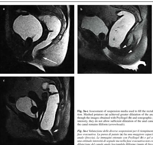
stata utilizzata la purea di patate con gadobutrolo si è ve-rifi cata una maggiore apertura del canale anale durante la fase defecatoria rispetto alle altre due sospensioni, verosi-milmente in relazione ad una maggiore densità e consisten-za del materiale (Fig. 3).
Tutte le patologie osservate sono state classifi cate in classi di gravità. Abbiamo valutato il prolasso degli organi rispetto alla linea H, la lassità del pavimento pelvico ed il rettocele anteriore e confrontati i risultati ottenuti nei due decubiti. the three suspensions used to opacify the rectum showed
that there were no substantial differences in signal intensity (Fig. 2). We noted, however, that the use of mashed potatoes with gadobutrol resulted in a greater opening of the anal ca-nal during the defecation phase compared with the other two suspensions, probably due to the greater density and thicker texture of the material (Fig. 3).
All disorders observed were classifi ed according to sever-ity. We evaluated organ prolapse with respect to the H line,
Table 2 Evaluation of signal-to-noise ratio
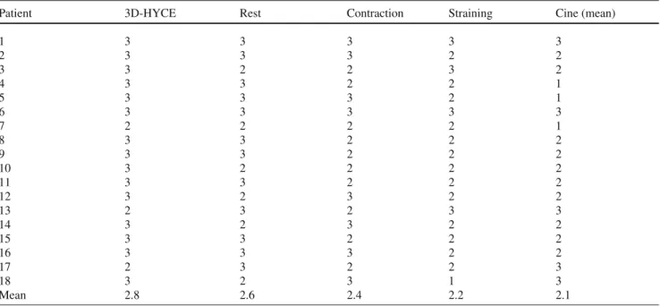
Patient 3D-HYCE Rest Contraction Straining Cine (mean)
1 3 3 3 3 3 2 3 3 3 2 2 3 3 2 2 3 2 4 3 3 2 2 1 5 3 3 3 2 1 6 3 3 3 3 3 7 2 2 2 2 1 8 3 3 2 2 2 9 3 3 2 2 2 10 3 2 2 2 2 11 3 3 2 2 2 12 3 2 3 2 2 13 2 3 2 3 3 14 3 2 3 2 2 15 3 3 2 2 2 16 3 3 3 2 2 17 2 3 2 2 3 18 3 2 3 1 3 Mean 2.8 2.6 2.4 2.2 2.1
HYCE, hybrid contrast-enhanced sequence
Tabella 2 Valutazione del rapporto segnale/rumore
Paziente 3D-HYCE Rilassamento Contrazione Ponzamento Cine (media)
1 3 3 3 3 3 2 3 3 3 2 2 3 3 2 2 3 2 4 3 3 2 2 1 5 3 3 3 2 1 6 3 3 3 3 3 7 2 2 2 2 1 8 3 3 2 2 2 9 3 3 2 2 2 10 3 2 2 2 2 11 3 3 2 2 2 12 3 2 3 2 2 13 2 3 2 3 3 14 3 2 3 2 2 15 3 3 2 2 2 16 3 3 3 2 2 17 2 3 2 2 3 18 3 2 3 1 3 Media 2,8 2,6 2,4 2,2 2,1
Abbiamo osservato una sottostima per tutte le patologie nella valutazione in decubito supino: in particolare, 6 prolassi ve-scicali misconosciuti in decubito supino sono stati stimati di grado lieve in ortostasi, mentre 2 stimati di grado moderato in ortostasi sono risultati lievi in supino. Abbiamo riscontra-to 10 prolassi rettali severi in orriscontra-tostasi 3 dei quali valutati moderati in decubito supino (Fig. 4), 5 prolassi moderati in ortostasi, 1 dei quali è risultato lieve in decubito supino e 3 prolassi lievi in ortostasi misconosciuti in decubito supi-pelvic fl oor laxity and anterior rectocele, and compared the
results obtained in the orthostatic and supine position. The supine position resulted in an underestimation of all disor-ders: in particular, six bladder prolapses were missed, which were graded as mild in the orthostatic position, whereas two moderate prolapses in the orthostatic position were grad-ed as mild in the supine position. We identifi grad-ed ten severe rectal prolapses in the orthostatic position, three of which were graded as moderate in the supine position (Fig. 4); fi ve
Table 3 Evaluation of artefacts
Patient 3D-HYCE Rest Contraction Straining Cine (mean)
1 1 1 2 2 3 2 1 1 1 1 1 3 1 1 2 1 2 4 1 1 1 2 2 5 2 2 2 2 3 6 1 1 1 1 1 7 1 1 1 1 2 8 2 2 1 1 3 9 2 1 1 2 3 10 1 2 2 1 2 11 2 2 1 1 2 12 1 2 2 2 2 13 2 2 3 2 2 14 1 2 2 1 2 15 2 1 2 1 1 16 2 2 2 2 1 17 2 1 2 2 1 18 1 2 2 1 2 Mean 1.4 1.5 1.6 1.4 1.9
HYCE, hybrid contrast-enhanced sequence
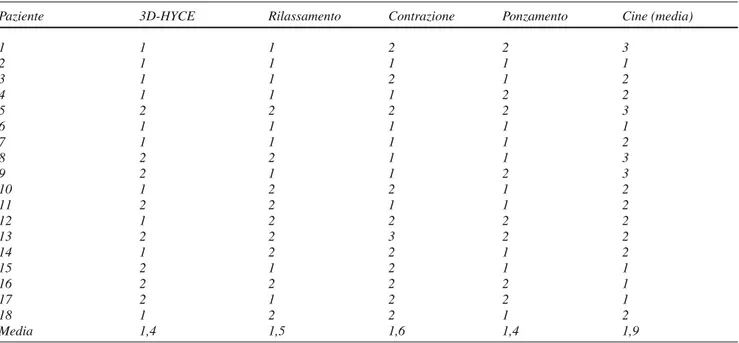
Tabella 3 Valutazione del livello di artefatti
Paziente 3D-HYCE Rilassamento Contrazione Ponzamento Cine (media)
1 1 1 2 2 3 2 1 1 1 1 1 3 1 1 2 1 2 4 1 1 1 2 2 5 2 2 2 2 3 6 1 1 1 1 1 7 1 1 1 1 2 8 2 2 1 1 3 9 2 1 1 2 3 10 1 2 2 1 2 11 2 2 1 1 2 12 1 2 2 2 2 13 2 2 3 2 2 14 1 2 2 1 2 15 2 1 2 1 1 16 2 2 2 2 1 17 2 1 2 2 1 18 1 2 2 1 2 Media 1,4 1,5 1,6 1,4 1,9
no. Cinque lassità del pavimento pelvico sono risultate gravi in ortostasi ed 1 è stata sottostimata moderata in supino, 1 valutata di grado moderato in ortostasi è stata sottostimata lieve in decubito supino e 2 lievi in ortostasi misconosciute in supino. Degli 11 rettoceli anteriori osservati, classifi cati come gravi in decubito ortostatico, solo 8 sono altrettanto severi in supino; 3 gravi ed 1 moderato sono stati sottosti-mati, mentre 1 rettocele misconosciuto in decubito supino è risultato lieve in ortostasi (Tabella 4).
Discussione
La reale prevalenza delle disfunzioni del pavimento pelvico è sottostimata in quanto tali patologie sono spesso asinto-matiche per lunghi periodi o paucisintoasinto-matiche (dolore pel-vico, senso di peso, sensazione di incompleta evacuazione) soprattutto in caso di iniziale prolasso degli organi pelvici [1, 2, 8]. L’esame fi sico inoltre è spesso insuffi ciente e dif-fi coltoso per una completa valutazione anatomica e funzio-nale della pelvi [9].
moderate prolapses in the orthostatic position, one of which was graded as mild it the supine position; and three mild prolapses in the orthostatic position, which were all missed in the supine position. Five cases of severe pelvic fl oor lax-ity were graded as severe in the orthostatic position, one of which was underestimated as moderate in the supine posi-tion; one case of moderate laxity in the orthostatic position was graded as mild in the supine position; and two cases graded as mild laxity in the orthostatic position were missed in the supine position. Of the 11 anterior rectoceles classi-fi ed as severe in the orthostatic position, only eight were also graded as severe in the supine position; three severe and one moderate anterior rectocele were underestimated, whereas one mild rectocele in the orthostatic position was missed in the supine position (Table 4).
Discussion
The true prevalence of pelvic fl oor disorders is underes-timated, as these disorders tend to remain asymptomatic
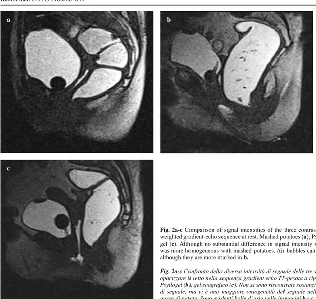
Fig. 2a-c Comparison of signal intensities of the three contrast agents used in the
T1-weighted gradient-echo sequence at rest. Mashed potatoes (a); Psyllogel (b); sonographic gel (c). Although no substantial difference in signal intensity was observed, the signal was more homogeneous with mashed potatoes. Air bubbles can be seen in both b and c, although they are more marked in b.
Fig. 2a-c Confronto della diversa intensità di segnale delle tre sospensioni utilizzate per opacizzare il retto nella sequenza gradient echo T1-pesata a riposo. Purea di patate (a), Psyllogel (b), gel ecografi co (c). Non si sono riscontrate sostanziali differenze di intensità di segnale, ma vi è una maggiore omogeneità del segnale nell’immagine ottenuta con purea di patate. Sono evidenti bolle d’aria nelle immagini b e c ma soprattutto in b.
a b
Il planning pre-operatorio si basa sulla visita clinica e sui dati forniti dalla defecografi a tradizionale, coadiuvati talvolta da quelli ottenuti con la defeco-RM. L’imaging gio-ca quindi un ruolo fondamentale e fornisce la possibilità di ridurre le recidive post-intervento soprattutto nei pazienti con patologia multi-compartimentale [10]. I lavori riportati in letteratura sull’argomento sono stati eseguiti su magnete chiuso ad alto campo (1,5 T) ed acquisizioni T2 pesate su multipli piani dello spazio, con paziente in decubito supino o posto sul fi anco a ginocchia fl esse [10–12]. La realizza-zione di magneti aperti da 0,5 T, dotati di supporti dedicati e apertura del gantry suffi cientemente ampia, ha permesso di eseguire lo studio defeco-RM con paziente in posizione seduta [13, 14].
Gli studi fi nora pubblicati in letteratura per la valuta-zione comparativa degli esami defeco-RM in due differenti decubiti utilizzano pertanto magneti a diversa intensità del campo magnetico stazionario (1,5 T per il decubito supino e 0,5 T per la posizione seduta). Questo, a nostro parere, rap-presenta un limite per l’impossibilità di compararne i risul-tati a causa delle differenti performance dei due magneti e or poorly symptomatic (pelvic pain, sense of weight,
feel-ing of incomplete evacuation) for years, especially in the early stages of pelvic organ prolapse [1, 2, 8]. Moreover, physical examination is often diffi cult and inadequate for a complete anatomical and functional assessment of the pelvis [9].
Preoperative planning is based on clinical assessment and data provided by conventional defecography, in some cases supplemented by MRD. Imaging therefore plays a key role in these disorders and provides an opportunity to reduce postoperative relapses, especially in patients with multicompartmental disorders [10]. Previous published studies have been conducted with closed-confi guration, high-fi eld (1.5-T) systems and T2-weighted multiplanar ac-quisitions, with the patient in the supine position or lateral decubitus with fl exed knees [10–12]. The development of open-confi guration 0.5-T systems with dedicated supports and suffi ciently wide gantry aperture has allowed MRD ex-aminations of the patient in a sitting position [13, 14]. All published studies comparing MRD performed with the two patient positions have therefore been conducted using
dif-a
c
b
Fig. 3a-c Assessment of suspension media used to fi ll the rectal ampulla during
defeca-tion. Mashed potatoes (a) achieved greater dilatation of the anal sphincter (arrow). Al-though the images obtained with Psyllogel (b) and sonographic gel (c) show good signal intensity, they do not allow suffi cient dilatation of the anal canal during defecation, and the canal remains fi liform (arrowheads).
Fig. 3a-c Valutazione delle diverse sospensioni per il riempimento dell’ampolla rettale in fase evacuativa. La purea di patate (a) ha una maggiore capacità di far aprire il canale anale (freccia). Le immagini ottenute con Psyllogel (b) e gel ecografi co (c) forniscono una ottimale intensità di segnale ma nella fase evacuativa non consentono una suffi ciente dilatazione del canale anale lasciandolo fi liforme (punte di freccia).
delle sequenze utilizzate [13, 15]. Nel nostro studio abbiamo utilizzato un magnete permanente aperto di nuova concezio-ne con campo magconcezio-netico stazionario di 0,25 T, equipaggia-to con tavolo ribaltabile da 0° a 90°, che ha consentiequipaggia-to di eseguite lo stesso protocollo d’esame nei due differenti de-cubiti. L’esame sotto carico è stato eseguito tuttavia non in posizione seduta, ma in ortostatismo. L’esame defeco-RM in ortostasi è stato inoltre acquisito con tavolo inclinato a 80° per aumentare la stabilità del soggetto in esame e ridurre gli artefatti da movimento indotti dalla stazione eretta.
Analogamente allo studio defecografi co convenzionale, sono state acquisite sequenze statiche in fase di riposo, con-trazione e ponzamento. La possibilità di studiare la fase eva-ferent magnetic fi eld strengths (1.5 T for supine decubitus
and 0.5 T for the sitting position). This is a limitation, in our opinion, that precludes comparison of results owing to the different performance of the two magnets and the se-quences used [13, 15].
In our study, we used a new open-confi guration per-manent magnet with a 0.25-T stationary magnetic fi eld equipped with a 0°-–90° tilting table, which allowed us to apply the same imaging protocol in the two patient posi-tions. The examination under weight-bearing conditions was performed, however, not in the sitting position but in the orthostatic position. In addition, MRD in the orthostatic position was acquired with the table at an 80° tilt to increase
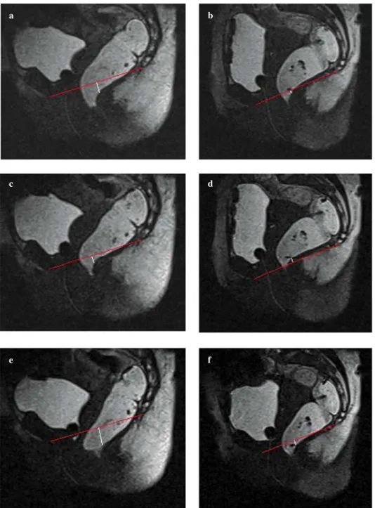
Fig. 4a-f Rectal prolapse. Images
ob-tained in the orthostatic position at rest (a), during contraction (c) and straining (e) and during the same phases in the supine position (b,d,f). The pubococ-cygeal line was drawn on the images, and the distance from this line to the anorectal junction was measured. The prolapse is clearly evidenced during straining in the orthostatic position only (arrows).
Fig. 4a-f Paziente con prolasso retta-le. Immagini ottenute in ortostatismo a riposo (a), durante contrazione (c) e ponzamento (e) ed in posizione supina nelle medesime condizioni (b,d,f). Sul-le immagini è stata tracciata la linea pubo-coccigea ed è stata misurata la distanza dalla linea pubo-coccigea alla giunzione ano-rettale. Il prolasso risulta evidente in fase di ponzamento ma solamente in ortostatismo (frecce).
a b
c d
cuativa appare indispensabile per aumentare l’accuratezza diagnostica e fornire un completo quadro clinico ed un cor-retto inquadramento terapeutico del paziente, poiché alcuni reperti patologici risultano sottostimati nelle acquisizioni statiche in fase di ponzamento. È stato inoltre fondamentale, per una ottimale valutazione della fase evacuativa, utilizzare la sospensione idonea per una corretta apertura del cana-le anacana-le. Le tre sospensioni utilizzate, hanno una differente densità e consistenza e nonostante risulti possibile utilizzare tutte e tre le sospensioni abbiamo dimostrato come la purea di patate abbia una maggiore capacità di far aprire il ca-nale aca-nale, consentendo una valutazione più accurata delle eventuali patologie presenti. Le sospensioni composte dal gel ecografi co e dallo Psyllogel danno una ottimale intensità di segnale, ma nella fase evacuativa non dilatano suffi cientemente il canale anale lasciandolo fi liforme, ciò rende diffi -coltoso, seppur possibile, fare le dovute valutazioni secondo i metodi utilizzati. Grazie probabilmente alla sua maggiore densità e consistenza, la purea di patate apre il canale anale in maniera ottimale e agevola la valutazione della patologia. È questa sua caratteristica che lo rende nella nostra espe-rienza il mezzo di opacizzazione migliore.
Non essendo per il momento disponibili sul magnete utilizzato sequenze di tipo steady state free precession (ba-lanced), attualmente in voga per la fase evacuativa, è stato necessario adattare le sequenze turbo fi eld echo (TFE) T1 pesate. Un compromesso ragionevole fra la risoluzione spa-ziale, risoluzione temporale e l’intensità del segnale ha con-sentito l’ottimizzazione della sequenza dinamica utilizzata nella fase evacuatoria con tempi di acquisizione di 3 s/im-magine. Una migliore risoluzione temporale potrebbe pro-patient stability and reduce movement artefacts resulting
from the standing position.
As in conventional defecography, static sequences were acquired with the patient at rest, during contraction and while straining. The possibility of studying the evacuation phase appears essential for increasing diagnostic accuracy of the examination, providing complete clinical assessment and guiding appropriate patient management, given that some pathological fi ndings are underestimated in the stat-ic images acquired during straining. Moreover, in order to achieve optimal evacuation phase evaluation, it is important to use a suitable suspension medium allowing correct open-ing of the anal canal. The three suspensions we used have different density and texture and, although all three can be used, we found that mashed potatoes achieve greater open-ing of the anal canal and thus a more accurate assessment of any disorder. The suspensions composed of sonographic gel and Psyllogel have optimal signal intensity but fail to dilate the anal canal suffi ciently in the evacuation phase, leaving it fi liform, which makes it diffi cult, albeit possible, to carry out assessments according to the methods used. Probably as a result of its greater density and thicker texture, mashed potatoes provides optimal opening of the anal canal and fa-cilitates evaluation of the disorder. In our experience, this feature makes it the best medium by which to obtain opaci-fi cation in our experience.
Because the MR system we used does not provide steady-state free precession (balanced) sequences, which are commonly used for the evacuation phase, we adapted the T1-weighted turbo fi eld-echo sequences. A reasonable
Table 4 Results obtained in the upright and supine positions
Condition Mild Moderate Severe Total
Upright Supine Upright Supine Upright Supine Upright Supine
Bladder prolapse 6 2 2 0 0 0 8 2
Vaginal prolapse 0 0 0 0 0 0 0 0
Rectal prolapse 3 1 5 7 10 7 18 15
Pelvic fl oor laxity 2 1 3 3 5 4 10 8
Anterior rectocele 1 1 4 6 11 8 16 15
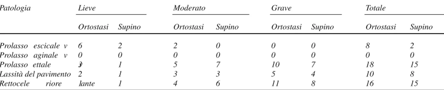
Tabella 4 Valutazione dei reperti ottenuti rispettivamente in ortostasi ed in posizione supina
Patologia Lieve Moderato Grave Totale
Ortostasi Supino Ortostasi Supino Ortostasi Supino Ortostasi Supino
Prolasso vescicale 6 2 2 0 0 0 8 2
Prolasso vaginale 0 0 0 0 0 0 0 0
Prolasso rettale 3 1 5 7 10 7 18 15
Lassità del pavimento 2 1 3 3 5 4 10 8
babilmente essere facilmente ottenibile grazie alla costru-zione di sequenze specifi che per l’esame defeco-RM esenti dalla calibrazione fra un’immagine e l’altra e con rapporto segnale/rumore suffi ciente a ridurre i tempi di acquisizione e dalla messa a punto di sequenze dinamiche di tipo steady state free precession (balanced). L’utilizzo di sequenze 3D HYCE GE bilanciate, ha consentito invece lo studio morfo-logico preliminare del pavimento pelvico e degli organi in esso contenuti con acquisizioni statiche multiplanari. Le im-magini orientate sul piano assiale hanno inoltre permesso di valutare lo slargamento dello iato pubo-rettale in caso di lassità del pavimento pelvico.
Nella nostra casistica, in accordo con i dati in letteratu-ra, la comparazione dei risultati nei due differenti decubiti, ha mostrato nel decubito supino una sottostima della gra-vità per tutte le patologie osservate (Tabella 4) [7, 11]. Un ulteriore limite del decubito supino consiste nella diffi coltà ad effettuare la fase evacuativa, che risulta invece di fon-damentale importanza per un corretto inquadramento cli-nico-diagnostico del paziente. Nella nostra casistica infatti solamente 3 soggetti su 8 arruolati sono stati in grado di completare l’esame defeco-RM in decubito supino, mentre tutti i soggetti sono riusciti ad effettuare la fase evacuativa in ortostatismo.
Per l’analisi quantitativa delle immagini ottenute è sta-to utilizzasta-to il sistema HMO (linea H, linea M e prolasso degli organi pelvici rispetto alla linea H), al fi ne di defi -nire, differenziare e valutare la gravità del prolasso degli organi pelvici e la lassità della muscolatura del pavimen-to pelvico stesso (Tabella 2) [10–13, 16]. Confrontando la gravità dei prolassi e dei rettoceli anteriori riscontrati nei due decubiti, abbiamo osservato una sottostima del decubi-to supino in tutti i pazienti (Tabella 4). I differenti risultati fra gli esami RM nei due decubiti sono da correlare verosi-milmente alla direzione della forza gravitazionale che viene esercitata secondo un asse verticale in ortostasi, gravando interamente sulle strutture muscolo-fasciali del pavimento pelvico, e secondo un asse antero-posteriore in decubito su-pino. In quest’ultimo decubito la forza gravitazionale non viene esercitata direttamente sul pavimento pelvico e la sot-tostima dei reperti è legata al fatto che l’unica forza diretta verso il pavimento pelvico in senso cranio-caudale è quella del torchio addominale durante ponzamento ed evacuazio-ne. I nostri dati dimostrano che lo studio RM in posizione ortostatica, sebbene non fi siologica, consente una migliore valutazione delle patologie del pavimento pelvico rispetto all’esame RM con paziente in decubito supino.
Il principale limite del nostro studio è stato l’impossi-bilità di valutare il paziente in posizione seduta in quanto la conformazione della macchina, concepita per lo studio dinamico osteo-articolare, ha impedito, a causa dell’esiguo spazio (35 cm), di adattare un supporto dedicato. Un ulte-riore limite è legato alle sequenze utilizzate ed alla relativa bassa intensità del campo magnetico, in particolare per lo studio cinematico durante evacuazione dell’ampolla rettale. compromise between spatial resolution, temporal resolution
and signal intensity allowed us to optimise the dynamic se-quence used in the evacuation phase with acquisition time of 3 s/image. A better temporal resolution could probably be obtained by constructing specifi c MRD sequences that do not require calibration between one image and the next and with a suffi cient SNR to reduce acquisition times, as well as by developing dynamic (balanced) steady-state free precession sequences. The use of 3D HYCE balanced gradi-ent echo sequences allowed for preliminary morphological study of the pelvic fl oor and pelvic organs with static mul-tiplanar acquisitions. Moreover, axial images allowed wid-ening of the puborectal hiatus to be assessed in evaluating pelvic fl oor laxity.
In agreement with previous reports, the comparison of results obtained with the two different patient positions showed that the supine decubitus resulted in an underesti-mation of the severity of all disorders [7, 11] (Table 4). A further limitation of the supine decubitus is the diffi culty in performing the evacuation phase, which is essential for cor-rect clinical and diagnostic assessment of the patient. In our series, only three out of 18 patients were able to complete the MRD examination in the supine position, whereas all patients succeeded in performing the evacuation phase in the orthostatic position.
For quantitative image analysis, we used the H line, M line and organ prolapse with respect to the H line (HMO) system in order to defi ne, differentiate and grade the sever-ity of organ prolapse and pelvic fl oor musculature laxsever-ity (Table 2) [10–13, 16]. Comparison of prolapses and ante-rior rectocele severity seen in the two imaging positions showed that the supine position resulted in an underestima-tion in all patients (Table 4). The different results of the MRD examination in the two positions are most likely re-lated to the direction of the force of gravity exerted along a vertical axis, bearing completely on the muscular–fascial structures of the pelvic fl oor in the orthostatic position, and along an anteroposterior axis in the supine position. In the latter position, the force of gravity is not exerted directly on the pelvic fl oor, and the underestimation of fi ndings is related to the fact that the only force bearing on the pelvic fl oor in the craniocaudal direction is that of bearing down with the abdominal muscles during straining and evacua-tion. Our fi ndings demonstrate that MRD in the orthostatic position, even though not physiological, allows for better evaluation of pelvic fl oor disorders compared with the pa-tient in the supine position.
The main limitation of our study was the inability to evaluate the patient in a sitting position. This was due to the confi guration of the MR system, which was devised for dynamic osteoarticular studies and is not compatible with a dedicated support due to lack of space (35 cm). A further limitation is related to the sequences used and the relatively
Le sequenze dinamiche adattate allo scopo non possiedono una ottimale risoluzione temporale e la presenza di un inter-vallo di calibrazione tra un frame ed il successivo potrebbe determinare una sottostima dei reperti più fugaci. La possi-bilità di acquisire un solo strato di 5,5 mm in fase dinamica ha inoltre reso obbligatoria non solo l’assoluta immobilità del paziente durante tutto l’esame, ma anche una estrema precisione dell’operatore nell’impostazione dei parametri geometrici al fi ne di centrare perfettamente l’asse del cana-le anacana-le. L’utilizzo di sequenze T1 pesate nello studio dina-mico ha reso, inoltre, indispensabile l’opacizzazione della vescica, della vagina e del retto, con conseguente aumento dei costi dell’esame, del disagio dei pazienti e del tempo complessivo dell’esame.
Conclusioni
Lo studio defeco-RM risulta fattibile e di elevata qualità dia-gnostica con un magnete permanente da 0,25 T. Lo studio in posizione ortostatica risulta più accurato nella valutazione delle patologie del pavimento pelvico rispetto all’esame in decubito supino, talvolta infi ciato dalla diffi coltà a comple-tare la fase evacuatoria. Il sistema di misurazione HMO nella valutazione delle disfunzioni del pavimento pelvico appare, a nostro parere, il metodo più affi dabile perché, valutando separatamente due entità patologiche distinte e spesso coesistenti (lassità del pavimento e prolasso degli organi), consente un più corretto inquadramento clinico e fornisce quindi informazioni indispensabili per la scelta della migliore strategia terapeutica.
Questo studio preliminare, eseguito fondamentalmente per la messa a punto della migliore tecnica di imaging ne-cessita di casistiche più ampie per valutare il reale impatto clinico della metodica. Sicuramente i continui miglioramen-ti tecnologici soprattutto delle sequenze dinamiche T2, dedi-cate a questo tipo di studio, potranno migliorare la qualità diagnostica delle immagini, riducendo i tempi di esame. low magnetic fi eld strength, in particular for the cine study
during evacuation of the rectal ampulla. The specifi cally adapted dynamic sequences do not have optimal temporal resolution, and the presence of a calibration interval be-tween frames could lead to an underestimation of the more transient fi ndings. In addition, the possibility of acquiring only one 5.5-mm slice during the dynamic phase required not only absolute patients immobility throughout the ex-amination, but also extreme operator precision in setting the geometrical parameters to achieve perfect centring of the axis of the anal canal. The use of T1-weighted sequences in the dynamic study made it necessary to opacify the blad-der, vagina and rectum, resulting in increased costs, greater patient discomfort and longer examination time.
Conclusions
MRD with a permanent 0.25-T magnet is feasible and pro-vides high diagnostic quality. The study in the orthostatic position is more accurate for evaluating pelvic fl oor disor-ders compared with the supine position, which may be ham-pered by diffi culties completing the evacuation phase. The HMO measurement system in evaluating pelvic fl oor disor-ders appears to be the most reliable method; by evaluating separately two distinct and often coexisting pathological en-tities (pelvic fl oor relaxation and organ prolapse), it allows a more accurate clinical assessment and therefore provides information that is vital for selecting the most appropriate treatment strategy.
The fi ndings of this preliminary study, performed mainly to identify the best examination technique, warrant larger series to evaluate the real clinical impact of the modality. The continuous technological improvements, especially in dedicated dynamic T2 sequences, will improve image diag-nostic quality while reducing examination times.
Confl ict of interest None References/Bibliografi a
1. Nygaard I, Barber MD (2008) Prevalence of symptomatic pelvic fl oor disorders in US women. JAMA 300:1311–1316
2. Lawrence JM, Lukacz ES (2008) Prevalence and co-occurrence of pelvic fl oor disorders in community-dwelling women. Obstet Gynecol 111:678–685 3. Jelovsek JE, Barber MD (2006) Women
seeking treatment for advanced pelvic organ prolapsed have decreased body image and quality of life. Am J Obstet Gynecol 194:1455–1461
4. Soligo M, Salvatore S (2002) Prevalence of urinary incontinence in 1.200.000 women after menopause. Urogynaecologia Int J s16:46 5. Shorvon PJ, Marshall MM (2005)
Evacuation proctography. In: Wexner SD, Zbar AP, Pescatori M (eds) Complex anorectal disorders: investigation and management. Springer, Berlin Heidelberg New York, pp 171–198
6. Dobben AC, Wiersma TG (2005) Prospective assessment of interobserver agreement for defecography in fecal incontinence. AJR Am J Roentgenol 185:1166–1172
7. Barbaric ZL, Marumoto AK (2001) Magnetic resonance imaging of the perineum and pelvic fl oor. Top Magn Reson Imaging 12:83–92
8. DeLancey JO (2005) The hidden epidemic of pelvic fl oor dysfunction: achievable goals for improved prevention and treatment. Am J Obstet Gynecol 192:1488–1495
9. Terra MP, Stoker J (2006) The current role of imaging techniques in faecal incontinence. Eur Radiol 16:1727–1736 10. Boyadzhyan L, Raman SS (2008) Role
of static and dynamic MR Imaging in surgical pelvic fl oor disfunction. Radiographics 28:949–967
11. Pannu HK, Kaufman HS (2000) Dynamic MR imaging of pelvic organ prolapse: spectrum of abnormalities. Radiographics 20:1567–1582 12. Broekhuis SR, Kluivers KB (2009)
Dynamic magnetic resonance imaging: reliability of anatomical landmarks and reference lines used to assess pelvic organ prolapse. Int Urogynecol J Pelvic Floor Dysfunct 20:141–148
13. Mortele KJ, Fairhurst J (2007) Dynamic MR defecography of the posterior compartment: Indications, techniques and MRI features. Eur J Radiol 61:462–472
14. Schoenenberger AW, Debatin JF (1998) Dynamic MR defecography with a superconducting, open-confi guration MR system. Radiology 206:641–646 15. Fielding JR (2002) Practical MR
Imaging of female pelvic fl oor weakness. Radiographics 22:295–304 16. Bertschinger KM, Hetzer FH (2002)
Dynamic MR imaging of pelvic fl oor performed with patient sitting in an open-magnet unit versus with patient supine in a closed-magnet unit. Radiology 223:501–508