Università di Bologna
Scuola di Scienze
Corso di Laurea Magistrale in Biologia Marina
In vivo and in vitro effects of genistein as
obesogenic/anti-obesogenic compound in the lipid
metabolism of rainbow trout (Oncorhynchus mykiss)
Tesi di laurea in
Adattamenti degli animali all’ambiente marino
Relatore Presentata da
Prof. Elena Fabbri Michela di Gennaro
Correlatore
Index
Index
………...2
Abstract
………...5
1. Introduction
...6
1.1 Oncorhynchus mykiss………..9
1.1.1 Taxonomy…………...9
1.1.2 Morphology………...9
1.1.3 Biology and habitat………...10
1.1.4 Farm techniques………...12
1.1.5 Fry production………...13
1.1.6 Fattening techniques-intensive farming………...15
1.1.7 Nutrition of farmed trout………...16
1.1.8 Main producer countries………...18
1.2 Lipid metabolism into liver and adipose tissue……...18
1.2.1 An introduction to lipids………...18
1.2.2 Hormonal regulation of lipid metabolism and its
dependence on diet………...21
1.2.3 Fatty acids synthesis and uptake………...25
1.2.8 Adipose tissue and adipogenesis………...34
1.2.9 The adipocyte life cycle………...35
1.3 Apoptosis and autophagy………...37
1.4 The Phytochemicals………...44
1.4.1 Flavonoids………...44
1.5 Genistein: sources, structure and metabolism………...45
1.6 Genistein: a compound with pleiotropic effects….…...48
1.6.1 Effects of genistein on adipocytes………...50
2. Aims of research
………...57
3. Material and methods
………...58
3.1 Animals (Rainbow trout: Oncorhynchus mykiss)………...58
3.2 Experimental design in vivo: intraperitoneal injection...58
3.3 Gene expression analysis in adipose tissue, liver and white
muscle………...59
3.3.1 RNA isolation………...59
3.3.2 RNA extraction………...60
3.3.3 Complementary DNA synthesis………...62
3.3.4 Real-time quantitative reverse transcription PCR (Real-time
qRT-PCR)………...64
3.4 Experimental design in vitro: cell culture………...67
3.5 Cytotoxicity assay: MTT assay for the cellular viability
………...73
3.6 Cytochemical assay: ORO (Oil red oil) assay………....76
4. Results
………...79
4.1 In vivo results: Gene expression analysis in adipose tissue and
liver………...79
4.2 In vivo results: Gene expression analysis in white muscle
………....83
4.3 In vitro results: MTT assay………...86
4.4 In vitro results: Oil red oil assay (ORO)………..87
5. Discussion
………...88
5.1 Effects in adipose tissue and liver………....88
5.2 Effects in white muscle………....94
6. Conclusions
………...102
7. Bibliography
………...104
Abstract
Nowadays, soy is one of the most used ingredients in the formulation of fish feed, due to the ample market supply, lower market price, high protein concentration and favorable amino acid composition. Nevertheless, soybean meal products are rich and primary diet source of phytoestrogens, as genistein, which may have a potential negative impact on growth, hormonal regulation and lipid metabolism in fish. The principal aim of this study was to better understand in vivo and in vitro genistein’s effects on lipid metabolism of rainbow trout. In adipose tissue it was showed an unclear role of genistein on lipid metabolism in rainbow trout, and in liver an anti-obesogenic effect, with an up-regulation of autophagy-related genes LC3b (in adipose tissue) and ATG4b (in liver and adipose tissue), a down-regulation of apoptosis-related genes CASP3 (in adipose tissue) and CASP8 (in liver). An increase of VTG mRNA levels in liver was also observed. Genistein partially exerted these effects via estrogen- receptor dependent mechanism. In white muscle, genistein seemed to promote lipid turnover, up-regulating lipogenic (FAS and LXR) and lipolytic (HSL, PPARα and PPARβ) genes. It seemed that genistein could exert its lipolytic role via autophagic way (up-regulation of ATG4b and ATG12l), not through an apoptotic pathway (down-regulation of CASP3). The effects of genistein on lipid-metabolism and apoptosis-related genes in trout muscle were not dose-dependent, only on autophagy-related genes ATG4b and ATG12l. Moreover, a partial estrogenic activity of this phytoestrogen was also seen. Through in vitro analysis (MTT and ORO assay), instead, it was observed an anti-obesogenic effect of genistein on rainbow trout adipocytes, and this effect was not mediated by ERs. Both in vivo and in vitro, genistein exerted its effects
1. Introduction
World aquaculture production has increased significantly in the last 50 years , with an average annual growth rate of 6.1% in volume between 2004 and 2006 (FAO, 2009). Fish meal and fish oil are the main raw materials used in the formulation of fish feeds. Due to the expansion of aquaculture, marine fisheries will not be able to sustain the needs of aquaculture in the not too distant future. The global demand for fishmeal for aquafeeds may exceed total available supplies around the year 2020 and for fish oil well before 2010 (New& Wijkstroem, 2002). Thus, alternatives to the use of marine materials in fish feeds must be found. Several studies have investigated the replacement of fish oil by vegetable oils in fish feed. Partial replacement of fish oils by vegetable oils such as rapeseed, soybean, linseed or palm oils in fish feeds has no negative impacts on growth and survival of Atlantic salmon (Rosenlund et al., 2001), brook char (Guillou et al., 1995), gilthead sea bream and European seabass (Izquierdo et al., 2003) and rainbow trout (Greene & Selivonchick,1990; Caballero et al., 2002). Regarding protein supply, soybean meal (SBM) and soy protein concentrate (SPC) are ingredients currently incorporated in aquafeeds to partially replace fish meal without negative effects on growth performance (Kaushik et al., 1995; Refstie et al., 2010).
A class of compounds of high concentration in soy are isoflavones that, for their capacity of binding to and activating estrogen receptors (Latonnelle et al., 2002), were classified as phytoestrogens. The phytoestrogen of greatest
associated with oxidative damage (Anthony et al., 1998; Hertog et al.,1998) . Different hypotheses have been suggested to explain these health benefits, such as the involvement of the two major isoflavones in soy foods, genistein and daidzein, whose different biological activities have been documented in vitro and in vivo studies. For example, it has been proposed that the protective effect exerted by genistein against atherosclerosis could be related to its antioxidant properties (Ferretti et al., 2003); in fact, genistein is able to inhibit lipid peroxidation induced in vitro by several pro-oxidant agents on model and natural membranes (Jha et al., 1985), on cultured cells (Guo et al., 2002; Ho et al.,2003), and on low density lipoproteins (LDL) (Kerry et al., 1998; Wilson et al., 2002).
On the other hand, it has been reported, for example, that genistein can impact negatively the growth performance. This happen if, considering that a maximum concentration of genistein in soy products is ~ 5900 µg/g, this is added to a concentration of 3000µg/g of genistein, which is more or less equal to genistein levels when there is a total replacement of fish meal by soybean meal (Chen et al., 2014). So, it has been observed that the high dietary levels of genistein (3000 µg/g) can depress the growth performance in fishes, as Nile Tilapia, while lower levels of it (0-300µg/g) haven’t effect on growth performance (Chen et al., 2014); similar results have also been reported in rainbow trout (Oncorhynchus mykiss) (Catherine et al., 2001) for a dietary genistein supplementation of 500µg/g. Here, this suppressing effect of genistein can be partly due to its capacity of inhibit the activity of major digestive enzymes: stomach protease and intestine amylase. In vivo and in vitro studies indicate genistein (and dadzein) is capable of binding to and activating estrogen receptors in rainbow trout (Latonnelle et al., 2000; Bennetau-Pelissero et al., 2001; Denny et al., 2005; Cosnefroy et al., 2009; Cleveland, 2014 ). This ability suggests that they may have negative effects on growth- related mechanisms
that are parallel to those of estradiol. In fact, in salmonids E2 negatively affects physiological and metabolic processes that support anabolic growth, as down- regulating the growth hormone (GH)/insulin-like growth factor (IGF) axis (Holloway and Leatherland, 1998; Norbeck and Sheridan, 2011), and promoting catabolic effects on protein turnover in skeletal muscle (reducing rates of protein synthesis and increasing rates of protein degradation). Cleveland and Weber, in 2011, demonstrated that also phytoestrogens (as genistein) are capable of these effects. For example, phytoestrogens have effects on protein turnover and cell proliferation in rainbow trout white muscle, in particular, high concentrations of these have negative effects on skeletal growth via estrogen receptor-dependent and –independent mechanisms (Cleveland, 2014). Many studies have been done on the effects that phytoestrogens can have on metabolic processes in mammals, as on lipid metabolism and glucose tolerance, through estrogen receptor-dependent and -independent mechanisms, the latter including AMPK and PPAR activation and inhibition of tyrosine kinase activity (Orgaard and Jensen, 2008; Arunkumar and Anuradha, 2012; Palacios-Gonzalez et al., 2014). But few of these studies have been done on the effects of phytoestrogens and their mechanisms of action in fish adipose tissue.
Thus, in this study we want to examine the in vivo effects of the phytoestrogen, genistein, on lipid metabolism in adipose tissue primarily, but also in liver and white muscle, and its in vitro effects on adipocytes in culture from rainbow trout (Oncorhynchus mykiss). In addition, in vivo effects of genistein on apoptosis- related genes expression and autophagy-related genes expression in all three
1.1 Oncorhynchus mikyss (common name “Rainbow trout”)
1.1.1 Taxonomy Class: Actinopterygii Order: Salmoniformes Family: Salmonidae Genre: OncorhynchusSpecies: O. mykiss Walbaum, 1792
1.1.2 Morphology
The rainbow trout has an elongate body, the length of which is generally 5 times greater than its height. The head has a conical shape and the mouth is slightly oblique, with the maxillary bone which extends to the posterior edge of the eye. The trout teeth are arranged in 1 or 2 series and are present only on the stem of the ploughshare. The lateral line is nearly horizontal and back, before the tail
fin, is an adipose fin with a black edge. There aren’t nuptial tubercles, but changes take place about the head, mouth and colour in spawning males. The body colour of the rainbow trout may vary according to habitat, the size of the specimens and their stage of sexual maturation. Generally the body of this salmonid has a green-blue shade into dorsal part, while the hip area shows a pigmentation which gradually tends to clear and take silvery reflections. On the sides there is a pink band along the lateral line which assumes more intense shades tending to iridescent when arrives the breeding season. The abdomen of the trout has further a colour lighter, almost whitish.
1.1.3 Biology and habitat
Rainbow trout is a species native to north America, that since 1874 it has been introduced in most of the rivers on the planet, for recreational fishery and aquaculture. This species has an anadromous life cycle, in fact, it spends a few years of life at sea, then, returns to freshwater when spawning begins. These animals are characterize by a very rapid growth and, are be able to, in just three years, accumulate from 7 to 10 kg of weigh, while normal rainbow trout, in the same time, increase by a maximum of 4.5kg. The rainbow trout was introduced in Italy about a century ago, and, as a result of its outstanding ability to adapt to various environmental conditions, the presence of this salmonid can be found in most of the rivers and lakes on the national territory, nowadays. It is a species that is easily reproduced in captivity, and, is characterized by very short rates of growth.
growth of trout takes place in water temperatures that can vary from 6° to 20° C with a thermal optimum between 14° and 18° C , while as regards the reproductive activity , the optimal thermal levels are between 10° and 12°C. Usually, the maturation of sexual organs takes place on reaching the 3rd-4th year of age, but acting on the diet and thermal conditions of the environment, this can be anticipated. The eggs of the trout have a diameter ranging from 3 to 7 mm, and, the females can also produce up to 2000 per kg of body weight. In breeding, changing some environmental parameters or through hormonal treatments depending on broodstock, it is possible to obtain only females or sterile individuals. These last show development similar to that of females, but more rapid than that of males. Spawning occurs in the natural environment in spring (January-May), but in breeding, this stage can be moved in time, acting on nutrition, on the conditions of photoperiod or through hormonal induction. In the wild, adult trout feed on aquatic and terrestrial insects, molluscs, crustaceans, fish eggs, minnows, and other small fishes, but the most important food is freshwater shrimp, containing the carotenoid pigments responsible for the orange-pink colour in the flesh. In aquaculture, the inclusion of the synthetic pigments astaxanthin and canthaxanthin in aquafeeds causes this pink colouration to be produced.
Fig. 1 Production cycle of Oncorhynchus mykiss.
1.1.4 Farm techniques
The breeding of the trout is generally practiced within intensive structure, in which it must constantly be the opportunity to take advantage of waters of excellent quality (without aeration - 1 l/min/kg of trout without aeration or 5 l/sec/tonne of trout with aeration), that meets a number of criteria:
Trout is very sensitive to the presence of nitrogenous substances which are derived from the catabolism of proteins, such as , ammonia, nitrates and nitrites. A parameter that must always be kept under control in the plants of trout farming that use water (with dissolved nitrogen) pumped from underground wells , is the oversaturation gaseous water, which must be avoided because it causes the formation of gas bubbles in the blood of the fish. Alternatively, river water can be used, but temperature and flow fluctuations alter production capacity. Trout are animals that can tolerate an higher alkalinity and hardness of water compared to acidity, but in any case, it is good practice to use neutral or medially alkaline water with a pH between 6.5 and 8.5. However, when the above criteria are met, trout are generally on-grown in raceways or ponds supplied with flowing water, but some are produced in cages and recirculating systems.
1.1.5 Fry production
Trout will not spawn naturally in culture systems; thus juveniles must be obtained either by artificial spawning in a hatchery or by collecting eggs from wild stocks. Larvae are well developed at hatching. The breeders, males and females, after being selected, are generally kept separate. The choice of their number depends on the number of fry required, and, the ratio which is most frequently adopted between males and females is 1:3. The females don’t spawn naturally in captivity environment, and when are fully mature, to induce the release of the eggs, it is necessary human intervention. Trout are subjected to an anesthetic treatment, and subsequently, eggs are removed manually from females by applying a slight pressure on the abdomen from the pelvic fins to the vent area. To reduce the stress of the animals, it has been ideated another method of extraction, “air spawning” ; it consists of insertion of a hypodermic needle about 10 mm into the body cavity near the pelvic fins and air pressure (2 psi) expels the eggs. The air is removed from the body cavity by massaging the
sides of the fish. Up to 2000 eggs/kg body weight are collected in a dry pan and kept dry, improving fertilization. Males are stripped in the same way as females; the semen is collected into containers by pressing, and then, added to eggs. Water is added to activate the sperm and allow the eggs to increase in size by about 20 percent by filling the perivitelline space between the shell and yoke; a process known as 'water-hardening'.
Fertilized eggs are incubated in hatcheries until the eyed stage is reached; the others (non-viable eggs) are removed.A single water source flows (3-4 L/min) up through the eggs, spills over into the tray below, thus becoming aerated, allowing large numbers of eggs to hatch in a minimal amount of space and water. Time required to hatch varies according to the water temperature. With a temperature of 3.9°C it takes 100 days , while with 14.4°C requires 21 days, for a total of about 370 degrees/day. Fry can remain in trays until swim-up at about 10 to 14 days after hatching. Hatching of the batch of eggs usually takes 2-3 days, during which time all eggshells are regularly removed, as well as dead and deformed fry. Eggs incubated separately from rearing troughs are transferred to rearing troughs after hatching.
After hatching, the trays are removed and trough water depth is kept shallow (8-10 cm) with a reduced flow until fry reach 'swim-up' stage, the yolk sac is absorbed, and active food searching . Larvae are able to absorb the yolk sac in a period ranging from 2 to 6 weeks , depending on the temperature conditions, and, when the yolk sac has been absorbed for about 2/3, begins the artificial administration of food with frequent meals and diets properly formulated. In
important the efficiency of the water exchange; in case of poor availability of water, it is appropriate to reduce the loading of the animals in the tanks up to 10 kg / m3, so as to ensure at least 6 changes of water per day.
During this period and also in the subsequent phases, it is extremely important to calibrate fishes to form groups of uniform size, to facilitate the management, and, to reduce losses due to cannibalism. Fry remain in these tanks until the completion of ossification of cartilaginous tissues, which usually occurs around the 12th week of life.
1.1.6 Fattening techniques-intensive farming
When fry reach 8-10 cm in length (250 fish/kg), they are transferred into fattening facilities, as concrete raceways, ponds or cages. The raceways are the most used in the traditional trout farming, which, usually, are rectangular tanks 2-3 m wide, 12-30 m long and 1-1.2 m deep. The amount of food that is given, the amount of water exchange, and, the amount of oxygen to add in the water body, will have to be appropriate to bred biomass and average size of trout. The sustainable load into tanks must be established as a function of environmental conditions and water temperature. The factor that has the greatest impact on growth of trout is the water temperature, which should never deviate too much from the optimal temperature levels. With temperatures between 10°C and 14°C, the trout can reach a weight of 80-100g and a length of 15-20cm in only 6-8 months after hatching, and using diets high in energy, they reach the size of commercialization (250-300 g) in 10-12 months. Alternative on-growing systems for trout include cage culture (6 m by 6 m by 4-5 m deep) production systems where fish (up to 100 000) are held in floating cages in freshwater and marine environments, ensuring good water supply and sufficient dissolved oxygen. This method uses existing water bodies at a lower capital cost than flow-through systems. However, on one hand, stocks are vulnerable to external
water quality problems and fish eating predators (rats and birds), and growth rates depend on ambient temperature; on the other, high stocking densities can be achieved (30-40 kg/m²) and fish transferred to marine cages have faster growth rates, reaching larger market size. Fry of about 70 g weight can attain 3 kg in less than 18 months.
1.1.7 Nutrition of farmed trout
Feeds for rainbow trout have been modified over the years.Rainbow trout feeds have undergone a shift since the 1970s (fig. 2); in the same period, the percentage of digestible protein has increased, making modern trout feeds much more efficient and less polluting.
An additional change in trout feeds has been a reduction of the percentage of protein provided by fishmeal and a corresponding increase in the contribution of
Fig.2 Changes in percent protein, digestible protein and fat in rainbow trout feed. (FAO)
About feed ingredients used in rainbow trout feed formulations, these are similar throughout the world; in Tab.2, are shown some examples of rainbow trout feed formulations:
Ingredient composition
(%)
Life stages/ size class
Early fry Fry Fingerling Growe
r
Broodstock
Fish meal 68 68 46 30 34
Corn gluten meal 0 0 2 4 4
Poultry byproduct meal 2 2 5 6 8
Feather meal 0 0 4 6 5
Soybean meal 0 0 5 12 10
Blood meal, avian 1 1 2 4 4
Ground wheat 17 17 20 22 20
Soybean oil 0 0 0 5 0
Fish oil 10 10 12 9 10
Vitamin 1.5 1.5 1.5 1.5 1.5
Mineral 0.5 0.5 0.5 0.5 0.5
Tab.2 Feed formulae (ingredient composition) of commonly used feed for different life stages of rainbow trout in intensive farming structures.
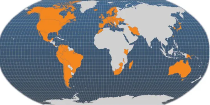
1.1.8 Main producer countries
Main areas where it is practiced aquaculture trout are located in Europe, North America, Chile, Japan and Australia.
Fig. 3 Main producer countries of Oncorhynchus mykiss (FAO Fishery Statistics, 2006).
1.2 Lipid metabolism in liver and adipose tissue
1.2.1 An introduction to lipids
a number of major roles in all organisms. Perhaps most importantly, they are structural components of cell membranes and are essential for energy provision and storage. Lipids and fatty acids, along with proteins, are the major macronutrients for fish (Sargent et al., 2002). Fish seem not to have evolved efficient carbohydrate utilization systems in contrast to terrestrial vertebrates. So, carbohydrates are less important as nutrients (quantitatively) for most fish, suggesting that fish may satisfy structural carbohydrate and storage carbohydrate (glycogen) requirements principally by catabolizing amino acids (Cowey and Walton, 1989). The types of lipids that are most vital to energy metabolism are fatty acids and triglycerides.Fatty acids are the most commonly stored and circulating forms of energy, and triglycerides are the most common non-toxic form of fatty acids. Fatty acids/triglycerides may originate from four sources (pool input): de novo fatty acids synthesis,cytoplasmic triacylglycerol stores, fatty acids derived from triglycerides of lipoprotein remnants directly taken up by the liver, and plasma non-esterifiedfatty acids (NEFA) released by adipose tissue. The relative importance of these sources depends on species differences and on short- and long-term nutritional status and energy balance. Fatty acids and triglycerides may also be used in different ways (pool output). Triglycerides may accumulate in hepatocytes (while NEFA or activated forms of NEFA may not) unless NEFA are oxidized (more or less completely) or triglycerides are exported as constituents of very low density lipoproteins (VLDL).The triacylglycerol content of hepatocytes is regulated by the activity of cellular molecules that facilitates hepatic fatty acid uptake, fatty acid synthesis, and esterification (‘input’) and hepatic fatty acid oxidation and triacylglycerol export (output). Moreover, fatty acids regulate lipid metabolism by binding nuclear receptors that modulate gene transcription. Peroxisome proliferator-activated receptors (PPAR) are ligand binding transcription factors of the nuclear receptor superfamily, which includes receptors for steroids,
thyroids and retinoids (Han et al., 2007; Sertznig et al., 2007). Three types of PPAR have been identified (a, b, ɤ), each encoded by distinct genes and expressed differently in many parts of the body (Sertznig et al., 2007). They form heterodimers with the retinoid X receptor, and these complexes subsequently bind to a specific DNA sequence, the peroxisome proliferating response element (PPRE) that is located in the promoter region of PPAR target genes and modulates their transcription (Tachibana et al., 2008). Gender and stage of life cycle influence expression levels of all the PPARs in brown trout; estrogen appears to play an important role in differential expression of PPARs (Batista-Pinto et al., 2009). In rainbow trout adipose tissue, the gene transcriptional levels of PPARs change differentially with the nutritional status (Cruz-Garcia et al., 2015). As in mammals, it was found in fish that PPARɤ is highly expressed in adipose tissue and acts as a promoter of fat storage and adipocyte differentiation (Bouraoui et al., 2008; Cruz-Garcia et al., 2009; Albalat et al., 2007). In concordance with this, it was seen that insulin administration upregulated this PPAR isotype highly significantly in trout adipose tissue in vivo and in in vitro in line with the pro-lipogenic actions of this hormone (Cruz-Garcia et al., 2015). PPARɤ gene expression increased during adipocyte differentiation in rainbow trout (Bouraoui et al., 2008), but not with differentiation of trout myocytes, in agreement with the myogenic development program (Rescan, 2008). PPARα and PPARβ are regulated by a transcriptional factor LXR (liver X receptor) in trout myocytes (Cruz-Garcia et al., 2011); PPARα is regulated by insulin in trout adipose tissue, not instead PPARβ
(Cruz-in adipogenesis, PPARɤ is an important transcriptional regulator of glucose and lipid metabolism, and is implicated in the regulation of insulin sensitivity, atherosclerosis, and inflammation (Lehrke et al., 2005; Semple et al., 2006). PPARγ affects transcription rates of a variety of lipogenic target genes such as FABP4 (fatty acid binding protein 4), CD36 (thrombospondin receptor), LPL (lipoprotein lipase), leptin, ACC (Acetyl-CoA carboxylase), FAS (fatty acid synthase), and SCD1 (Stearoyl-CoA desaturase 1) (Lee and Hossner, 2002). Additionally, PPARα is responsible for regulating fatty acid β-oxidation (Varga et al., 2011). Regarding PPARβ, little is known about it in the context of target tissues, target genes, lipid homeostasis, and functional overlap with PPARα, and PPARɤ. The activation of PPARβ in skeletal muscle cells programs a cascade of gene expression designed to activate catabolism, and energy expenditure (Dressel, et al., 2003). In adipose tissue, instead, even if PPARβ is not directly implicated in the control of adipogenesis, because alone is not able to promote lipogenesis (but only together with PPARɤ), however, it plays a role in the adaptive response of adipose tissue to dietary fatty acid content, (Neels and Grimaldi, 2014).
Definitely, fatty acids can be produced from acetyl CoA into “de novo” Lipogenesis, and can be broken down to acetyl CoA by β-oxidation, a cyclical pathway. Triglycerides, instead, accounted as the main storage form of fatty acids, are produced through the process known as Lipogenesis or synthesis of TG, while their hydrolysis is known as Lipolysis, with release of fatty acids.
1.2.2 Hormonal regulation of lipid metabolism and its dependence on diet.
Adipose tissue is a specialized organ that functions as one of the major storage sites for fat in the form of triglycerides and provides a buffer for energy imbalances. In mammals, the equilibrium between lipolytic and lipogenic pathways in adipose tissue is influenced by nutritional and endocrine factors
and by components of the immune response (Rosen et al., 2006). There is limited knowledge on the hormonal control of lipid turnover in fish adipose tissue (Albalat et al., 2005; Polakof et al., 2011). It has been seen that in mammals, GH reduces body fat, inhibits adipocyte differentiation, decreases lipogenesis, and increases lipolysis (Herrington et al.,2001; Xu et al., 2009). Also in isolated trout adipocytes, GH enhances lipolysis and inhibits, at least in part, lipogenesis. In the first case, through the direct modulation of HSL activity probably, not at a transcriptional level; in the second, through the down-regulation of FAS expression during fasting (Cruz-Garcia et al., 2015). It has been reported that growth hormone (GH), in addition to being a growth promoter, exerts a lipolytic effect in gilthead sea bream adipocytes too (Albalat et al., 2005; Salmeron et al., 2013). GH transgenic coho salmon (Leggatt et al., 2009) are shown to increase their utilization of lipids for synthetic roles to maintain accelerated growth, but the specific effects in adipose tissue of GH overexpression remain unknown.
Insulin acts as a promoter of carbohydrate and lipid deposition by reservoir tissues during the post-feeding period in fish, although its concentration decreases during fasting periods (Navarro et al., 2004), with an increase in circulating GH (Cruz-Garcia et al., 2015). During fasting, insulin down-regulates PPARα expression, suggesting a reduction in fatty acids oxidation associated with the enhanced use of fatty acids for re-esterification and triglyceride synthesis. Studies have demonstrated the anabolic role of insulin in rainbow trout adipocytes and myocytes, where it stimulates glucose and fatty
Insulin-like growth factor (IGF)-I is structurally and functionally similar to insulin but is more potent as a growth factor and a metabolic controller in rainbow trout and gilthead sea bream myocytes and adipocytes (Salmeròn et al., 2013; Bouraoui et al., 2010; Codina et al., 2008; Castillo et al., 2004). In vertebrates, many of the growth-promoting actions of GH are known to be mediated indirectly through the stimulation of IGF transcription, mainly by the liver, or locally by extra hepatic tissues (Chia, 2014), but the mechanisms of action involved in GH proliferative and metabolic effects in fish are not well known (Bergan et al., 2013).
In contrast, tumor necrosis factor a (TNFα), secreted by adipose tissue, that a part from acts as a pro-inflammatory cytokine, it also regulates lipid cell uptake and degradation of triglycerides because it is lipolytic in fish and mammalian adipose tissue (Saera-Vila et al., 2007; Albalat et al., 2005; Zhang et al., 2002). TNFα has been described as a limiting factor for adiposity in gilthead sea bream (Bouraoui et al., 2008; Saera-Vila et al., 2007), and it has been shown to promote lipolysis in isolated adipocytes of rainbow trout and gilthead sea bream (Albalat et al., 2005; Cruz-Garcia et al., 2009). The mechanisms underlying the action of TNFa on lipolysis are complex, and modulation of the expression of peroxisome proliferator-activated receptors (PPARs) appears to be significant (Cruz-Garcia et al., 2009). In fact, it has been reported by Cruz-Garcia that TNFα decreases PPARɤ expression in adipocytes in line with its anti-fat storage effects and anti-insulin actions described in relation to the lipid metabolism of mammals and fish (Bou et al., 2015; Nieto-Vazquez et al., 2008). Regarding PPARα expression after TNFα treatment in isolated cells, it remains to be elucidated, but points to a possible pathway for the inhibition of fatty acid oxidation in adipose tissue.
The management of fat deposition has become a key area of interest in fish farming in the quest to obtain a high-quality product with good nutritional value
and to maintain fish health. Regarding the growth in fin-fish aquaculture, it has been made possible by the development of artificial diets or feeds formulated to satisfy essential requirements (amino acids, fatty acids, vitamins and minerals, etc.), and provide macronutrients (protein, lipid, carbohydrate) and energy in balance to optimize growth. Thus, it has been the requirements of aquaculture that have driven research into fish nutrition, including lipid and fatty acid metabolism and its regulation. Dietary lipid and fatty acids can have three primary fates in fish. They can be incorporated into cell membranes and hence the flesh of the fish, they can be oxidized to provide energy, or lipid can be deposited in adipose or other tissues as energy storage (Tocher, 2003). Therefore, from an aquaculture perspective, the lipid (fat) content and the fatty acid composition of the diet must be optimized to enable high growth rates, ensure fish health, and, at the same time, maintain the nutritional benefits of fish for the human consumer (Sargent et al., 2002; Tocher, 2003). The lipid content of pelleted diets has increased greatly in recent years due in part to the technical advancements in the production of feed. This increase is driven by the observation that more energy can be supplied by dietary lipid, less protein will be used for energy, and so more dietary protein can be “spared” for synthesis of new tissue/flesh (Hemre and Sandnes, 1999). However, although protein sparing by dietary lipid is widely accepted, the limits to its effectiveness, or the mechanisms by which it might occur, have not been accurately defined for any fish (see Company et al., 1999). Consequently, dietary formulations have maximized lipid content in order to satisfy commercial pressure to increase
fatty acid profile of the diets (Watanabe, 1982), studies have been conducted into the lipid metabolic disorders associated with the content and type of lipids in the diet (Turchini et al., 2009; Benedito-Palos et al., 2008) and the dietary effects on macrophage function and stress susceptibility (Sitjà-Bobadilla et al.,2005;Gjoen et al.,2004). Unfortunately, while in higher vertebrates, many studies have shown that changes in dietary fatty acid composition can induce modification of hepatic lipogenesis (Blake & Clarke,1990; Clarke et al. 1990, Salati & Amir-Ahmady, 2001), lipid transport in blood (Grundy & Denke, 1990; Fernandez & West, 2005) and tissue lipid uptake (Montalto & Bensadoun, 1993; Raclot et al., 1997), a few of them have been done on fishes as on rainbow trout. One of these studies on rainbow trout, conducted by Richard et al., in 2006, has shown that a dietary vegetable oils can have a little effects on hepatic lipogenesis, lipid transport and tissue lipid uptake; in particular, it was seen that hepatic lipogenesis and lipid uptake in perivisceral adipose tissue, white muscle and liver weren’t modified; only regarding the lipid composition of plasma, it was seen a decrease in plasma cholesterol and LDL (low density lipoproteins), because of expression of LDL receptor gene in the liver that was down-regulated.
1.2.3 Fatty acids synthesis and uptake
Two major tissues produce fatty acids in the body: the liver and the adipose tissue. Fatty acids synthesized in the liver are exported through lipoprotein production, and thus provide an energy source and structural components for membrane building. In adipose tissue, de novo synthesis of fatty acid directly contributes to in situ fat deposition and long-term energy storage. Fatty acids synthesis occurs in the cytosol and uses acetyl CoA to build a long chain fatty acid. As shown in fig.4, the first step of fatty acids synthesis consists in a conversion of two-carbon acetyl CoA into a three-carbon malonyl CoA (the substrate of the multiprotein enzyme fatty acid synthase), this thanks to an
enzyme, acetyl CoA carboxylase. Fatty acids synthase uses malonyl CoA (and reducing energy of NADPH) to extend the length of the fatty acid, adding two carbons in a series of four reactions: 1) condensation, 2) reduction, 3) dehydration, 4) reduction. At the end of one cycle (5), the fatty acid shifts to the initial position to allow the next malonyl CoA to bind. After seven cycles, when the fatty acid has grown to 16 carbons, palmitate has been produced; this is a common component of fatty acid stores and from it can be produced fatty acids with longer chains. (Fig. 4).
Besides the synthesis capacity of adipose tissue and liver, these tissues take up NEFA from the blood in proportion to their concentration. NEFA enter cells via transporters (fatty acid transport protein (FATP) or fatty acid translocase (FAT))
However the cells with these fatty acids, rapidly, assimilate them into neutral and polar lipids, and some are oxidized. The result of these metabolic pathways is to keep intracellular NEFA and fatty acyl-CoA very low.
1.2.4 Β-oxidation of fatty acids
The catabolism of fatty acids can occur by β-oxidation in two different organelles in the cells, mitochondria and peroxisomes. Red muscle, liver, and heart are generally regarded as the most important tissues for β-oxidation in fish. The fatty acid oxidation pathway happens primarily into mitochondria and results in the production of acetyl CoA. Depending on conditions, this acetyl-
CoA can enter the TCA cycle or be destined to other pathways. Because the substrate for β-oxidation is fatty acyl CoA, cells must first convert fatty acids to their CoA esters using a fatty acyl CoA synthase. While short and medium chain fatty acids are able to enter the mitochondria directly (where they are activated by a mitochondrial fatty acyl CoA synthase), Palmitate (and others as it) that cannot cross outer mitochondrial membrane, is activated by an extramitochondrial fatty acyl CoA synthase. Because mitochondria aren’t able to import any fatty acyl CoA directly, they use a multienzyme complex ( known
Fig. 5 Fatty acid transport into mitochondria.(Animal Physiolology, Moyes and Schulte,
as carnitine palmitoyl transferase, CPT) and this transport process is called the “carnitine shuttle” (Fig.5). After that fatty acyl CoA has crossed the outer mitochondrial membrane, is converted to fatty acyl carnitine by the enzyme CPT-1 releasing coenzyme A (CoASH). After that fatty acyl carnitine is transported across the inner mitochondrial membrane by the carnitine-acyl carnitine translocase, the enzyme CPT-2
converts it into fatty acyl CoA. This elaborate transport process provides an additional level of control for the oxidation of long chain fatty acids as Palmitate. Once inside the mitochondria, fatty acids enter the β-oxidation pathway (Fig.6). This is a cyclical pathway that sequentially cuts pairs of carbons of fatty acid to form acetyl CoA. Β- oxidation consists of 4 steps:
1. Oxidation 2. Hydration 3. Oxidation 4. Thiolysis
This cycle is repeated until all the acyl CoA of the fatty acid are converted to acetyl CoA.
1. The first step provides a dehydrogenation of the fatty acyl CoA (a loss of hydrogen), forming double bind between carbon 1 and carbon 2 of the molecule; this reaction is catalysed by enzyme acyl CoA dehydrogenase (present on mitochondrial inner membrane) that uses as coenzyme FAD that, in turn, gaining the hydrogen atoms dissociated, become FADH2.
The oxidation reaction is the following:
Fatty acyl CoA acyl CoA dehydrogenase Enoyl CoA + FADH2
2. The second step is a reaction of hydration catalyzed by the enzyme Enoyl CoA hydratase, with the addition of a H2O molecule to double bind
formed to have β-hydroxyl CoA, according to this reaction:
Enoyl CoA β-hydroxyl CoA
H2O
3. The third step is an another oxidation reaction catalyzed by enzyme β-hydroxyacyl dehydrogenase that has as co-factor NAD+; this reaction of dehydrogenation provides a transformation of the hydroxyl group on the C3 into a carbonyl group, with the loss of two hydrogen atoms, one of which is gained by NAD+ that become, in turn, NADH according to the following reaction:
Β-hydroxyl CoA β-hydroxyacyl dehydrogenase β-ketoacyl CoA
4. The last step is a thiolysis reaction that provides a separation of the remaining carbonyl group; it is catalyzed by an enzyme thiolase that uses
as co-enzyme CoASH which, in turn, acts as lytic agent to form an acetyl CoA plus a fatty acyl CoA, following the reaction:
β-ketoacyl CoA thiolase Acyl CoA + Acetyl CoA
CoASH
About 30% of the energy liberated from fatty acids derives from reducing equivalents (FAD and NAD+) produced in β-oxidation. The remaining 70% derives, instead, from oxidation of acetyl CoA in the TCA cycle (citric acid cycle, also known as Krebs cycle). Mitochondrial β-oxidation isn’t the only pathway trough which it happens fatty acids breakdown, but many cells have other pathways that are complementary to the mitochondrial. In the peroxisomes occurs a β-oxidation that processes very long chain fatty acids (more than 22 carbons), which aren’t efficiently oxidized in the mitochondrial pathway; this process occurs through a few cycles, releasing the shortened fatty acids into the cytoplasm where they can be oxidized by mitochondrial β-oxidation. A third pathway of fatty acid oxidation is called ω-oxidation and occurs in the endoplasmic reticulum of liver and kidney. However, this process isn’t important for the production of energy but can used in order to synthetize other metabolites.
lipogenesis have less ability to secrete triacylglycerol from the liver compared with species in which the liver is a major or moderate source of lipogenesis (Pullen et al., 1990). In these species, fatty acids could preferentially be esterified into phospholipids that would be incorporated into membranes, then transferred to pre-high-density lipoprotein particles (Yokoyama, 2006). However, in some cases, the liver can also synthesize triglycerides when high concentrations of NEFA are present and phospholipid transfer to membranes is exceeded. Triglyceride synthesis, or lipogenesis, is a multistep pathway through which from glycerol 3-phosphate (produced from glycolysis), fatty acids (activated into their CoA ester by fatty acyl CoA synthase) are added sequentially to carbon 1 then carbon 2 to form phosphatidic
acid; after removal of the phosphate group, 1,2-diacylglicerol is formed. At the end with the addition of a third fatty acid, triglyceride is formed (Fig.7). Triacylglycerol synthesis is under the control of transcription factors and nuclear receptors such assterol regulatory element binding proteins SREBP-1c, carbohydrate regulatory element binding protein (ChREBP) (Dentin et al., 2006), peroxisome proliferator-activated receptor c (PPARɤ), liver X receptors (LXRs) and their ligands. These play an important role together with hormonal and nutritional regulators, such as insulin, carbohydrate, and fatty acids (Coleman and Lee, 2004).
Fig.7 Triglyceride synthesis. (Animal
1.2.6 Triglyceride export (VLDL synthesis and secretion)
The liver synthetize VLDL (very low density lipoproteins), enclosing inside them a large amount of triglycerides. VLDL are lipoproteins that, like the other (chylomicrons, IDL, LDL, HDL), are constituted of a central core with lipids inside, and an outer shell constituted of one or more apoproteins, phospholipids with polar groups on the outside and free cholesterol; but, differ from the other lipoproteins in lipid composition and apoproteins, which subserve both the lipid transport (allowing them to move in the aqueous smoothly), and the regulation of lipoproteins metabolism, through interaction with plasma enzymes and with specific cellular receptors. In particular, chylomicrons provide the transport of exogenous lipids from the intestine to the tissues; LDL and HDL are characterized by an high content in cholesterol, carrying it into blood; but, while the first give it to the tissues, the latter, instead, remove superfluous cholesterol from pheripheral tissues and transport it to the liver.
VLDL, synthetized into liver, transport endogenous triglycerides from liver to the tissues.Apoprotein B100 (apoB100; and apoB48 in a few species) is the key component whose rate of synthesis in the rough endoplasmic reticulum controls the overall rate ofVLDL production. Lipid components that are synthesized in the smooth endoplasmic reticulum are added by the microsomal triacylglycerol transfer protein to apoprotein B (White et al., 1998). After being carried to the Golgi apparatus in transport vesicles, the apoproteins are glycosylated.
so, firstly, become IDL (intermediate density lipoprotein), then, losing triglycerides again, LDL (low density lipoprotein). There are important species differences in the ability to export triglycerides from the liver as VLDL despite similar rates of esterification of fatty acids to triglycerides. It has been suggested that among different species, the rate of export of triglycerides as VLDL from the liver is proportional to its lipogenic capacity. For example, some animals that don’t synthetize fatty acids in the liver also have low rates of triglycerides export from the liver,with an increased risk of fat accumulation in this tissue; instead, others, in which lipogenesis occurs predominantly in the liver (chicken and fish), or in adipose tissue and liver (rabbits and rats) produce very high/intermediate rates of VLDL (Pullen et al., 1990). The origin of the fatty acids, that constitute triglycerides, can affect the rate of VLDL export. In obese mice, de novo fatty acids synthesis in the liver does not stimulate VLDL production (Wiegman et al., 2003). Rather, plasma exogenous NEFA, seem to play an important role in enhancing hepatic esterification and stimulating VLDL production (Julius, 2003), as it has been seen in rats.
1.2.7 Lipolysis
Triglyceride breakdown, or lipolysis, needs enzymes called lipases that attack triglyceride molecule, breaking the bond between the fatty acid and glycerol. There are two types of lipases, an hormone-sensitive lipase (HSL) that breaks off two fatty acid from triglyceride molecule to form diacylglyceride, and, monocylglyceride lipase that completes the breakdown of triglyceride, releasing the last fatty acid and separating it from glycerol. The liberated fatty acids are either used directly into the cell or introduced into circulation for uptake by other tissues (such as liver or muscle) that can use them for energy metabolism.
1.2.8 Adipose tissue and “Adipogenesis”
The mesenteric adipose tissue is an organ of fat accumulation in fish which, together with the muscle and liver, controls the lipid homeostasis and energetic balance of the animal (Jobling and Johansen, 2003; Sheridan and Kao, 1998), and, at the same time, it also acts as an endocrine organ secreting adipokines which act as potent messengers to distant organs such as the liver and muscle to maintain the body's energy balance (Gregor and Hotamisligil, 2007).
Adipose tissue stores lipids and provides energy from lipid stores. Triglycerides, which come from the diet, are hydrolyzed by lipoprotein lipase (LPL) and the fatty acids released are taken up by the adipocytes and accumulated in droplet form. In response to energy demands, (HSL), after phosphorylation by protein kinase A, can access the lipid droplet and hydrolyze the triglycerides into glycerol and fatty acids (Lafontan and Langin, 2009).In fish as in mammals, the development of adipose tissue is a continuous process which includes the hypertrophy of existing adipocytes and the proliferation of new ones. The training process of new adipocytes from undifferentiated mesenchymal cells is called adipogenesis (fig.8); this occurs thanks to a transcriptional factor PPARɤ (Peroxisome Proliferator Activated Receptor gamma,), that activates other transcriptional factors, and by which, the precursor cells become lipoblast, and then, pre-adipocytes; the latter, then, proliferate and differentiate into adipocytes, which, in turn, accumulate lipid droplets.
Fig.8 Adipogenesis
These processes are known to be affected by diet in mammals, but how dietary changes affect the capacity for enlargement of adipocytes or the differentiation of new ones is poorly understood in fish. For example, it was seen by Cruz-Garcia et al. in a study conducted in 2011 on gilthead sea bream (Sparus Aurata
L.) that fish oil substitution by 66% vegetable oils in a diet results in an increase
of lipolytic activity and adipocyte cell size. Therefore, dietary vegetable oil, causing changes in tissue fatty acids composition, can affect the metabolism of gilthead sea bream adipocytes and the proliferation of new cells which could potentially affect other organs such as the liver. It has been suggested that excessive lipid accumulation in the liver or steatosis is due in part to the increased hepatic uptake of fatty acids released from adipose tissue with enhanced lipolysis (Benedito-Palos et al., 2008).
1.2.9 The adipocyte life cycle
Adipocytes are derived from mesenchymal stem cells, which have the potential to differentiate into myoblasts, chondroblasts, osteoblasts or adipocytes. The adipocyte life cycle includes alteration of cell shape and growth arrest, clonal storage of lipid and finally cell death (Gregoire, 2001) (fig. 9). During the growth phase, preadipocytes resemble fibroblasts morphologically. Pref-1, a
Preadipocytes Mature
adipocytes
Day 0 Day 7 Day 10
preadipocyte-secreted factor serves as a marker for preadipocytes and is extinguished during adipocyte differentiation (Wang, et al., 2006). At confluence, preadipocytes enter a resting phase called growth arrest before undergoing the differentiation process. Two transcription factors, CCAAT/enhancer binding protein (C/EBPα), and peroxisome proliferator-activated receptor PPARγ were shown to be involved in the preadipocyte growth arrest that is required for adipocyte differentiation (Umek, et al., 1991). Following growth arrest, preadipocytes must receive an appropriate combination of mitogenic and adipogenic signals to continue through the subsequent differentiation steps. During the process of differentiation, preadipocytes undergo one round of DNA replication leading to clonal amplification of committed cells (Pairault, et al., 1979). The induction of differentiation also results in drastic change in cell shape as the cells convert from fibroblastic to spherical shape. Following induction, a dramatic decrease in Pref-1 expression accompanies a rapid increase in the expression of C/EBPβ, followed by expression of C/EBPα and PPARγ (Rosen, et al., 2002). During the terminal stages of differentiation, the mRNA levels for enzymes involved in triacylglycerol metabolism like glycerol-3-phosphate dehydrogenase, fatty acid synthase and glyceraldehyde-3-phosphate dehydrogenase, increase to a great extent (Paulauskis, et al., 1988; Spiegelman, et al., 1983). Finally, the total number of adipocytes change throughout life, and new adipocytes can be formed or can be removed by the process of apoptosis.
Fig.9 Mesenchymal stem cells are the precursors of several different types of cells, including myoblasts, chondroblasts, osteoblasts and preadipocytes. Once preadipocytes are triggered to mature, they begin to change shape and undergo a round of cell division known as clonal expansion, followed by initiation of the genetic program that allows them to synthesize and store triglycerides. Mature adipocytes can continue storing lipid when energy intake exceeds output, and they can mobilize and oxidize lipid when energy output exceeds input. Mature adipocytes can also undergo apoptotic cell death under certain conditions.
1.3 Apoptosis and autophagy
Cell death has been subdivided into the categories apoptosis (Type I), autophagic cell death (Type II), and necrosis (Type III). The boundary between Type I and II has never been completely clear and perhaps does not exist due to intrinsic factors among different cell types and the crosstalk among organelles within each type. Apoptosis can begin with autophagy, autophagy can end with apoptosis, and blockage of caspase activity can cause a cell to default to Type II cell death from Type I.
Fig.9 Types of cell deaths. (Richard A. Lockshin and Zahra Zakeri, 2004)
The controlled cells deaths frequently display substantial caspase-independent autophagy or they are predominantly apoptotic. Most apoptotic deaths are caspase-dependent, but there are claims of apoptotic morphology in situations in which caspase activity is equivocal. Caspase activation can occur by means of ligation of a membrane-bound receptor or by means of metabolic changes resulting in depolarization of mitochondria and release of cytochrome c and APAF-1. Other metabolic means of activating apoptosis include UPR (unfolded protein response) that is a cellular stress response related to the “ER (endoplasmatic reticulum) stress; this process is usually activated in response to an accumulation of unfolded or misfolded proteins in the lumen of the endoplasmatic reticulum. If it doesn’t restore the normal function of the cell, the UPR process can lead to apoptosis (Lockshin and Zakeri, 2004). The “Apoptosis” process was described for the first time by Kerr et al., in 1972, who asserted that apoptosis is a mechanism of controlled cell deletion, which appears to play a complementary but opposite role to mitosis in the regulation of animal
histologically by the formation of small, roughly spherical or ovoid cytoplasmic fragments. Electron microscopy shows that the structural changes in apoptosis take place in two discrete stages (Fig. 10) (Kerr et al., 1972): the first comprises the formation of apoptotic bodies, the second their phagocytosis and degradation by other cells. The condensation is presumably a consequence of the extrusion of water, but its mechanism is still unknown. The formation of apoptotic bodies involves marked condensation of both nucleus and cytoplasm, nuclear fragmentation, and separation of protuberances that form on the cell surface (Fig. 10) (Kerr, 1971) to produce many membrane compact, but otherwise well preserved cell remnants of greatly varying size. The initial morphological events have not been identified: apoptotic cells have already condensed and separated from their neighbours, and the nuclear chromatin is aggregated in dense masses beneath the nuclear envelope (Kerr, 1971). Fully developed apoptotic bodies show closely packed organelles, which may themselves be condensed, but which are apparently intact, both chemically (Kerr, 1965, 1967; Ballard and Holt, 1968) and structurally. Lucent cytoplasmic vacuoles and dense masses of nuclear material are seen in some bodies.
The content of an apoptotic body depends on the cellular constituents that happened to be present in the cytoplasmic protuberance that gave rise to it; small bodies thus occasionally consist almost entirely of condensed nuclear chromatin , whereas others are composed only of cytoplasmic elements. It is difficult to determine precisely the time taken for the sequence of events described above, even when augmented by various stimuli, the process appears to start in individual cells of the same organ or tissue at different times. However, examination of the serial changes that take place in several experimental models (Kerr, 1971; Crawford et al., 1972; Wyllie et al., 1972b) suggests that the process is completed fairly rapidly: bodies may form and disappear within 24 hours.
Fig.10 Morphological features of apoptosis. (Kerr et al. 1972).
Currently, the morphology and behaviour of apoptotic cells is largely explained by activation of caspases (cysteinyl aspartate specific proteinase), and apoptosis is considered to be nearly synonymous with caspases activation. Among them, caspase-3 is a frequently activated death protease, catalyzing the specific cleavage of many key cellular proteins. However, the specific requirements of this (or any other) caspase in apoptosis have remained largely unknown until now. Pathways to caspase-3 activation have been identified that are either dependent on or independent of mitochondrial cytochrome c release and caspase-9 function. Caspase-3 is required for some typical hallmarks of
death appears to be a release from inhibition. So, it is assumed that classical apoptosis is a caspase-dependent form of cell death, whether triggered by extrinsic (cell surface receptor) or intrinsic (mitochondrial depolarization) means. So, the vast majority of maturing or mature cells possess the machinery for self-destruction in the form of inactive proenzymes (pro-caspases) as well as machinery for regulating or adjusting the level at which the proenzymes can be activated. Cells normally hold the machinery in abeyance, and default to its activation when any of numerous conditions define an imperfect situation for the cells. Finally, we can say that as an antagonist of cellular proliferation, apoptosis contributes to maintaining homeostasis between cell production and elimination of superfluous cells produced, that elude mechanisms of control of cell development or that undergo genetic damage.
About autophagy process, this is usually defined as a non-selective vacuolar degradative pathway conserved in eukaryotic cells, starting with the formation in the cytoplasm of a multi-membrane-bound compartment named autophagosome. The discovery of autophagy was contemporary to that of lysosomes by de Duve and Wattiaux (de Duve & Wattiaux, 1966). At the same time, the term autophagic cell death or type II programmed cell death (PCD II) was introduced to describe a cell death different from apoptosis or type I programmed cell death (PCD I) (Schweichel & Merker, 1973) and reviewed by Clarke (1990). From these data it appeared that autophagy is a cell response to stress, which under certain circumstances can lead to cell death. However, the precise role of autophagy as a cell-death mechanism remains to be explored (Lipinski, & Degterev, 2003). Autophagy is an evolutionarily conserved lysosomal pathway involved in the turnover of long-lived proteins and organelles (Dunn, 1994; Klionsky & Ohsumi, 1999; Seglen & Bohley, 1992). Autophagy starts with the formation of a multilayer-membrane bound vacuole (autophagosoma), in this step, cytoplasmic constituents, including organelles,
are sequestered by a unique membrane called the phagophore or isolation membrane, which is a very flat organelle like a Golgi cisterna. Complete sequestration by the elongating phagophore results in formation of the autophagosome, which is typically a double-membraned organelle. This step is a simple sequestration, and no degradation occurs. The autophagosomal membrane is derived from a pre-autophagosomal structure of uncertain origin (Mizushima et al., 2001; Suzuki et al., 2001). A first step towards the formation of the autophagosome is the expansion of the pre-autophagosomal membrane. This step is dependent upon signaling molecules that modulate the activity and the expression of some “autophagy genes”. In the next step, autophagosomes fuse with lysosomes (in metazoan cells) or vacuoles (in yeast and plant cells). The inner membrane of the autophagosome and the cytoplasm-derived materials contained in the autophagosome are then degraded by lysosomal/vacuolar hydrolases. These degrading structures are often called “autolysosomes” or “autophagolysosomes”. In mammalian cells, most autophagosomes receive input from endocytic compartments (Stromhaug & Seglen, 1993; Liou, Geuze, Geelen, & Slot, 1997) to form a hybrid organelle called the amphisome (Stromhaug & Seglen, 1993). Studies in rat hepatocytes showed that all lysosomes are able to fuse with autophagic vacuoles to degrade the sequestered material (Fengsrud et al., 1995). In some cases direct fusion of autophagosomes with lysosomes was observed (Lawrence & Brown, 1992). However, once macromolecules have been degraded in the lysosome/vacuole, monomeric units (e.g., amino acids) are exported to the cytosol for reuse; for example, amino
remodeling of the cytoplasm. But autophagy can be also stimulated in response to different situations of stress, such as starvation, change in cell volume, oxidative stress, accumulation of misfolded protein, hormonal signaling, irradiation and xenobiotic treatment. About role of autophagy in adaptation to starvation, it was seen that the breakdown of proteins by autophagy produces amino acids and other elements needed for intermediary metabolism and for biosynthetic pathways. For this, it’s clear that amino acids are regulators of autophagy. Although autophagy is considered to be a non-selective process, selective sequestration of organelles can be observed in various pathophysiological and/or stress situations. Definitively, in addition to diseases as myopathies, neurodegenerative disorders and cancer, where alterations in the autophagic pathway and/or deficiency in autophagy genes are involved, the role of autophagy during physiological processes falls into three categories: (1) autophagy is involved in remodeling during development and differentiation; (2) autophagy is involved in the production of amino acids when nutrients fall short; (3) autophagy is involved in the elimination of unwanted and damaged organelles and molecules, which is likely to be an important function during the adult life span.
Free fatty acids (FFAs) are taken up by hepatocytes and converted into triglycerides (TGs) for storage with cholesterol in lipid droplets (LDs). LD-sequestered TGs continually undergo hydrolysis, generating FFAs that are predominantly re-esterified back into TGs for storage. Nutrient deprivation upregulates TG hydrolysis to supply FFAs for oxidation to meet cellular energy demands. An alternative energy source in times of nutrient scarcity is provided by the breakdown of cellular components by autophagy. In 2009, this new role of autophagy was discovered by a group of researchers (Singh, et al., 2009); they saw that lipid droplets and autophagic components associated during nutrient deprivation, and inhibition of autophagy in cultured hepatocytes and
mouse liver increased triglyceride storage in lipid droplets. So, the regulatory and functional similarities between autophagy and lipolysis, along with the capability of lysosomes to degrade lipids, indicated that autophagy may contribute to LD and TG breakdown.
1.4 The phytochemicals
Phytochemicals are a large group of plant-derived compounds that are commonly found in fruits, vegetables, beans, cereals and plant-based beverages such as tea and wine (Arts & Hollman, 2005). Based on their chemical structure, phytochemicals can principally be categorized into alkaloids, flavonoids, pigments, phenolics, terpenoids, steroids and essential oils.
1.4.1 Flavonoids
Flavonoids are diphenylpropanes that constitute one of the most characteristic classes of secondary metabolites in plants (Cao et al., 1997). It has been shown that flavonoids are potent antioxidants, capable of removing hydroxyl radicals, superoxide anions and lipid peroxy radicals, and have been reported as having antibacterial, anti-inflammatory, antiallergic, antimutagenic, antiviral, antineoplastic, anti-thrombotic and vasodilatory actions (Yao et al., 2004; Chakraborty & Hancz, 2011). The structural components common to these molecules include two benzene rings on either side of a 3-carbon ring, and multiple combinations of hydroxyl groups, sugars, oxygens and methyl groups attached to these structures create the various classes of flavonoids: flavanols, flavanones, flavones, flavan-3-ols (catechins), anthocyanins and isoflavones.
(EDCs) (Ng et al., 2006; Cheshenko et al., 2008). Among Phytochemicals, flavonoids are an important group of endocrine disrupting chemicals. Among them there are genistein and dadzein (isoflavones), often called phytoestrogens, because of their structural resemblance with 17b-oestradiol and estrogenic and/or anti-estrogenic properties. In particular, there are numerous reports of genistein exerting estrogenic effects in fish. For example, Bennetau-Pelissero et al. (2001) reported increased plasma vitellogenin concentrations in male and female fish fed diets containing either 500 or 1000 ppm genistein until spawning. Male fish fed a diet with 500 ppm genistein showed a slight decrease in plasma levels of FSH and LH at the end of spermatogenesis, testicular development was accelerated, and sperm motility and concentration were decreased in a dose-dependent way at spawning. Female fed a diet containing 50ppm of genistein showed decreased FSH and LH levels, delayed spawning and impaired gamete quality. Contrarily, in yellow perch, Perca flavescens, Koet al. (1999) reported that genistein (0.75 and 7.5 mg g 1 diet) haven’t apparent estrogenic effects on reproductive function. This happens because of dual role of genistein as not only acts as an estrogenic agonist, but also as an antagonist blocking estrogen’s action. About mechanism of action of phytoestrogens as endocrine disruptors, it has been seen that phytoestrogens, including isoflavones such as genistein and daidzein, bind weakly to estrogen receptors so that they can produce or inhibit estrogenic effects.
1.5 Genistein: sources, structure and metabolism
Epidemiological data strongly suggested that consumption of plant-based foods rich in isoflavones provides beneficial effects for human health (Ososki and Kennelly, 2003). Thus, much attention has been given to investigating the effects of these compounds, especially genistein, in human physiological and pathological states. Soybean is the main source of isoflavones in the human diet, it contains between 0.6 and 3.8 g isoflavones/kg fresh weight (Cassidy et al.,
2000). The three principal isoflavones found in soy are genistein, daidzein and glycetein, generally in a concentration ratio of 1:1:0.2 (Manach et al., 2004).
Fig.11 Chemical structure of genistein, genistin, daidzein and 17b estradiol and are presentation of genistein metabolism. (Behloul et al., 2013).
The particular similitude of isoflavones to 17β-estradiol, enables them to bind to estrogen receptors and thus can at least partly explain their effects. After ingestion, genistein is released from the glucoside genistin by acid hydrolysis in the stomach or by microflora hydrolysis in the intestine. The resulting aglycone can either be absorbed or further metabolized to specific metabolites (Dihydrogenistein,5-hydroxy-equol) (Matthies et al., 2012). Genistein