1
UNIVERSITÀ DI PISA
Scuola di dottorato in Fisiopatologia Clinica e Scienze del Farmaco
Dottorato di Ricerca in Fisiopatologia Medica e Farmacologia
Direttore Prof. E. Ferrannini
TESI DI DOTTORATO DI RICERCA
2009-2011
“Neurocognitive impairment in patients with Fibromyalgia
and Chronic Fatigue Syndrome”
Relatore
Candidato
2
Index
Page
Abstract 3 Chapter 1: Introduction 5 1 Fibromyalgia 52 Chronic Fatigue Syndrome 7
3Central sensitization 9
4 Neurocognitive impairment 11
5. The peptide β Amyloid 14
Chapter 2: Objectives 16
Chapter 3: Materials and Methods 17
1. Subject 17 2. Measures 17 3. Statistical analysis 22 Chapter 4: Results 23 Chapter 5: Discussion 34 Chapter 6: Conclusions 39 References 40 Acknowledgments 47
3
Abstract
Background: Fibromyalgia (FM) and chronic fatigue syndrome (CFS), two diseases
that are frequently associated, are known to share many symptoms, which include pain - usually more pronounced in FM - and fatigue, together with mood, sleep and neurocognitive disorders. Symptoms as clouded mentation, forgetfulness and difficulty concentrating are very common and contribute to the disability of the disorders. These complaints are termed “fibrofog” and suggest that the central nervous system may be involved in the pathophysiology of the syndroms. Coexisting psychological distress or a psychiatric disorder also may contribute to neurocognitive deficits. Moreover literature data suggest that the peptide β amyloid, major constituent of amyloid plaques, is involved in neurodegenerative and psychiatric disorders as Alzheimer and depression.
Objective: The aim of the study was to investigate neurocognitive disorders in FMS
and CFS patients also examining the influence of many clinical variables (i.e. pain, fatigue, mood and sleep disorders, drug assumption). Secondary objective was to evaluate the levels of β amyloid to better understand neurodegenerative impairments in these syndromes.
Methods: Forty patients with a diagnosis of fibromyalgia and 45 patients with a
diagnosis of chronic fatigue syndrome were consecutively recruited. All patients were asked to complete a set of questionnaires on paper and perform a battery of neurocognitive computerized (CNS Vital Signs©). Subsequently in a subgroup of 25 FMS, 25 CFS patients and 25 healthy subjects we evaluated the levels of beta amyloid (isoforms Aβ 40 and Aβ 42 and their ratio) using a commercial ELISA kit.
Results: Patients with fibromyalgia were compared to chronic fatigue syndrome, and in
the first group female sex was prevalent (97.5% vs 51.1%). Moreover, they were different for duration of illness and pain perception. Although patients from both groups similarly complained about neurocognitive problems of concentration or/and attention, thanks to the use of CNS Vital Signs© test battery we found that fibromyalgia patients
4
resulted to have more compromised neurocognitive function and neurocognitive impairments were found to correlate with pain and illness duration. Concerning beta amyloid no difference in Aβ 40 and Aβ 42 and their ratio was found between patient and control, moreover we did not find any correlation between Aβ40 and Aβ42 levels and cognitive impairment. In the FM group we found a statistically significant negative correlation between Aβ40/42 ratio and disease duration (p=0.0056; r= -0.53) and a statistically significant negative correlation between Aβ40 levels and FIQ values (p=0.037; r=-0.42) and FACIT scores (p=0.0069; r=-0.52). Moreover in FM we found a negative correlation between Aβ42 levels and age (p=0,03, r= -0,43)
Conclusion: In patient affected by fibromyalgia or/and chronic fatigue syndrome,
neurocognitive impairments should never be underestimated, because they could disclose a more severe condition affecting the central nervous system. The cognitive impairment referred by FM and CFS patients and evaluated by CNS vital signs© don’t correlate with the Aβ levels. These observation suggest that the cognitive impairment referred by the patients, was probably related to the health status, instead of a damage of central nervous system.
5
Chapter 1: Introduction
1. Fibromyalgia
Fibromyalgia syndrome (FMS) is a common extra-articular rheumatic disorder characterized by chronic widespread musculoskeletal pain, stiffness and presence of multiple points tenderness to palpation (tender points). FMS affects approximately 2% to 5% of the population and its etiology is uncertain. It is most frequent in females aged 20 to 50 years. Pain is the defining characteristic of FMS, it is usually described as fluctuating and always associated with hyperalgesia and/or allodynia but the disorder is frequently associated with other symptoms, such as stiffness, fatigue, anxiety, depression, mental confusion and sleep disturbances. Moreover patients frequently report diminished cognitive performance such as memory and attention deficit.
FMS varies in severity, however quality of life in FMS patients is reduced both for physical and emotional involvement. Approximately 50% of all patients have difficulty with routine daily activities, while 30-40% have to stop work or change their employment.
The etiology of FM has not yet been fully understood. Several authors (Kindler LL et al, 2011; Yunus MB, 2008); agree on the role of central sensitization as a mechanism of hyperalgesia induction, but the mechanism by which this sensitization occurs is less clear.
According to the 1990 American College of Rheumatology (ACR) classification criteria the diagnosis of fibromyalgia require tenderness on pressure in at least 11 of 18 specified sites and the presence of widespread pain, defined as axial pain, left and right-sided pain, and upper and lower segment pain (Wolfe F et al, 1990).
In the last years, many objections have been expressed in relation to these criteria particularly because tender point count was rarely or incorrectly performed in primary care and the symptoms were not given the right consideration. Furthermore approximately 25% of FMS patients did not satisfy the ACR 1990 classification criteria even though they were considered to have fibromyalgia by their physicians.
6
The previous objections, together with the real need to find a common definition and classification for FMS, led Wolfe et al. in 2010 to propose simple, practical criteria for a clinical diagnosis of fibromyalgia. (Wolfe F et al, 2010)
These new preliminary criteria had been thought to be suitable for use and did not require a tender point examination, providing instead a severity scale for characteristic fibromyalgia symptoms.
The authors identified two variables that best defined fibromyalgia and its symptom spectrum: the widespread pain index and the composite symptom severity scale, a composite variable composed of physician-rated cognitive problems, unrefreshing sleep, fatigue and somatic symptoms.
Furthermore, in 2011, Wolfe published a modification of the ACR preliminary Diagnostic Criteria for Fibromyalgia that allowed their use in epidemiologic and clinical studies without the requirement for an examiner. Practically, the author modified the symptom severity scale by substituting the somatic symptoms item with a 0–3 item that represented the sum of 3 items: headaches, pain or cramps in liver, abdomen or depression symptoms during the previous 6 months. However, it is important to remark that, although simple to use, the new criteria are not thought to be used for self-diagnosis (Wolfe F et al, 2011).
In summary we can say that in recent years the importance of symptoms other than pain has become very important in the assessment of the disease, in particular the presence of cognitive impairment is one of the three symptoms of the symptom severity scale.
7
2. Chronic fatigue syndrome
Fatigue is a subjective feeling of low vitality, common in the community, with up to half of the general population reporting fatigue in large surveys. (Pawlikowska T., 1994) (Bates D., 1993)
Complaints of fatigue are common in major chronic illness diseases (neoplasms, infections, etc) and are a significant component of rheumatologic disorders (Evans EJ., 1999). Typically fatigue is transient and self-limiting, however, a minority of persons experience persistent and debilitating fatigue. When the fatigue cannot be explained by a medical condition it may represent chronic fatigue syndrome (CFS). CFS is a debilitating condition characterized by fatigue which, by exclusion, cannot be ascribed to another illness; it is defined as a persisted or recurrent condition of fatigue, that has lasted at least 6 months, not relieved by rest and resulting in a substantial reduction of previous levels of occupational, educational, social, or personal activities (Fukuda K. et al, 1994). In addition to the chronic fatigue, widespread and persisyent pain is common in individuals with CFS. Other symptoms that are frequently present in CFS are headaches, recurrent sore throats, fever, muscle and joint pain and neurocognitive complaints.
It is difficult to establish the prevalence of CFS, since it depends on the diagnostic criteria used and the study population; initial research suggested a prevalence between 0.002% and 0.04%. However, latest epidemiological studies in the USA and in the United Kingdom show prevalence rates ranging from 0.007% to 2.5% of the general population.(Price R., 1992) (Jason LA., 1999)
Despite more than a decade of research, the etiology and pathophysiology of CFS remains unknown. The onset is often sudden and precipitated by an infection, but in some patients, onset is more insidious and can be preceded by negative, stressful life events. The latter may explain the malfunctioning of the short-term (autonomic nervous system) and long-term (hypothalamus-pituitary-adrenal axis) stress response systems. (Nijs Jo, 2011)
Many theories for the pathophysiology have been suggested, in particular:
- the involvement of a bacterial or viral pathogen as the precipitating event in an immunogenetically vulnerable host
8
- the alterations of the immune system activity characterized by a defict in NK-cell lytic activity and an imbalance in cytokine production (Klimas NG., 1990) (Carlo-Stella N., 2006)
- the involvement of the hypothalamic pituitary adrenal axis having stress as a precipitating factor. (Narita M., 2003)
9
3. Central sensitisation
Concerning studies on fibromyalgia, central sensitization and impaired descending pain modulation are generally accepted as the two major underlying mechanisms causing widespread hypersensitivity to pain (Staud R. and Rodriguez ME., 2006).
Central sensitisation is defined as an increased central neuronal responsiveness. Several mechanism may lead to central sensitisation:
(i) action potential windup, that results from repeated stimulation of afferents nerve fibers in the dorsal root
(ii) expansion of receptor field, that results from activation of neurotransmitter receptor
(iii) hyperexcitable neuronal response, that results from the release of substances that increase neuronal sensitivity (substance P, glutamate, aspartate)
(Dadabhoy D. et al, 2008;)
The proposed neuroplastic changes that result from central sensitization have been visualized in FMS patients with functional magnetic resonance imaging (fMRI). Gracely and colleagues (Gracely RH, 2002) investigated fMRI changes in FMS patients and healthy controls while applying slow, controlled pressure to the thumb nail; FMS patients rated the experience as significantly more painful and demonstrated activation of significantly more pain related brain areas as compared to the controls. These data demonstrated the presence of brain neuroplastic changes.
Furthermore, a PET study performed without noxious stimuli found a significant hyperperfusion in regions of the brain involved in the sensory dimension of pain processing, while hypoperfusion was noted in areas associated with the affective-attentional dimension (Guedj E. et al., 2007).
Moreover FMS is characterized by a dysfunction in descending pain inhibition (Vierck JCJ, 2006). Studies report low levels of the serotonin metabolite 5 hydroxy-indoleacetic acid (5HIAA) in the cerobrospinal fluid of FM patients and an overall dysfunction in serotonergic neurotransmission (Coaccioli S. et al., 2008). Serotonin plays an important role in modulating the descending inhibitory system.
10
Clinically central sensitisation is often manifested as hyperalgesia (exaggerated perception of painful stimuli) and allodynia (a perception of innocuous stimuli as painful) (Lidbeck J., 2002). Tender points are manifestations of allodynia.
A direct evidence of assignment of CSF to a CNS disorder is still scarse even if symptoms like fatigue, non-refreshing sleep, concentration difficulties, impairments in short-term memory, and widespread pain are suggestive of CNS involvement.
In the literature there are evidences that support the importance of widespread pain in CFS and often chronic pain is more disabling than chronic fatigue but only little progress has been made in understanding chronic widespread pain in patients with CFS. There are no direct evidence supporting the central sensitization hypothesis in CFS patients, but the present knowledge is suggestive of a central process similar to that seen in FMS, given the great overlap between the two diseases and the observed similarities. In fact hyperalgesia and lower pain thresholds are reported in patients with CFS and, similar to FM, the lack of peripheral tissue damage and the lack of a distinct localization of the pain complaints are suggestive of a central abnormality responsible for the chronic widespread pain. (Mira Meeus & Jo Nijs, 2007)
11
4. Neurocognitive impairment
FMS and CFS are known to share many symptoms, which include pain - usually more pronounced in FM - and fatigue, together with mood, sleep and neurocognitive disorders.
Symptoms as clouded mentation, forgetfulness and difficulty concentrating affect up to 70% of patents and contribute to the overall disability of the disorders. These complaints are termed “fibrofog”. It is unclear whether this psychological “sensation” of abnormal cognitive ability has related objective changes in neurocognitive function in fact cognitive complaints may mark the presence of truly impaired cognitive functioning or they may represent the patient’s perception of impairment where none exists. (McCracken LM and Iverson GL, 2001). In this context, the evidence suggests chronic pain as the symptom more related to cognitive impairments, although there is little consensus on how pain brings about the observed decrement in cognitive function (Roth RS et al, 2005).
However, clinical observations of self-reported cognitive deficits were confirmed by a study on 100 women with FMS and CFS demonstrating a 95% incidence of concentration difficulties and a 93% incidence of failing memory. Forgetfulness and concentrations problems were the fifth and sixth greatest problems reported after pain, fatigue, muscle tension or stiffness, and sleep problems (Zachrisson O et al, 2002).
Fibromyalgia
There are only a few studies that have specifically investigated neuropsychological issues in patients suffering from FMS and results remain heterogeneous, particularly it is unclear how much of the sensation of abnormal cognition in FM can be attribuited to objective dysfunction.
Some studies have described significant cognitive deficits, mostly in working memory performance (Park DC., 2001, Leavitt F. and Katz RS., 2006); others studies have either failed to detect such differences (Walitt B., 2008) or found that differences between groups disappeared after correcting for fatigue, pain and depression (Suhr JA., 2003)
12
Park et al in 2001 analyzed three small groups of female subjects (23 FM, 23 healthy age-matched controls and 22 older adults) evaluating the following parameters: speed of information processing, working memory function, free recall, recognition memory, verbal fluency and vocabulary. Speed and working memory are the building blocks of cognitive function and predict long-term memory and reasoning. Speed of information processing is measured by how rapidly an individual can make simple mental operations, and working memory is measured by how much information a person can simultaneously store and process-it is an index of the “mental horsepower” that an individual brings to any given situations. Free recall is a measure of long-term memory and the ability to actively retrieve past episodic events. Recognition memory is the ability to recognize a previously studied item. Verbal fluency is the ability to quickly and efficiently retrieve information from their existing knowledge stores. In this study the authors reported memory and vocabulary deficits with intact processing speed in FM patients.
In 2008 Walitt investigated the relations between objective cognitive function in FM using an automated neuropsychological assessment metrics (ANAM). ANAM is a computerized test of neurocognitive function that analyzes information processing, speed, complex attention, working memory and short-term memory. 27 FM patients, 27 healthy controls and 18 muscoloskeletal pain patients were evaluated. No differences between subjects with pain disorder and pain-free controls were found concerning short-term memory, cognitive efficiency, concentration and reaction time and no relation was found between FM symptoms severity and cognitive function. Moreover despite clinical improvement in FM patients after treatment there was no concomitant improvement in ANAM performance.
In contrast some existing data suggest that the influence of psychological variables such as depression, pain and fatigue, must be considered as factors contributing to neuropsychological presentation in FM. Suhr in 2003 suggested that most of the neuropsychological deficits found in FM can be explained by depression and fatigue, particularly depression was significantly related to memory performance and self-reported fatigue was related to psychomotor speed.
13 Chronic Fatigue Syndrome
Cognitive complaints, as poor concentration, decreased memory for recent events and poor word-finding ability, are reported frequently also by CFS patients and these contribute considerably to their social and occupational dysfunction. Three major explanations have been considered in the literature which may contribute to cognitive dysfunctions in CFS:
1) a CNS involvement,
2) effect of subjectively experienced fatigue on performance, 3) effect of psychopatological factors on performance.
These explanations may possible interact. MRI studies demonstrated aspecific abnormalities in the cerebral white matter of CFS patients similar to patients with depression.
In a review of Michiels 2001, the author showed that the most prominent findings in objective cognitive testing in CFS evidenced that information processing speed, learning and working memory are impaired and that CFS patients show intact performance on several aspects of attention and memory function. Moreover the evidence suggests that subjectively experienced fatigue is not related to cognitive performance. (Michiels V. and Cluydts R., 2001)
Certainlymany variables can influence neurocognitive functioning since FMS and CFS are commonly associated with both a number of co-morbidities and treatment factors that can adversely affect cognitive function, for example affective disorders, such as depression, anxiety, and panic, all of which are known risk factors for cognitive disorder among pain conditions (McCracken LM and Iverson GL, 2001; Roth RS et al, 2005).
Moreover, also disturbed sleep and excessive fatigue are known to correlate with cognitive impairment (Cote KA and Moldofsky H, 1997; Suhr AJ, 2003).
Fortunately in the last decade the new technologies opened the way to the development of more sophisticated softwares for the assessment of neurocognitive disorders, thus allowing standardized and more objective evaluations (Gualtieri CT and Johnson LG, 2006)
14
5. The peptide β Amyloid
The peptide β amyloid (Aβ) is the major constituent of amyloid plaques, and originates from the protein Amyloid Precursor Protein (APP). The majority of APP produced is degraded during the transport process on the cell surface. This is indicative of a fine regulation of the activity of this protein. The process of degradation of APP involves three enzymes: α, β and γ-secretase. The last two give way to the so-called "amyloidogenic pathway" that leads to the formation of the two isoforms Aβ 40 and Aβ 42. The literature shows that these two peptides are involved in neurodegenerative and psychiatric disorders. Amyloid-β is normally present in the brain, cerebrospinal fluid and peripheral blood. In patients with Alzheimer’s disease, Aβ 42 aggregates and deposits in the brain, forming senile plaques that are one of the pathological hallmarks of the disease.
In cerebrospinal fluid the concentration of Aβ 42 is reduced in patients with Alzheimer’s disease and in those with the mild cognitive impairment that precedes the disease, suggesting an association with selective deposition of Aβ 42 in the brain. (Andreasen N., 2001); most of the studies reported also an increase of Aβ 40 and Aβ 40/ Aβ 42 ratio.
Some studies showed that patients suffering from mood disorder with multiple affective episodes present a greater risk for developing cognitive deficit and overt demential forms. (Kessing LV., Andersen PK, 2004)
In particular literature data suggest that depression may increase the risk for Alzheimer’s disease; concerning depression, elevated plasma Aβ 42 levels and a lower Aβ 40/Aβ 42 ratio have been reported in patients with late-life depression, on the contrary lower plasma Aβ 42 levels and a higer Aβ 40/ Aβ 42 ratio have been reported in elderly individuals with depression. (Pomara N., 2006).
In a study Baba et al showed a significantly higher serum Aβ 40/Aβ 42 ratio in patients affected by major depressive disease than controls in all age groups (young, middle-aged and elderly) (Baba H., 2012)
In a recent work pubblished by Piccinni et al, the authors underlined that the changes in plasma levels of different Aβ peptides might represent a useful tool to identify the risk for cognitive decline in bipolar patients confirming the presence of lower Aβ 42 levels and higher Aβ 40/ Aβ 42 ratio in patients respect to healthy subjects . They also
15
demonstrated a significant negative correlation between Aβ 42 levels and the duration of illness. (Piccinni A., 2012)
16
Chapter 2 : Objectives
The aim of the study was to assess and compare neurocognitive disorders in FMS and CFS patients, also examining the influence that several clinical variables – i.e. pain, fatigue, mood and sleep disorders, drug assumption - have on the impairment of cognitive functioning, evaluated by means of a standardized and computerized test battery. We also assessed the levels of the two isoforms of Aβ amyloid (40 and 42) and their ratio and correlate this results with cognitive impairment investigated by an objective method.
17
Chapter 3 : Materials and methods
Subjects
40 patients affected by FMS (according ACR 1990 criteria (Wolfe F et al, 1990) and 45 affected by CFS (according to Fukuda criteria of 1994 (Fukuda K. et al, 1994) were consecutively recruited from March 2010 to December 2011. Patients who were at least 18 years old, could read and write Italian, and completed all measures for this study were selected. The Ethical Committee of the Azienda Ospedaliero-Universitaria Pisana approved all recruitment and assessment procedures. Eligible subjects provided written informed consent after receiving a complete description of the study and having an opportunity to ask questions.
Measures
All subjects provided information about demographic and clinical characteristics, medication use, and pain treatment history. Tender Points (TP) count was performed in each participants, and a TP index (TPi) was calculated according to a standardized procedure previously reported (Okifuji A et al, 1997). All patients completed the Fibromyalgia Impact Questionnaire (FIQ) (Burckhardt CS et al, 1991; Sarzi-Puttini P. et al, 2003), in which pain and fatigue severity and level of depression and anxiety were assessed with 10-cm visual analog scales, (0= “better” and 10= “worst”) and the health assessment questionnaire (HAQ) evaluating the quality of life
Fatigue was assessed also by means of the Functional Assessment of Chronic Illness Therapy-Fatigue Scale (FACIT) (Webster K. et al, 2003).
Psychiatric diagnoses were made through the Structured Clinical Interview for the DSM-IV Axis I disorders (SCID-I/P) (First et al 1997), administered by psychiatrists. The following questionnaires were also administered: Self-raiting anxiety scale and the Self-raiting depression scale (SAS and SDS respectively) (Zung WW, 1971; Zung WW, 1965).
Finally, each subject was asked to refer the presence of sleep disorders, auto-referred complaints and the presence of memory and attention impairments following the onset
18
of the disease. Moreover, we considered the VAS from the FIQ asking if the patient felt rested upon awaking during the last week.
All patients also completed the CNS Vital Signs©, a computerized neurocognitive assessment platform that enables the objective evaluation and characterization of patients neurocognitive function. This test battery is well validated and a large amount of scientific papers concerning its use in different diseases have been published. It is comprised of seven tests: verbal and visual memory, finger tapping, symbol digit coding, the Stroop Test, a test of shifting attention and the continuous performance test. The neurocognitive index (NCI) is an average score derived from the domain scores or a general assessment of the overall neurocognitive status of the patient.
These scores are auto‐scored using an algorithm based on a normative data set of 1900+ subjects, ranging from ages 8 – 90, that represents the “average” score (Gualtieri CT and Johnson LG 2006). The test battery lasts approximately 30 minutes. The exercises included in the software are:
Verbal memory (VBM). Fifteen words are presented, one by one, on the screen
every two seconds. For immediate recognition, the participant has to identify those words nested among fifteen new words. Then, after six more tests, there is a delayed recognition trial. This test measures verbal learning, memory for words, word recognition and immediate and delayed recall.
Visual memory (VIM). This test is analogue to the previous, except for the fact that
geometric figures are presented. This test measures visual learning, memory for geometric shapes, geometric shapes recognition, immediate and delayed recall.
Finger tapping test (FTT). This test requires athletes to press the Space Bar with
their right index finger as many times as they can in 10 seconds. They do this once for practice, and then there are three test trials. The test is repeated with the left hand. This test measures motor speed and fine motor control.
Symbol Digit Coding (SDC). This test consists of serial presentations of screens,
each of which contains a bank of eight symbols above and eight empty boxes below. The participant types in the number that corresponds to the symbol that is highlighted. With this test information processing speed, complex attention, visual‐perceptual speed and information processing speed are evaluated.
19
Stroop test (ST). It consists of three parts. In the first part, the words RED,
YELLOW, BLUE, and GREEN (printed in black) appear at random on the screen, and the participant presses the space bar as soon as the athlete sees the word. In the second part, the words RED, YELLOW, BLUE, and GREEN appear on the screen, printed in color. The participant is asked to press the space bar when the color of the word matches what the word says. In the third part, the words RED, YELLOW, BLUE, and GREEN appear on the screen, printed in color. The participant is asked to press the space bar when the color of the word does not match what the word says. With this test it is possible to assess executive function, simple and complex reaction time, speed‐accuracy trade‐off, information processing speed and inhibition/disinhibition.
Shifting Attention (SAT) test. It is a measure of ability to shift from one instruction
set to another quickly and accurately. Participants are instructed to match geometric objects either by shape or by color. Three figures appear on the screen, one on top and two on the bottom. The top figure is either a square or a circle. The bottom figures are a square and a circle. The figures are either red or blue (mixed randomly). The participant is asked to match one of the bottom figures to the top figure. The rules change at random (i.e., match the figures by shape, for another, by color). This test evaluates executive function (shifting sets), reaction time, information processing speed, and speed‐accuracy trade‐off.
Continuous Performance (CPT). It is a measure of vigilance or sustained attention
or attention over time. The athlete is asked to respond to the target stimulus “B” but not to any other letter. The stimuli are presented at random. With this test sustained attention, choice reaction time, and impulsivity are measured.
20
CLINICAL DOMAINS
CLINICAL DOMAIN DESCRIPTION Neurocognitive
Index (NCI)
Measure: An average score derived from the domain scores or a general
assessment of the overall neurocognitive status of the patient.
Relevance: Summary views tend to be most informative when evaluating
a population, a condition category, and outcomes.
Composite Memory
Measure: How well subject can recognize, remember, and retrieve
words and geometric figures.
Relevance: Remembering a scheduled test, recalling an appointment,
taking medications, and attending class.
Verbal Memory
Measure: How well subject can recognize, remember, and retrieve
words. Relevance: Remembering a scheduled test, recalling an appointment, taking medications, and attending class.
Visual Memory
Measure: How well subject can recognize, remember and retrieve
geometric figures.
Relevance: Remembering graphic instructions, navigating, operating
machines, recalling images, and/or remember a calendar of events.
Processing Speed
Measure: How well a subject recognizes and processes information i.e.,
perceiving, attending/responding to incoming information, motor speed, fine motor coordination, and visual‐perceptual ability.
Relevance: Ability to recognize and respond/react i.e., fitness‐to-drive,
occupation issues, possible danger/risk signs or issues with accuracy and detail.
Executive Function
Measure: How well a subject recognizes rules, categories, and manages
or navigates rapid decision making.
Relevance: Ability to sequence tasks and manage multiple tasks
simultaneously as well as tracking and responding to a set of instructions.
Psychomotor Speed
Measure: How well a subject perceives, attends, responds to
visual‐perceptual information, and performs motor speed and fine motor coordination.
Relevance: Ability to perform simple motor skills and dexterity through
cognitive functions i.e., use of precision instruments or tools, performing mental and physical coordination i.e., driving a car, playing a musical instrument.
Reaction Time
Measure: How quickly the subject can react, in milliseconds, to a simple
and increasingly complex direction set.
Relevance: Driving a car, attending to conversation, tracking and
responding to a set of simple instructions, taking longer to decide what response to make.
Complex Attention
Measure: Ability to track and respond to information over lengthy
periods of time and/or perform mental tasks requiring vigilance quickly and accurately. Relevance: Self‐regulation and behavioral control.
Cognitive Flexibility
Measure: How well subject is able to adapt to rapidly changing and
21
information. Relevance: Reasoning, switching tasks, decision‐making, impulse control, strategy formation, attending to conversation.
Table 1 CNS Vital Signs© domains and their descriptions.
Subsequently in a subgroup of 25 FMS and 25 CFS patients the levels of beta amyloid were evaluated using a commercial ELISA kit (Invitrogen Inc) and compared with 25 age and sex matched healthy controls
22 Statistical analysis
The statistical analysis of differences between the two groups was performed using the two-tailed T-test. The correlations between two variables were determined by using linear regressions and Pearson correlations. Also a multivariate analysis was performed in order to adjust comparison for independent variables (age, sex, illness duration and pain perception), that varied between the two groups of patients. The comparison between dichotomous variables was performed by means of the Chi-squared test. In order to establish a coefficient of concordance between neurocognitive disorders complained and established by the CNS Vital Signs©, the contingency analysis was performed. Significance for the results was set at p < 0.05. All statistical analyses were carried out using the Graph Pad Prism 5.0 and SPSS 14.0 softwares
23
Chapter 4 : Results
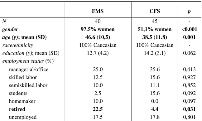
The demographic characteristics of patients recruited are summarized in Table 2. FMS and CFS were significantly different in terms of age and sex (p=0,001 and p=0,0001 respectively), in accordance with the literature (Neumann L. and Buskila D., 2003)
FMS CFS p
N 40 45 -
gender 97.5% women 51,1% women <0.001
age (y); mean (SD) 46.6 (10,5) 38.5 (11.8) 0.001
race/ethnicity 100% Caucasian 100% Caucasian - education (y); mean (SD) 12.7 (4.2) 14.2 (3.1) 0.062 employment status (%) managerial/office 25.0 35.6 0,413 skilled labor 12.5 15.6 0,927 semiskilled labor 10.0 11.1 0,852 students 2.5 15.6 0,092 homemaker 10.0 0.0 0,097 retired 22.5 4.4 0,031 unemployed 17.5 17.8 0,801
Table 2 Demographic characteristics
FMS and CFS patients did not differ by race and years of education but about employment status, a prevalence of retired people is present in FMS group, that depended of the higher mean age. About 18% of both FMS and CFS patients was unemployed and most of them referred to have left work because of the disease.
Concerning co-morbidity we analyzed only the presence of autoimmune thyroiditis and no difference was found in the two groups (27% in FM vs 29% in CFS, p 0,93).
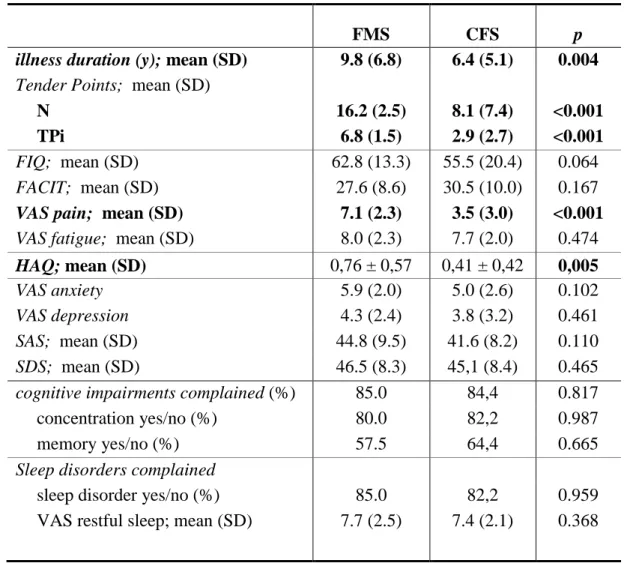
Clinical characteristics of the disease, questionnaire scores and scales are shown in Table 3
24
FMS CFS p
illness duration (y); mean (SD) 9.8 (6.8) 6.4 (5.1) 0.004
Tender Points; mean (SD)
N 16.2 (2.5) 8.1 (7.4) <0.001
TPi 6.8 (1.5) 2.9 (2.7) <0.001
FIQ; mean (SD) 62.8 (13.3) 55.5 (20.4) 0.064 FACIT; mean (SD) 27.6 (8.6) 30.5 (10.0) 0.167
VAS pain; mean (SD) 7.1 (2.3) 3.5 (3.0) <0.001
VAS fatigue; mean (SD) 8.0 (2.3) 7.7 (2.0) 0.474
HAQ; mean (SD) 0,76 ± 0,57 0,41 ± 0,42 0,005
VAS anxiety 5.9 (2.0) 5.0 (2.6) 0.102
VAS depression 4.3 (2.4) 3.8 (3.2) 0.461 SAS; mean (SD) 44.8 (9.5) 41.6 (8.2) 0.110 SDS; mean (SD) 46.5 (8.3) 45,1 (8.4) 0.465 cognitive impairments complained (%) 85.0 84,4 0.817 concentration yes/no (%) 80.0 82,2 0.987
memory yes/no (%) 57.5 64,4 0.665
Sleep disorders complained
sleep disorder yes/no (%) 85.0 82,2 0.959 VAS restful sleep; mean (SD) 7.7 (2.5) 7.4 (2.1) 0.368
Table 3 Clinical characteristics, questionnaire scores and visual analogue scales
FMS patients had a longer duration of illness compared to CFS patients (p=0.004). FMS patients showed a higher number of TP and TP index (p<0.0001) and a higher value of pain (p<0.0001) and it is in agreement with the definitions of the disease.
A significant difference was found about HAQ which is higher in FMS patients (p=0,005) to suggest that they have a poorer quality of life. No difference was found between the two groups with respect to fatigue (evaluated by means of VAS scale and FACIT ).
No statistically significant difference was also found for auto-referred feelings of anxiety and depression (assessed with visual scales and questionnaires SAS and SDS), cognitive impairments complained (concentration and memory) and sleep disorders.
25
No difference was found concerning ANA positivity in the two groups (36,6% in FMS vs 41,3% in CFS, p 0.917).
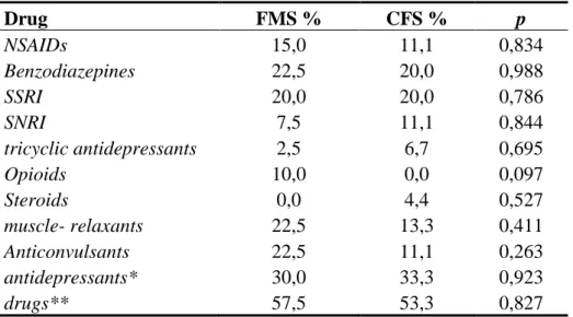
Concerning drug assumption, no statistically significant difference was found between FMS and CFS patients, even if FM patients used more drugs, in particular opioids, than CFS, as shown in Table 4. Drug FMS % CFS % p NSAIDs 15,0 11,1 0,834 Benzodiazepines 22,5 20,0 0,988 SSRI 20,0 20,0 0,786 SNRI 7,5 11,1 0,844 tricyclic antidepressants 2,5 6,7 0,695 Opioids 10,0 0,0 0,097 Steroids 0,0 4,4 0,527 muscle- relaxants 22,5 13,3 0,411 Anticonvulsants 22,5 11,1 0,263 antidepressants* 30,0 33,3 0,923 drugs** 57,5 53,3 0,827
Table 4 Comparison of drug assumption between FMS
and CFS patients (NSAIDs, non steroidal anti-inflammatory drugs; SSRI, selective serotonin reuptake inhibitors; SNRI, serotonin noradrenalin reuptake inhibitors). * SSRI + SNRI + tricyclic antidepressants; ** patients who assumed at least one drug.
Psychiatric comorbidity
No difference was observed between FMS and CFS patients in terms of axis-I psychiatric disorders, assessed by means of SCID-I (Table 5). The prevalent psychiatric condition in both groups was a lifetime (LT) disorder characterized by generalized anxiety (GAD) and panic attacks (PD). Bipolar type-II disorder (BD II) was instead observed in a larger number of CFS patients (17.78%) as a lifetime condition compared to FMS patients (2.50%), although the difference was not statistically significant (p=0.053). Only one CFS patient and none of the FMS patients, still had a BD II disorder at the observation time. Small percentages of patients from both groups
26
resulted to have psychiatric LT or current disorders such as depression (D; 10.00% LT and 5.56% current in FMS versus 8.89% and 2.22% in CFS), LT eating disorders such as anorexia and bulimia (ED; 2.50% in FMS and 4.44% in CFS), and current obsessive-compulsive disorder (OCD; present only in 1 FMS patients, 2.50%).
LT (%) current (%) FMS CFS p FMS CFS p GAD/PD 37.50 20.0 0,12 11,11 6.7 0,817 D 10.00 8.9 0,79 5.56 2.2 0,917 BD II 2.50 17.8 0,053 0.00 2.2 0,953 ED 2.5 4.4 0,92 0.00 0.0 - OCD 2.5 0.0 0,95 2.5 0.0 0,953
Table 5 Lifetime (LT) and current psychiatric
comorbidity assessed by SCID-I (DSM-IV) (GAD, generalized anxiety disorder; PD, panic disorder; D, depression; BD II, bipolar disorder II; ED, eating disorders; OCD, obsessive-compulsive disorder).
Neurocognitive functioning assessment
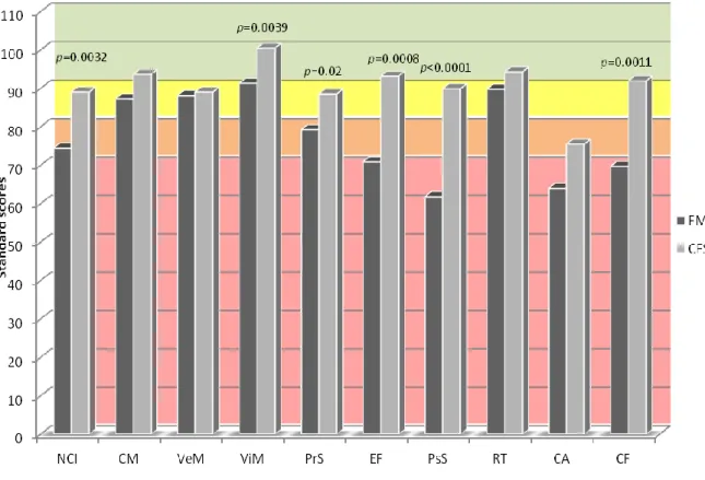
As assessed by CNS Vital Signs© test battery, FMS patients resulted to have more compromised neurocognitive function compared to CFS patients.
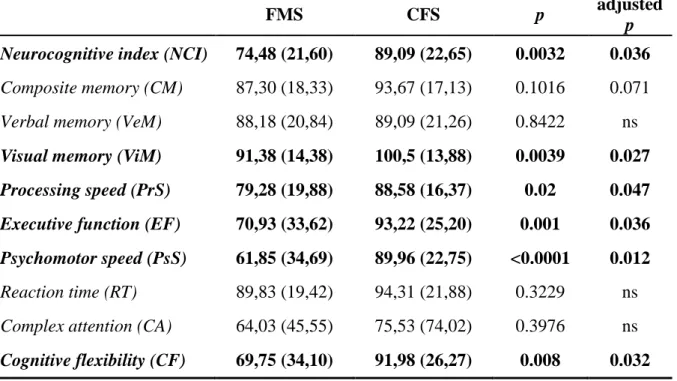
The neurocognitive index (NCI) was lower in FMS than in CFS patients (p=0.0032), particulary single items such as visual memory (ViM; p=0.0039), processing speed (PrS; p=0.02), executive function (EF; p=0.001), psychomotor speed (PsS; p<0.0001) and cognitive flexibility (CF; p=0.0011) were lower in FMS compared to CFS patients. Since FMS and CFS patients differed in terms of age, sex, disease duration and pain perception (VAS pain and TPi), also multivariate analysis was performed in order to adjust results for all possible confounders (table 6) (figure 1).
27
Figure 1 Comparison of CNS Vital Signs© mean standard scores between FMS and CFS patients (standard scores ranges: >110=above; 110-90 (light-green)=average; 89-80 (yellow)=low average; 79-70 (orange)=low; <70 (red)=very low).
28
FMS CFS p adjusted
p
Neurocognitive index (NCI) 74,48 (21,60) 89,09 (22,65) 0.0032 0.036
Composite memory (CM) 87,30 (18,33) 93,67 (17,13) 0.1016 0.071 Verbal memory (VeM) 88,18 (20,84) 89,09 (21,26) 0.8422 ns
Visual memory (ViM) 91,38 (14,38) 100,5 (13,88) 0.0039 0.027
Processing speed (PrS) 79,28 (19,88) 88,58 (16,37) 0.02 0.047
Executive function (EF) 70,93 (33,62) 93,22 (25,20) 0.001 0.036
Psychomotor speed (PsS) 61,85 (34,69) 89,96 (22,75) <0.0001 0.012
Reaction time (RT) 89,83 (19,42) 94,31 (21,88) 0.3229 ns Complex attention (CA) 64,03 (45,55) 75,53 (74,02) 0.3976 ns
Cognitive flexibility (CF) 69,75 (34,10) 91,98 (26,27) 0.008 0.032
Table 6 mean CNS vital signs scores (SD). (ns, not significant).
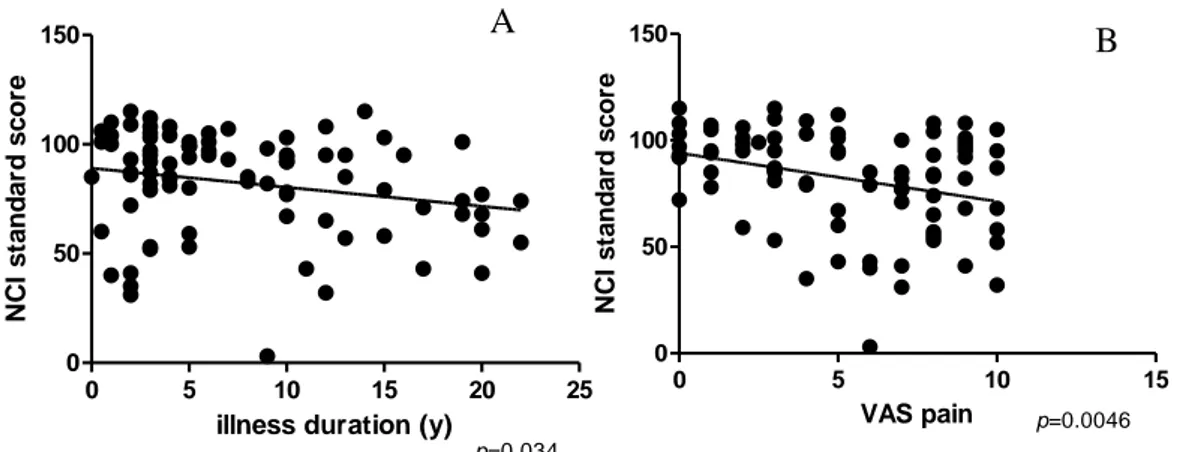
Neurocognitive function correlated with the duration of illness. In particular, duration of illness negatively correlated with NCI (r=-0.23; p=0.034), ViM (r=-0.28; p=0.0099) and PsS (r=-0.45; p<0.0001) (see Figure 2A).
NCI did not correlate with any clinical parameters, although in CFS group EF negatively correlated with FIQ (r=-0.36; p=0.016) and FACIT (r=-0.30; p=0.048), and CF negatively correlated with FIQ (r=-0.318; p=0.035) and FACIT (r=-0.374; p=0.011). In FMS group instead ViM negatively correlated with VAS pain (r=-0.475; p=0.008) and with FIQ (r=-0.374; p=0.042).
29
Figure 2 A) Negative correlation between illness duration and NCI. B) Negative correlation between VAS pain and NCI.
Moreover, considering the two groups together, also VAS pain was found to negatively correlate with NCI standard score (p=0.0046; r=-0.31) and with single items of the CNS vital signs, such as ViM (p=0.0066; 0.30), PrS (p=0.008; 0.29), EF (p=0.01; r=-0.28), PsS (p=0.007; r=-0.30), and CF (p=0.007; r=-0.30) (see Figure 2B). On the contrary, VAS fatigue was not found to correlate with any item of the CNS Vital Signs©. However, when the two groups were considered separately, the significant negative correlation of VAS pain and neurocognitive disorders was lost, both in FMS and in CFS groups. This finding is probably due to the fact that, within each group of patients, VAS pain scores did not spread on the entire scale, while when patients were taken together, we could observe a wider range of VAS pain scores. Furthermore, while in FMS patients VAS pain correlated with VAS fatigue (p=0.003; r=0.47), in CFS patients did not.
Neurocognitive standard scores were also analyzed by comparing both FMS and CFS patients with a psychiatric disorder versus those who had not. In particular, we found statistically significant differences between patients who had or had not a lifetime psychiatric disorder in terms of NCI (mean ± SD: 75.0±25.8 vs 90.3±16.8; p=0.002), PrS (mean ± SD: 80.4±19.1 vs 88.5±17.3; p=0.043), EF (mean ± SD: 76.0±34.6 vs 90.2±25.7; p=0.036), PsS (mean ± SD: 68.2±35.8 vs 86.3±24.3; p=0.008), CF (mean ± SD: 74.1±35.4 vs 89.9±25.7; p=0.022). The more impaired function in patients with a lifetime psychiatric disorder was psychomotor speed, whose mean standard score was
0 5 10 15 20 25
0 50 100 150
illness duration (y)
NC I s ta n d a rd s c o re p=0.034 0 5 10 15 0 50 100 150 VAS pain N C I s ta n d a rd s c o re p=0.0046 A B
30
well under the range of normality (very low score), while the other functions seemed to be less impaired. As for presence of a current psychiatric disorder, no difference was instead observed between patients who had and patients who had not.
Further analysis showed that neurocognitive function, measured with the NCI, also differ between patients assuming or not antidepressants (p=0.015), particularly in terms of complex attention (p=0.008), which decreased well under the normality range in patients taking antidepressants (mean ± SD: 44.07 ± 94.81 and 82.24 ± 33.58 respectively). Considering instead patients under one or more pharmacological treatments versus patients not treated at the observation time, the only difference highlighted was related to processing speed (PrS), which was slightly lower in patients assuming drugs (p=0.04).
Finally we calculated the k-coefficient of concordance between complained (subjective) and objective neurocognitive disorders in both group of patients. We found in both groups a lack of concordance between complained and real deficits, although in CFS the difference resulted more marked, as shown in Table 7.
Table 7 Analysis of complained versus real neurocognitive impairments
FMS CFS
NCD subjective % 85.0% 84.4% NCD objective % 42.5% 22.2% concordant 21/40 15/45 k-coefficient 0.0082 -0.009
31 Beta amyloid
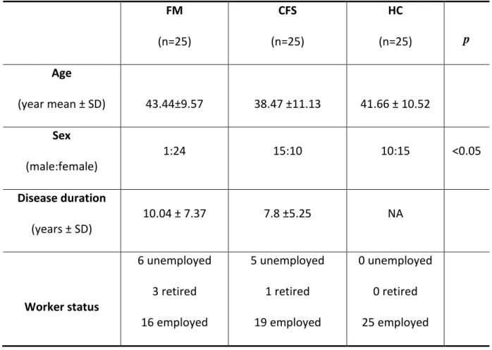
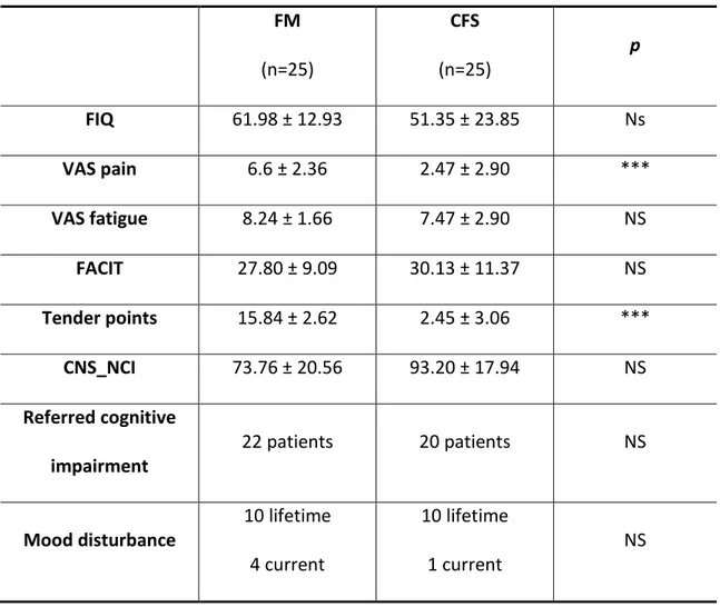
In a subgroup of 25 FMS and 25 CFS patients we analyzed levels of β amyloid (Aβ ) 40 and 42 and then we compared these results with a sample of 25 healthy subjects (HC). Demographic and clinical characteristics of our cohort are showed in table 8 and 9. We observed a difference in gender, with a prevalence of male in CFS group, no differences was found concerning age and onset of disease.
FM (n=25) CFS (n=25) HC (n=25) p Age (year mean ± SD) 43.44±9.57 38.47 ±11.13 41.66 ± 10.52 Sex (male:female) 1:24 15:10 10:15 <0.05 Disease duration (years ± SD) 10.04 ± 7.37 7.8 ±5.25 NA Worker status 6 unemployed 3 retired 16 employed 5 unemployed 1 retired 19 employed 0 unemployed 0 retired 25 employed
Table 8: patients and control demographics characteristics; in the p column we showed
32 FM (n=25) CFS (n=25) p FIQ 61.98 ± 12.93 51.35 ± 23.85 Ns VAS pain 6.6 ± 2.36 2.47 ± 2.90 *** VAS fatigue 8.24 ± 1.66 7.47 ± 2.90 NS FACIT 27.80 ± 9.09 30.13 ± 11.37 NS Tender points 15.84 ± 2.62 2.45 ± 3.06 *** CNS_NCI 73.76 ± 20.56 93.20 ± 17.94 NS Referred cognitive impairment 22 patients 20 patients NS Mood disturbance 10 lifetime 4 current 10 lifetime 1 current NS
Table 9: clinical characteristics of patients and healthy volunteers
The levels of Aβ 40 and Aβ 42 and Aβ 40/ 42 ratio were showed in table 10. We did not observe any difference in Aβ 40, Aβ 42 and their ratio in our population. Moreover we did not find any correlation between Aβ40 and Aβ42 levels and all cognitive domains assessed by CNS vital signs© in patients and controls. No difference in Aβ levels was found concerning psychiatric co-morbidities.
33 A pg/ml) A pg/ml) A A FM 20,35±11,37 4,25±0,72 4,72±2,32 CFS 18,30±11,91 4,25±0,86 4,18±2,37 HC 19,46±13,41 4,40±1,14 4,57±3,19
Table 10: Aβ 40 and 42 and their ratio in patients and controls
FM CFS HC 0 20 40 60 80 A 40 (p g /m l) FM CFS HC 0 2 4 6 8 10 A 42 (p g /m l) FM CF S HC 0 5 10 15 20 A 4 0 /4 2
Fig 1: graph representation of level of Aβ 40, Aβ 42 and Aβ 40/ Aβ 42 in our
population: no significant differences were found
In the FM group we found a statistically significant negative correlation between Aβ40/42 ratio and disease duration (p=0.0056; r= - 0,53). In the FM group we found a statistically significant negative correlation between Aβ40 levels and FIQ values (p=0.037; r=-0.42) and FACIT scores (p=0.0069; r=-0.52). Moreover in FM we found a negative correlation between Aβ42 levels and age (p=0,03, r= -0,43).
34
Chapter 5 : Discussion
Patients with chronic pain often complain of difficulties with cognitive functioning. Chronic pain is commonly associated with both a number of co-morbidities and treatment factors that can adversely affect cognitive function. For example affective distress, depression, anxiety, disturbed sleep and fatigue are known risk factors for cognitive disturbance. Cognitive complaints may mark the presence of truly impaired cognitive functioning or they may represent the patient’s perception of impairment where none exists.
Accumulating evidence indicates that a number of chronic pain and stress-related disorders, including chronic low back pain, FMS and CFS, are characterized by changes in brain morphology, in particular gray matter reductions (Apkarian AV et al, 2004; de Lange FP et al, 2005; Kuchinad A. et al, 2007) and abnormal cerebral perfusion (MacHale SM., 2000). In FMS patients, gray matter loss occurred mainly in regions related to stress (parahippocampal gyrus) and pain processing (cingulated, insular and prefrontal cortices) and the changes in these systems could contribute to the maintenance of pain and appears consistent with cognitive deficit characteristic of FMS (Kuchinad A et al, 2007). Moreover Kuchinad and colleagues (2007) found that FMS patients have brain gray matter atrophy with a yearly decrease in gray matter volume more than three times than that of age-matched controls. Concerning CFS some magnetic resonance imaging (MRI) studies have detected significantly more abnormalities in the subcortical white matter of chronic fatigue syndrome subjects, compared to healthy or trauma comparison subjects (Sullivan PF, 2000), while in other MRI studies the results for subjects with this syndrome did not differ from those for healthy or depressed subjects (Cope H., 1996). In addition, MRI abnormalities have not been associated with neurocognitive performance (Cope H., 1995). Other studies using single photon emission computed tomography (SPECT) scans have found that CFS patients have lower levels of regional cerebral blood flow throughout the brain, compared to healthy subjects. CNS perfusion abnormalities, typically hypoperfusion, also have been found more often on SPECT scans in these patients than in healthy or depressed comparison subjects, although no specific anatomic pattern has emerged and
35
the effect of co-morbid major depression is difficult to ascertain (Ichise M, 1992). Moreover some studies showed that an abnormal cerebral perfusion pattern is present in CFS subjects non depressed and that is it similar but not identical to those in patients with depressive illness. (MacHale SM, 2000). Conversely, a recent rigorously controlled study detected no difference in cerebral blood flow between twins with chronic fatigue syndrome and their healthy co-twins (Lewis D., 2001). Overall, MRI and SPECT studies are generally consistent in demonstrating some abnormalities in chronic fatigue syndrome patients. However, the functional significance and clinical utility of these findings remain uncertain.
The results of the present study showed the importance of chronic pain in neurocognitive impairments in FMS and CFS patients, as resulted by the correlation of neurocognitive index and other CNS Vital Signs© domains with VAS pain and illness duration, as well as by a worse impairment in FMS compared to CFS patients.
No difference was observed between FMS and CFS patients in terms of axis-I psychiatric disorders and thank to these observations we can suppose that, at least in our cohort of patients, neurocognitive functioning should be independent of psychiatric comorbidity.
Unlike other studies (McCracken, 2001; Roth RS, 2005; Suhr JA, 2003) we did not find any correlation between neurocognitive impairment and depression and fatigue (both assessed by VAS) although some neurocognitive items –in particular psychomotor speed- were lower in patients with a lifetime psychiatric disorder compared to patients without. The absence of correlation with depression is probably related to the small number of depressed patient in our cohort.
Moreover, concerning the frequently found prevalence of neurocognitive impairments in female gender - reported by the previously mentioned authors - our data suggested instead that in FMS and CFS patients gender did not influence neurocognition, since the gender-adjusted multivariate analysis still remained significant, and no difference was highlighted by comparing male and female patients within CFS group. However, unlike these studies, in the present one neurocognitive functioning was assessed by means of computerized system, more objective and accurate in evaluating every single ability.
36
We have already shown in Figure 1 how evident is the neurocognitive function impairment in FMS patients, in particular, FMS patients, aside from the NC index, which is only a composite index that globally indicate how compromised are cognitive faculties, reported very low standards score - listed in order of severity- in psychomotor speed, complex attention (low also in CFS), cognitive flexibility, executive functioning, and processing speed (low).
Psycomotor speed means being able to coordinate thinking fast with doing something fast i.e., use of precision instruments or tools, performing mental and physical coordination i.e., driving a car, playing a musical instrument. A deficit in complex attention make instead problematic the performance of mental tasks that require vigilance, quickly and accurately Cognitive flexibility is the ability to switch behavioral response according to the context of the situation, for example an impaired in this function can cause difficulties in reasoning, switching tasks, decision‐making, etc… Executive functioning is the ability to sequence tasks and manage multiple tasks simultaneously, as well as tracking and responding to a set of instructions. Finally, processing speed involves the ability to automatically and fluently perform relatively easy or over-learned cognitive tasks, especially when high mental efficiency is required. Unlike the literature we showed a slow information processing speed in FMS compared to CFS (Glass JM, 2008). These results are obviously insufficient to justify such a discrepancy, nevertheless it is reasonable to emphasize that, despite the other studies, information processing speed was here assessed with a computerized procedure which calculated the scores coming from several different exercises (i.e. symbol digit coding, Stroop and shifting attention tests).
Of particular interest are the negative correlations of EF, CF and ViM with FIQ, FACIT and VAS scales whose values increase together with disease severity. CFS patients who had a more severe disease (higher FIQ and FACIT scores) also had more impaired executive function and cognitive flexibility, confirming the possibility of a dysfunction at prefrontal level, as reported by neuroimaging studies (MacHale SM et al, 2000). FMS patients who had more severe disease (higher FIQ and VAS pain) reported instead poor performance in terms of visual memory, suggesting the possible involvement of
37
brain areas such as medial temporal lobe and occipital cortex in the pathophysiology of the syndrome (Khan ZU et al, 2011).
Globally considering the reported findings, in FMS patients attention and concentration generally seemed more compromised than memory and it is in accordance with the recent literature in fact several writers have invoked the role of disturbed attentional process to explain the adverse impact of chronic pain on cognitive function. (Glass JM, 2006).
It is important to underline that complex attention was found reduced by the use of antidepressants (SSRI, SNRI and/or tricyclic compounds), as previously reported also by McCracken and colleagues (2001). Processing speed instead seemed to be more impaired in patients who were assuming at least one drug among those which are normally prescribed for the treatment of FMS and CFS. Further investigations on the relationship between neurocognitive disorders and drugs commonly prescribed for the treatment of FMS - and also on the link among the various affective disorders - could be useful to understand if the impairment of neurocognitive function could be at least partially ascribed to the use of some of these ones. However that fact that no difference was found in pharmacological therapies between FMS and CFS patients let us suppose that the neurocognitive impairment in FMS is not due to assumed drugs.
It is interesting that FMS patients were more compromised but less frequent aware of their cognition problems on the contrary CFS patients usually complained without having a real deficit. A possible explanation of the this observation could be related to personality traits of CFS patients, such as self-criticism and perfectionism (Kempke S. et al, 2011), which can lead to excessive worries about their performance (e.g., concern over mistakes and doubt about actions).
Another important aspect highlighted by the present research is the one concerning the relation between β amyloid levels and cognitive impairment. Actually, there are not validated biomarkers of these diseases, in particular for FMS which is a common disease, (Bazzichi L., 2010) and many researchers tried to found a putative biomarker.(Ang DC, 2011; Heidari B., 2010; Bazzichi L, 2010). Since literature data showed a relation between β amyloid levels and cognitive deficit (Kessing L.V.,
38
Andersen PK, 2004), the second aim of this study was to evaluate for the first time the levels of these peptides in FMS and CFS patients in order to find a possible correlation with objective cognitive impairment evaluated by a standardized methods. In the literature Aβ peptides was found altered in many conditions, such as mild cognitive impairment (MCI) and Alzheimer disease (AD) (Graff-Radford NR, 2007). No data are available in literature concerning the levels of these peptides in FMS and CFS patients. The present study showed a normal levels of Aβ in the serum of FMS and CFS patients. The cognitive impairment referred by FMS and CFS patients don’t correlate with the Aβ levels. Also the cognitive impairment evaluated by CNS vital signs, don’t correlate with the amyloid peptides. These observations suggest that the cognitive impairment referred by the patients, was probably related to the health status, instead of a damage of central nervous system. In contrast with literature, we did not find any differences in patients concerning psychiatric comorbidities, in particular depression. These observation is probably related to the small number of depressed patient in our cohort (only 5 patients).
The observation of normal levels in Ab42 suggest a non-improvement FMS related in AD development. However the correlation between Ab40 levels and disease duration, FIQ and FACIT, suggest a possible alteration in Amyloid metabolism, related to Fibromyalgia and this observation might suggest a relationship with chronic pain. Further studies are necessary to confirm these data, in particular to understand the CNS structures involved in FM.
39
Chapter 6 : Conclusions
The main findings of the present work have been:
1) the more elevated prevalence of neurocognitive disorders in FM than in CFS patients, despite the more frequent complaint of the latter;
2) the tight relation between neurocognitive impairments and chronic pain, which is independent of psychiatric comorbidity.
3) the lack of correlation between cognitive impairment and Aβ levels
4) the possible alteration in Amyloid metabolism in FMS related in particular to disease duration, FIQ and FACIT
Concerning neurocognitive impairments in FMS patients compared to CFS patients, we demonstrated that this kind of disorder - in particular attention and concentration deficits - is prevalent in FMS patients and it is mainly related to chronic pain conditions. Moreover, CFS patients seemed more frequently complained about memory and attention difficulties without having a severe impairment.
However, in these diseases, it is important not to underestimate attention and memory disorders complained by patients, because they contribute to the disability of the disorders and could disclose a more severe condition affecting the CNS, including a decreased gray matter density.
40
References
Andreasen N, Minthon I, Davidsson P, et al. Evaluation of CFS-tau and CFS-Aβ42 as diagnostic markers for Alzheimer disease in clinical practice. Arch Neurol, 2001;58:373-379.
Ang DC, Moore MN, Hilligoss J, Tabbey R. MCP-1 and IL-8 as pain biomarkers in fibromyalgia: a pilot study. Pain Med. 2011 Aug;12(8):1154-61.
Apkarian AV, Sosa Y, Sonty S, Levy RM, Harden RN, Parrish TB, Gitelman DR. Chronic back pain is associated with decreased prefrontal and thalamic gray matter density. J Neurosci 2004;24:10410 –10415.
Baba H, Nakano Y, Maeshima H, Satomura E, Kita Y, Suzuki T, Arai H. J Clin Psychiatry 2012; 73 (1):115-120.
Bates D, Schmitt W, Buchwald D et al.: Prevalence of fatigue and chronic fatigue syndrome in a primary care practice. Arch Intern Med 1993; 153:2759–2765
Bazzichi L, Rossi A, Giacomelli C, Bombardieri S. Exploring the abyss of fibromyalgia biomarkers. Clin Exp Rheumatol. 2010 Nov-Dec;28 (6 Suppl 63) :S125-30.
Bazzichi L, Ciregia F, Giusti L, Baldini C, Giannaccini G, Giacomelli C, Sernissi F, Bombardieri S, Lucacchini A. Detection of potential markers of primary fibromyalgia syndrome in human saliva. Proteomics Clin Appl. 2009 Nov;3(11):1296-304.
Bennett R. Fibromyalgia: From present to future. Current Rheumatology Reports. 2005;7(5), 371–376.;
Burckhardt CS, Clark SR, Bennett RM: The fibromyalgia impact questionnaire: development and validation. J Rheumatol 1991, 18:728-33.
Carlo-Stella N, Badulli C, De Silvestri A, Bazzichi L, Martinetti M, Lorusso L, et al. A first study of cytokine genomic polymorphisms in CFS: Positive association of TNF-857 and IFNgamma 874 rare alleles. Clin Exp Rheumatol 2006; 24: 179-82.
41
Coaccioli S, Varrassi G, Sabatini C, Marinangeli F, Giuliani M, Puxeddu A. Fibromyalgia: Nosography and therapeutic perspectives. Pain Practice 2008;8(3):190– 201.
Cope H, David AS: Neuroimaging in chronic fatigue syndrome. J Neurol Neurosurg Psychiatry 1996; 60:471–473
Cope H, Pernet A, Kendall B: Cognitive functioning and magnetic resonance imaging in chronic fatigue. Br J Psychiatry 1995; 176:86–94
Cote KA and Moldofsky H. Sleep, daytime symptoms, and cognitive performance in patients with fibromyalgia. J Rheumatol 1997;24:2014-23.
Dadabhoy D, Crofford LJ, Spaeth M, Russell IJ, and Clauw DJ. Biology and therapy of fibromyalgia. Evidence-based biomarkers for fibromyalgia syndrome. Arthritis Research & Therapy. 2008;10(4), 211.
De Lange FP, Kalkman JS, Bleijenberg G, Hagoort P, van der Meer JW, Toni I. Gray matter volume reduction in the chronic fatigue syndrome. NeuroImage. 2005;26:777– 781.
Evans EJ. Subjective fatigue and self-care in individuals with chronic illness. Nursing 1999; 8: 363-71
First et al 1997. In M.B. First, R.L. Spitzer, M. Gibbon and J.B.W. Williams, Editors, Structured Clinical Interview for DSM-IV Axis I Disorders-Patient Edition (SCID-I/P, Version 2.0, 4/97 revision) (1997) Biometrics Research Department, New York State Psychiatric Institute.
Fukuda K, Straus SE, Hickie I, Sharpe MC, Dobbins JG, Komaroff A. The chronic fatigue syndrome: a comprehensive approach to its definition and study. International Chronic Fatigue Syndrome Study Group. Ann Intern Med. 1994 Dec 15;121(12):953-9. Glass JM. Cognitive dysfunction in fibromyalgia and chronic fatigue syndrome: new trends and future directions. Curr Rheumatol Rep. 2006 Dec;8(6):425-9. Review.
