The polyphenols resveratrol and epigallocatechin-3-gallate restore the
severe impairment of mitochondria in hippocampal progenitor cells
from a Down syndrome mouse model
Daniela Valenti
a,⁎
,1, Lidia de Bari
a,1, Domenico de Rasmo
a, Anna Signorile
b, Alexandra Henrion-Caude
c,d,
Andrea Contestabile
e, Rosa Anna Vacca
a,⁎
a
Institute of Biomembranes and Bioenergetics, National Council of Research, Bari, Italy
b
Department of Basic Medical Sciences, Neuroscience and Sense Organs, University of Bari, Italy
c
INSERM UMR1163, Université Paris Descartes, Institut Imagine, Paris, France
dHôpital Necker-Enfants Malades, Paris, France e
Neuroscience and Brain Technologies Department, Istituto Italiano di Tecnologia, Genova, Italy
a b s t r a c t
a r t i c l e i n f o
Article history:
Received 23 December 2015
Received in revised form 26 February 2016 Accepted 4 March 2016
Available online 7 March 2016
Mitochondrial dysfunctions critically impair nervous system development and are potentially involved in the pathogenesis of various neurodevelopmental disorders, including Down syndrome (DS), the most common ge-netic cause of intellectual disability. Previous studies from our group demonstrated impaired mitochondrial ac-tivity in peripheral cells from DS subjects and the efficacy of epigallocatechin-3-gallate (EGCG) – a natural polyphenol major component of green tea– to counteract the mitochondrial energy deficit. In this study, to gain insight into the possible role of mitochondria in DS intellectual disability, mitochondrial functions were an-alyzed in neural progenitor cells (NPCs) isolated from the hippocampus of Ts65Dn mice, a widely used model of DS which recapitulates many major brain structural and functional phenotypes of the syndrome, including im-paired hippocampal neurogenesis. We found that, during NPC proliferation, mitochondrial bioenergetics and mi-tochondrial biogenic program were strongly compromised in Ts65Dn cells, but not associated with free radical accumulation. These data point to a central role of mitochondrial dysfunction as an inherent feature of DS and not as a consequence of cell oxidative stress. Further, we disclose that, besides EGCG, also the natural polyphenol resveratrol, which displays a neuroprotective action in various human diseases but never tested in DS, restores oxidative phosphorylation efficiency and mitochondrial biogenesis, and improves proliferation of NPCs. These ef-fects were associated with the activation of PGC-1α/Sirt1/AMPK axis by both polyphenols. This research paves the way for using nutraceuticals as a potential therapeutic tool in preventing or managing some energy deficit-associated DS clinical manifestations.
© 2016 Elsevier B.V. All rights reserved.
Keywords: Down syndrome Mitochondrial bioenergetics Mitochondrial biogenesis Nutraceuticals PGC-1α AMPK 1. Introduction
Down syndrome is a genetic disorder caused by trisomy of chro-mosome 21. Mild to severe intellectual disabilities were observed in DS patients for whom the extra chromosome 21 causes various neuroanatomical abnormalities, such as reduction in brain size and weight[1]. Likewise, abnormal neuronal density and distribution were documented using induced pluripotent stem cell-derived DS neurons[2].
To analyze the mechanisms involved in DS-associated intellectual disability and the neuronal development impairment in DS, over the past decades, several trisomic DS mouse models have been generat-ed, recapitulating the essential genetic and cognitive deficits of human DS, as reviewed in [3]. The most widely used model is Ts65Dn mouse carrying a partial triplication of mouse chromosome 16, the portion that is analogous to 21q21–21q22.3. The triplication
Abbreviations: AA, antimycin; AICAR, 5-aminoimidazole-4-carboxamide-1-b-D -ribofuranoside; AMPK, 5′ AMP-activated protein kinase; Ap5A, P1
,P5
-Di(adenosine-5′) pentaphosphate; ASC, ascorbate; BrdU, bromodeoxyuridine; CC, Compund C; CN−, cyanide; DCF, dichlorofluorescein; DCFH-DA, 2′,7′-dichlorofluorescein diacetate; DS, Down syndrome; EGCG, epigallocatechin-3-gallate; FCCP, carbonyl cyanide p-trifluoromethoxyphenylhydrazone; G6P-DH, glucose 6 phosphate dehydrogenase; GAPDH, glyceraldeide 3-phosphate dehydrogenase; gDNA, genomic DNA; GLU/MAL, glu-tamate plus malate; HK, hexokinase; HOVA, homovanillic acid; MRC, mitochondrial respi-ratory chain; mtDNA, mitochondrial DNA; NPCs, neural progenitor cells; NRF-1, nuclear respiratory factor 1; OXPHOS, oxidative phosphorylation; OLIGO, oligomycin; PGC-1α, peroxisome proliferator-activated receptor-gamma coactivator 1 alpha; POX, horseradish peroxidase; ROS, reactive oxygen species; ROS-ds, ROS-detecting system; ROT, rotenone; RSV, resveratrol; SOD, superoxide dismutase; SUCC, succinate; T-FAM, mitochondrial tran-scription factor A; TMPD, N,N,N′,N′-tetramethyl-p-phenylenediamine; wt, wild type.
⁎ Corresponding authors at: Institute of Biomembranes and Bioenergetics, National Council of Research, 70126 Bari, Italy.
E-mail addresses:[email protected](D. Valenti),[email protected](R.A. Vacca).
1
Equallyfirst authors.
http://dx.doi.org/10.1016/j.bbadis.2016.03.003
0925-4439/© 2016 Elsevier B.V. All rights reserved.
Contents lists available atScienceDirect
Biochimica et Biophysica Acta
of these genes reproduces many of the cognitive deficits associated with pediatric development and later Alzheimer disease pathology associated with the syndrome[4]. Reduced adult neurogenesis has been shown to mainly depend on impaired Ts65Dn proliferation of neural progenitor cells (NPCs) in the Ts65Dn hippocampal dentate gyrus[5]. NPCs have self-renewal and proliferative abilities and are capable of differentiating into neurons and astrocytes, and thus are widely used to study neurogenesis in vitro[6]. Although defective GABAergic signaling has been proposed to contribute, at least par-tially, to the overall impairment of cognitive functions in Ts65Dn mice[7,8], decreased neurogenesis is also believed to play a role in DS phenotype[5]. However, the underlying mechanism involved in defective proliferation of Ts65Dn neuronal precursors cells still re-mains unknown.
Brain mitochondria, by generating energy and regulating subcellular calcium and redox homoeostasis, are essential for neural development processes including self-renewal and differentiation of neural stem cells, axonal and dendritic growth, and synaptic formation and reorga-nization[9]. Therefore, dysfunctional mitochondria and alterations in energy metabolism negatively affect neural precursor proliferation and neuronal development[10–12].
While the maintenance of mitochondrial bioenergetics seems re-quired for NPC proliferation and neurogenesis in mouse[13,14], there is no information on the possible link between mitochondrial function and defective neurogenesis in DS mouse models. However, we have previously known that mitochondrial dysfunction has a cru-cial role in human peripheral DS cells[15,16]. In the present study, we explored whether and how mitochondrial functions are also affected in hippocampal NPCs obtained from Ts65Dn mice and whether mitochondrial targeting molecules would improve NPC proliferation. Therefore, we tested in this in vitro model of neurogenesis, the effect of two polyphenols the epigallocatechin-3-gallate (EGCG) and resveratrol (RSV), natural pharmacological tools known to improve mitochondrial functions in a variety of neu-ronal cell types and diseases[17–23].
EGCG, a green tea catechine, has been extensively studied as inter-esting drug candidate for DS treatment. This polyphenol appears to res-cue brain functions and improve some cognitive phenotypes in Ts65Dn mouse model and in adults with DS[24]. We have demonstrated in human DS cell cultures the efficacy of EGCG in counteracting oxidative stress and mitochondrial energy deficit[25]and recently reported that a dietary supplementation of EGCG plusfish oil omega-3 in a DS child is safe, rescues mitochondrial dysfunction and improves some behavior-al deficits[26]. Although many modalities of action have been recently suggested for this catechine (reviewed in Ref.[17]), the mechanistic basis underlying its effects in DS is not completely established at the molecular level.
RSV, a natural polyphenolic compound found in a wide variety of plant species, induces expression of genes involved in mitochondri-al biogenesis, oxidative phosphorylation and endogenous antioxi-dant defense by modulation of cell signaling pathways that control cell homeostasis[27,28]. Recently, neuroprotective efficacy of RSV against prenatal stress induced impaired postnatal hippocampal neurogenesis has been reported[29]. Although thousands research paper and review articles have been published related to its phar-macological activities in cardiovascular, inflammation, cancer and neurological diseases (for recent reviews see[30,31]), no study has been performed on the effect of this polyphenol in Down syndrome.
The results of the present work show that both EGCG and RSV reverse the severe impairment of mitochondrial bioenergetics and biogenesis in Ts65Dn-derived hippocampal progenitor cells rescuing the in vitro impaired neurogenesis. This is likely linked to the activation of the PGC-1α/Sirt1/AMPK axis suggesting that besides EGCG, a potential beneficial action of resveratrol for treat-ment in DS.
2. Materials and methods 2.1. Adult hippocampal NPC cultures
The NPC cell lines were previously isolated from the dentate gyrus of adult (6–8 weeks) Ts65Dn mice, carrying a partial trisomy of chromo-some 16 or wild type (wt) littermates[5].
Adult NPC cell lines were obtained from pooled dentate gyrus tissue obtained from 11 to 12 mice (mixed male and female) for each geno-type. Cells were cultured as a monolayer on poly-D-lysine (PDL; Sigma-Aldrich) and laminin-coated (Roche)flasks in Neurobasal medi-um containing 2% B27 (minus vit A), 1% GlutaMAX, and 1% penicillin– streptomycin solution (all from Invitrogen) supplemented with recom-binant FGF-2 and EGF (20 ng/ml, PeproTech), as previously described [32]. NPCs were passaged at 70–80% confluence by harvesting with Accutase (PAA Laboratories) and re-plating at 104cells/cm2. After initial isolation, NPCs were expanded for 3–4 passages and aliquots of cell sus-pension in medium containing 10% DMSO, were kept frozen in liquid ni-trogen, as previously described[33]. All cells were positive for the NPC markers nestin and Sox2[5]. All experiments were done using cells ob-tained after 2–3 passages following thawing. For all experiments, cells were grown for 48 h in medium containing 2 ng/ml FGF-2 and EGF be-fore analysis. Cell cultures were kept in a 5% CO2humidified atmosphere at 37 °C.
2.2. EGCG and RSV treatments
EGCG– extract from green tea leaves with a purity N95% (Sigma-Al-drich)– and resveratrol from grape skin N 99% (Sigma-Aldrich) were prepared at 100 mM concentration in DMSO and stocked in aliquots at −20 °C. For each experiment, NPCs, cultured for 48 h in medium con-taining 2 ng/ml FGF-2 and EGF, were added with EGCG and RSV, freshly diluted in cell culture medium at a concentration of 20μM and 10 μM, respectively, and incubated for 24 h. In both wt and NPC non-treated cells an equal volume of DMSO was added (vehicle).
2.3. Measurement of oxygen consumption in digitonin-permeabilized NPCs Oxygen consumption measurements were carried out using a Gilson 5/6 oxygraph with a Clark electrode, essentially as reported in[15]. NPCs (1 mg protein) were pre-incubated with digitonin (0.01% w/v) at 37 °C for 5 min, in 1.5 ml of the respiration medium consisting of 210 mM mannitol, 70 mM sucrose, 3 mM MgCl2, 20 mM Tris/HCl, 5 mM KH2PO4/K2HPO4, (pH 7.4) plus 5 mg/ml BSA. The oxygen con-sumption was monitored in the presence of the sequential addition of the respiratory substrates glutamate plus malate (GLU/MAL, 5 mM each), succinate (SUCC, 5 mM) and ascorbate (ASC, 5 mM) plus N,N,N′,N′-tetramethyl-p-phenylenediamine (TMPD, 0.5 mM) under uncoupled conditions i.e. in the presence of carbonyl cyanide p-trifluoromethoxyphenylhydrazone (FCCP, 1.25 μM). Where indi-cated, rotenone (ROT, 3μM), antimycin (AA, 2 μM) and cyanide (CN−, 0.3 mM) were added to inhibit the complex I, III and IV activ-ities, respectively. The sensitivity of the instrument was set to allow the measurement of oxygen consumption as low as 0.2 natO/min. The rate of oxygen consumption, obtained from the tangent to the initial part of the progress curve, was expressed as natO/min × mg cell protein.
2.4. Measurement of mitochondrial ATP production rate in NPCs
The rate of ATP production by OXPHOS was determined in digitonin-permeabilized cells, essentially, as previously described[15]. Briefly, NPCs (0.3–0.5 mg protein) were incubated at 37 °C in 2 ml of respiratory medium pH 7.4 in the presence of the ATP detecting system consisting of glucose (2.5 mM), hexokinase (HK, 2 e.u.), glucose 6-phosphate de-hydrogenase (G6P-DH, 1 e.u.) and NADP+(0.25 mM) in the presence
of GLU plus MAL (5 mM each) or SUCC (5 mM) plus ROT (3μM), as energy sources, plus 10μM diadenosine pentaphosphate (Ap5A), used to specif-ically inhibit adenylate kinase. After 5 min of incubation with digitonin (0.01% w/v), ADP (0.5 mM) was added to start the reaction and the reduc-tion of NADP+in the extra-mitochondrial phase was monitored as an in-crease in absorbance at 340 nm. As a control, the ATP synthase inhibitor oligomycin (OLIGO, 5μg/10 μl) was added in course of reaction to show the inhibition of the mitochondrial ATP production. Care was taken to use enough HK/G6P-DH coupled enzymes to ensure a non-limiting ADP-regenerating system for the measurement of ATP production. 2.5. Measurement of ATP andL-lactate content in NPCs
NPCs were detached from plate, washed with PBS and cellular ATP was extracted by using the boiling water procedure, as described in Ref.[34]. The amount of intracellular ATP was determined enzymatical-ly in the extracts, as described in Ref.[15].
L-lactate concentration was measured in culture medium by using
theL-lactate dehydrogenase method that gives a reliable estimate ofL -lactate production inside the cells[35].
2.6. NPC proliferation assay
For the proliferation assay, NPCs were dissociated and plated at a den-sity of 0.5 × 10⁵cells/ml in 96-well plates. Cell proliferation was deter-mined by the amount of incorporated bromodeoxyuridine (BrdU) using a labeling and detection kit (Millipore) according to the manufacturer's instructions. The extent of BrdU intake was determined by immunostain-ing for BrdU,fixing the cells, denaturing the DNA and measuring the absorbance at the measure-reference wavelengths of 450–595 nm, re-spectively using a spectrometric plate reader. To ensure validity of the ex-periment, for each time point, wells with only the culture media (no cells) and cells without BrdU label were included in the assay.
2.7. Measurement of mitochondrial respiratory chain complex activities Measurements of MRC complex activities were carried out in mito-chondrial membrane-enriched fractions obtained from NPCs. For isola-tion of mitochondrial membrane-enriched fracisola-tions, pellets obtained by NPCs werefirst frozen at −80 °C, then thawed at 2–4 °C, suspended in 1 ml of 10 mM TRIS–HCl (pH 7.5) plus 1 mg/ml BSA and exposed to ultrasound energy, as described in Ref.[25]. Measurement of MRC com-plex activities were performed, essentially as in Ref.[36], by three assays which rely on the sequential addition of reagents to measure the activ-ities of: i) NADH:ubiquinone oxidoreductase (complex I) followed by ATP synthase (complex V), ii) succinate:ubiquinone oxidoreductase (complex II) and iii) cytochrome c oxidase (complex IV) followed by cy-tochrome c oxidoreductase (complex III).
2.8. Reactive oxygen species (ROS) production in NPCs
Production of superoxide anion and hydrogen peroxide by mito-chondria was measured (without discriminating between them) as H2O2production rate in the presence of endogenous and exogenous (70 e.u.) superoxide dismutase (SOD). H2O2production rate was mea-sured using homovanillic acid (HOVA, 200μM) and horseradish perox-idase (POX, 8 e.u.) forming a fluorescent dimer monitored at the excitation/emission wavelengths of 312/420 nm. In each experiment the arbitraryfluorescence units were converted to amounts of H2O2 by measuring the increase influorescence after the addition of known amounts of H2O2in the presence of POX, HOVA and SOD (ROS-detecting system, ROS-ds)[37]. NPCs were detached from plate, washed with PBS and 1.3 mg of digitonin-permeabilized cells were incubated at 37 °C in a final volume of 2 ml of assay medium consisting of 145 mM KCl, 30 mM Hepes–Tris, 5 mM KH2PO4, 3 mM MgCl2, 0.1 mM EGTA, and 0.1%
fatty-acid-free albumin (pH 7.4). The mitochondrial production of H2O2was detected after the addition of the respiratory substrates glutamate/ma-late (GLU/MAL, 5 mM each). The rate of H2O2generation was obtained from the tangent to the progress curve and expressed as pmol of H2O2 formed/min × mg of mitochondrial proteins.
Quantitative analysis of intracellular ROS levels was performed by means of LS50 Perkin Elmer spectrofluorimeter using 2′,7′-dichloro-fluorescein diacetate (DCFH-DA), a non-fluorescent dye which is hydro-lyzed in cells and reacts with multiple types of ROS, mainly H2O2, to give thefluorescent product, dichlorofluorescein (DCF)[38]. Cultured cells were incubated with 5μM DCFH-DA for 30 min under growth condi-tions, washed and suspended in PBS. Fluorescence increase was record-ed at excitation-emission wavelengths of 488–520, respectively, and normalized to the protein content to determine the relative cell ROS amount.
2.9. Immunoblot analysis
NPCs were lysed with 0.1% Triton in PBS in the presence of a protease inhibitor cocktail (Sigma-Aldrich). Cell lysate (0.05 mg protein) was re-solved by a 10–12%-SDS-NuPAGE Bis/Tris gel (Life Technologies), de-pending on the molecular weight, and transferred to a polyvinylidene difluoride membrane (Millipore). Membranes were blocked by TBS-T (50 mM Tris, 150 mM NaCl, 0.02% Tween 20, pH 7.5) containing 3% BSA and probed with the following primary antibodies overnight at 4 °C: MitoProfile OXPHOS cocktail (1:1000 dilution, MitoSciences); anti-PGC-1α (1:500, Calbiochem); anti-NRF-1 (1:200, Santa Cruz); T-FAM (1:200, Abcam); porin (1:1000, MitoSciences); anti-Sirt1 (1:500, Cell Signaling); anti-hystone H3 (1:10,000, Millipore); anti-Acetyl-Hystone H3 (1:500, Millipore); anti-AMPK (1:1000, Cell Signaling); and anti P-AMPK (Thr 172) (1:1000, Cell Signaling). Immunoblot analysis was performed, using horseradish peroxidase-conjugated anti-mouse or anti-rabbit antibodies and enhanced chemi-luminescence western blotting reagents (Amersham, Pharmacia Bio-tech). Membranes were also probed with anti-actin antibody (1:2000 dilution, Sigma Aldrich) as internal loading control and densitometry value of immunoreactive bands for each sample was normalized versus the corresponding densitometry value of actin.
2.10. Quantitative analysis of mtDNA content
Total genomic DNA was extracted from NPCs using the NucleoSpin kit (Macherey-Nagel) and quantitative real-time PCR reactions were performed by using the Applied Biosystems™ 7900HT and the SYBR-Green PCR Master Mix (Qiagen). Triplicate reactions were performed using primers for mitochondrial DNA (mtDNA) sequence (forward: 5 ′-CCGCAAGGGAAAGATGAAAGA-3′; reverse: 5′-TCGTTTGGTTTCGGGGTT TC-3′) and for the genomic DNA (gDNA) sequence of glyceraldeide 3-phosphate dehydrogenase (GAPDH. Forward: 5′-GAACATCATCCCTGCA TCCA-3′; reverse: 5′-CCAGTGAGCTTCCCGTTCA-3′) housekeeping gene, as described in Ref.[15].
2.11. Statistical analysis
All data are expressed as mean ± standard deviation (SD). Statistical evaluation of the differential analysis between groups was performed by one-way ANOVA and Bonferroni post hoc test or Student's t test as ap-propriate. The threshold for statistical significance was set at P b 0.05. 3. Results
3.1. Mitochondrial bioenergetics is impaired in NPCs isolated from the hip-pocampus of Ts65Dn mice
To investigate the bioenergetics status in NPCs isolated from hippo-campus of Ts65Dn, we functionally evaluated both mitochondrial
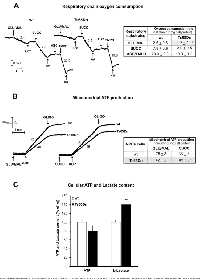
Fig. 1. Impairment of mitochondrial bioenergetics and reduced cell energy status in Ts65Dn NPCs. Measurements of mitochondrial respiratory chain oxygen consumption (A), ATP production rate via OXPHOS (B) and cellular ATP content andL-lactate level (C) were carried out in digitonin-permeabilized (A, B) or cell extracts (C) of wild type (wt) and Ts65Dn NPCs. (A) Representative oxygen consumption traces with rates expressed as natO/min × mg cell protein. NPCs (1 mg cell protein) were added with the uncoupler FCCP (1.25μM) and subsequently GLU/MAL (5 mM each), ROT (3μM), SUCC (5 mM), AA (2 μg/10 μl), ASC (5 mM), TMPD (0.5 mM) and CN−(0.5 mM) were added to cell suspension. Statistical
analysis is reported in the table as mean oxygen consumption rates ± SD of four independent experiments on wt and Ts65Dn NPCs. (B) Representative mitochondrial ATP production traces. NPCs (0.3 mg of cell protein) were added with the ATP detecting system plus 10μM Ap5A in the presence of the respiratory substrates GLU/MAL (5 mM each) or SUCC (5 mM) plus ROT (3μM). Where indicated, ADP (0.5 mM) was added. Numbers along curves are rates of ATP production expressed as nmol/min × mg cell protein. Statistical analysis is reported in the table as mean ATP production rates ± SD of four independent experiments on wt and Ts65Dn NPCs. (C) Cellular ATP content andL-lactate level in Ts65Dn are expressed as a percentage of wt NPCs. Data are reported as the mean values ± SD from three independent experiments carried out on wt and Ts65Dn cell lines. Asterisks indicate significant differences between wt and Ts65Dn samples calculated with Student's t test (* = P b 0.05; ** = P b 0.01).
respiration (Fig. 1A) and ATP production via OXPHOS (Fig. 1B) in permeabilized neuronal progenitors from Ts65Dn with respect to wild type (wt) cells.
As shown by typical experimental traces reported inFig. 1A and the statistical analysis of data in the related table, respiration rate induced by the addition of complex I substrates GLU/MAL to Ts65Dn NPCs was 52 ± 4% lower than that found in wt NPCs. As expected, the respiration rate was blocked in both cell types by complex I inhibitor ROT. On the contrary, the rates of oxygen consumption measured in the presence of the complex II substrate SUCC and of complex IV substrate pair ASC/ TMPD were comparable to those measured in wt. As expected, AA and CN−efficiently inhibited complex III and complex IV, respectively.
Consistently with the results shown inFig. 1A, a 44 ± 4% decrease in the rate of mitochondrial ATP synthesis was found when complex I sub-strates GLU plus MAL were used as energy sources (Fig. 1B and table in-side). Notable, differently from SUCC-dependent respiration (Fig. 1A), the rate of SUCC-dependent mitochondrial ATP synthesis was reduced by 30 ± 3% in Ts65Dn with respect to wt NPCs, suggesting also an im-pairment of the mitochondrial ATP synthesis machinery downstream of complex II.
We then sought whether the impairment of mitochondrial ATP pro-duction found in Ts65Dn NPC influenced cell energy status by measur-ing the cellular ATP pool (Fig. 1C). ATP content was slightly but significantly reduced in Ts65Dn cells (20 ± 5%, P b 0.05) with respect to wt. Consistently with a compensatory enhancement of glycolysis, the level ofL-lactate was higher in Ts65Dn (40 ± 8%, Pb 0.01) than in
wt (Fig. 1C), suggesting that, despite the shift toward glycolysis occur-ring in Ts65Dn to meet the cellular demand for ATP, the OXPHOS-dependent energy deficit remains not fully compensated.
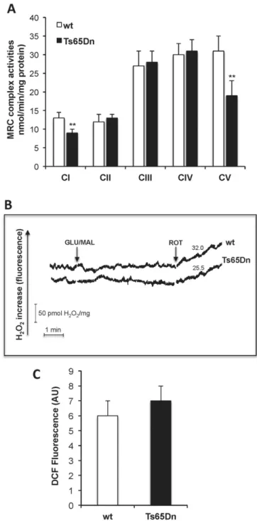
For further investigation, all MRC complex activities were then mea-sured in Ts65Dn and compared to wt NPCs (Fig. 2A). Both complex I and ATP synthase (complex V) activities were significantly inhibited in Ts65Dn with respect to control values (36 ± 4%, and 40 ± 8% of inhibi-tion, respectively, Pb 0.01), whereas no significant difference was de-tected in the activity of complexes II, III and IV.
Since complex I is a well-established source of ROS[16,39], we sought whether complex I impairment in Ts65Dn led to a change in ROS production. We directly monitored mitochondrial ROS production in permeabilized NPCs following the addition of the complex I respirato-ry substrates GLU and MAL. Interestingly, as a result of three separate experiments, in spite of complex I activity deficit, no detectable complex I-dependent ROS production by mitochondria was found in Ts65Dn cells as well as in wt, as shown by typical experimental traces reported inFig. 2B. As a control, the addition of ROT, inhibiting the ubiquinone binding site of complex I, induced, as expected, GLU/MAL-dependent ROS production by mitochondria in both wt and Ts65Dn cells at compa-rable rates. In parallel, intracellularfluorescence analysis revealed that cellular ROS levels, measured by DCF, was not significantly different in Ts65Dn NPCs from that observed in wt cells (Fig. 2C, PN 0.05), and, as wt cells, mitochondrial ROS were below detection when using the mito-chondrialfluorescent probe MitoSox (not shown), indicating the ab-sence of ROS overproduction in Ts65Dn cells during NPC proliferation. 3.2. Polyphenol treatment restores mitochondrial OXPHOS capacity and cell energy deficit and enhances proliferation of Ts65Dn NPC cells
To test the potential of polyphenols EGCG and RSV to improve the impaired OXPHOS capacity in Ts65Dn cells, we measured MRC complex activities and mitochondrial ATP production by respiratory substrates in NPCs cultured in the presence of either 20μM EGCG, 10 μM RSV or vehi-cle, for 24 h. Both EGCG and RSV concentrations were chosen according to preliminary tryouts showing neither cytotoxicity nor ROS production after 24 h treatment with 20μM of EGCG (as in Ref.[40]) and 10μM RSV (as in Ref.[41]) of both wt and Ts65Dn cultured NPCs (data not shown). As shown inFig. 3A, exposure of Ts65Dn cells to either EGCG or RSV completely prevented the specific impairment of mitochondrial
Fig. 2. Impairment of complex I and ATP synthase activities in Ts65Dn NPCs is not associated to ROS overproduction. (A) MRC complex activities. The activities of complex I (NADH:ubiquinone oxidoreductase), complex II (succinate:ubiquinone oxidoreductase), complex III (cytochrome c reductase), complex IV (cytochrome c oxidase) and complex V (ATP synthase) were measured photometrically in mitochondrial membrane-enriched fractions (0.1–0.3 mg protein) from NPCs. Data are reported as the mean values ± SD from four independent experiments carried out on wt and Ts65Dn NPCs. Asterisks indicate significant differences between wt and Ts65Dn calculated with Student's t test (** = Pb 0.01). (B) Representative traces of H2O2production rate in digitonin-permeabilized NPCs isolated from
Ts65Dn and wt mice expressed as pmol/min/mg of cellular protein. NPCs (1.3 mg cell protein) were incubated at 37 °C in 2 ml of ROS-ds (for details seeMaterials and methods) and the reaction was started by the addition of the respiratory substrates GLU/MAL (5 mM each). When indicated, ROT (5μg/10 μl) was added to the cell suspension during the reaction. (C) Quantitative analysis of intracellular radical production carried out as reported in Material and Methods section and expressed in arbitrary units (AU) of DCFfluorescence at λex 488 nm and λem 520 nm wavelengths. Histograms represent the mean values ± SD from three independent experiments carried out on wt and Ts65Dn NPCs.
complex I and ATP synthase (complex V) activities in Ts65Dn cells (Fig. 3A) with no effect on complex II. No significant differences in these activities were found between untreated and treated wt cells
with the polyphenols (not shown). Interestingly, both EGCG and RSV treatments significantly prevented the impairment of mitochondrial ATP production via OXPHOS (Fig. 3B) and conferred to Ts65Dn cells
Fig. 3. EGCG and RSV rescue the deficit of MRC complex activities, mitochondrial ATP synthesis and cell energy, and enhance proliferation of Ts65Dn NPC cells. Ts65Dn NPCs were incubated in the absence (Ts65Dn) or presence of either 20μM EGCG (Ts65Dn + EGCG) or 10 μM RSV (Ts65Dn + RSV) for 24 h. (A) MRC complex activities. The activity of the complex I (CI), complex II (CII) and ATP synthase (CV) were measured spectrophotometrically in mitochondrial membrane enriched fractions (0.1 mg protein) and expressed as percentage of wt. (B) Mitochondrial ATP production. The rate of mitochondrial ATP production via OXPHOS was measured in wt and Ts65Dn samples as inFig. 1B. Data are mean rates ± SD obtained from three independent experiments, expressed as a percentage of wt NPCs. (C) Cellular ATP content andL-lactate level. The mean values ± SD obtained from three independent experiments are reported as a percentage of wt NPCs. (D) BrdU incorporation in NPCs. Results are mean values of optic density (OD) ± SD at the measure-reference wavelengths of 450–595 nm, respectively, obtained from three independent ELISA assay experiments carried out on wt and Ts65Dn NPCs, incubated in the absence or presence of EGCG or RSV. Significant differences, calculated with one-way ANOVA and Bonferroni test, are indicated as follow: wt NPCs vs. Ts65Dn NPCs, α = P b 0.05; αα = P b 0.01; Ts65Dn NPCs vs. Ts65Dn NPCs treated with polyphenols, * = Pb 0.05; ** = P b 0.01.
the capability to maintain the intracellular basal ATP content compara-ble to that of wt cells (Fig. 3C).
To investigate the relative contribution of both EGCG and RSV on tri-somic cell proliferation in vitro, we determined NPC proliferative capac-ity in untreated and EGCG- or RSV-treated Ts65Dn NPCs by BrdU incorporation experiments. As shown inFig. 3D, Ts65Dn NPC cells showed a 37 ± 13% reduction of proliferation as compared to wt cul-tures, in agreement with[5]. Both EGCG and RV treatment for 24 h in-duced a significant increase of proliferation capacity in Ts65Dn cells similar to the level seen in wt cells (Fig. 3D).
3.3. Mitochondrial biogenesis is impaired in Ts65Dn NPCs. EGCG and RSV modulate PGC-1α and restore mtDNA content
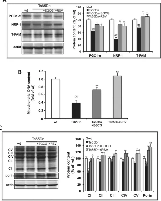
Control of mitochondrial energetic functions can be attained through the regulation of a number of transcriptional factors and cofac-tors among which the peroxisome proliferator-activated receptor-γ co-activator (PGC)-1α[42,43], the master regulator of mitochondrial biogenesis[44]. Analysis of the protein levels of the PGC-1α, and the tar-get genes, the nuclear respiratory factor 1 (NRF-1) and the mitochondri-al transcription factor A (T-FAM)[45]by immunoblotting, revealed a
Fig. 4. EGCG and RSV activate mitochondrial biogenesis in Ts65Dn NPCs. Ts65Dn NPCs were incubated in the absence (Ts65Dn) or presence of either EGCG (Ts65Dn + EGCG) or RSV (Ts65Dn + RSV) for 24 h. (A) Representative immunoblot and densitomentric analysis of protein levels of PGC-1α, NRF-1 and T-FAM measured in cell extracts (0.05 mg protein). Data are mean values (± SD) of three independent experiments on wt, Ts65Dn, Ts65Dn + EGCG and Ts65Dn + RSV NPCs. Results of Ts65Dn samples are expressed as percentage of untreated wt. (B) Real-time PCR analysis of mtDNA content. The mtDNA marker (COX II) and a genome DNA marker (GAPDH) were used. The relative amplification of mtDNA markers in Ts65Dn samples versus wt NPCs was calculated upon normalization to the reference GAPDH as described inMaterials and methods. Values are the mean ± S.E. of three independent experiments. (C) Representative immunoblot and densitomentric analysis of protein levels of 20-kDa subunit of complex I (CI), 30-kDa subunit of complex II (CII), core 2 protein of complex III (CIII), COX I of complex IV (CIV) andα subunit of F1 ATPase (CV) in cell extract (0.05 mg protein); protein levels of porin and β-actin were also analyzed as mitochondrial and cytosolic protein markers, respectively. Data are mean values (±SD) of three independent experiments on Ts65Dn, Ts65Dn + EGCG, Ts65Dn + RSV and wt NPCs, expressed as percentage of wt. Significant differences, calculated with one-way ANOVA and Bonferroni test, are indicated as follow: wt NPCs vs. Ts65Dn NPCs, α = P b 0.05; αα = Pb 0.01; Ts65Dn NPCs vs. Ts65Dn NPCs treated with polyphenols, * = P b 0.05; ** = P b 0.01
decrease of all three transcriptional factors in Ts65Dn as compared to wt cells (Fig. 4A). Consistently, a strong decrease in relative mtDNA content (mtDNA/gDNA) measured by real-time PCR (about 2.5-fold) was found in Ts65Dn with respect to wt (Fig. 4B). These results demonstrate that an impairment of mitochondrial biogenesis pathway also occurred in Ts65Dn NPCs, which could account for the decreased OXPHOS efficiency.
Remarkably, both EGCG and RSV treatments of Ts65Dn cells induced a significant enhancement of mtDNA restoring the wt-like levels, partic-ularly in RSV-treated cells (Fig. 4B). In addition the amount of some mi-tochondrial proteins found reduced in Ts65Dn cells, such as porin and some OXPHOS subunits, were found significantly increased in Ts65Dn cells treated with both polyphenols (Fig. 4C).
We then verified whether the promoting effect of EGCG and RSV on mitochondrial biogenesis in Ts65Dn NPCs was the consequence of the activation of PGC-1α, as in other studies[46,47]. Both EGCG and RSV treatments induced not only a slight increase of the PGC-1α protein content but also a strong transcriptional up-regulation of its down-stream target proteins NRF-1 and T-FAM (Fig. 4A), therefore suggesting a possible effect of the polyphenols on the PGC-1α activity at a post-translational level rather than through its expression.
3.4. Polyphenol treatment restore mitochondrial OXPHOS capacity though modulation of Sirt1 and AMPK activities
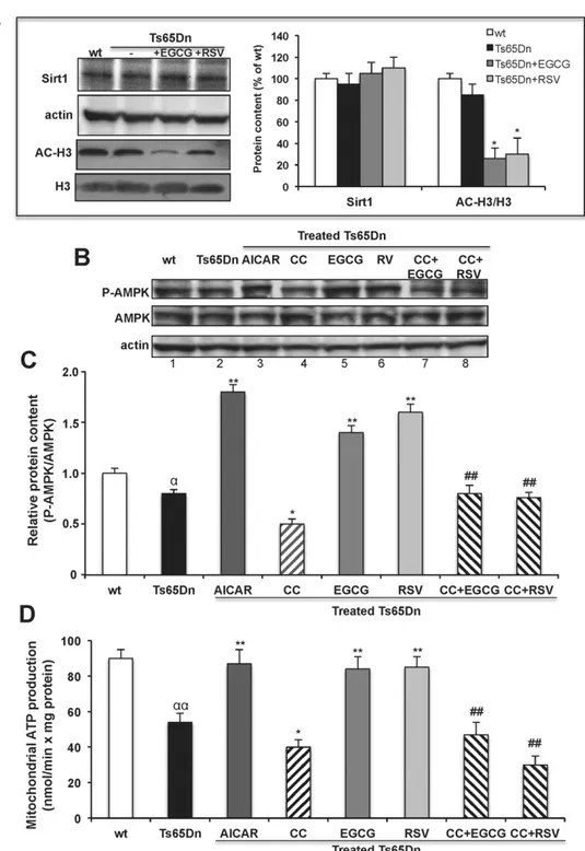
Post-translational modifications which regulate PGC-1α activity consist mainly in Sirt1-mediated deacetylation and AMPK-mediated phosphorylation[48]. The low level of PGC-1α protein in NPCs did not allow us to evaluate directly the acetylation state of the protein. The ef-fect of EGCG and RSV on Sirt1 activity was then assessed by monitoring the acetylated state of the histone 3 (H3), one of Sirt1 main downstream target, as reported in[25,49]. No significant differences in Sirt1 protein levels among untreated and polyphenol-treated NPC cells and not sig-nificant acetylation of H3 was found in basal DS cells respect to wt (Fig. 5A). However, both EGCG and RSV treatment induced a significant strong decrease of acetylated H3 content in Ts65Dn cells compared to the untreated cells (Fig. 5A), indicating an increase of the Sirt1 deacetylase activity after polyphenol treatment of Ts65Dn neuronal progenitor cells.
In addition to Sirt1 activation, both EGCG and RSV have also been shown to target 5′ AMP-activated protein kinase (AMPK)[50,51]. Whether there are changes in AMPK pathway and the effect of these polyphenols on AMPK phosphorylation is completely unknown in Ts65Dn and in DS in general.
Therefore, we monitored the Thr172-phosphorylation, an indicator of AMPK activation and AMPK protein levels using specific antibodies in untreated and treated Ts65Dn cells (Fig. 5B).Fig. 5C shows that the basal p-AMPK/AMPK ratio was slightly but significantly lower in Ts65Dn as compared with control cells (0.8 ± 0.04, Pb 0.05;Fig. 5B, lanes 1 and 2). Treatment of both EGCG and RSV (Fig. 5B, lane 5 and 6) induced a significant increase of the P-AMPK/AMPK ratio (Fig. 5C) in Ts65Dn cells as compared to untreated cells, without affecting total AMPK protein levels (Fig. 5B), indicating that both EGCG and RSV cause AMPK activation in neuron progenitor cells of the mouse model of DS.
To test whether and how the modulation of AMPK activity would have an impact on oxidative phosphorylation, we analyzed the effect of a specific AMPK activator, 5-aminoimidazole-4-carboxamide-1-b-D -ribofuranoside (AICAR, 1 mM) and the specific AMPK inhibitor, com-pound C (CC, 10μM) on mitochondrial ATP production rate. Treatment of Ts65Dn cells with AICAR, which stimulates AMPK phosphorylation (Fig. 5C and B lane 3), reversed ATP production impairment though OXPHOS (Fig. 5D); treatment with CC inhibited both AMPK phosphory-lation (Fig. 5C and B lane 4) and mitochondrial ATP production (Fig. 5D). Co-treatment with CC and polyphenols, abolished both polyphenol-in-duced AMPK-phosphorylation (Fig. 5C and B lanes 7 and 8) and ATP
production increases (Fig. 5D) thus suggesting a correlation between AMPK activation and the restoration of oxidative phosphorylation by both EGCG and RSV treatment in DS cells.
4. Discussion
In this manuscript, we provide for thefirst time evidence of a severe and multi-level impairment occurring at mitochondria during prolifera-tion of cultured adult hippocampal progenitors from the Ts65Dn mouse model of DS and shed light on how mitochondrial dysfunctions could contribute to impaired adult neurogenesis in DS and to the intellectual disability thereon. We further show that treatment of Ts65Dn NPCs by EGCG and RSV reactivates mitochondria bioenergetics and biogenesis and promote neuronal progenitors cell proliferation.
EGCG has been already tested in human and in mouse models of DS [24,52,53]and considered an interesting candidate drug for therapeutic treatment of DS. EGCG is multimodal molecule acting on several cell sig-naling pathways[17]and also a specific inhibitor of the kinase activity of the chromosome 21-encoded DYRK1A, a protein involved in brain de-velopment and in the control of synaptic plasticity[54,55]. Many reports display the positive effect of this polyphenol on hippocampal develop-ment and cognitive feature in DS as exclusively due to the modulation of DYRK1A activity[56–58]albeit we have already demonstrated the positive modulation of EGCG on mitochondrial function in DS both in vitro[25]and in vivo[26]in humans. As far as resveratrol, despite its neuroprotective effect in many neurological diseases[19–22]due to its multiple-target action[59], which includes mitochondrial func-tions[18], but excludes at our knowledge DRK1A, it has never been test-ed in DS. Interestingly we show here that not only EGCG but also resveratrol reversed both mitochondrial energy deficit and the impaired NPC proliferation in Ts65Dn, this strongly suggests that a link occurs be-tween mitochondrial dysfunctions and impairment of NPC proliferation which could account for the reduced neurogenesis in Ts65Dn, in agree-ment with previous studies[13,60].
It is widely reported that mitochondrial bioenergetics, distribution and shape exert modulatory function over maturation of adult-born hippocampal neurons and on the regulation of neuronal plasticity[12, 61,62]and that mitochondrial dysfunction strongly affects neuronal progenitor proliferation and neuronal function, survival and differentia-tion[63]. We provide the evidence of a severe bioenergetic deficit dur-ing Ts65Dn NPC proliferation resultdur-ing in an impairment of cell energy status in spite of an apparent glycolytic compensation. This involves both respiration and ATP synthesis through OXPHOS, due to a defect in MRC complex activity selectively ascribe to complex I and ATP syn-thase. Interestingly, deficit in ATP synthase and complex I activities is present in many other cell types and tissues from DS patients and mouse models comprising Ts16 mouse brain cortex[64], fetal and adult human skinfibroblasts[15,16]lymphocytes from a DS children [26]and fetal heartfibroblasts[65]. Notably, we show here, differently from human skinfibroblasts, that neural progenitor cells from Ts65Dn mice exhibit neither change in intracellular ROS level nor ROS overpro-duction by mitochondria as a result of MRC complex I impairment. This observation prompts to argue that dysfunction of MRC complexes is an in-herent feature of DS and not a consequence of chronic cell oxidative stress. In line with observations in the brain cortex of the Ts16 mouse model of DS[64], we found a decrease in complex I-dependent activity coupled to a decrease in the protein level of complex I subunit that likely leads to no consequence on electron leak-dependent ROS overproduction. We show here that not only mitochondrial bioenergetics was com-promised in NPC cells, but also mitochondrial biogenic program was al-tered, as assessed by decreased mitochondrial DNA and protein levels of the co-activator and transcription factors PGC-1α, NRF-1 and T-FAM which regulate the transcription of nuclear- and mitochondrial-encoded genes and the mitochondrial DNA replication[45]. Genes map-ping to human chromosome 21, such as DYRK1A and the regulator of calcineurin 1 (RCAN1) were demonstrated to control PGC-1α via the
calcineurin/nuclear factor of activated T-cells (NFAT) pathway, largely through the binding of the cytoplasmic NFATc to the PGC-1α promoter [66]. Thus, the concurrent overexpression of DRK1A and RCAN1 genes,
already observed in Ts65Dn mouse model of DS[67], could also account for the reduced PGC-1α protein level in Ts65Dn NPCs and the reduced mitochondrial biogenesis and functions thereon. Since PGC-1α has
Fig. 5. EGCG and RSV increase SIRT1 deacetylase activity and AMPK phosphorylation. (A) Activation of Sirt1 activity by the polyphenols. Ts65Dn NPCs were incubated in the absence (Ts65Dn) or presence of either EGCG (Ts65Dn + EGCG) or RSV (Ts65Dn + RSV) for 24 h. Representative immunoblot and densitomentric analysis of protein levels of SIRT1, acetylated histone 3 (AC-H3) and histone 3 (H3) measured in cell extracts (0.05 mg protein). Protein level ofβ-actin was also analyzed as cytosolic protein marker. Data are mean values (±SD) of three independent experiments on wt, Ts65Dn, Ts65Dn + EGCG and Ts65Dn + RSV NPCs. Results of Ts65Dn samples are expressed as percentage of wt. (B and C) Levels of phospho-/total-AMPK measured by western blotting. (B) Representative immunoblotting and (C) densitometric analysis of protein level of AMPK and its (Thr 172) phosphorylated form (P-AMPK) detected in wt NPCs and in Ts65Dn incubated for 24 h in the absence or presence of the specific AMPK activator, AICAR (1 mM), or the specific AMPK inhibitor, compound C (CC, 10μM). Ts65Dn NPCs were also incubated with either EGCG or RSV in in the absence or presence of CC pre-incubated 1 h with the cells before adding the polyphenols. The level of AMPK phosphorylation is calculated as ratio of P-AMPK/AMPK (C). Data are mean values (±SD) of three independent experiments. (D) Mitochondrial ATP production modulated by AMPK activation/inhibition. The rate of mitochondrial ATP production via OXPHOS was measured in wt NPCs and in Ts65Dn, Ts65Dn + EGCG and Ts65Dn + RSV NPCs incubated in the absence or presence of AICAR (1 mM) or CC (10μM) as described in (C). Data are mean rates ± SD obtained from three independent experiments, expressed as nmol/min × mg of cell protein. Significant differences, calculated with one-way ANOVA and Bonferroni test, are indicated as follow: wt NPCs vs. Ts65Dn NPCs,α = P b 0.05; αα = P b 0.01; Ts65Dn NPCs vs. treated-Ts65Dn NPCs, * = P b 0.05; ** = P b 0.01; polyphenol-treated Ts65Dn vs. CC + polyphenols-treated Ts65Dn NPCs, ## = Pb 0.01.
also been proposed to modulate mitochondrial functions such as respi-ration, oxidative metabolism and mitochondrial biogenesis[42,43], we could suggest that the reduced level of PGC-1α in NPCs could account for its reduced activity and for the reduced oxidative metabolism in these proliferating cells.
Many exogenously supplied factors including polyphenols may be able to drive mitochondrial biogenesis and function by promoting PGC-1α expression and activity through augmenting signaling re-sponses[27,68]. It is well known that PGC-1α activity is not only deter-mined by its protein levels, but also by a number of post-translational modifications, such as AMP-activated protein kinase (AMPK) phosphor-ylation[69], acetylation[70]and methylation[71]. The impact of AMPK and Sirt1 on the phosphorylation/acetylation status of PGC-1α and other transcriptional regulators, leads to mitochondrial biogenesis and improved mitochondrial function[72].
Here we show that in Ts65Dn NPC cells both RSV and EGCG up-regulates NRF-1 and T-FAM and increase the level of mitochondrial pro-teins and mtDNA copy number. This promoting effect exerted by both polyphenols occur in part by the up-regulation of PGC-1α protein ex-pression, which however remains not fully rescued to wt levels, but also could be due to stimulation of Sirt1 and AMPK activities, as shown by the strong increase of both the histone deacetylation activity and P-AMPK phosphorylation level after EGCG and RSV treatments.
AMPK, one of the central regulators of cellular metabolism in eu-karyotes, is activated when intracellular ATP level is lower[73]. It is in-teresting to note that in our DS model, in spite of a reduced cell energy status, AMPK fails to be activated as revealed by the slightly, but signif-icantly reduced P-AMPK level in basal NPC Tn65Dn cells. Why and how this important“metabolic master sensors” fails to be activated in DS and the analysis upstream targets of AMPK merit a further investigation. Here we provide thefirst evidence of a correlation between activation of AMPK and enhancement of mitochondrial ATP synthesis and show the occurrence of a link between AMPK activation and the beneficial ef-fect of RSV and EGCG on mitochondrial function in Ts65Dn cells. Indeed we show that specific activation or inhibition of AMPK by AICAR or com-pound C, respectively, results in activation or inhibition of ATP synthesis by OXPHOS and the positive polyphenols' effects on both AMPK phos-phorylation and ATP synthesis is abolished by AMPK inhibition.
The modulation of intracellular pathways by both EGCG and RSV, which ultimately reactivates mitochondrial functions, could explain, at least partly, the effects of these nutraceutics in activation of neurogenesis and neuroplasticity processes. Preclinical study in several mouse models of neurological diseases have showed that both EGCG and RSV not only improved hippocampal neurogenesis, but also hippo-campal functional connectivity increasing the number and maturation of dendritic spines in granular cells of the dentate girus with beneficial effect on the learning and memory processes[74,75].
As far as the pharmacological relevance of EGCG and RSV in DS, our present data in the context of adult neurogenesis support the idea that both nutraceutics could have interesting positive implications. Of course, the concentrations of EGCG (165–275 μM) and resveratrol (19–34 μM) present in tea extracts and red wine, respectively[76,77] are very low with respect to the dose resulting effective and safe in human i.e. 10–50 mg/kg body weight/die for EGCG[24,26]and about 20 mg/kg body weight/die for RSV[78]. Thus, enriched extracts of EGCG and RSV, whose bioavailability, pharmacokinetics, pharmacody-namics and ability to cross the blood brain barrier has been already established in humans[79–81], are currently commercially available and could be used as pharmacological tools.
In conclusion, the present report gives new indication on the molec-ular mechanisms leading to energy deficit and altered mitochondrial biogenesis. Since disturbances in mitochondrial function and signaling have been generally associated with impaired neuroplasticity and neurogenesis[9–11,61,62], mitochondria dysfunction could be strongly associated to the impaired proliferation of neural precursors in the Ts65Dn mouse model of DS. In fact our study demonstrate that the
polyphenols EGCG and RSV can sustain and enhance mitochondrial functions by up-regulating PGC1a/Sirt1/AMPK axis and promote neural precursor proliferation from Ts65Dn. Thus, ourfindings support the continuation of clinical research on EGCG and suggest resveratrol as a new drug to be tested in vivo as potential therapeutic tool to promote mitochondrial functions, accelerate neurogenesis and ultimately coun-teract some of the Down syndrome clinical features.
Disclosure
None of the authors declaresfinancial interests or potential conflict of interests.
Transparency document
TheTransparency documentassociated with this article can be found in the online version.
Acknowledgments
This study was partially supported by grants from the Italian Minis-try of Instruction, University and Research (MIUR)-Programma FIRB-MERIT (1-RBNE08HWLZ-012) and from Fondation Jerome Lejeune (VACCA/1093-VR2012B) to R.A. Vacca and A. Henrion-Caude.
References
[1] F. Carducci, P. Onorati, C. Condoluci, G. Di Gennaro, P.P. Quarato, A. Pierallini, M. Sarà, S. Miano, R. Cornia, G. Albertini, Whole-brain voxel-based morphometry study of children and adolescents with Down syndrome, Funct. Neurol. 28 (2013) 19–28.
[2] Y. Hibaoui, I. Grad, A. Letourneau, M.R. Sailani, S. Dahoun, F.A. Santoni, S. Gimelli, M. Guipponi, M.F. Pelte, F. Béna, S.E. Antonarakis, A. Feki, Modelling and rescuing neurodevelopmental defect of Down syndrome using induced pluripotent stem cells from monozygotic twins discordant for trisomy 21, EMBO Mol. Med. 6 (2014) 259–277.
[3] A. Contestabile, F. Benfenati, L. Gasparini, Communication breaks-Down: from neurodevelopment defects to cognitive disabilities in Down syndrome, Prog. Neurobiol. 91 (2010) 1–22.
[4] R.H. Reeves, N.G. Irving, T.H. Moran, A. Wohn, C. Kitt, S.S. Sisodia, C. Schmidt, R.T. Bronson, M.T. Davisson, A mouse model for Down syndrome exhibits learning and behaviour deficits, Nat. Genet. 11 (1995) 177–184.
[5] A. Contestabile, B. Greco, D. Ghezzi, V. Tucci, F. Benfenati, L. Gasparini, Lithium res-cues synaptic plasticity and memory in Down syndrome mice, J. Clin. Invest. 123 (2013) 348–361.
[6] R. Lin, L. Iacovitti, Classic and novel stem cell niches in brain homeostasis and repair, Brain Res. (2015) (pii: S0006–8993(15)00325-X).
[7] F. Fernandez, W. Morishita, E. Zuniga, J. Nguyen, M. Blank, R.C. Malenka, C.C. Garner, Pharmacotherapy for cognitive impairment in a mouse model of Down syndrome, Nat. Neurosci. 10 (2007) 411–413.
[8] G. Deidda, M. Parrini, S. Naskar, I.F. Bozarth, A. Contestabile, L. Cancedda, Reversing excitatory GABAAR signaling restores synaptic plasticity and memory in a mouse model of Down syndrome, Nat. Med. 21 (2015) 318–326.
[9] O. Kann, R. Kovács, Mitochondria and neuronal activity, Am. J. Physiol. Cell Physiol. 292 (2007) C641–C657.
[10] J.M. Xavier, C.M. Rodrigues, S. Solá, Mitochondria: major regulators of neural devel-opment, Neuroscientist 6 (2015) (pii: 1073858415585472).
[11]J. Wakabayashi, Z. Zhang, N. Wakabayashi, Y. Tamura, M. Fukaya, T.W. Kensler, M. Iijima, H. Sesaki, The dynamin-related GTPase Drp1 is required for embryonic and brain development in mice, J. Cell Biol. 186 (2009) 805–816.
[12] D. Valenti, L. de Bari, B. De Filippis, A. Henrion-Caude, R.A. Vacca, Mitochondrial dys-function as a central actor in intellectual disability-related diseases: an overview of Down syndrome, autism, Fragile X and Rett syndrome, Neurosci. Biobehav. Rev. 46 (2014) 202–217.
[13] Y. Lee, S.B. Oh, H.R. Park, H.S. Kim, M.S. Kim, J. Lee, Selective impairment on the pro-liferation of neural progenitor cells by oxidative phosphorylation disruption, Neurosci. Lett. 535 (2013) 134–139.
[14] A. Oruganty-Das, T. Ng, T. Udagawa, E.L. Goh, J.D. Richter, Translational control of mi-tochondrial energy production mediates neuron morphogenesis, Cell Metab. 16 (2012) 789–800.
[15]D. Valenti, A. Tullo, M.F. Caratozzolo, R.S. Merafina, P. Scartezzini, E. Marra, R.A. Vacca, Impairment of F1F0-ATPase, adenine nucleotide translocator and adenylate kinase causes mitochondrial energy deficit in human skin fibroblasts with chromo-some 21 trisomy, Biochem. J. 431 (2010) 299–310.
[16] D. Valenti, G.A. Manente, L. Moro, E. Marra, R.A. Vacca, Deficit of complex I activity in human skinfibroblasts with chromosome 21 trisomy and overproduction of reac-tive oxygen species by mitochondria: involvement of the cAMP/PKA signalling path-way, Biochem. J. 435 (2011) 679–688.
[17]H. Kim, H.J. Quon, J.A. Kim, New insights into the mechanisms of polyphenols be-yond antioxidant properties; lessons from the green tea polyphenol, epigallocate-chin 3-gallate, Redox Biol. 2 (2014) 187–195.
[18]R.T. Abib, K.C. Peres, A.M. Barbosa, T.V. Peres, A. Bernardes, L.M. Zimmermann, A. Quincozes-Santos, H.D. Fiedler, R.B. Leal, M. Farina, C. Gottfried, Epigallocatechin-3-gallate protects rat brain mitochondria against cadmium-induced damage, Food Chem. Toxicol. 49 (2011) 2618–2623.
[19] S. Bastianetto, C. Ménarda, R. Quirion, Neuroprotective action of resveratrol, Biochim. Biophys. Acta 1852 (2015) 1195–1201.
[20] S. Bastianetto, S. Krantic, J.G. Chabot, R. Quirion, Possible involvement of pro-grammed cell death pathways in the neuroprotective action of polyphenols, Curr. Alzheimer Res. 8 (2011) 445–451.
[21] S. Schaffer, H. Asseburg, S. Kuntz, W.E. Muller, G.P. Eckert, Effects of polyphenols on brain ageing and Alzheimer's disease: focus on mitochondria, Mol. Neurobiol. 46 (2012) 161–178.
[22] A. Ferretta, A. Gaballo, P. Tanzarella, C. Piccoli, N. Capitanio, B. Nico, T. Annese, M. Di Paola, C. Dell'Aquila, M. De Mari, E. Ferranini, V. Bonifati, C. Pacelli, T. Cocco, Effect of resveratrol on mitochondrial function: implications in parkin-associated familiar Parkinson's disease, Biochim. Biophys. Acta 1842 (2014) 902–915.
[23]S. Gorlach, J. Fichna, U. Lewandowska, Polyphenols as mitochondria-targeted anti-cancer drugs, Cancer Lett. 366 (2015) 141–149.
[24]R. De la Torre, S. De Sola, M. Pons, A. Duchon, M.M. de Lagran, M. Farré, M. Fitó, B. Benejam, K. Langohr, J. Rodriguez, M. Pujadas, J. Charles Bizot, A. Cuenca, N. Jane, S. Catuara, M.I. Covas, H. Blehaut, Y. Herault, J.M. Delabar, M. Dierssen, Epigallocate-chin-3-gallate, a DYRK1A inhibitor, rescues cognitive deficits in Down syndrome mouse models and in humans, Mol. Nutr. Food Res. 58 (2014) 278–288.
[25] D. Valenti, D. De Rasmo, A. Signorile, L. Rossi, L. de Bari, I. Scala, B. Graneseg, S. Papa, R.A. Vacca, Epigallocatechin-3-gallate prevents oxidative phosphorylation deficit and promotes mitochondrial biogenesis in human cells from subjects with Down's syndrome, Biochim. Biophys. Acta 1832 (2013) 542–552.
[26]R.A. Vacca, D. Valenti, Green tea EGCG plusfish oil omega-3 dietary supplements rescue mitochondrial dysfunctions and are safe in a Down's syndrome child, Clin. Nutr. 34 (2015) 783–784.
[27] M. Lagouge, C. Argmann, Z. Gerhart-Hines, H. Meziane, C. Lerin, F. Daussin, N. Messadeq, J. Milne, P. Lambert, P. Elliott, B. Geny, M. Laakso, P. Puigserver, J. Auwerx, Resveratrol improves mitochondrial function and protects against meta-bolic disease by activating SIRT1 and PGC-1α, Cell 127 (2006) 1109–1122.
[28] X. Zhou, M. Chen, X. Zen, J. Yang, H. Deng, L. Yi, M-t. Mi, Resveratrol regulates mito-chondrial reactive oxygen species homeostasis through Sirt3 signaling pathway in human vascular endothelial cells, Cell Death Dis. 5 (2014), e1576.
[29] S. Madhyastha, S. Sekhar, G. Rao, Resveratrol improves postnatal hippocampal neurogenesis and brain derived neurotrophic factor in prenatally stressed rats, Int. J. Dev. Neurosci. 31 (2013) 580–585.
[30] E.J. Park, J.M. Pezzuto, The pharmacology of resveratrol in animals and humans, Biochim. Biophys. Acta 1852 (2015) 1071–1113.
[31] S.S. Kulkarni, C. Cantó, The molecular targets of resveratrol, Biochim. Biophys. Acta 1852 (2015) 1114–1123.
[32] H. Babu, G. Cheung, Kettenmann, T.D. Palmer, G. Kempermann, Enriched monolayer precursor cell cultures from micro-dissected adult mouse dentate gyrus yield func-tional granule cell-like neurons, PLoS One 2 (2007), e388.
[33] H. Babu, j.H. Claasen, S. Kannan, A.E. Rünker, T. Palmer, G. Kempermann, A protocol for isolation and enriched monolayer cultivation of neural precursor cells from mouse dentate gyrus, Front. Neurosci. 5 (2011) 89.
[34] N.C. Yang, W.M. Ho, Y.H. Chen, M.L. Hu, A convenient one-step extraction of cellular ATP using boiling water for the luciferin–luciferase assay of ATP, Anal. Biochem. 306 (2002) 323–327.
[35] W. Walz, S. Mukerji, Lactate production and release in cultured astrocytes, Neurosci. Lett. 86 (1988) 296–300.
[36] P. Bénit, S. Goncalves, E. Philippe Dassa, J.J. Brière, G. Martin, P. Rustin, Three spectro-photometric assays for the measurement of thefive respiratory chain complexes in minuscule biological samples, Clin. Chim. Acta 374 (2006) 81–86.
[37]B. De Filippis, D. Valenti, L. de Bari, D. De Rasmo, M. Musto, A. Fabbri, L. Ricceri, C. Fiorentini, G. Laviola, R.A. Vacca, Mitochondrial free radical overproduction due to respiratory chain impairment in the brain of a mouse model of Rett syndrome: pro-tective effect of CNF1, Free Radic. Biol. Med. 83 (2015) 167–177.
[38] C.P. LeBel, H. Ischiropoulos, S.C. Bondy, Evaluation of the probe 2′,7-dichlorofluorescin as an indicator of reactive oxygen species formation and oxida-tive stress, Chem. Res. Toxicol. 5 (1992) 227–231.
[39] R. Fato, C. Bergamini, S. Leoni, G. Lenaz, Mitochondrial production of reactive ox-ygen species: role of complex I and quinone analogues, Biofactors 32 (2008) 31–39.
[40]D. Valenti, L. de Bari, G.A. Manente, L. Rossi, L. Mutti, L. Moro, R.A. Vacca, Negative modulation of mitochondrial oxidative phosphorylation by epigallocatechin-3 gal-late leads to growth arrest and apoptosis in human malignant pleural mesothelioma cells, Biochim. Biophys. Acta 1832 (2013) 2085–2096.
[41] H.R. Park, K.H. Kong, B.P. Yu, M.P. Mattson, J. Lee, Resveratrol inhibits the prolifera-tion of neural progenitor cells and hippocampal neurogenesis, J. Biol. Chem. 287 (2012) 42588–42600.
[42]A. Wagatsuma, N. Kotake, K. Mabuchi, S. Yamada, Expression of nuclear-encoded genes involved in mitochondrial biogenesis and dynamics in experimentally dener-vated muscle, J. Physiol. Biochem. 67 (2011) 359–370.
[43] S. Austin, J. St-Pierre, PGC1α and mitochondrial metabolism emerging concepts and relevance in ageing and neurodegenerative disorders, J. Cell Sci. 125 (2012) 4963–4971.
[44] P.J. Fernandez-Marcos, J. Auwerx, Regulation of PGC-1α, a nodal regulator of mito-chondrial biogenesis, Am. J. Clin. Nutr. 93 (2011) 884S–890S.
[45]R.C. Scarpulla, Transcriptional paradigms in mammalian mitochondrial biogenesis and function, Physiol. Rev. 88 (2008) 611–638.
[46]D.C. Nieman, D.A. Henson, K.R. Maxwell, A.S. Williams, S.R. McAnulty, F. Jin, R.A. Shanely, T.C. Lines, Effects of quercetin and EGCG on mitochondrial biogenesis and immunity, Med. Sci. Sports Exerc. 41 (2009) 1467–1475.
[47]M. Pallàs, G. Casadesús, M.A. Smith, A. Coto-Montes, C. Pelegri, J. Vilaplana, A. Camins, Resveratrol and neurodegenerative diseases: activation of SIRT1 as the potential pathway towards neuroprotection, Curr. Neurovasc. Res. 6 (2009) 70–81.
[48] C. Cantó, J. Auwerx, PGC-1alpha, SIRT1 and AMPK, an energy sensing network that controls energy expenditure, Curr. Opin. Lipidol. 20 (2009) 98–105.
[49] Y. Wu, X. Li, J.X. Zhu, W. Xie, W. Le, Z. Fan, J. Jankovic, T. Pan, Resveratrol-activated AMPK/SIRT1/autophagy in cellular models of Parkinson's disease, Neurosignals 19 (2011) 163–174.
[50] M. Gasparrini, F. Giampieri, J.M. Alvarez Suarez, L. Mazzoni, T.Y. Forbes Hernandez, J.L. Quiles, P. Bullon, M. Battino, AMPK as a new attractive therapeutic target for dis-ease prevention: the role of dietary compounds, Curr. Drug Targets (Jun 15 2015) (Epub ahead of print).
[51] D. Chen, S. Pamu, Q. Cui, T.H. Chan, Q.P. Dou, Novel epigallocatechin gallate (EGCG) analogs activate AMP-activated protein kinase pathway and target cancer stem cells, Bioorg. Med. Chem. 20 (2012) 3031–3037.
[52]A.C. Costa, J.J. Scott-McKean, Prospects for improving brain function in individuals with Down syndrome, CNS Drugs 27 (2013) 679–702.
[53] W. Xie, N. Ramakrishna, A. Wieraszko, Y.W. Hwang, Promotion of neuronal plasticity by (−)-epigallocatechin-3-gallate, Neurochem. Res. 33 (2008) 776–783.
[54]J. Park, W.J. Song, K.C. Chung, Function and regulation of Dyrk1A: towards under-standing Down syndrome, Cell. Mol. Life Sci. 66 (2009) 3235–3240.
[55]F. Guedja, P. Lopes Pereirab, S. Najasc, M.J. Barallobrec, C. Chaberta, B. Soucheta, C. Sebriee, C. Verneyf, Y. Heraultb, M. Arbonesc, J.M. Delabar, DYRK1A: a master regu-latory protein controlling brain growth, Neurobiol. Dis. 46 (2012) 190–220.
[56]M. Dierssen, M.M. de Lagrán, DYRK1A (dual-specificity tyrosine-phosphorylated and -regulated kinase 1A): a gene with dosage effect during development and neurogenesis, ScientificWorldJournal (Jun 6 2006) 1911–1922.
[57] F. Guedj, D.W. Bianchi, J.M. Delabar, Prenatal treatment of Down syndrome: a real-ity? Curr. Opin. Obstet. Gynecol. 26 (2014) 92–103.
[58] F. Stagni, A. Giacomini, S. Guidi, E. Ciani, R. Bartesaghi, Timing of therapies for Down syndrome: the sooner, the better, Front. Behav. Neurosci. 9 (2015) 265.
[59] J.M. Smoliga, J.A. Baur, H.A. Hausenblas, Resveratrol and health— a comprehensive review of human clinical trials, Mol. Nutr. Food Res. 55 (2011) 1129–1141.
[60] B. Su, X. Wang, D. Bonda, G. Perry, M. Smith, X. Zhu, Abnormal mitochondrial dynamics—a novel therapeutic target for Alzheimer's disease? Mol. Neurobiol. 41 (2010) 87–96.
[61] M.P. Mattson, M. Gleichmann, A. Cheng, Mitochondria in neuroplasticity and neuro-logical disorders, Neuron 60 (2008) 748–766.
[62] A. Cheng, Y. Hou, M.P. Mattson, Mitochondria and neuroplasticity, ASN Neuro 2 (2010), e00045.
[63]E.I. Rugarli, T. Langer, Mitochondrial quality control: a matter of life and death for neurons, EMBO J. 31 (2012) 1336–1349.
[64] L.L. Bambrick, G. Fiskum, Mitochondrial dysfunction in mouse trisomy 16 brain, Brain Res. 1188 (2008) 9–16.
[65] C. Piccoli, A. Izzo, R. Scrima, F. Bonfiglio, R. Manco, R. Negri, G. Quarato, O. Cela, M. Ripoli, M. Prisco, F. Gentile, G. Calì, P. Pinton, A. Conti, L. Nitsch, N. Capitanio, Chronic pro-oxidative state and mitochondrial dysfunctions are more pronounced in fibro-blasts from Down syndrome foeti with congenital heart defects, Hum. Mol. Genet. 22 (2013) 1218–1232.
[66] M. Rachidi, C. Lopes, Mental retardation and associated neurological dysfunctions in Down syndrome: a consequence of dysregulation in critical chromosome 21 genes and associated molecular pathways, Eur. J. Paediatr. Neurol. 12 (2008) 168–182.
[67] M. Raveau, J.M. Lignon, V. Nalesso, A. Duchon, Y. Groner, A.J. Sharp, D. Dembele, V. Brault, Y. Hérault, The App-Runx1 region is critical for birth defects and electrocar-diographic dysfunctions observed in a Down syndrome mouse model, PLoS Genet. 8 (2012) (2012), e1002724.
[68] H. Li, J. Wang, P. Wang, Y. Rao, L. Chen, Resveratrol reverses the synaptic plasticity deficits in a chronic cerebral hypoperfusion rat model, J. Stroke Cerebrovasc. Dis. (2015) (pii: S1052–3057(15)00490–5).
[69]S. Jäger, C. Handschin, J. St-Pierre, B.M. Spiegelman, AMP-activated protein kinase (AMPK) action in skeletal muscle via direct phosphorylation of PGC-1alpha, Proc. Natl. Acad. Sci. U. S. A. 104 (2007) 12017–12022.
[70]J.T. Rodgers, C. Lerin, W. Haas, S.P. Gygi, B.M. Spiegelman, P. Puigserver, Nutrient control of glucose homeostasis through a complex of PGC-1alpha and SIRT1, Nature 434 (2005) 113–118.
[71] C. Teyssier, H. Ma, R. Emter, A. Kralli, M.R. Stallcup, Activation of nuclear receptor co-activator PGC-1alpha by arginine methylation, Genes Dev. 19 (2005) 1466–1473.
[72] C. Cantó, Z. Gerhart-Hines, J.N. Feige, M. Lagouge, L. Noriega, J.C. Milne, P.J. Elliott, P. Puigserver, J. Auwerx, AMPK regulates energy expenditure by modulating NAD+ metabolism and SIRT1 activity, Nature 458 (2009) 1056–1060.
[73] M.M. Mihaylova, R.J. Shaw, The AMPK signalling pathway coordinates cell growth, autophagy and metabolism, Nat. Cell Biol. 13 (2011) 1016–1023.
[74] C. Menard, S. Bastianetto, R. Quirion, Neuroprotective effects of resveratrol and epi-gallocatechin gallate polyphenols are mediated by the activation of protein kinase C gamma, Front. Cell. Neurosci. 7 (2013) 281.
[75]M. Torres-Pérez, R.I. Tellez-Ballesteros, L. Ortiz-López, M. Ichwan, N.M. Vega-Rivera, M. Castro-García, A. Gómez-Sánchez, G. Kempermann, G.B. Ramirez-Rodriguez, Resveratrol enhances neuroplastic changes, including hippocampal neurogenesis, and memory in Balb/C mice at six months of age, PLoS One 10 (2015), e0145687.
[76] D. Del Rio, A.J. Stewart, W. Mullen, J. Burns, M.E. Lean, F. Brighenti, A. Crozier, HPLC-MSn analysis of phenolic compounds and purine alkaloids in green and black tea, J. Agric. Food Chem. 52 (2004) 2807–2815.
[77]S.N. Hashim, L.J. Schwarz, R.I. Boysen, Y. Yang, B. Danylec, M.T. Hearn, Rapid solid-phase extraction and analysis of resveratrol and other polyphenols in red wine, J. Chromatogr. A. 1313 (2013) 284–290.
[78] Y. Sakata, H. Zhuang, H. Kwansa, R.C. Koehler, S. Doré, Resveratrol protects against experimental stroke: putative neuroprotective role of heme oxygenase 1, Exp. Neurol. 224 (2010) 325–329.
[79] F. Martel, R. Monteiro, C. Calhau, Effect of polyphenols on the intestinal and placen-tal transport of some bioactive compounds, Nutr. Res. Rev. 23 (2010) 47–64.
[80] D.N. Sarma, M.L. Barrett, M.L. Chavez, P. Gardiner, R. Ko, G.B. Mahady, R.J. Marles, L.S. Pellicore, G.I. Giancaspro, T. Low Dog, Safety of green tea extracts: a systematic re-view by the US Pharmacopeia, Drug Saf. 31 (2008) 469–484.
[81] C. Sergides, M. Chirilă, L. Silvestro, D. Pitta, A. Pittas, Bioavailability and safety study of resveratrol 500 mg tablets in healthy male and female volunteers, Exp. Ther. Med. 11 (2016) 164–170.