Contents lists available atScienceDirect
Journal of Dentistry
journal homepage:www.elsevier.com/locate/jdent
Long-term bond strength and endogenous enzymatic activity of a
chlorhexidine-containing commercially available adhesive
Tatjana Maravi
ć
a, Allegra Comba
a, Sandra Ribeiro Cunha
b, Valeria Angeloni
c, Milena Cadenaro
d,
Erika Visinitini
e, Chiara Ottavia Navarra
f, Stefano Salgarello
g, Lorenzo Breschi
a,⁎,
Annalisa Mazzoni
aaDepartment of Biomedical and Neuromotor Sciences, DIBINEM, University of Bologna - Alma Mater Studiorum, Via San Vitale 59, 40125, Bologna, Italy bDepartment of Restorative Dentistry, School of Dentistry, University of São Paulo, Av. Prof. Lineu Prestes 2227, 05508-000, São Paulo, Brazil cPrivate Practice, Via Martiri della Foce 20, 17031, Albenga, Italy
dDepartment of Medical Sciences, University of Trieste, Piazza dell’Ospitale 1, 34129, Trieste, Italy
eS.C. Clinica di Chirurgia Maxillofacciale e Odontostomatologia AsuiTS, Piazza dell’Ospitale 1, 34129, Trieste, Italy fDepartment of Medical, Surgical and Health Sciences, University of Trieste, Piazza dell’Ospitale 1, 34129, Trieste, Italy
gDipartimento di Specialità Medico-Chirurgiche, Scienze Radiologiche e Sanità Pubblica - Università degli Studi di Brescia, P.le Spedali Civili 1, 25123, Brescia, Italy
A R T I C L E I N F O
Keywords: Chlorhexidine Dental bonding systems Hybrid Layer Aging Dentin
A B S T R A C T
Objectives: The aim of this study was to investigate, by the means of microtensile bond strength (μTBS) test, gelatin and in situ zymography, the influence of 0.2% CHX contained within a commercially available adhesive on long-term bond strength and endogenous enzymatic activity.
Methods: Non-carious teeth were subjected toμTBS test (N = 15 for each group) and stressed until failure. μTBS was evaluated immediately and after 12-month storage in artificial saliva at 37 °C. Dentin powder was obtained from additional teeth (N = 7) for gelatin zymography, while for in situ zymography, 3 teeth for each group were selected. Gelatin and in situ zymography were performed in dentin powder and slices of dentin, respectively, to assess the ability of 0.2% CHX blended within the adhesive to inhibit endogenous enzymatic activity. Results:μTBS bond strength was higher in the CHX-containing groups, immediately as well as after aging. The bond strength significantly decreased after 12-month aging. The activation of endogenous MMPs was found to be related to the presence of CHX within the adhesive system and the bonding strategy employed.
Conclusions: Under this perspective 0.2% CHX blended within Peak Universal adhesive monomer seems to in-crease immediate bond strength, to preserve bond strength over time and to efficiently inhibit endogenous enzymatic activity in dentin. Hence, blending the CHX in low concentrations within the adhesive could be recommended as a feasible technique in every-day clinical practice.
Clinical significance: Using CHX-containing adhesives could be recommended due to several benefits: it seems to increase the longevity of the hybrid layer; the inhibitor appears to be efficiently delivered to the dentinal substrate and to inhibit endogenous enzymatic activity, without prolonging chair time.
1. Introduction
In spite of rapid development in thefield of dental materials, the issue of time affected degradation of the hybrid layer has still not been resolved. Dentinal endogenous enzymes, such as MMPs and cysteine cathepsins can accelerate the aging process of the hybrid layer by de-grading the collagen fibrils exposed after certain dental procedures [1–3]. MMPs are Zn2+- and Ca2+-dependent enzymes with the
capability of degrading almost all components of the dentinal extra-cellular matrix [4]. So far, several MMPs have been identified in dentin (MMP-2, -3, -8, -9 and -20) [5–9]. These enzymes are active during the development of dentin [10], while later in life, they stay trapped within the mineralized dentin and are inactive. However, they can be re-activated by adhesive procedures or caries [11]. Furthermore, the sy-nergistic effect of the dentinal MMPs and cysteine cathepsins is thought to contribute to the this process [12].
https://doi.org/10.1016/j.jdent.2019.03.004
Received 19 November 2018; Received in revised form 5 March 2019; Accepted 7 March 2019
⁎Corresponding author.
E-mail addresses:[email protected](T. Maravić),[email protected](A. Comba),[email protected](S.R. Cunha),
[email protected](V. Angeloni),[email protected](M. Cadenaro),[email protected](E. Visinitini),[email protected](C.O. Navarra), [email protected](S. Salgarello),[email protected](L. Breschi),[email protected](A. Mazzoni).
0300-5712/ © 2019 Elsevier Ltd. All rights reserved.
Several approaches have been investigated to improve the pre-servation of the hybrid layer. Among these is the use of MMPs in-hibitors, such as chlorhexidine (CHX), galardin, tetracycline, bispho-sphonates, quaternary ammonium compounds [13]. CHX, a widely used antimicrobial agent in dentistry, is one of the most investigated MMP inhibitors, due to its ability to inhibit MMP-2, -8 and 9 in very low concentrations (total inhibition of MMP-2 at a CHX concentration as low as 0.0001%, MMP-9 at 0.002% and MMP-8 at 0.02% CHX) [14], as well as cysteine cathepsins [15]. It is assumed that the ability of CHX to inhibit MMPs derives from its binding properties [4,14]. CHX has been investigated in numerous in vitro and in vivo studies, used as either as a separate primer in water solution [16,17], or incorporated in one of the components of etch-and-rinse (E&R) or self-etch (SE) adhesive systems, within the primer [18–20] or the adhesive [21–25] agent.
So far, CHX showed mixed outcomes. Several authors found bene-ficial effect of the use of CHX strength of the adhesive layer [16–18,21–23,26–28], while others found no difference between the treated and untreated groups [24,25,29–31]. These studies differ in methodology, materials used, as well as aging time, which could be the cause of the discrepancies among the available results. CHX water so-lution used as a separate primer has shown good results. However, there has been a tendency to avoid the use of CHX as a separate primer since it prolongs chair-time, but rather to incorporate it into one of the components of the adhesive system. Apart from the experimental sys-tems used in in vitro studies, an adhesive system containing 0.2% CHX has been released on the market for clinical use (Peak Universal Bond, Ultradent Products Inc., South Jordan, UT).
Hence, the aim of this study was to investigate, by the means of microtensile bond strength test (μTBS), gelatin and in situ zymography, the influence of 0.2% CHX contained within Peak Universal Bond (PUB), applied in E&R or self-etch SE mode, on the preservation of aged hybrid layers.
The null hypotheses tested were: (1) no differences in immediate bond strength exist between the tested groups, (2) presence of CHX within the adhesive system does not affect the stability of the tested adhesive interfaces after 12-month-ageing in artificial saliva at 37 °C, (3) activation of endogenous MMPs is not related to the presence of CHX within the adhesive system or the bonding strategy.
2. Materials and methods
2.1. Microtensile (μTBS) bond strength test
Non-carious molars (N = 15 for each group) stored in 0.5% chlor-amine in water at 4 °C were used within 1 month after extraction. The occlusal enamel was removed using a low speed diamond saw under water irrigation (Micromet, Remet, Bologna, Italy) and dentin was ex-posed to create aflat surface for conventional bonding. Specimens were assigned to treatment groups and bonded according to manufacturer’s instructions (Table 1):
1 Dentin + Peak Universal Bond (PUB, Ultradent, South Jordan, UT, USA) adhesive system in E&R mode (containing 0.2% CHX) 2 Dentin + Adper Scotchbond Universal (SBU, 3 M ESPE, St. Paul,
MN, USA) adhesive system in the E&R mode– control
3 Dentin + PUB adhesive system in SE mode (containing 0.2% CHX) 4 Dentin + SBU adhesive system in SE mode– control
Further, 4-mm thick resin composite build-ups were created (Filtek Z250, 3 M ESPE) and polymerized for 40 s using a light-emitting diode light-curing unit. Non-trimming technique [32] was used to obtain resin-dentin sticks with cross-sectional area of approximately 0.9 mm2.
Each stick was measured, and the dimensions recorded for bond strength calculation. Within each of the groups, the sticks were equally divided into two parts and stored at 37 °C in artificial saliva prepared in accordance with Pashley et al. [1], for 24 h (T0) or for 12 months (T12).
After aging,μTBS sticks were stressed until failure using a simplified universal testing machine at a crosshead speed of 1 mm/min (Bisco Inc., Schaumburg, IL, USA). The evaluation of failure modes and classi fica-tion as cohesive (C), adhesive (A), or mixed (M) failures was done ac-cording to Breschi et al. [16]. Although recorded, the number of pre-maturely debonded sticks per each group was not included in the statistical analysis, due to the fact that all premature failures occurred during the cutting procedure, which was performed at time zero and did not exceed 3% of the total number of tested specimens.
2.2. Gelatin zymography
The zymographic essay on dentin extracts was performed according to the protocol of Mazzoni et al. [33] to investigate the expression of MMP-2 and -9. Enamel, cement and pulp were removed from 7 sound human molars and dentin was reduced to powder using a Retsch mill (Model MM400, Retsch GmbH, Haan, Germany). The powder from all teeth was mixed together and a pool of dentin powder was created. Aliquots of dentin powder were divided into 6 groups: G1– mineralized dentin powder (control); G2– demineralized dentin powder treated with 10 wt% phosphoric acid for 10 min (control); G3– demineralized as G2 and treated with Peak Universal Bond for 30 min; G4– demi-neralized as G2 and treated with SBU for 30 min; G5– mineralized dentin powder treated with Peak Primer for 30 min followed by Peak Universal Bond application for 30 min; G6– mineralized dentin powder treated with SBU for 30 min. The groups treated with the adhesives or primer were subjected to a series of three suspensions in 1 mL of acetone followed by centrifuges (20.800xg for 20 min), to properly remove the adhesive resin [34]. Further, the specimens were re-suspended in the extraction buffer (50 mM Tris–HCl pH 6, containing 5 mM CaCl2, 100 mM NaCl, 0.1% Triton X-100, 0.1% nonionic de-tergent P-40, 0.1 mM ZnCl2, 0.02% NaN3) and kept for 24 h at 4 °C under constant agitation, after which they were sonicated for 10 min (at ≈ 30 pulses) and centrifuged (20.800 g; 20 min; 4 °C). The supernatant was separated and re-centrifuged two times in order to remove traces of the powder. The protein content was concentrated by centrifugation in Vivaspin concentrators (10,000 kDa cut-off; Vivaspin Sartorius Stedim Biotech, Goettingen, Germany) for 30 min at 25 °C (15,000 g, 3 times) and the protein concentration was determined using Bradford assay (Bio-Rad, Hercules, CA, USA). The extracted protein aliquots were di-luted in Laemmli sample buffer in a 4:1 ratio. Sodium dodecyl sulfate-polyacrylamide gel electrophoresis (SDS-PAGE, 10%) was performed under non-reducing conditions. The gel contained 1 mg/mL of fluor-escently labelled (2-methoxy-2,4-diphenyl-3(2 H)-furanone) gelatin. Pre-stained low-range molecular weight SDS-PAGE standards (Bio-Rad) were used as molecular-weight markers. After electrophoresis, the gels were washed for 1 h in 2% Triton X-100, incubated in activation solu-tion (50 mmol/L Tris–HCl, 5 mmol/L CaCl2, pH 7.4) for 48 h and photographed under long wavelength ultraviolet light illumination (Gel Doc XR System, Bio-Rad). The zymographic assay was performed and analyzed in triplicate.
2.3. In-situ zymography of resin-dentin interfaces
One-millimeter-thick slabs of middle/deep dentin were obtained from extracted human third molars (N = 3) using the low-speed Micromet saw with water-cooling. Each slab was further divided into four parts to test all the investigated groups on the same substrate (Fig. 1). Silicon-carbide paper (600-grit) was used to create a standar-dized smear layer on each dentin surface. One surface of each quarter of a slab was treated with the adhesive systems as described forμTBS test. This was followed by a 1-mm build-up withflowable composite (Filtek 250flow; 3 M ESPE); the composite was polymerized for 40 s using a light-emitting diode light-curing unit (Curing Light 2500; 3 M ESPE). After completion of those procedures, the bonded assemblies were sectioned vertically into 1-mm-thick specimens to expose the
adhesive-dentin interfaces. Each specimen was glued to a microscope slide, ground down approximately to the thickness of 50μm and polished.
In-situ zymography was performed following the protocol reported by Mazzoni et al. [35]. Briefly, self-quenched fluorescein-conjugated gelatin mixture (E-12055; Molecular Probes, Eugene, OR, USA) was placed on the specimen covering the polished resin-dentin surfaces and then protected with a coverslip. The specimens were incubated for 12 h at 37 °C in a humid chamber avoiding direct contact with water, or exposure to light. Confocal laser scanning microscope was used to ex-amine the specimens after incubation (excitation wavelength, 488 nm; emission wavelength, 530 nm; Model A1-R; Nikon, Tokyo, Japan). For each assembly, a series of images were made (one image per each 1μm into the depth of the sample) to show the hydrolysis of the quenched fluorescein-conjugated gelatin substrate, presented as green fluores-cence. ImageJ software (National Institutes of Health, Bethesda, MD, USA) was used to measure integrated density of thefluorescence sig-nals. The differences in the intensity of the fluorescence between the experimental and the control groups were used as a relative measure-ment of the differences in the enzymatic activity of the hybrid layer between the tested groups.
2.4. Statistical analysis
μTBS test results were analyzed using the two-way ANOVA test followed, when significant, by pair-wise comparisons using the Tukey Test. Since the in situ zymography data failed to comply with normality requirements (Shapiro-Wilk test), Kruskal Wallis test was used to compare the density of thefluorescence signal within the different in-vestigated groups. The significance threshold was set at p < 0.05. All the analyses were performed in Sigma plot v. 12.0 (Systat Software Inc.).
3. Results
3.1. Microtensile bond strength test
The mean values and standard deviations of theμTBS are listed in Table 2. Bond strength was significantly higher in the experimental compared to the control groups, immediately, as well as after aging (p < 0.05). The bond strength after 12-month aging was significantly lower in all tested groups compared to immediate bond strength (p < 0.05), apart from SBU SE group where the values were lower Table 1
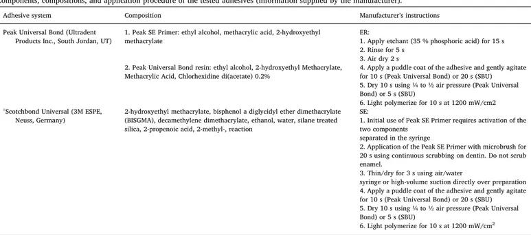
Components, compositions, and application procedure of the tested adhesives (information supplied by the manufacturer).
Adhesive system Composition Manufacturer’s instructions
Peak Universal Bond (Ultradent Products Inc., South Jordan, UT)
1. Peak SE Primer: ethyl alcohol, methacrylic acid, 2-hydroxyethyl methacrylate
ER:
1. Apply etchant (35 % phosphoric acid) for 15 s 2. Rinse for 5 s
3. Air dry 2 s 2. Peak Universal Bond resin: ethyl alcohol, 2-hydroxyethyl Methacrylate,
Methacrylic Acid, Chlorhexidine di(acetate) 0.2%
4. Apply a puddle coat of the adhesive and gently agitate for 10 s (Peak Universal Bond) or 20 s (SBU) 5. Dry 10 s using ¼ to ½ air pressure (Peak Universal Bond) or 5 s (SBU)
6. Light polymerize for 10 s at 1200 mW/cm2
aScotchbond Universal (3M ESPE,
Neuss, Germany)
2-hydroxyethyl methacrylate, bisphenol a diglycidyl ether dimethacrylate (BISGMA), decamethylene dimethacrylate, ethanol, water, silane treated silica, 2-propenoic acid, 2-methyl-, reaction
SE:
1. Initial use of Peak SE Primer requires activation of the two components
separated in the syringe
2. Application of the Peak SE Primer with microbrush for 20 s using continuous scrubbing on dentin. Do not scrub enamel.
3. Thin/dry for 3 s using air/water
syringe or high-volume suction directly over preparation 4. Apply a puddle coat of the adhesive and gently agitate for 10 s (Peak Universal Bond) or 20 s (SBU) 5. Dry 10 s using ¼ to ½ air pressure (Peak Universal Bond) or 5 s (SBU)
6. Light polymerize for 10 s at 1200 mW/cm2
a In the SE mode, the procedure starts from step 4.
Fig. 1. Schematic overview of tooth cutting procedure for in situ zymography, making sure that all the groups are tested on the same substrate.
Table 2
μTBS obtained by applying Scotchbond Universal or Peak Universal Bond ad-hesive system on the dentin surface. Values are mean ± standard deviation [number of premature failed sticks/number of intact sticks tested]. T0 and T12 indicate specimens that were tested after storage of 24 h and 12 months, re-spectively.
T0 T12
PUB E&R (0.2%CHX) 50.1 ± 11.0aAMPa
[7/102]
40.7 ± 8.6bAMPa
[6/98] SBU E&R (control) 38.9 ± 12.8aBMPa
[5/93] 21.6 ± 7.7bCMPa [4/109] PUB SE (0.2%CHX) 53.0 ± 11.3aAMPa [7/92] 43.9 ± 11.7bAMPa [5/108] SBU SE (control) 35.8 ± 13.0aBMPa
[4/103]
33.6 ± 11.1aBMPa
[8/96]
Premature failures due to preparation procedures were not included in the statistical analysis (p > 0.05). Different superscript lowercase letters (in rows) indicate statistical difference in storage time. Different superscript uppercase letters (in columns) indicate statistical difference in different adhesive proto-cols.
after 12 months, but not significantly.
Failure mode distribution of the de-bonded specimens is shown in Table 3. The predominant failure mode was the mixed, followed by the adhesive failure mode in all the tested groups.
3.2. Gelatin zymography
Gelatinolytic activity of the investigated groups is shown inFig. 2a. Mineralized dentin powder (G1; Lane 1) showed week enzymatic ac-tivity in the corresponding to the molecular weights of MMP-2 and -9. Demineralized dentin extract (G2; Lane 2) showed multiple forms of gelatinolytic enzymatic activity, with the weak MMP-2 active and pro-form at 66 kDa and 72 kDa, as well as a stronger signal of MMP-9 active and pro-forms at 86 kDa and 92 kDa, respectively. The active enzyme forms were more pronounced. In both E&R and SE Peak groups (Lanes 3 and 5, respectively), there is an absence of the activity of MMP-2, while the activity of MMP-9 seems to be reduced. On the other hand, enzy-matic activity in the E&R and SE SBU groups is more pronounced compared to the controls (Lane 4 and 6, respectively). Quantification of enzymatic activity is shown inFig. 1b and corresponds to the qualita-tive results.
3.3. In situ zymography
The results obtained on the confocal microscope revealed di ffer-ences in thefluorescence signal exhibited by different tested groups (Fig. 3). The level of enzymatic activity corresponds to the density of the greenfluorescence signal (Fig. 3a–d). Quantification of the density of thefluorescence (Fig. 3i) corresponded to qualitative findings and demonstrated a reduction in enzymatic activity in the hybrid layers created with PUB used in the E&R mode compared to the control SBU used in the same mode (p < 0.05). Both adhesives used in the SE mode showed a similar level offluorescence (p > 0.05), while they exhibited lower enzymatic activity compared to the same adhesives used in E&R mode. Interestingly, Peak used in the E&R mode showed a similar ac-tivity as SBU used in SE mode.
4. Discussion
The results of the presented study showed higher immediate bond strength in the experimental compared to control groups. Hence, the first null hypothesis should be rejected. The bond strength after 12-month-aging was lower compared to the immediate bond strength, but significantly higher in the CHX-containing groups compared to the controls, which requires the rejection of the second hypothesis. The activation of endogenous MMPs was found to be related to the presence of CHX within the adhesive system and the bonding strategy, which supports the rejection of the third hypothesis.
Since the hybrid layer is composed of organic dentinal matrix, the remnants of hydroxyapatite, resin monomers, and solvents, the changes in any of these components can influence the longevity of resin-dentin bonds [36]. Neither E&R nor SE adhesives are able to fully infiltrate the collagen network, creating different degrees of infiltration in different depths of the hybrid layer [13]. This leaves part of the collagenfibrils exposed and prone to degradation over time. The degradation of the hybrid layer can mainly be contributed to the hydrolysis of the resin and/or collagenfibrils and to the disorganization of the collagen fibrils [37]. Pashley et al. were the first to show that endogenous dentinal enzymes can degrade exposed collagenfibrils over time in the absence of bacteria, and that this effect can be diminished by the use of CHX [1]. CHX has an excellent substantivity to dentin [38] and it binds to mineralized as well as demineralized dentin [39]. It is well known that low concentrations of CHX can inhibit MMPs and cysteine cathepsins [14,15]. Several authors established the beneficial effect of CHX water solution used as a primer in the preservation of the μTBS [16,17,26,27,40]. On the other hand, certain authors found no influ-ence of CHX water solution on bonding performance [29], or a negative influence on mechanical properties of the hybrid layer after the use of this inhibitor [41]. Nevertheless, the majority of the available research is in favor of the use of CHX. In every-day dental practice, it is of great importance for patients, as well as dental practitioners, to make chair-time as short as possible. Therefore, there is a tendency to avoid ad-ditional steps in the restorative procedure, thus efforts have been made to incorporate CHX within one of the components of bonding systems. Since adhesives act like semipermeable membranes [16], it was as-sumed that the adhesive layer would serve as a reservoir of CHX, re-leased into the hybrid layer over time, which could contribute to the durability of the adhesive bond.
Therefore, in the present study, the authors tested a commercially available adhesive containing 0.2% CHX. The resin system containing 0.2% CHX revealed significant improvement in bond stability, im-mediately, as well as after aging. After a 12-month storage in artificial saliva, a general decrease in bond strength was noted, however, the CHX-containing groups showed high bond strength values, in fact, higher than the T0values in the control group. Failure modes found in
this study are relatively evenly distributed between mixed and adhesive one, with the mixed mode being the most prevalent in all investigated groups, regardless of the aging. This means that the failures usually Table 3
Percentages of failure modes after microtensile test.
Adhesive system T0 T12
PUB E&R (0.2%CHX) 35% A 65% M
45% A 55% M SBU E&R (control) 50% A
50% M 50% A 50% M PUB SE (0.2%CHX) 35% A 5%CD 65% M 60% A 40% M SBU SE (control) 30% A 10%CD 60% M 45% A 55% M
Fractures were classified as: A, adhesive; CD, cohesive failure in dentin; CC, cohesive failure in resin composite; M, mixed failure.
Fig. 2. (a) Gelatin zymography: Figure showing the differences in the enzy-matic activity between the investigated groups presented as light bands in the area of the molecular weights of pro- and active MMP-2 and MMP-9; (b) Graph illustrating the densitometric evaluation of bands obtained from the zymo-graphic analysis of proteins extracted from dentin powder. STD = Standard; MIN = Mineralized dentin powder; DEM = Demineralized dentin powder; E&R = Etch-and-Rinse Mode; SE = Self-Etch Mode; SBU = Scotchbond Universal.
started in the composite and propagated through the adhesive layer into dentin. These results differ from the results of a similar recent study [25] in which the same commercial adhesive system was used as the experimental group. Sabatini et al. found no difference between the control and experimental group in shear bond stress, regardless of aging (6 months), or the bonding mode (E&R/SE). Further, Sabatini et al. [25] confirmed the efficacy of CHX in inhibiting the dentinal MMPs using a zymographic assay testing only the effect of CHX aqueous solution. This finding is in accordance with our results, however, in our study, gelatin zymography was for the first time performed following all the steps required by the manufacturers for the use of Peak Universal and SBU systems in E&R and SE mode. Hence, the results of Sabatini et al. [25] are not fully comparable to our study, due to the different methodology, control group and aging time. Perhaps the aging of 6 months was not long enough to reveal differences between the tested groups. Further-more, the control group in the study of Sabatini et al. was Peak LC Bond, which is a material similar to PUB, since they are produced by the same manufacturer, with the difference that Peak LC Bond does not contain 0.2% CHX. Since a different system was used as the control group in the present study (SBU), the possibility that PUB is superior to SBU as an adhesive system and that the differences are not driven by the presence of CHX cannot be excluded. However, since the present study did not only test theμTBS, but also showed, using two different zymographic assays, that the PUB system presents lower dentinal en-zymatic activity compared to SBU, especially in the E&R mode, and having in mind previous studies that have shown preservation of the hybrid layer and collagen matrix with the use of CHX, we could hy-pothesize that there is a correlation between the CHX within PUB and better bond strength. Indeed, future studies should include observation of the bonded interfaces under a transmission electron microscope to investigate whether there are differences in the preservation of the
collagen matrix between the tested groups and validate the results of the present study.
The results of other studies with CHX incorporated into one of the parts of the adhesive system varied, especially in SE mode. Certain studies showed that the incorporation of CHX within adhesive blends, used in the E&R mode could contribute to the preservation of bond strength after a 12-month water storage [21,22]. Yiu et al. [23] found mixed results where the effect of CHX was correlated with the com-position of the adhesive used in the E&R mode. Zhou et al. found that the incorporation of 0.1%-1% CHX into a SE primer could preserve dentin bond strength after a 12-month aging [18]. On the other hand, a study incorporating 2% CHX into a SE adhesive found no influence of CHX on bonding performance after 6 months [24]. It is thought that acid etching during E&R procedures can denaturize enzymes within the etched dentin, due to the low pH values of the phosphoric acid. How-ever, Mazzoni et al. [2] showed that enzymatic activity in dentin could be reactivated after acid etching. The authors hypothesized that acid-etching inactivates the superficial layer of the enzymes, while acti-vating the residual latent enzymes within the underlying demineralized dentin. The SE adhesives, on the other hand, have a pH 1.5– 2.7, which means that the more acidic adhesives could denaturize the enzymes, while the ones with a higher pH value could contribute to the activation of enzymatic activity [3]. Moreover, CHX binds to calcium chloride released from dentinal tissue by the influence of acids (especially the primers of SE adhesives) [13,14], which could diminish the inhibitory effect of CHX. Furthermore, in the SE systems, the hybrid layer is cre-ated simultaneously as the acidic monomer etches into the dentin. Therefore, it is assumed that there are less denuded collagenfibrils at the bottom of the hybrid layer compared to the E&R system, and con-sequently less amount of activated MMPs [33]. Perhaps this is why CHX might have less influence on dentinal enzymatic activity in SE Fig. 3. Resin-dentin interfaces bonded with Peak Universal or SBU, in E&R or SE mode, incubated with quenchedfluorescein-labeled gelatin; (a,b,c,d) Images acquired in green channel, showingfluorescence (identifying intense endogenous enzymatic activity) in dentinal tubules and within the HL created using different protocols; (e,f,g,h) Images of resin-dentin interfaces created using different protocols, obtained by merging differential interference contrast image (showing the optical density of the resin-dentin interface) and image acquired in green channel (showing enzymatic activity); (i) Graph illustrating the quantification of the enzymatic activity of the tested groups. E&R = Etch-and-Rinse mode; SE = Self-Etch mode; SBU = Scotchbond Universal; D = Dentin; HL = Hybrid Layer; R = Resin Composite. (For interpretation of the references to colour in thisfigure legend, the reader is referred to the web version of this article).
adhesives. This corresponds to the in situ zymography results of the present study, since the activity in the SE groups was much lower compared to the E&R groups, regardless of the presence of CHX in the bonding system. All the aforementioned differences could be re-sponsible for the discrepancies in the results found in different studies investigating E&R and SE dentin bonding strategies.
Further, the aging time used in this study could be considered short, and in vitro and clinical studies with a longer monitoring time should be conducted to assess the efficacy of this adhesive system. However, the clinical studies conducted thus far have shown mixed results on the protective effect of CHX on the hybrid layer. The study by Araújo et al. [30] investigated whether there was a clinical advantage of the use of 1% CHX primer over the course of 2 years, while Sartori et al. [31] conducted a 5-year clinical follow up, using a 2% CHX primer. Both studies investigated non-carious cervical lesions and neither found a difference in survival in the CHX group as compared to the control group. Non-carious lesions of the V class are quite specific, there the dentin is hyper-mineralized and structurally different compared to sound or caries-affected dentin, which might have influenced the re-sults. Carrilho et al. [32], on the other hand, found that the use of a CHX primer preserves bond strength in restorations of the I class, after 14 months of intra-oral use. These differences demonstrate the im-portance of the type of cavity and dentinal substrate in in vivo evalua-tions, and in general, the importance of standardizing clinical research requirements and methods, and hence making the studies more com-parable.
5. Conclusions
The CHX-containing adhesive tested in the present study seems to perform better in terms of bond strength preservation and protease inhibition compared to the adhesive system that does not contain CHX. It could be hypothesized that this effect is due to the CHX, but the possibility of the influence of the adhesive system itself must not be completely excluded. Within the limitations of this study, it seems that the use of CHX-blended adhesive could be recommended as a feasible technique in every-day clinical practice, since the inhibitor seems to be efficiently delivered to the dentinal substrate, without prolonging chair time.
Declarations of interest None.
Acknowledgments
The authors wish to thank Mr. Aurelio Valmori for technical assis-tance. The study was partially supported with MIUR grants (Italy). References
[1] D. Pashley, F. Tay, C. Yiu, M. Hashimoto, L. Breschi, R. Carvalho, Collagen de-gradation by host-derived enzymes during aging, J. Dent. Res. 83 (2004) 216–221. [2] A. Mazzoni, D.H. Pashley, Y. Nishitani, L. Breschi, F. Mannello, L. Tjäderhane,
M. Toledano, E.L. Pashley, F.R. Tay, Reactivation of inactivated endogenous pro-teolytic activities in phosphoric acid-etched dentine by etch-and-rinse adhesives, Biomaterials 27 (2006) 4470–4476,https://doi.org/10.1016/j.biomaterials.2006. 01.040.
[3] Y. Nishitani, M. Yoshiyama, B. Wadgaonkar, L. Breschi, F. Mannello, A. Mazzoni, R. Carvalho, L. Tjäderhane, F. Tay, D. Pashley, Activation of gelatinolytic / col-lagenolytic activity in dentin by self etching adhesives, Eur. J. Oral Sci. 114 (2006) 160–166.
[4] A. Mazzoni, L. Tjäderhane, V. Checchi, R. Di Lenarda, T. Salo, F.R. Tay, D.H. Pashley, L. Breschi, Role of dentin MMPs in caries progression and bond sta-bility, J. Dent. Res. 94 (2015) 241–251,https://doi.org/10.1177/
0022034514562833.
[5] A. Mazzoni, F. Mannello, F.R. Tay, G.A.M. Tonti, S. Papa, G. Mazzotti, R. Di Lenarda, D.H. Pashley, L. Breschi, Zymographic analysis and characterization of MMP-2 and -9 forms in human sound dentin, J. Dent. Res. 86 (2007) 436–440,
https://doi.org/10.1177/154405910708600509.
[6] A. Mazzoni, V. Papa, F. Nato, M. Carrilho, L. Tjäderhane, A. Ruggeri, P. Gobbi, G. Mazzotti, F.R. Tay, D.H. Pashley, L. Breschi, Immunohistochemical and bio-chemical assay of MMP-3 in human dentine, J. Dent. 39 (2011) 231–237,https:// doi.org/10.1016/j.jdent.2011.01.001.
[7] J. Santos, M. Carrilho, T. Tervahartiala, T. Sorsa, L. Breschi, A. Mazzoni, D. Pashley, F. Tay, C. Ferraz, L. Tjäderhane, Determination of matrix metalloproteinases in human radicular dentin, J. Endod. 35 (2009) 686–689,https://doi.org/10.1016/j. joen.2009.02.003.
[8] M. Sulkala, T. Tervahartiala, T. Sorsa, M. Larmas, T. Salo, L. Tjäderhane, Matrix metalloproteinase-8 (MMP-8) is the major collagenase in human dentin, Arch. Oral Biol. 52 (2007) 121–127,https://doi.org/10.1016/j.archoralbio.2006.08.009. [9] M. Sulkala, M. Larmas, T. Sorsa, T. Salo, L. Tjäderhane, The localization of matrix
metalloproteinase-20 (MMP-20, enamelysin) in mature human teeth, J. Dent. Res. 81 (2002) 603–607.
[10] H. Palosaari, C. Pennington, M. Larmas, L. Tjäderhane, T. Salo, Expression profile of matrix metalloproteinases (MMPs) and tissue inhibitors of MMPs in mature human odontoblasts and pulp tissue, Eur. J. Oral Sci. 111 (2003) 117–127.
[11] A.R. Hannas, J.C. Pereira, J.M. Granjeiro, L. Tjäderhane, A.R. Hannas, J.C. Pereira, J.M. Granjeiro, L. Tjäderhane, The role of matrix metalloproteinases in the oral environment, Acta Odontol. Scand. 65 (2007) 1–13,https://doi.org/10.1080/ 00016350600963640.
[12] F.D. Nascimento, C.L. Minciotti, S. Geraldeli, M.R. Carrilho, D.H. Pashley, F.R. Tay, H.B. Nader, T. Salo, L. Tjäderhane, I.L.S. Tersariol, Cysteine cathepsins in human carious dentin, J. Dent. Res. 90 (2011) 506–511,https://doi.org/10.1177/ 0022034510391906.
[13] A. Frassetto, L. Breschi, G. Turco, G. Marchesi, R. Di Lenarda, F.R. Tay, D.H. Pashley, M. Cadenaro, Mechanisms of degradation of the hybrid layer in ad-hesive dentistry and therapeutic agents to improve bond durability– a literature review, Dent. Mater. 32 (2016) e41–e53,https://doi.org/10.1016/j.dental.2015. 11.007.
[14] R. Gendron, D. Grenier, T. Sorsa, D. Mayrand, Inhibition of the activities of matrix metalloproteinases 2, 8, and 9 by chlorhexidine, Clin. Diagn. Lab. Immunol. 6 (1999) 437–439.
[15] P.M.C. Scaffa, C.M.P. Vidal, N. Barros, T.F. Gesteira, A.K. Carmona, L. Breschi, D.H. Pashley, L. Tjaderhane, I.L.S. Tersariol, F.D. Nascimento, M.R. Carrilho, Chlorhexidine inhibits the activity of dental cysteine cathepsins, J. Dent. Res. 91 (2012) 420–425,https://doi.org/10.1177/0022034511435329.
[16] L. Breschi, F. Cammelli, E. Visintini, A. Mazzoni, M. Carrilho, M. Cadenaro, S. Foulger, F.R. Tay, Influence of chlorhexidine concentration on the durability of etch-and-rinse dentin bonds: a 12-month in vitro study, J. Adhes. Dent. 11 (2009) 191–198.
[17] L. Breschi, A. Mazzoni, F. Nato, M. Carrilho, L. Tjäderhane, A. Ruggeri Jr, F.R. Tay, E. De Stefano, Chlorhexidine stabilizes the adhesive interface: a 2 year in vitro study, Dent. Mater. 26 (2010) 1–12,https://doi.org/10.1016/j.dental.2009.11.153. Chlorhexidine.
[18] J. Zhou, J. Tan, L. Chen, D. Li, Y. Tan, The incorporation of chlorhexidine in a two-step self-etching adhesive preserves dentin bond in vitro, J. Dent. 37 (2009) 807–812,https://doi.org/10.1016/j.jdent.2009.06.011.
[19] J. Zhou, J. Tan, X. Yang, C. Cheng, X. Wang, L. Chen, Effect of chlorhexidine ap-plication in a self-etching adhesive on the immediate resin-dentin bond strength, J. Adhes. Dent. 12 (2010) 27–31,https://doi.org/10.3290/j.jad.a17543.
[20] J. Zhou, J. Tan, X. Yang, X. Xu, D. Li, L. Chen, MMP-inhibitory effect of chlor-hexidine applied in a self-etching adhesive, J. Adhes. Dent. 13 (2011) 111–115,
https://doi.org/10.3290/j.jad.a18783.
[21] E.M. da Silva, C.U.F. de Sá Rodrigues, M.P. de Oliveira Matos, T.R. de Carvalho, G.B. dos Santos, C.M. Amaral, Experimental etch-and-rinse adhesive systems con-taining MMP-inhibitors: physicochemical characterization and resin-dentin bonding stability, J. Dent. 43 (2015) 1491–1497,https://doi.org/10.1016/j.jdent. 2015.10.004.
[22] R. Stanislawczuk, F. Pereira, M.A. Muñoz, I. Luque, P.V. Farago, A. Reis, A.D. Loguercio, Effects of chlorhexidine-containing adhesives on the durability of resin-dentine interfaces, J. Dent. 42 (2014) 39–47,https://doi.org/10.1016/j.jdent. 2013.11.002.
[23] C.K.Y. Yiu, N. Hiraishi, F.R. Tay, M. King, Effect of chlorhexidine incorporation into dental adhesive resin on durability of resin-dentin bond, J. Adhes. Dent. 14 (2012) 355–362,https://doi.org/10.3290/j.jad.a25674.
[24] C. Pomacóndor-Hernández, A. Nogueira, D.G. Antunes, Effect of replacing a com-ponent of a self-etch adhesive by chlorhexidine on bonding to dentin, Braz. Dent. J. 24 (2013) 335–339.
[25] C. Sabatini, Effect of a chlorhexidine-containing adhesive on dentin bond strength stability, Oper. Dent. 38 (2013) 609–617,https://doi.org/10.2341/12-239-L. [26] E.A. Campos, G.M. Correr, D.P. Leonardi, F. Barato-Filho, C.C. Gonzaga, J.C. Zielak,
Chlorhexidine diminishes the loss of bond strength over time under simulated pulpal pressure and thermo-mechanical stressing, J. Dent. 37 (2009) 108–114,
https://doi.org/10.1016/j.jdent.2008.10.003.
[27] A.D. Loguercio, V. Hass, M.F. Gutierrez, I.V. Luque-Martinez, A. Szezs, R. Stanislawczuk, M.C. Bandeca, A. Reis, Five-year effects of chlorhexidine on the in vitro durability of resin/dentin interfaces, J. Adhes. Dent. 18 (2016) 35–43,https:// doi.org/10.3290/j.jad.a35514.
[28] S. Talungchit, J.L.P. Jessop, D.S. Cobb, F. Qian, S. Geraldeli, D.H. Pashley, S.R. Armstrong, Ethanol-wet bonding and chlorhexidine improve resin-dentin bond durability: quantitative analysis using raman spectroscopy, J. Adhes. Dent. 16 (2014) 441–450,https://doi.org/10.3290/j.jad.a32695.
[29] A.P. Manso, R.H.M. Grande, A.K. Bedran-Russo, A. Reis, A.D. Loguercio, D.H. Pashley, R.M. Carvalho, Can 1% chlorhexidine diacetate and ethanol stabilize resin-dentin bonds? Dent. Mater. 30 (2014) 735–741,https://doi.org/10.1016/j.
dental.2014.04.003.
[30] M.S.R.G. Araújo, L.C. Souza, F.M. Apolonio, L.O. Barros, A. Reis, A.D. Loguercio, V.P.A. Saboia, Two-year clinical evaluation of chlorhexidine incorporation in two-step self-etch adhesive, J. Dent. 43 (2015) 140–148,https://doi.org/10.1016/j. jdent.2014.07.010.
[31] N. Sartori, S.C. Stolf, S.B. Silva, G.C. Lopes, M. Carrilho, Influence of chlorhexidine digluconate on the clinical performance of adhesive restorations: a 3-year follow-up, J. Dent. 41 (2013) 1188–1195,https://doi.org/10.1016/j.jdent.2013.09.004. [32] M.R.O. Carrilho, S. Geraldeli, F. Tay, M. de Goes, R. Carvalho, L. Tjäderhane, In
vivo preservation of hybrid layer by chlorhexidine, J. Dent. Res. 86 (2007) 529–533.
[33] A. Mazzoni, P. Scaffa, M. Carrilho, L. Tjäderhane, R. Di Lenarda, A. Polimeni, A. Tezvergil-Mutluay, F.R. Tay, D.H. Pashley, L. Breschi, Effects of etch-and-rinse and self-etch adhesives on dentin MMP-2 and MMP-9, J. Dent. Res. 92 (2013) 82–86,https://doi.org/10.1177/0022034512467034.
[34] A. Mazzoni, F. Nascimento, M. Carrilho, I. Tersariol, V. Papa, L. Tjaderhane, R. Di Lenarda, F. Tay, D. Pashley, L. Breschi, MMP activity in the hybrid layer detected with in situ zymography, J. Dent. Res. 91 (2012) 467–472,https://doi.org/10. 1177/0022034512439210.
[35] A. Mazzoni, F.M. Apolonio, V.P.A. Saboia, S. Santi, V. Angeloni, V. Checchi, R. Curci, R. Di Lenarda, F.R. Tay, D.H. Pashley, L. Breschi, Carbodiimide in-activation of MMPs and effect on dentin bonding, J. Dent. Res. 93 (2014) 263–268,
https://doi.org/10.1177/0022034513516465.
[36] L. Breschi, A. Mazzoni, A. Ruggeri, M. Cadenaro, R. Di Lenarda, E. De Stefano Dorigo, Dental adhesion review: aging and stability of the bonded interface, Dent. Mater. 24 (2008) 90–101,https://doi.org/10.1016/j.dental.2007.02.009. [37] M. Hashimoto, H. Ohno, H. Sano, M. Kaga, H. Oguchi, In vitro degradation of
resin-dentin bonds analyzed by microtensile bond test, scanning and transmission elec-tron microscopy, Biomaterials 24 (2003) 3795–3803,https://doi.org/10.1016/ S0142-9612(03)00262-X.
[38] M.R. Carrilho, R.M. Carvalho, E.N. Sousa, J. Nicolau, L. Breschi, A. Mazzoni, L. Tjäderhane, F.R. Tay, K. Agee, D.H. Pashley, Substantivity of chlorhexidine to human dentin, Dent. Mater. 26 (2010) 779–785,https://doi.org/10.1016/j.dental. 2010.04.002.
[39] J. Kim, T. Uchiyama, M. Carrilho, K.A. Agee, A. Mazzoni, L. Breschi, R.M. Carvalho, L. Tj??derhane, S. Looney, C. Wimmer, A. Tezvergil-Mutluay, F.R. Tay, D.H. Pashley, Chlorhexidine binding to mineralized versus demineralized dentin powder, Dent. Mater. 26 (2010) 771–778,https://doi.org/10.1016/j.dental.2010. 04.001.
[40] P. Zheng, M. Zaruba, T. Attin, A. Wiegand, Effect of different matrix metallopro-teinase inhibitors on microtensile bond strength of an etch-and-rinse and a self-etching adhesive to dentin, Oper. Dent. 40 (2014) 80–86,https://doi.org/10.2341/ 13-162-L.
[41] N. Hiraishi, C.K.Y. Yiu, N.M. King, F.R. Tay, Effect of 2% chlorhexidine on dentin microtensile bond strengths and nanoleakage of luting cements, J. Dent. 37 (2009) 440–448,https://doi.org/10.1016/j.jdent.2009.02.002.