Role of the lipid rafts in the life cycle of canine
coronavirus
Annamaria Pratelli and Valeriana Colao
Correspondence Annamaria Pratelli [email protected]
Received 8 August 2014 Accepted 2 November 2014
Department of Veterinary Medicine, University of Bari, Bari, Italy
Coronaviruses are enveloped RNA viruses that have evolved complex relationships with their host cells, and modulate their lipid composition, lipid synthesis and signalling. Lipid rafts, enriched in sphingolipids, cholesterol and associated proteins, are special plasma membrane microdomains involved in several processes in viral infections. The extraction of cholesterol leads to disorganization of lipid microdomains and to dissociation of proteins bound to lipid rafts. Because cholesterol-rich microdomains appear to be a general feature of the entry mechanism of non-eneveloped viruses and of several coronaviruses, the purpose of this study was to analyse the contribution of lipids to the infectivity of canine coronavirus (CCoV). The CCoV life cycle is closely connected to plasma membrane cholesterol, from cell entry to viral particle production. The methyl-b-cyclodextrin (MbCD) was employed to remove cholesterol and to disrupt the lipid rafts. Cholesterol depletion from the cell membrane resulted in a dose-dependent reduction, but not abolishment, of virus infectivity, and at a concentration of 15 mM, the reduction in the infection rate was about 68 %. MbCD treatment was used to verify if cholesterol in the envelope was required for CCoV infection. This resulted in a dose-dependent inhibitory effect, and at a concentration of 9 mM MbCD, infectivity was reduced by about 73 %. Since viral entry would constitute a target for antiviral strategies, inhibitory molecules interacting with viral and/or cell membranes, or interfering with lipid metabolism, may have strong antiviral potential. It will be interesting in the future to analyse the membrane microdomains in the CCoV envelope.
INTRODUCTION
Coronaviruses, a genus in the family Coronaviridae, are large,
enveloped, positive-sense RNA viruses, 27.6–31 kb in length,
responsible for highly prevalent diseases in humans, birds and
domestic animals. The one-third 39 section of the genome
contains ORFs encoding the major structural proteins, spike,
envelope, membrane, haemoagglutinin-esterase and
nucleo-capsid proteins. These ORFs are interspersed with several
ORFs encoding different non-structural proteins, most of
which have unknown functions (Lai & Holmes, 2001; Pratelli,
2006, 2011). In rooted trees, the members of the coronavirus
genus consistently form three distinct monophyletic groups,
referred to as phylogroups 1, 2 and 3. Canine coronaviruses
(CCoVs) are included in phylogroup 1. In view of the recent
increase in the number of newly discovered coronaviruses,
and ensuing debates and confusion in the literature
concern-ing coronavirus taxonomy, the unofficial, but widely accepted,
nomenclature has been proposed to the International
Committee on Taxonomy of Viruses (ICTV) Executive
Committee, and phylogroups 1–3 were converted into genera
designated Alpha-, Beta- and Gammacoronavirus, respectively
(Pratelli, 2011). Deltacoronavirus is a new genus proposed in
July 2013 (ictvonline.org/virusTaxonomy.asp) (Table 1).
Lipid rafts are special plasma membrane microdomains
with an increased structural order, which are designated
liquid ordered domains in model membranes. Lipid rafts,
enriched in sphingolipids, cholesterol and associated
proteins, play a critical role in different biological aspects
of the life cycle of several viruses, and are involved in
many processes in viral infection. In particular, the tight
packaging of the sphingolipids is maintained by the
pres-ence of cholesterol, a major constituent of the lipid
rafts, and several proteins partition into these membrane
domains (Imhoff et al., 2007). Extraction of cholesterol
destroys this order, leading both to disorganization of the
lipid raft microdomains and to the dissociation of proteins
bound to the lipid rafts (Barman & Nayak, 2007).
The role of cholesterol in the entry of non-enveloped viruses
was demonstrated for simian virus 40, rotavirus, rhinovirus
and enterovirus (Anderson et al., 1996; Suzuki & Suzuki,
2006). Successful entry of enveloped viruses requires binding
to specific cellular receptors and fusion of the viral
membrane with the cell membrane. Accumulating evidence
suggests that enveloped virus entry may require cholesterol
in either, or both, of the two membranes involved. Human
immunodeficiency virus (HIV) type 1 infection requires
cholesterol both in the target cell membrane and in the viral
envelope (Guyader et al., 2002; Liao et al., 2001, 2003).
Cholesterol in both membranes is also required for bovine
herpesvirus 1 infection of Madin Darby Bovine Kidney
(MDBK) cells (Zhu et al., 2010). For other viruses in the
subfamily Alphaherpesvirinae of the family Herpesviridae,
such as herpes simplex virus 1, varicella-zoster virus and
porcine pseudorabies virus, cell membrane cholesterol is
required during virus entry (Bender et al., 2003; Hambleton
et al., 2007; Desplanques et al., 2008). Other viruses are
sensitive to cholesterol depletion from the cell membrane,
such as Semliki Forest virus, murine leukaemia virus, Ebola
virus and Marburg virus (Ahn et al., 2002; Bavari et al., 2002;
Lu et al., 2002; Phalen & Kielian, 1991). For influenza virus
and duck hepatitis B virus, the presence of cholesterol in the
viral envelope is critical, but it is not essential in the target
cell membrane (Sun & Whittaker, 2003; Funk et al., 2008),
and recently it has been demonstrated that canine distemper
virus also requires cholesterol in the viral envelope (Imhoff
et al., 2007). In contrast, in the case of vesicular stomatitis
virus, replication is not affected by cholesterol depletion,
and numerous strains of the family Flaviviridae, i.e. dengue
virus and yellow fever virus, enter and infect cells
indepen-dent of cholesterol (Umashankar et al., 2008).
It is known that coronaviruses differ in their tissue tropism,
and different cellular receptors are involved in virus entry.
The depletion of cellular and viral cholesterol inhibits entry
of several coronaviruses: mouse hepatitis virus (MHV)
(Choi et al., 2005), severe acute respiratory syndrome
coronavirus (SARS-CoV) (Li et al., 2007), human
corona-virus (HCoV)-229E (Nomura et al., 2004), transmissible
gastroeneteritis virus (TGEV) (Ren et al., 2008) and avian
infectious bronchitis virus (IBV) (Imhoff et al., 2007). In
the present study we investigated the role of cholesterol in
the viral envelope and the cell membrane in CCoV
infection of A72 cells. Methyl-b-cyclodextrin (MbCD), a
cholesterol-binding agent, was employed to remove
cholesterol and to disrupt the lipid rafts.
RESULTS
Infection efficiency after cholesterol depletion
from the cell membrane
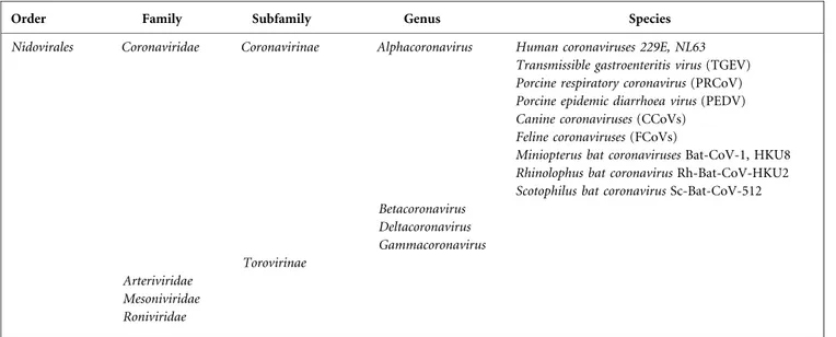
To investigate if cellular cholesterol was essential for CCoV
entry into susceptible cells, A72 monolayers were mock
pretreated or pretreated with various concentrations of
MbCD and subsequently infected with CCoV strain SE/97.
Cells were cultured and virus yield was determined with a
virus titration assay. MbCD treatment of A72 cells resulted
in an abatement of virus production in a dose-dependent
manner, suggesting that cell membrane cholesterol is
necessary at the entry stage for CCoV infection. At a
concentration of 15 mM, the reduction in the infection
rate was about 68 % (Fig. 1a).
To confirm that the inhibitory effects for CCoV replication
at the entry stage were due to cholesterol depletion, cell
membrane cholesterol was replenished with different
con-centrations of exogenous cholesterol, and the recovery of
virus infection was analysed. Cholesterol-depleted cells
(pretreated with 15 mM MbCD) were incubated with
exogenous cholesterol, infected with CCoV and virus yield
was investigated with a virus titration assay. As shown in Fig.
1(b), the inhibitory effect was reversed with cholesterol
replenishment and virus production was partially restored to
values close to those observed prior to MbCD treatment. At a
concentration of 700 mg ml
21, infectivity was restored to a
mean of 77 % compared to the mock-treated cells.
The concentration of MbCD and cholesterol employed in
this study did not cause significant adverse effects on cell
viability (data not shown).
Table 1. Coronaviridae classification and important viruses in the genus Alphacoronavirus
Order Family Subfamily Genus Species
Nidovirales Coronaviridae Coronavirinae Alphacoronavirus Human coronaviruses 229E, NL63
Transmissible gastroenteritis virus (TGEV) Porcine respiratory coronavirus (PRCoV) Porcine epidemic diarrhoea virus (PEDV) Canine coronaviruses (CCoVs)
Feline coronaviruses (FCoVs)
Miniopterus bat coronaviruses Bat-CoV-1, HKU8 Rhinolophus bat coronavirus Rh-Bat-CoV-HKU2 Scotophilus bat coronavirus Sc-Bat-CoV-512 Betacoronavirus Deltacoronavirus Gammacoronavirus Torovirinae Arteriviridae Mesoniviridae Roniviridae
Infection efficiency after cholesterol depletion
from the viral membrane
To analyse whether cholesterol in the viral envelope is
required for CCoV entry into susceptible cells, the virus
was mock treated or treated with different
concentra-tions of MbCD prior to infection. Cell monolayers were
incubated with non-treated and MbCD-treated viral
suspensions and virus yield was determined with a virus
titration assay. As reported in Fig. 2(a), the exposure of
CCoV to MbCD resulted in a dose-dependent inhibitory
effect on virus infectivity. In particular, at a concentration
of 9 mM MbCD, virus yield was reduced by about 73 %.
To verify whether the effect of cholesterol depletion was
reversible, exogenous cholesterol at various concentrations
was added to virus suspensions pretreated with 9 mM
MbCD. Cholesterol replenishment resulted in an increase
of the infectivity of CCoV, and at concentration of 700 mg
ml
21, infectivity reached about 82 % of the value observed
prior to cholesterol depletion (Fig. 2b).
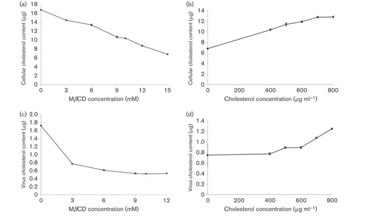
Cellular and viral cholesterol measurements
A72 cells were treated with various concentrations of
MbCD and cellular cholesterol was determined. MbCD
treatment resulted in a dose-dependent reduction of the
cholesterol content in the lipid raft microdomains of the
A72 plasma membrane. In particular, 15 mM of MbCD
reduced the amount of cellular cholesterol by about 60 %
(Fig. 3a). A72 pretreated with 15 mM of MbCD were
analysed after cholesterol replenishment by addition of
exogenous cholesterol in increasing amounts. As shown in
Fig. 3(b), 700 mg ml
21of exogenous cholesterol restored
the cholesterol values of the cell membranes to nearly the
values determined prior to MbCD treatment.
0 0 TCID 50 ml –1 (log 10 ) 5 4 3 2 1 6 (a) 12 9 6 MβCD concentration (mM) 3 15 00 TCID 50 ml –1 (log 10 ) 5 4 3 2 1 (b) Cholesterol concentration (mg ml–1) 600 400 200 800
Fig. 1. CCoV infection efficiency after cholesterol depletion and replenishment from cellular membrane. (a) MbCD treatment of A72 cells reduced infectivity of CCoV in a dose-dependent manner, and at a concentration of 15 mM, viral titres suffered a reduction of about 68 %. (b) Cholesterol-depleted cells were replenished with exogenous cholesterol and virus production was partially restored. The 100 % infectivity value corresponds to the original titre of the stock virus. Each point indicates the mean value and the error bars represent the standard deviation.
0 0 TCID 50 ml –1 (log 10 ) 5 4 3 2 1 6 (a) 9 6 MβCD concentration (mM) 3 12 00 TCID 50 ml –1 (log 10 ) 5 4 3 2 1 (b) Cholesterol concentration (mg ml–1) 600 400 200 800
Fig. 2. CCoV infection efficiency after cholesterol depletion and replenishment from viral membrane. (a) MbCD treatment of CCoV reduced infectivity in a dose-dependent manner, and at a concentration of 9 mM, viral titres suffered a reduction of about 73 %. (b) Replenishment of cholesterol in the viral membrane resulted in an increase in CCoV infectivity. The 100 % infectivity value corresponds to the original titre of the stock virus. Each point indicates the mean value and the error bars represent the standard deviation.
Viral cholesterol was also measured. MbCD was used to
deplete cholesterol, and increasing drug concentrations
resulted in a dose-dependent decrease in cholesterol
content in the viral membrane. At a concentration of
9 mM MbCD, viral cholesterol was reduced by about 70 %
(Fig. 3c). Cholesterol-depleted virions were replenished
with exogenous cholesterol in increasing amounts and
virus pellets were used for cholesterol measurements.
Exogenous cholesterol (700 mg ml
21) restored the
choles-terol values of the viral membranes to nearly the values
determined prior to MbCD treatment (Fig. 3d).
DISCUSSION
Viruses are intracellular parasites entirely dependent upon
the host cell system for replication and spreading. In the
case of enveloped viruses, viral nucleocapsid is surrounded
by a lipid membrane derived from the infected cell,
where glycoproteins are fixed supporting the functions of
entry into target cells and/or fusion between viral and
cell membranes. The lipid composition of animal
mem-branes is complex, and three main categories of lipids can
be distinguished: glycerophospholipids, sphingolipids and
sterols. Sphingolipids are main components of animal cell
membranes, and sphingomyelin at the plasma membrane is
known to be enriched in lipid microdomains forming the
so-called lipid rafts, together with cholesterol (Blaising &
Pe´cheur, 2013). These lipids contribute to viral infection by
modulating the properties of viral and/or cell membranes
during infection, and can thus play a role through their
preferential partitioning into the membrane
microdo-mains. Specifically, viral entry brings together virions and
host cells that will interact in a subtly controlled
step-by-step process. Each step-by-step, therefore, relies on a paired
combination of lipids and proteins (Blaising & Pe´cheur,
2013).
Viruses have evolved complex relationships with their host
cells and many viruses modulate lipid composition, lipid
synthesis and signalling of their host cell (Blaising &
Pe´cheur, 2013). In particular, lipids are essential for the life
cycle of several coronaviruses. The depletion of cellular
cholesterol inhibits entry of MHV (Thorp & Gallagher,
2004; Choi et al., 2005), SARS-CoV (Li et al., 2007; Glende
et al., 2008), HCoV-229E (Nomura et al., 2004) and IBV
(Thorp & Gallagher, 2004; Nomura et al., 2004; Li et al.,
2007; Imhoff et al., 2007). Ren et al. (2008) showed
the importance of cholesterol in both the cell and viral
membranes for TGEV infection, and in addition a
func-tional analysis suggested that cholesterol depletion affects a
post-adsorption step in the TGEV entry process (Yin et al.,
00 16 14 12
Cellular cholesterol content (
m g) 10 8 6 4 2 18 (a) 12 9 6 MβCD concentration (mM) 3 15 00 14 12
Cellular cholesterol content (
m g) 10 8 6 4 2 (b) Cholesterol concentration (mg ml–1) 600 400 200 800 0 0 1.8 1.6 1.4 1.2 1.0
Virus cholesterol content (
m g) 0.8 0.6 0.4 0.2 2.0 (c) 9 6 MβCD concentration (mM) 3 12 00 1.4 1.2 1.0
Virus cholesterol content (
m g) 0.8 0.6 0.4 0.2 (d) Cholesterol concentration (mg ml–1) 600 400 200 800
Fig. 3. Cholesterol content determination after depletion and replenishment of cholesterol from the cell membrane (a, b) and from the viral membrane (c, d). (a) Cellular cholesterol depletion with various concentrations of MbCD. (b) Recovery of cellular cholesterol after exogenous cholesterol replenishment. (c) Viral cholesterol depletion with various concentrations of MbCD. (d) Recovery of viral cholesterol after exogenous cholesterol replenishment. Each point indicates the mean value and the error bars represent the standard deviation.
2010). Therefore, the importance of cholesterol-rich
microdomains appears to be a general feature of the entry
mechanism of different viruses and, as far as coronaviruses
are concerned, the purpose of this study was to analyse the
contribution of lipids in the infectivity of CCoV, and in
particular whether cholesterol was important as a
con-stituent of the virus, the host cells, or both. The CCoV life
cycle appears to be closely connected to plasma membrane
cholesterol. In the case of TGEV and HCoV-229E, the
cholesterol dependence is consistent with the presence of
porcine and human aminopeptidase N, respectively (Ren
et al., 2008). Conversely, MHV and SARS-CoV use
different receptors, MHVR and ACE2, respectively, which
are non-raft proteins (Thorp & Gallagher, 2004; Warner
et al., 2005).Our analysis did not provide evidence that the
activities of the S protein, binding to sialic acids and to
aminopeptidase N, were reduced in cholesterol-depleted
virions. However, optimal infectivity of CCoV requires
cholesterol in the plasma membrane. In particular,
choles-terol depletion resulted in a reduction, but not
abolish-ment, of virus infectivity, and virus entry may occur also at
lower cholesterol levels, but increased cholesterol makes
this process more efficient. At a concentration of 700 mg
ml
21, infectivity was restored to an average of 77 %
compared to the mock-treated cells, confirming that the
reduction was due to the cholesterol depletion, and that the
inhibitory effect was partially reversible.
Interestingly, our study also demonstrated the role of
cholesterol in the viral membrane. This data is particularly
important because coronaviruses mature by a budding
process in the early compartments of the secretory pathway
(Tooze et al., 1984), where the content of cholesterol and
sphingolipids is lower than in the plasma membrane
(Sevlever et al., 1999). As observed for TGEV by Ren et al.
(2008), our results confirmed the possibility that lipid
microdomains exist in the membrane of CCoV, and the low
concentration of cholesterol may explain why the infectivity
of CCoV in vitro is affected by MbCD concentrations
lower than those that affect infectivity of other viruses, like
HIV and influenza virus. It will be interesting in future
studies to analyse the membrane microdomains in the
CCoV envelope.
Lipids and receptors for lipids are therefore key players in
the early stages of CCoV infection, i.e. entry and fusion.
These stages are amenable to antiviral strategies, and
molecules inhibiting CCoV entry and/or fusion could be
likely to act on extracellular targets, thereby limiting
virus-induced cell damage. By analogy, as observed for hepatitis
C virus by Blaising & Pe´cheur (2013), molecules targeting
lipids or their receptors could be considered as CCoV-entry
inhibitors, and the virus could be employed in an animal
model to test coronaviruses antiviral. Viral entry is a key
target for antiviral strategies, and molecules and/or drugs
interacting with viral and/or cell membranes, or interfering
with the function of lipid metabolism regulators, could be
considered potential antivirals, and could constitute potent
therapeutics against coronavirus infections combined with
existing strategies.
Future work may be aimed at addressing the question of
whether cholesterol facilitates coronavirus entry by
affect-ing membrane fluidity, or whether other molecular
interac-tions depend on an increased content of cholesterol.
METHODS
Cell and viruses.The A72 canine fibroma cell line, established from a tumour surgically removed from a female 8-year-old Golden Retriever dog (Binn et al., 1980), was employed. The cells were maintained in Dulbecco’s minimal essential medium (DMEM) supplemented with 5 % FCS, and were passaged twice a week. CCoV strain SE/97 (‘SE’ stands for ‘Seeing Eye dogs’, Pennsylvania isolate) was employed throughout the study. The virus, supplied by Professor L. E. Carmichael (Cornell Vet, Ithaca, NY), was isolated on A72 cells from an adult dog with mild enteritis and recovered within a week. SE/97 was propagated on A72 cells and grown in serum-free medium. The viral titre was determined in 96-well microtitration
plates with A72 cells and was expressed as TCID5050 ml21calculated
using the Reed-Muench formula (Reed & Muench, 1938). CCoV-induced cytopathic effect on infected cells was determined based on the appearance of enlarged, bizarrely shaped cells followed by focal
cell detachment. The infectivity titre of the stock virus was 105.5
TCID5050 ml21.
Reagents.Methyl-b-cyclodextrin (MbCD) (C4555, Sigma-Aldrich) is a strictly surface-acting drug that can selectively and rapidly remove cholesterol from the plasma membrane in preference to other membrane lipids (Barman & Nayak, 2007). This cholesterol depletion reagent has been widely employed in studying the effect of both cholesterol depletion and lipid raft disassembly, and current data indicate that it inhibits entry of several viruses (Choi et al., 2005; Li et al., 2007; Nomura et al., 2004; Imhoff et al., 2007; Ren et al., 2008). To remove the plasma membrane cholesterol, concentrations of 3, 6, 9, 10, 12, 15 mM of MbCD in DMEM were prepared.
Water-soluble cholesterol (C4951, Sigma-Aldrich) was employed to replenish cholesterol after extraction of cellular and viral cholesterol using MbCD.
Cholesterol depletion and replenishment from cellular mem-brane at the virus-entry stage.To remove cholesterol from cellular membranes, cell monolayers seeded in 24-well plates, containing approximately 300 000 A72 cells per well, were washed three times
with DMEM and incubated for 30 min at 37uC in a CO2incubator
with serum-free DMEM in the absence (mock cells) or in the presence (treated cells) of MbCD at concentrations of 3, 6, 9, 10, 12 or 15 mM. To determine if cellular cholesterol depletion at the virus-entry stage affects virus replication, MbCD-treated or mock cells were washed three times with DMEM to remove MbCD and incubated with 100
TCID5050 ml21of virus suspension at 37uC for 1 h. Fresh DMEM
was then applied and the cells were incubated for 48 h in a CO2
incubator. To investigate the infection efficiency, MbCD-treated or mock cells were frozen and thawed three times, and subjected to virus titration in A72 cells as described above.
For cholesterol replenishment, monolayers of A72 cells in 24-well plates, containing approximately 300 000 A72 cells per well, were mock pretreated or pretreated with 15 mM MbCD for 30 min at
37uC to remove cellular membrane cholesterol as described above.
The concentration of 15 mM was selected because in the cholesterol depletion test, it was the optimal MbCD concentration which did not produce collateral effects for A72 cells.
The cells were then washed three times with DMEM, replenished with different concentrations of water-soluble cholesterol in DMEM
ranging from 400 to 800 mg ml21and incubated for 1 h at 37uC.
Mock cells were replenished with serum-free medium. For cell infection analysis after cellular cholesterol replenishment, the cells were washed three times with DMEM, and viral suspensions
containing 100 TCID5050 ml21were applied to the cell monolayers.
The plates were incubated for 1 h at 37uC (Zhu et al., 2010), fresh
DMEM was applied and the cells were incubated for 48 h in a CO2
incubator. To investigate the infection efficiency, treated or mock cells were frozen and thawed three times, and subjected to virus titration in A72 cells as described above.
The reduction and the restoration of viral infectivity were converted to percentages to quantify the reduced and restored amounts, respectively.
All experiments were repeated twice under the same conditions. Cholesterol depletion from viral membranes and effects on virus infectivity. For viral cholesterol extraction, 1 ml of viral
suspensions containing 100 TCID50 50 ml21 were incubated with
MbCD at concentrations of 3, 6, 9, 10 or 12 mM for 1 h at 37uC. To
determine if the virus cholesterol was essential for CCoV infectivity, after cholesterol depletion from viral membranes, cell monolayers were washed three times with DMEM and then incubated with
MbCD-treated viral suspensions at 37uC for 1 h. To avoid negative
effects of MbCD on A72 cells, inocula were diluted 1 : 3 in DMEM before infection. The controls were mock treated. Finally, treated cells and controls were washed three times with DMEM and incubated for
48 h in a CO2incubator. To analyse the infection efficiency after viral
cholesterol depletion, the monolayers were frozen and thawed three times, and subjected to virus titration in A72 cells as described above. For cholesterol replenishment, 1 ml of CCoV suspensions were mock
treated or treated with 9 mM MbCD for 30 min at 37uC, then
replenished with different concentrations of water-soluble cholesterol
in DMEM ranging from 400 to 800 mg ml21and incubated for 1 h at
37uC. The concentration of 9 mM was selected because in the
cholesterol depletion test, it was the optimal MbCD concentration which did not produce collateral effects for CCoV. Mock cells were replenished with serum-free medium. For cell infection analysis after viral cholesterol replenishment, the cells were washed three times with DMEM, and cholesterol-replenished or non-replenished (control) viral suspensions were applied to the cell monolayers and incubated at
37uC for 48 h (Ren et al., 2008). To investigate the infection
efficiency, samples were frozen and thawed three times, and subjected to virus titration in A72 cells as described above.
The reduction and the restoration of viral infectivity were converted to percentages to quantify the reduced and restored amounts, respectively.
All experiments were repeated twice under the same conditions. Cellular and viral cholesterol content measurements.Cellular and viral cholesterol were measured using an Amplex Red Cholesterol Assay kit (A12216, Invitrogen/Life Technologies) according to the manufacturer’s instructions and according to protocols reported by Ren et al. (2008).
To determine cellular cholesterol, confluent monolayers of A72 cells grown in six-well plates were treated with different concentrations of MbCD ranging from 3 to 15 mM. At the same time, monolayers of A72 cells in six-well plates pretreated with 15 mM of MbCD were replenished with various concentrations of exogenous cholesterol
ranging from 400 to 800 mg ml21. All the monolayers were then
washed three times with DMEM, trypsinized with EDTA, centrifuged
at 800 g at 4uC for 5 min to remove cellular debris and the pellets
were suspended in PBS. The cellular cholesterol concentration was determined in triplicates with an Amplex Red Cholesterol Assay kit. Non-treated A72 cells were used as a control.
To determine viral cholesterol, 1 ml of two different viral suspensions
(105.5 TCID
50 ml21 each) were treated in parallel with MbCD for
cholesterol depletion, specifically one suspension with different concentrations of the drug from 3 to 12 mM and the other with 9 mM. Both suspensions were then treated with exogenous water-soluble cholesterol by applying final concentrations ranging from 400
to 800 mg ml21. The suspensions were centrifuged at 800 g at 4uC for
5 min to remove cellular debris and then ultracentrifuged at
140 000 r.p.m. for 1 h at 4uC. The pellets were suspended in PBS
and subjected to cholesterol concentration determination in tripli-cates with an Amplex Red Cholesterol Assay kit. Non-treated virus was employed as a control.
All experiments were repeated twice under the same conditions. It should be noted that CCoV was grown in serum-free medium, to avoid cholesterol measurements being affected by serum cholesterol.
ACKNOWLEDGEMENTS
The authors would like to thank their colleagues at the Department of Veterinary Medicine, Section of Infectious Diseases at the University of Bari, Italy, whose collaboration has been essential to accomplish the present study.
REFERENCES
Ahn, A., Gibbons, D. L. & Kielian, M. (2002).The fusion peptide of Semliki Forest virus associates with sterol-rich membrane domains. J Virol 76, 3267–3275.
Anderson, H. A., Chen, Y. & Norkin, L. C. (1996).Bound simian virus 40 translocates to caveolin-enriched membrane domains, and its entry is inhibited by drugs that selectively disrupt caveolae. Mol Biol Cell 7, 1825–1834.
Barman, S. & Nayak, D. P. (2007).Lipid raft disruption by cholesterol depletion enhances influenza A virus budding from MDCK cells. J Virol 81, 12169–12178.
Bavari, S., Bosio, C. M., Wiegand, E., Ruthel, G., Will, A. B., Geisbert, T. W., Hevey, M., Schmaljohn, C., Schmaljohn, A. & Aman, M. J. (2002).Lipid raft microdomains: a gateway for compartmentalized trafficking of Ebola and Marburg viruses. J Exp Med 195, 593–602. Bender, F. C., Whitbeck, J. C., Ponce de Leon, M., Lou, H., Eisenberg, R. J. & Cohen, G. H. (2003).Specific association of glycoprotein B with lipid rafts during herpes simplex virus entry. J Virol 77, 9542– 9552.
Binn, L. N., Marchwicki, R. H. & Stephenson, E. H. (1980). Establishment of a canine cell line: derivation, characterization, and viral spectrum. Am J Vet Res 41, 855–860.
Blaising, J. & Pe´cheur, E.-I. (2013).Lipids: a key for hepatitis C virus entry and a potential target for antiviral strategies. Biochimie 95, 96– 102.
Choi, K. S., Aizaki, H. & Lai, M. M. (2005). Murine coronavirus requires lipid rafts for virus entry and cell-cell fusion but not for virus release. J Virol 79, 9862–9871.
Desplanques, A. S., Nauwynck, H. J., Vercauteren, D., Geens, T. & Favoreel, H. W. (2008).Plasma membrane cholesterol is required for efficient pseudorabies virus entry. Virology 376, 339–345.
Funk, A., Mhamdi, M., Hohenberg, H., Heeren, J., Reimer, R., Lambert, C., Prange, R. & Sirma, H. (2008).Duck hepatitis B virus
requires cholesterol for endosomal escape during virus entry. J Virol 82, 10532–10542.
Glende, J., Schwegmann-Wessels, C., Al-Falah, M., Pfefferle, S., Qu, X., Deng, H., Drosten, C., Naim, H. Y. & Herrler, G. (2008). Importance of cholesterol-rich membrane microdomains in the interaction of the S protein of SARS-coronavirus with the cellular receptor angiotensin-converting enzyme 2. Virology 381, 215–221. Guyader, M., Kiyokawa, E., Abrami, L., Turelli, P. & Trono, D. (2002). Role for human immunodeficiency virus type 1 membrane choles-terol in viral internalization. J Virol 76, 10356–10364.
Hambleton, S., Steinberg, S. P., Gershon, M. D. & Gershon, A. A. (2007).Cholesterol dependence of varicella-zoster virion entry into target cells. J Virol 81, 7548–7558.
Imhoff, H., von Messling, V., Herrler, G. & Haas, L. (2007).Canine distemper virus infection requires cholesterol in the viral envelope. J Virol 81, 4158–4165.
Lai, M. M. C. & Holmes, K. V. (2001).Coronaviridae: the viruses and their replication. In Fields Virology, pp. 1163–1185. Edited by D. M. Knipe, P. M. Howley, D. E. Griffin, R. A. Lamb, M. A. Martin, B. Roizman & S. E. Strais. Philadelphia: Lippincott Williams & Wilkins. Li, G. M., Li, Y. G., Yamate, M., Li, S. M. & Ikuta, K. (2007).Lipid rafts play an important role in the early stage of severe acute respiratory syndrome-coronavirus life cycle. Microbes Infect 9, 96–102.
Liao, Z., Cimakasky, L. M., Hampton, R., Nguyen, D. H. & Hildreth, J. E. (2001). Lipid rafts and HIV pathogenesis: host membrane cholesterol is required for infection by HIV type 1. AIDS Res Hum Retroviruses 17, 1009–1019.
Liao, Z., Graham, D. R. & Hildreth, J. E. (2003).Lipid rafts and HIV pathogenesis: virion-associated cholesterol is required for fusion and infection of susceptible cells. AIDS Res Hum Retroviruses 19, 675–687. Lu, X., Xiong, Y. & Silver, J. (2002). Asymmetric requirement for cholesterol in receptor-bearing but not envelope-bearing membranes for fusion mediated by ecotropic murine leukemia virus. J Virol 76, 6701–6709.
Nomura, R., Kiyota, A., Suzaki, E., Kataoka, K., Ohe, Y., Miyamoto, K., Senda, T. & Fujimoto, T. (2004).Human coronavirus 229E binds to CD13 in rafts and enters the cell through caveolae. J Virol 78, 8701– 8708.
Phalen, T. & Kielian, M. (1991).Cholesterol is required for infection by Semliki Forest virus. J Cell Biol 112, 615–623.
Pratelli, A. (2006).Genetic evolution of canine coronavirus and recent advances in prophylaxis. Vet Res 37, 191–200.
Pratelli, A. (2011). The evolutionary processes of canine corona-viruses. Adv Virol 2011, 562831.
Reed, L. J. & Muench, H. (1938).A simple method of estimating fifty percent endpoints. Am J Hyg 27, 493–497.
Ren, X., Glende, J., Yin, J., Schwegmann-Wessels, C. & Herrler, G. (2008).Importance of cholesterol for infection of cells by transmis-sible gastroenteritis virus. Virus Res 137, 220–224.
Sevlever, D., Pickett, S., Mann, K. J., Sambamurti, K., Medof, M. E. & Rosenberry, T. L. (1999).Glycosylphosphatidylinositol-anchor inter-mediates associate with triton-insoluble membranes in subcellular compartments that include the endoplasmic reticulum. Biochem J 343, 627–635.
Sun, X. & Whittaker, G. R. (2003).Role for influenza virus envelope cholesterol in virus entry and infection. J Virol 77, 12543–12551. Suzuki, T. & Suzuki, Y. (2006).Virus infection and lipid rafts. Biol Pharm Bull 29, 1538–1541.
Thorp, E. B. & Gallagher, T. M. (2004).Requirements for CEACAMs and cholesterol during murine coronavirus cell entry. J Virol 78, 2682–2692.
Tooze, J., Tooze, S. & Warren, G. (1984).Replication of coronavirus MHV-A59 in sac-cells: determination of the first site of budding of progeny virions. Eur J Cell Biol 33, 281–293.
Umashankar, M., Sa´nchez-San Martı´n, C., Liao, M., Reilly, B., Guo, A., Taylor, G. & Kielian, M. (2008).Differential cholesterol binding by class II fusion proteins determines membrane fusion properties. J Virol 82, 9245–9253.
Warner, F. J., Lew, R. A., Smith, A. I., Lambert, D. W., Hooper, N. M. & Turner, A. J. (2005).Angiotensin-converting enzyme 2 (ACE2), but not ACE, is preferentially localized to the apical surface of polarized kidney cells. J Biol Chem 280, 39353–39362.
Yin, J., Glende, J., Schwegmann-Wessels, C., Enjuanes, L., Herrler, G. & Ren, X. (2010).Cholesterol is important for a post-adsorption step in the entry process of transmissible gastroenteritis virus. Antiviral Res 88, 311–316.
Zhu, L., Ding, X., Tao, J., Wang, J., Zhao, X. & Zhu, G. (2010).Critical role of cholesterol in bovine herpesvirus type 1 infection of MDBK cells. Vet Microbiol 144, 51–57.