http://www.stockton-press.co.uk/leu
PILOT STUDY
Clonal stability in children with acute lymphoblastic leukemia (ALL) who relapsed
five or more years after diagnosis
L Lo Nigro1, G Cazzaniga2, A Di Cataldo1, A Pannunzio3, E D’Aniello2, G Masera2, G Schiliro´1and A Biondi2
1Divisione di Ematologia e Oncologia Pediatrica, Universita` di Catania;2Clinica Pediatrica Universita` di Milano, Monza; and3Cattedra di Ematologia, Istituto di Medicina Clinica, Policlinico, Bari, Italy
Although most relapses of childhood acute lymphoblastic leu-kemia (ALL) occur 24–36 months after first CR has been achi-eved, few patients relapse 5 or more years after CR achieve-ment. The assessment of clonality has proved to be useful in determining whether even those very late events represent the reoccurrence of the original clone or alternatively a secondary leukemia. To gain further information on clonal stability in such late relapse, we performed detailed comparative Southern blot-ting and PCR analyses of TcR␦and TcR␥gene rearrangements in five ALL at presentation and subsequent relapse which occurred more than 5 years after diagnosis. At least one stable rearranged allele of the TcR␦and TcR␥loci was traced in all cases at presentation and clinical relapse despite a wide het-erogeneity of the pattern of rearrangements. Our study extends to a larger series of patients previous findings which have sought to analyze the phenomenon of clonal evolution in chil-dren relapsed after more than 5 years of CCR. With respect to the potential pitfalls in monitoring minimal residual disease in childhood ALL for the presence of clonal evolution, our results highlight the combination of two target genes (such as TcR␥ and TcR␦) as a tool to reduce false negative MRD results. Keywords: late relapse; childhood ALL; clonality
Introduction
Children diagnosed with acute lymphoblastic leukemia (ALL) have a 70–75% long-term, relapse-free survival rate with cur-rent therapy.1In the remaining cases, unpredictable relapses occur mainly during treatment or during the first year after its suspension.2,3Only few patients relapse 5 or more years after complete remission (CR) has been achieved; thus, 5-year event-free survival is commonly considered a valuable end-point for clinical evaluation of treatment protocols.4
In the last decade, the assessment of Immunoglobulin (Ig) and T cell receptor (TcR) gene rearrangements as a marker of clonality in human lymphoid neoplasms, have provided insight into the mechanism(s) of relapsing children with ALL.5,6 Data have been obtained by comparative Southern blot (SB) and polymerase chain reaction (PCR) of Ig and TcR gene rearrangement patterns at diagnosis and subsequent relapse.7–10 Although different processes of clonal evolution between diagnosis and relapse have been described,11,12there is cumulative evidence that relapse in most cases indicates the persistence of the original leukemic clone that resists cytotoxic treatment.7,13,14However, in most cases only relapses occur-ring early duoccur-ring treatment or shortly after its interruption have been so far evaluated. Since adequate samples from long-term survivors may be difficult to obtain, only few studies have
Correspondence: A Biondi, Clinica Pediatrica Universita` di Milano, HS Gerardo, v Donizetti, 106–20052 Monza (MI) Italy; Fax: 39 39 2332167
Received 13 October 1998; accepted 22 October 1998
assessed clonality at presentation and recurrence in late-relapsing children with ALL.13,15However, since the PCR and SB are clone-specific rather than leukemia-specific, clonal evolution, clonal selection, emergence of independent new clones and secondary leukemias can all hamper detection of MRD.16
In order to gain further information on clonal stability in late relapses occurring in children with ALL, we performed detailed comparative Southern blotting and PCR analyses of TcR␦ and TcR␥ gene rearrangements in five ALL at presen-tation and subsequent relapse which occurred more than 5 years after diagnosis.
Materials and methods
Patients
After informal consent, bone marrow (BM) samples were obtained from five children with ALL (three precursor B-ALLs and two T-ALLs) at initial diagnosis and at first relapse occur-ring at least 60 months after presentation of childhood ALL. Diagnosis of ALL was made according to standard cyto-morphology, cytochemistry and immunological marker analyses.17
Southern blot analysis
High molecular weight DNA was isolated from mononuclear cells with standard phenol–chloroform extraction procedure. Fifteen micrograms of DNA was digested with EcoRI and Hin-dIII restriction enzymes (Boehringer Mannheim, Mannheim, Germany), size-fractionated in 0.8% agarose gels and trans-ferred on to nylon membranes (Genescreen Plus, New England Nuclear, Boston, MA, USA). Hybridization and wash-ing were performed as previously described.18 The configur-ation of the TcR␥ genes was analyzed by use of the PH 60 probe.19 TcR␦ gene rearrangements were detected with the TcRDJ1 probe (kindly provided by Dr JJM van Dongen, Rotter-dam, The Netherlands). Probes were labeled with the Mega-prime Kit (Amersham, Life Science, Buckinghamshire, UK) with 32P-␣-dCTP.
PCR amplification and sequencing of PCR products PCR was essentially performed as follow: a 100l reaction mixture contained 500 ng high molecular weight DNA, 200 mmol/l dNTP, 10 mmol/l Tris-HCl pH 8, 30 mmol/l MgCl2, 50 mmol/l KCl, 1 U Taq polymerase (Amplitaq; Perkin Elmer Cetus, Norwalk, CT, USA), 12 pmol of each
investigated for both TcR␦-PCR and TcR␥-PCR. For TcR␦ we used primers that have been previously published;20for TcR␥, according to the Southern blotting results, we used primers specific for each gene member of the V␥ family and primers for J␥1.3/2.3.20 The reaction mixture was first incubated at 94°C for 3 min to denature double-stranded DNA and then cooled at 55°C for 2 min. Primer extension was assessed to proceed for 3 min. Following this initial-round, denaturing, annealing and extension steps were performed at 94°C for 1 min, 55°C for 1 min, and 72°C for 2 min, respectively, for 35 cycles in an automatic PCR processor (DNA Thermal Cycler; Perkin Elmer Cetus).
Direct dideoxynucleotide sequencing of double-stranded PCR products was performed using the pMOS Blue T-Vector cloning system (Amersham, Life Science) and the Sequenase 2.0 Kit (Amersham, Life Science) with internal sequencing pri-mers (see Table 2). Sequencing products were run on a 8% denaturing polyacrylamide sequence gel, dried, and exposed for 12–72 h at−70°C.
Results
The main clinical and laboratory features of the five ALL patients included in the study are shown in Table 1. Length of first continued complete remission (CCR) ranged between 60 and 77 months. No significant changes were observed in immunophenotype between diagnosis and relapse (data not shown). By contrast, with the exception of case 2, a significant heterogeneity in TcR␥ and TcR␦ gene rearrangement patterns was found between presentation and recurrence of disease, as summarized in Table 2.
Patient 1
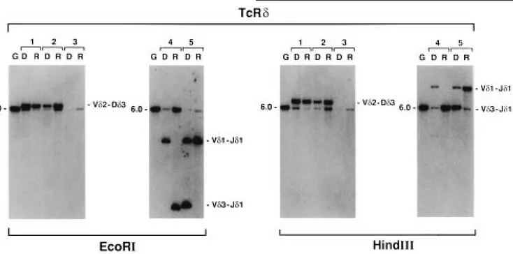
The same TcR␦ rearrangement (V␦2D␦3) was observed at diagnosis and relapse (Figure 1). However, when the V␦2D␦3 rearrangements were amplified by PCR and the amplification products used for direct sequence, a distinct junctional sequence was identified between DNA from presentation and Table 1 Clinical and laboratory features of the analyzed patients
Patient Sex/ Disease stage WBC Immunophenotype Leukemic Protocol Site of Clinical outcomeb
Age (months from ×109/l cells (%) treatmenta relapse
diagnosis)
1 M/9 D 36.1 common ALL 98 8602 2nd Rel. (+11 mo). Dead
R (69) 9.8 common ALL 95 9102-REC BM for progression (+12 mo)
2 M/3 D 126.0 common ALL 98 8503B Dead in 2nd CR for hesit
R (60) 14.0 common ALL 85 9103 BM failure (+6 mo)
3 M/5 D 5.4 common ALL 95 8802 PBSC-BMT (+4 mo).
R (72) 1.2 common ALL 90 9103 BM+CNS Dead for multi-organ
failure (+32 mo)
4 M/2 D 135.0 T-ALL 90 8503A Syngeneic BMT (+4 mo).
R (72) 27.0 T-ALL 70 VCR+ BM+CNS+ CCR (+5 y)
Epidoxorubicin Kidney
5 M/5 D 173.0 T-ALL 95 8503A Allo BMT (+6 mo). Dead
R (77) 141.8 T-ALL 80 REC89 BM for multi-organ failure (+3
mo) aDuration of front-line protocols was 24 months.
bTime points refer to the date of first relapse.
from the presentation bone marrow of this case showed germ-line bands, an equally intense rearranged band for V␥7 and a second weakly hybridizing band for V␥9. At relapse, the intense rearranged band (V␥7) was lost and a new, strongly hybridizing band (V␥2/4) appeared. The band at the position of the weakly hybridizing V␥9 rearranged band, shown at diagnosis, increased its intensity. PCR amplification and sequence analyses confirmed the nature of V␥7 and V␥4 for the two non-concordant rearrangements at diagnosis and relapse, respectively (data not shown). Sequence analysis of the junctional V–J region of the rearranged V␥9 demonstrated an identical junctional region at presentation and relapse (Table 2).
Patient 3
The analysis of TcR␦ gene displayed the same pattern at diag-nosis and at relapse. Southern blot analysis of DNA from pres-entation marrow after digestion with EcoRI and HindIII and the TcR␥ probe hybridization showed two germline and one rearranged band (V␥2/4) (Figure 1). At relapse both the germ-line band and the V␥2/4 rearranged band persisted, while a new rearranged band (V␥5) also appeared. Sequence analysis of the corresponding TcR␥ rearranged segment confirmed the identity of the same V␥4 rearrangement at diagnosis and at relapse.
Patient 4
Clonal variation at the TcR␦ locus was documented as a V␦1– J␦1 rearrangement at diagnosis whereas a V␦3–J␦1 was observed at recurrence (Figures 1 and 2). Of interest, when PCR amplification and sequence analyses were performed at presentation and relapse, the presence of the same V␦1–J␦1 rearrangement (detectable only at diagnosis by Southern blot) was confirmed even at the time of relapse (Table 2). Southern blot analysis of TcR␥ gene from the presentation bone marrow of this case showed a V␥2/4 rearranged band, whereas at relapse a second equally intense rearranged band
192 Table 2 Rearrangement patterns by Southern blotting, PCR analysis and sequencing of the ‘N’ region Patient Status Southern blotting PCR TcR gamma sequence TcR delta sequence TcR gamma TcR delta TcR gamma TcR delta 1D V ␥ 7–V ␥ 9V ␦ 2D ␦ 3V ␥ 9V ␦ 2D ␦ 3V ␥ 9 tgtgcctAGAGGtataagaaa V ␦ 2 cctgtgacgCCactggggg RV ␥ 2/4–V ␥ 9V ␦ 2D ␦ 3V ␥ 2, V ␥ 4V ␦ 2D ␦ 3V ␥ 4 tattacCCCTGGGACGGacaa V ␦ 2 cctgtTCGTactggggg V ␥ 9V ␥ 9 tgtgcctAGAGGtataagaaa 2D V ␥ 9V ␦ 2D ␦ 3N D V ␦ 2D ␦ 3N D V ␦ 2 tgtgaccccGAGGtggggg RV ␥ 9V ␦ 2D ␦ 3N D V ␦ 2D ␦ 3N D V ␦ 2 tgtgaccccGAGGtggggg 3D V ␥ 2/4 D/G V ␥ 2, V ␥ 4/ V ␥ 4 tgtgccaccCtataagaaa / RV ␥ 2/4–V ␥ 5 D/G V ␥ 2, V ␥ 4/ V ␥ 4 tgtgccaccCtataagaaa / V ␥ 5V ␥ 5 ggggtcCTGGGgaattatt 4D V ␥ 2/4 V ␦ 1J ␦ 1V ␥ 2, V ␥ 4V ␦ 1J ␦ 1V ␥ 4 gggaGCAGGTGGCGTCGatt V ␦ 1 aacCcctactgggggataTCGGaa RV ␥ 2/4–V ␥ 11 V ␦ 3J ␦ 1V ␥ 2, V ␥ 4V ␦ 3J ␦ 1V ␥ 2 acggTCACGGGAtataa V ␦ 3 cctttaTCGGTTtaaactc V ␥ 11 V ␦ 1J ␦ 1N D V ␦ 1 aacCcctactgggggataTCGGaa 5D V ␥ 2/4–V ␥ 8V ␦ 1J ␦ 1V ␥ 2, V ␥ 4V ␦ 1J ␦ 1V ␥ 2 acgggCGAGTTTTTGgaaa V ␦ 1 ggggaacAATGCATGCACAGCTcctt V ␦ 3J ␦ 1V ␥ 8V ␦ 3J ␦ 1V ␥ 8 gggataAAAGGTTGTCGTC ccACgggggatacgCAGGGTAGacc GGGgaaa V ␦ 3 cctttCAAGGTACTGAgaaaAAGccttccC GGAAGactgggggatacgGAAGACTcacc RV ␥ 2/4–V ␥ 8V ␦ 1J ␦ 1V ␥ 2, V ␥ 4V ␦ 1J ␦ 1V ␥ 2 acgggCGAGTTTTTGgaaa V ␦ 1 ggggaacAATGCATGCACAGCTcctt V ␥ 8V ␥ 8 gggataAAAGGTTGTCGTC ccACgggggatacgCAGGGTAGacc GGGgaaa
Figure 1 Southern blot analysis with the TcRDJ1 probe of EcoR1 and Hindlll cut DNA from bone marrow mononuclear cells of the five patients at diagnosis (D) and first relapse (R). G, germline pattern from placental DNA of a healthy donor. The samples are coded according to Table 1. Rearrangement patterns for V␦2–D␦3, V␦1–J␦1 and V␦3–J␦1 are indicated on the right of each panel. The size (kb) of the germline fragment is indicated on the left of each panel.
Figure 2 Southern blot analysis with the PH60 probe of EcoRl and Hindlll cut DNA from bone marrow mononuclear cells of the five patients at diagnosis (D) and first relapse (R). G, germline pattern from placental DNA of a healthy donor. The samples are coded according to Table 1. Rearrangement patterns for V␥2/4, V␥3, V␥5, V␥7, V␥8 and V␥9 V␥family members are indicated on the right of each panel. The sizes (kb) of the germline fragments are indicated on the left of each panel.
194
(corresponding to a V␥11 rearrangement) became apparent. PCR amplification and sequence analyses showed a V␥4–J␥1 rearrangement from the presentation marrow, whereas a V␥2– J␥1 was observed from the relapse marrow.
Patient 5
Rearranged bands at similar positions (corresponding to a V␦1–J␦1 rearrangement) were detected with the TcR␦ probe in both the presentation and relapse marrows, the former hav-ing an additional rearranged band correspondhav-ing to a V␦3– J␦1 rearrangement. PCR amplifications and sequence analyses showed the same junctional region of the V␦1–J␦1 rearranged allele at diagnosis and at relapse. No clonal variation was observed at the TcR␥ locus, a similar biallelic rearranged pat-tern (V␥2/4 and V␥8) being detected in both the presentation and relapse marrow by Southern blot (Figures 1 and 2) and further confirmed by PCR and sequence analyses.
Discussion
Even with modern front-line therapy protocols for childhood ALL, relapse remains the most important obstacle to over-come.1–3Although most events occur 24–36 months after first CR has been achieved,2few patients relapse 5 or more years after CR achievement. Such very late relapse is generally con-sidered a different biological and clinical entity, because of the long-term duration of first CR. In that context, the assess-ment of clonality has proved to be useful in determining whether even those very late events represent the reoccur-rence of the original clone or alternatively a secondary
leuke-mia.7,10,13,14Our study extends to a larger series of patients
previous findings which have sought to analyze the phenom-enon of clonal evolution in children relapsed after more than 5 years of CCR.13,15
At least one stable rearranged allele of the TcR␦ and TcR␥ loci was traced in all cases at presentation and clinical relapse despite a wide heterogeneity of the pattern of rearrangements. Differences at diagnosis and relapse with respect to TcR␥ locus occurred in these cases. In two cases (Nos 1 and 4) further rearrangements of V␥ genes, which in germline con-figuration are located upstream of the V␥ genes rearranged at the presentation, occurred at relapse. By contrast, the occur-rence of new clonal rearrangements can be observed at relapse in case Nos 3 and 4.
The existence of a significant heterogeneity of the TcR␦ gene leading to bi/oligoclonality in precursor B-ALLs, as reported by Ghali et al16might explain the pattern observed in case No. 1. Discordant junctional sequences of the V␦2– D␦3 rearrangement were found at presentation and relapse, suggesting either a different response to chemotherapy of the subclone present at diagnosis or a different proportional com-position. No experiments have been done to explore further the latter hypothesis. Clonal variation of VDJ rearrangements at the TcR␦ locus was documented in case Nos 4 and 5. Of interest, PCR analysis revealed the persistence in the relapse sample of the same V␦1–J␦1 rearrangement observed at diag-nosis, although undetectable by Southern blot.
The existence of at least one concordant rearranged allele favors the hypothesis that the new rearrangement observed at relapse probably originates from the presentation clone. A certain degree of clonal stability demonstrated in all cases of relapsing children after a long-term CCR (⬎5 years), is further
supported by the few reports indicating that even relapses occurring more than 10 years after diagnosis are due to true re-emergence of the original leukemic clone.13,15,21 It is intriguing to interpret a very long period of clinical remission with the persistence of minimal residual disease, being detect-able only by using very sensitive PCR techniques.22 What mechanism(s) can suppress the malignant clone for so long a time and which events can lead to its re-emergence, are still unanswered questions.
With respect to the potential pitfalls in monitoring minimal residual disease in childhood ALL for the presence of clonal evolution, our results highlight the combination of two target genes (such as TcR␥ and TcR␦) as a tool to reduce false nega-tive MRD results. Those findings further extend (even for late-relapse events) those recently reported in the largest series of children ALL prospectively analyzed for the prognostic impact of MRD detection.23
Acknowledgements
This work was partially supported by IBISCUS (LLN and ADC), by the Associazione Italiana per la Ricerca sul Cancro (AIRC) (AB), by the AIL project ‘30 ore per la vita’ (AP) and by Fondazione Tettamanti (GC, Ed’A and AB).
References
1 Pui CH. Childhood leukemias. New Engl J Med 1995; 332: 1618–1630.
2 Henze G, Fengler R, Hartmann R, Kornhuber B, Janka-Schaub G, Niethammer D, Riehm H. Six-year experience with a comprehen-sive approach to the treatment of recurrent childhood acute lym-phoblastic leukemia (ALL-REZ BFM 85). A relapse study of the BFM group. Blood 1991; 78: 1166–1172.
3 Conter V, Arico` M, Valsecchi MG, Rizzari C, Testi A, Miniero R, Di Tullio MT, Lo Nigro L, Pession A, Rondelli R, Messina C, San-toro N, Mori PG, De Rossi G, Tamaro P, Silvestri D, Biondi A, Basso G, Masera G for the ‘Associazione Italiana Ematologia Oncologia Pediatrica’ (AIEOP). Intensive BFM chemotherapy for childhood ALL: interim analysis of the AIEOP-ALL 91 study. Haematologica 1998; 83: 791–799.
4 Ochs J, Mulhern RK. Late effects of antileukemic treatment. Pediatr Clin North Am 1988; 35: 815–833.
5 Beishuizen A, Verhoeven MA, Hahlen K, van Wering ER, van Dongen JJ. Differences in immunoglobulin heavy chain gene rearrangement patterns between bone marrow and blood samples in childhood precursor B-acute lymphoblastic leaukemia at diag-nosis. Leukemia 1993; 7: 60–63.
6 Breit TM, Wolvers-Tettero IL, Beishuizen A, Verhoeven MA, van Wering ER, van Dongen JJ. Southern blot patterns, frequencies, and junctional diversity of T-cell receptor-delta gene rearrange-ments in acute lymphoblastic leukemia. Blood 1993; 82: 3063– 3074.
7 Beishuizen A, Verhoeven MA, van Wering ER, Hahlen K, Hooijkaas H, van Dongen JJ. Analysis of Ig and T-cell receptor genes in 40 childhood acute lymphoblastic leukemias at diagnosis and subsequent relapse: implications for the detection of minimal residual disease by polymerase chain reaction analysis. Blood 1994; 83: 2238–2247.
8 Marshall GM, Kwan E, Haber M, Brisco MJ, Sykes PJ, Morley AA, Toogood I, Waters K, Tauro G, Ekert H, Norris MD. Characteriz-ation of clonal immunoglobulin heavy chain and I cell receptor gamma gene rearrangements during progression of childhood acute lymphoblastic leukemia. Leukemia 1995; 9: 1847–1850. 9 Sterbergeen EJ, Verhagen OJHM, van Leeuwen EF, van der Berg
H, von dem Borne AEGK, van der Schoot CE. Frequent ongoing T-cell receptor rearrangements in B-precursor acute lymphoblastic leukemia: implications for monitoring minimal residual disease. Blood 1995; 86: 692–702.
K, Potter MN, Chessels JM, Oakhill A. A polymerase chain reac-tion study of the stability of Ig heavy-chain and T-cell receptor d rearrangements between presentation and relapse of childhood B-lineage acute lymphoblastic leukemia. Blood 1994; 83: 1355– 1362.
11 Davi F, Gocke C, Smith S, Sklar J. Lymphocytic progenitor cell origin and clonal evolution of human B-lineage acute lymphoblas-tic leukemia. Blood 1996; 88: 609–621.
12 Taylor JJ, Rowe D, Kylefjord H, Chessells J, Katz F, Proctor SJ, Middleton PG. Characterisation of non-concordance in the T-cell receptor gamma chain genes at presentation and clinical relapse in acute lymphoblastic leukemia. Leukemia 1994; 8: 60–66. 13 Levasseur M, Maung ZT, Jackson GH, Kernahan J, Proctor SJ,
Middleton PG. Relapse of acute lymphoblastic leukaemia 14 years after presentation: use of molecular techniques to confirm true re-emergence. Br J Haematol 1994; 87: 437–438.
14 Wassermann R, Yamada M, Ito Y, Finger LR, Reichard BA, Shane S, Lange B, Rovera G. VH gene rearrangement events can modify the immunoglobulin heavy chain during progression of B-lineage acute lymphoblastic leukemia. Blood 1992; 79: 223–228. 15 Frost L, Goodeve A, Wilson G, Peake I, Barker H, Vora A. Clonal
stability in late-relapsing childhood lymphoblastic leukaemia. Br J Haematol 1997; 98: 992–994.
16 Ghali DW, Panzer S, Fischer S, Argyriou-Tirita A, Haas OA, Kovar H, Gadner H, Panzer-Grumayer ER. Heterogeneity of the T-cell receptor delta gene indicating subclone formation in acute precur-sor B-cell leukemias. Blood 1995; 85: 2795–2801.
17 van der Does-van den Berg A, Bartram C, Basso G, Benoit YCM, Biondi A, Debatin KM, Haas OA, Harbott J, Kamps WA, Koller U, Lampert F, Kudwig WD, Niemeyer CM, van Wering E. Minimal requirements for the diagnosis, classification and evaluation of the
‘BFM Family’ Cooperative Group. Med Ped Oncol 1992; 20: 497–505.
18 Bottaro M, Berti E, Biondi A, Migone N, Crosti L. Heteroduplex analysis of T-cell receptor␥gene rearrangements for diagnosis and monitoring of cutaneous T-cell lymphomas. Blood 1994; 83: 3271–3278.
19 Lefranc MP, Forster A, Rabbitts TH. Rearrangement of two distinct T-cell gamma-chain variable-region genes in human DNA. Nature 1986; 319: 420–422.
20 Pongers-Willemse MJ, Seriu T, Stolz F, d’Aniello E, Gameiro P, Pisa P, Gonzales M, Bartram CR, Panzer-Grumayer ER, Biondi A, San Miguel JF, van Dongen JJM. Primers and protocols for stan-dardized detection of minimal residual disease in acute lymphobl-astic leukemia using Immunoglobulin and T cell receptor gene rearrangements and TAL1 deletions as PCR targets. Leukemia 1999; 13: 110–118.
21 Vora A, Frost L, Goodeve A, Wilson G, Ireland RM, Lilleyman J, Eden T, Peake I, Richards S. Late relapsing childhood lymphoblas-tic leukemia. Blood 1998; 92: 2334–2337.
22 Roberts WM, Estrov Z, Ouspenskaia MV, Johnston DA, McClain KL, Zipf TF. Measurement of residual leukemia during remission in childhood acute lymphoblastic leukemia. New Engl J Med 1997; 336: 317–323.
23 van Dongen JJM, Seriu T, Panzer-Grumayer ER, Biondi A, Pongers-Willemse MJ, Corral L, Stolz F, Schrappe M, Masera G, Kamps WA, Gadner H, van Wering ER, Ludwig WD, Basso G, de Brujin MAC, Cazzaniga G, Hettinger K, van der Does-van der Berg A, Hop WCJ, Riehm H, Bartram CR. Prognostic value of minimal residual disease in childhood acute lymphoblastic leukemia: a prospective study of the International BFM study group. Lancet 1998; 352: 1731–1738.