Università degli Studi di Salerno
Facoltà di Scienze Matematiche Fisiche e Naturali
DOTTORATO DI RICERCA IN CHIMICA
XIII Ciclo – Nuova serie
“Tesi di dottorato in:
Polymeric films with co-crystalline and
nanoporous crystalline phases:
orientations, chirality and possible
applications in photonic crystals”
Tutor PhD student
Dott.ssa Paola Rizzo Graziella Ianniello
Co-tutor
Matr. 8880700113
Prof. Vincenzo Venditto
Coordinatore
Prof. Gaetano Guerra
Introduction
Polymeric films with co-crystalline and nanoporous crystalline phases
5
References 12
Chapter
1
Disordered nanoporouscrystalline modifications and co-crystalline phase of syndiotactic polystyrene including carvacrol guest molecules
1.1 Introduction 14
1.2 Guest removal from intercalate forms 17
1.2.1 Guest removal from the triclinic
δ clathrate form with DBF 19
1.2.2 Guest sorption from the disordered
crystalline form obtained by guest (DBFTMB) removal from their
clathrate and intercalate forms 21
1.3 sPS\ carvacrol co-crystalline film 26
1.3.1 FTIR method to discriminate carvacrol
molecules in co-crystalline and amorphous
polymer phases 28
1.3.2 Partition of carvacrol molecules between
amorphous and crystalline phases of δ-form films
31
1.4 Conclusion 38
References 40
Chapter
2
Orientations in polymeric films2.1 Introduction 43
References 46
2.2 Poly(2,6-dimethyl-1,4-phenylene ether) (PPO)
48
2.2.1 Uniplanar orientations by
α-pinene-induced cocrystallization on amorphous films. 48
tetralin-induced cocrystallization of amorphous films.
54
2.2.3 Guest exchange in PPO cocrystalline
films with two different uniplanar orientations.
2.2.4 Uniplanar orientations by limonene
induced cocrystallization of amorphous films
56 60
2.2.5 Guest exchange in PPO/ limonene
cocrystalline films with tetralin 64
2.2.6 A preliminary model of PPO nanoporous
structure 67
2.2.7 Conclusion 70
References 71
2.3 Poly(L-lactide) (PLLA) 73
2.3.1 PLLA uniplanar orientation of ε form 73
2.3.2 Guest exchange in ε PLLA co-crystalline
phase
2.3.3 Effects of guest removal from ε form 2.3.4 Conclusion References 82 84 87 88
2.4 Correlation between shrinkage and orientation in sPS films 2.4.1 Syndiotactic polystyrene sPS 2.4.2 The shrinkage 2.4.3 Oriented α form 2.4.4 Amorphous sPS film 2.4.5 PP-sPS-PP film multilayer 2.4.6 Conclusion References 89 89 90 91 94 100 104 105 Chapter
3
Melt-extruded films of acommercial polymer with intense chiral optical response of achiral guests
3.1 Introduction 108
3.2 Induction of chirality in melt-extruded s-PS
with a nonracemic guest 110
3.3 Chiral optical polymer films with achiral
chromophores.
3.4 Considerations relative to g-value variations
with film thickness.
3.5 Conclusion References 114 120 123 124 Chapter
4
Photonic Crystals (PhC)4.1 Introduction 127
4.2 Cellulose Acetate (CA) and
Poly(2,6-dimethyl-1,4-phenyleneether)(PPO)
Photonic Crystals (PhC) 129
4.3 Thin films 133
4.3.1 Comparison of FT-IR and IRRAS analysis
of PPO thin films
133 4.3.2 Desorption of CCl4 141 4.3.3 Refractive indexes 144 4.4 Conclusion References 148 149 Summary 151
Chapter
5
Experimental section 5.1 Materials and Sample preparation 5.1.1 sPS5.1.2 PPO 5.1.3 PLLA 5.2 Techniques
5.2.1 X-ray diffraction analysis 5.2.2 IR Infrared spectra 5.2.3 TGA measurements References 155 155 156 157 157 157 159 162 162
List of Abbreviations
AN: Acetonitrile CA: Cellulose Acetate CCl4: Carbon tetrachloride
CD: Circular Dichroism CH2Cl2: Diclorometano
CPO: Cyclopentanone DBF: Dibenzofuran
DBR: Distributed Bragg Reflector DCE 1,2-dichloroethane
disi: disordered modification from the intercalate phase
dist: disordered modification obtained from the triclinic
clathrate phase
DMF: Dimethylformamide DMN: Dimethylnaphthalene DNB: 1,4-dinitrobenzene DOL: Dioxolane
g: Degree of circular polarization GBL: γ-butyrolactone
NA: paranitroaniline PhC: Photonic Crystal PLLA: Poly (L-lactide)
PPO: Poly (2,6-dimethyl-1,4-phenylene)oxide sPS: Syndiotactic Polystyrene
THF: Tetrahydrofuran
TMB: 1,3,5-trimethylbenzene
VCD: Vibrational Circular Dichroism XRD: X-ray Diffraction
I
ntroduction
Polymeric
films
with
co-crystalline
and
nanoporous crystalline phases
Polymers can crystallize in different crystalline forms; polymorphism is the term to indicate this ability. It is known that processing and physical properties of polymer-based materials are strongly affected by the occurrence of ‘‘polymorphism’’1and ‘‘metamorphism’’ (i.e., the occurrence
of ‘‘disordered’’ crystalline phases, characterized by a degree of structural organization that is intermediate between those identifying crystalline and amorphous phases)2a
My PhD thesis is focused on the study and on the characterization of polymer films with co-crystalline and nanoporous crystalline phases. Many polymers are able to form co-crystals i.e. molecules of low molecular weight (guest) trapped in the crystalline polymer lattice (host). Over the past two decades it has been observed that some polymers, with co-crystalline phases, such as syndiotactic polystyrene (sPS) and poly (2,6-dimethyl-1,4-phenylene oxide) (PPO) after guest removal can form nanoporous crystalline phases, able to absorb suitable guest molecules also at low activity. During this work, I have studied the possible molecular orientations that may be induced by solvents during co-crystallization process in polymeric films, (chapter 2); the development of chiro optical response, after co-crystallization with temporary chiral guest (chapter 3) and the possibility to realize photonic crystals by using polymers able to form nanoporous crystalline forms (chapter 4).
In detail, in chapter 1 the procedure to obtain disordered nanoporous crystalline phases in sPS films and their possible application is reported. This disordered nanoporous crystalline phase rapidly absorb low molecular mass molecules, also from very dilute aqueous solutions.
It is known in literature that nanoporous δ form of sPS is also able to absorb ethylene2b and carbon dioxide 2c-d, that have negatively effects for vegetable. Active packaging by
nanoporous-crystalline films, based on the removal of molecules generated by the vegetables being detrimental for their preservation 2e, could be complemented by the slow-release of antimicrobial molecules, which could be included as guest of the film crystalline cavities. Therefore the preparation of s-PS co-crystalline films that include guests with antimicrobial activity, in particular the carvacrol guest has been studied and reported in chapter 1. The kinetics of release, in variable concentrations of carvacrol in films with different thickness, has been analyzed. It was observed that the location of antimicrobial molecules mainly in the crystalline phase assure a decrease of desorption diffusivity and hence along-term antimicrobial release.
In chapter 2, the study of the possible molecular orientations that can be developed in polymer films able to form co-crystalline phases, are reported. This phenomenon has been observed only for sPS films until now. In particular, in my thesis has been shown that also other polymers, such as poly (2, 6-dimethyl-1, 4-phenylene oxide) (PPO) and poly (L-lactide) (PLLA), able to form co-crystalline phases, can develop orientations during the co-crystallization process with solvents. These orientations can be useful to the structural studies on PPO and PLLA co-crystalline forms.
We have also investigated on the shrinkage behavior developed in syndiotactic polystyrene (sPS) films after co-crystallization procedures leading to co-crystalline phases. High shrinkage values have been measured on sPS co-crystalline phase showing a co-crystalline phase orientation. In order to minimize this effect, novel procedures have been developed.
Another aspect of my work is focused on the study of chiro optical response of a racemic polymer crystallized with a temporary chiral guest, as reported in chapter 3. In particular, I evaluated the degree of circular polarization of different thickness sPS films, and of the achiral guests, such as azulene and 4-nitroaniline, included in the polymer crystalline phase after guest exchange procedure. These studies have been
useful to investigate on the nature of this phenomenon.
Finally, in chapter 4, a method to realize a photonic crystal (PhC) with polymeric materials is reported. A PhC is an object composed by two or more materials with different refractive index and an alternated periodicity. The main advantage to use polymers rather than inorganic materials is the ease and the speed to obtain thin films by spin coating and the low cost of materials.
In order to realize a photonic crystal, by using thin layers of PPO presenting nanoporous crystalline phase, it has been necessary to characterize amorphous as well as crystalline phases for this purpose. Techniques such as IRRAS and ellipsometry have been used (as reported in section 4.3 of chapter 4).
Characteristics of polymeric films presenting co-crystalline or nanoporous crystalline phases:
Syndiotactic polystyrene (sPS), Poly(2,6-dimethyl-1,4-phenylene ether) (PPO), Poly (L-lactide) (PLLA)
Syndiotactic polystyrene (sPS) exhibits a complex polymorphic behavior 3,4, which can be described in terms of two crystalline forms, α 5 and β 6, containing planar zigzag chains and three forms, γ7, δ8 and 9, containing s(2/1)2 helical chains (helical repetition of two structural units in one turn, the structural unit being formed by two monomeric units). Both β and α forms can exist in different modifications having different degrees of structural order, so that two limit-disordered modifications (β’ and α’) and two limit-ordered modifications (β’’ and α’’).10
8 and 9 forms are nanoporous-crystalline, i.e. exhibit
low-density packing of the polymer chains with some empty space regularly distributed in the crystalline phase and available for sorption of suitable guest molecules.11
Moreover, it is well known that s-PS can form co crystalline phases (clathrate3 and intercalate 4) with several low
molecular-mass molecules where the polymer always assumes its s(2/1)2 helical conformation. Melt-crystallization procedures lead only to the trans-planar α 5 and β 6 phases. On the other hand, solution crystallization and solvent-induced crystallization generally lead to the formation of helical cocrystalline and γ phases, although the thermodynamically favored trans-planar β phase is also obtained by solution casting at high temperatures as well as by annealing above 150 °C of cocrystalline and γ samples in the presence of solvents.12 The nanoporous δ and ε forms are obtained by guest removal from cocrystalline phases.12
Fig.1 Top and lateral views of the crystalline structures of the two nanoporous crystalline forms of s-PS. For the δ (upper figures) and ε (lower figures) forms, the porosity is distributed as cavities and channels, respectively. 4
Materials presenting the nanoporous δ crystalline phase have been deeply studied for their promising applications in chemical separations (mainly air/water purification) in sensorics or possible application as films for packaging, of particular economic relevance is the ability of the nanoporous δ form to absorb ethylene 2b and carbon dioxide 2c-d, they are
the molecules generated by the vegetables and detrimental for their preservation 2e. This ability could increase the shelf-life of vegetable. Films presenting s-PS/ active-guest co-crystals have been proposed as advanced materials, mainly for optical applications (e.g., as fluorescent, photoreactive) 12
Poly(2,6-dimethyl-1,4- phenylene ether) (PPO) is a polymer that exhibits nanoporous-crystalline phases, as s-PS.13 In most cases, his guest solubility is not only definitely higher than for the high free volume amorphous PPO phase but also higher than for the s-PS nanoporous crystalline forms. 13 Recently it was found that there is nearly a continuum of crystalline phases between two limit structures exhibiting highest and lowest 2θ values. The d values obtained with the different solvents clearly show that the solvent plays a fundamental role in determining the periodicity of the final crystalline phase. 13
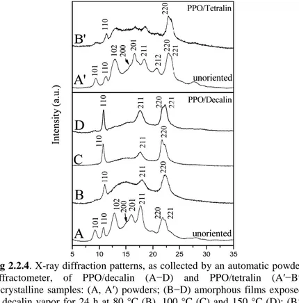
Moreover, since the end of the sixties of the last century, it is well known that PPO can co-crystallize with many guest molecules. 14 In fig.1.2 the X-ray diffraction pattern of clathrate structure PPO/α-pinene, reported in literature, 14b is reported.
Fig. 2 X-ray diffraction pattern of chlatrate structure PPO/α-pinene taken from Horikiri S., Journal of polymer Science 1972,(10), 1167-70
The only crystalline structure defined for PPO is the co-crystalline structure with -pinene, 14c whose unit-cell has been defined many years ago by electron diffraction of single crystals14d. Recently this structure has been refined. It was observed that the chiral guest is able to induce its chirality to the crystalline structure. The packing model of the
co-crystalline form of PPO with (1S)-(-)--pinene in a tetragonal unit cell (a=b=1.19 nm and c=1.71 nm) and according to the P43 space group is shown in Figure 1.3. 14c
Fig. 3 Packing model of the co-crystalline form of PPO with (1S)-(-)-α-pinene in the unit cell a= b = 1.19 nm and c = 1.71 nm, and the P43 space
group (A) Projection along c; (B) projection along a. None bonded distances are all longer than 0.36 nm.
Major applications of PPO are for automotive, business machine, and electrical industries.
Moreover PPO is an attractive material for the preparation of membranes due to its resistant against a number of chemical agents, including aqueous solutions of strong acids and bases. Poly (L-lactide) (PLLA) has different crystalline forms (α, β and γ).15
The α form of PLLA grows upon melt or cold crystallization, as well as from solution. The α form has two antiparallel chains in a left-handed 103 helical conformation (or distorted
103 helix) packed in an orthorhombic (or
pseudo-orthorhombic) unit cell with a = 1.066 nm, b = 0.616 nm, c = 2.888 16-17. The β form 18-19 (orthorhombic 18 or trigonal19)
is known to take a 31 helical conformation 20 the γ form is
obtained via epitaxial crystallization on hexamethylbenzene substrate. It is characterized by two antiparallel helices with
31 conformations packed in an orthorhombic unit cell with
a = 0.995 nm, b = 0.625 nm, c = 0.880 nm15
In addition to these crystalline forms, is known a metastable crystalline form called α', and it was observed that the formation of α' does not depend on the polymer molecular mass but from the crystallization temperature.16,17 Two different crystal modification, α' and α, are obtained at low and high crystallization temperature respectively21
The α' form has a 103 helix conformation and orthorhombic
(or pseudoorthorhombic) unit cell, like the α form, but the molecular packing within the unit cell of α ' is looser and more disordered than in the α form, due to the larger lattice dimension and weaker interchain interaction.21
Moreover, during heating the α’ crystals transform into α modification: the change of crystal structure from α' to α is a solid–solid transition that mainly involves slight rearrangements of the molecular packing within the unit cell to the more energy-favorable state, corresponding to a reduction of lattice dimensions.21
In addition in literature is reported that PLLA, treated with CO2 at different temperatures and pressures, can develop
different α, α' and α'' forms. The α'' form is expected to be nanoporous.22
Also PLLA is able to form co-crystals, as sPS and the PPO. It is known from literature that solvents such as CPO (cyclopentanone), DMF (dimethyl formamide), THF (tetrahydrofuran), DOL (dioxolane), GBL (γ-butyrolactone) are able to induce co crystalline ε form. 23
The ε co-crystalline structure including N,N-dimethylformamide (DMF) has been recently described in literature, in this model the PLLA chains show a 107 helical
conformation and are packed in the orthorhombic lattice with a=1.5-1.6 nm, b=1.2-1.3 nm and c=2.8-2.9 nm. 23
PLLA is a biodegradable and biocompatible polyester that can be produced by renewable resources. Being non-toxic to human body, PLLA is used in biomedical applications, like surgical sutures, bone fixation devices, or controlled drug
delivery. The mechanical and thermal properties of this semicrystalline polymer greatly depend on the crystal structure and morphology.21
References
1. a) Tashiro, K.; Tadokoro, H. In Encyclopedia of Polymer
Science and Engineering. Supplement 1989; p 187 b) Corradini, P; Guerra, G. Adv. Polym. Sci. 1992, 100, 183
2. a)Auriemma, F.; De Rosa, C.; Corradini, P. Adv. Polym.
Sci. 2005, 181, 1–74. b) A. R. Albunia, T. Minucci, G. Guerra, J. Mater. Chem. 2008, 18, 1046. c) Larobina D., Sanguigno L., Venditto V., Guerra G., Mensitieri G., Polymer
2004, 45, 429.
d) Annunziata L., Albunia A. R., Venditto V., Guerra G.,
Macromolecules ,2006, 39, 9166 e) P. Rizzo;C. Daniel; A. De
Girolamo Del Mauro; G. Guerra 2006 SA2006A22 Università di Salerno ID:1517898 Brevetto
3. Guerra, Vitagliano, De Rosa, Petraccone, Corradini
Macromolecules 1990, 23, 1539
4. Guerra, G., Daniel, C., Rizzo, P., Tarallo, O.:
J.Polym.Sci.Polym.Phys. 2012, 50, 305
5 De Rosa, C., Guerra, G., Petraccone, V., Corradini, P.:
Polym. J. 1991, 23, 1435
6. De Rosa, C., Rapacciuolo, M., Guerra, G., Petraccone, B.,
Corradini, P. Polymer, 1992, 33, 1423
7. Rizzo, P., Albunia, A. R., Guerra, G. 2005, 46, 9549
8. De Rosa, C., Guerra, G., Petraccone, V., Pirozzi, B,
Macromolecules 1997, 30, 4147
9. Rizzo, P., Daniel, C., De Girolamo Del Mauro, A., Guerra,
G. Chem. Mater., 2007, 19, 3864
10. Rizzo P., Lamberti M., Albunia A.R., Ruiz De
Ballestreros O. Guerra G. Macromolecules 2002, 35, 5854
11. Manfredi C., Del Nobile M.A., Mensitieri G., Guerra, G.,
Rapacciuolo, M. J. Polym. Sci., Part B: Polym. Phys. 1997, 35,133
12.AlexandraR. Albunia, Loredana AnnunziataGaetano Guerr
13. a) Daniel, C.; Longo, S.; Vitillo, J. G.; Fasano, G.; Guerra,
G. Chem. Mater. 2011, 23, 3195. b) Galizia, M.; Daniel, C.; Fasano, G.; Guerra, G.; Mensitieri, G. Macromolecules 2012, 45, 3604. c) Daniel, C.; Longo, S.; Cardea, S.; Vitillo, J. G.; Guerra, G. RSC Advances 2012, 2, 12011. d) Daniel, C.; Zhovner, D.; Guerra, G. Macromolecoles 2013, 46, 449.
14. a) Factor, A.; Heinsohn, G. E.; Vogt, L. H., Jr. Polym.
Lett.1969, 7, 205. b) Horikiri, S. J. Polym. Sci. A2 1972, 10, 1167 c) Tarallo, O.; Petraccone, V.; Daniel, C.; Fasano, G.; Rizzo, P.; Guerra, G. J. Mater. Chem. 2012, 22, 11672. d) Barrales-Rienda, M.; Fatou, J. M. G. Kolloid Z. Z. Polym.
1971, 244, 317. e) Hurek, J.; Turska, E. Acta Polym. 1984,
35, 201.
15. Di Lorenzo M. L., Cocca M., Malinconico M.
Thermochimica Acta 2011, 522, 110
16. Pan P., Kai W., Zhu B, Dong T., Inoue Y.
Macromolecules 2007, 40, 6898
17. Pan P., Zhu B, Kai W., Dong T., Inoue Y Journal of
applied Polymer Science, 2008,107, 54
18. Di Lorenzo ML. Eur Polym J 2005;41, 569.
19. Kawai T, Rahman N, Matsuba G, Nishida K, Kanaya T,
Nakano M, Macromolecules 2007, 40,9463.
20. Sawai D., Takahashi K., Sasashige A., Kanamoto T.
Macromolecules 2003, 36, 3601-3605
21. Cocca M., Di Lorenzo M.L., Malinconico M., Frezza V.
European Polymer Journal 2011, 47, 1073
22. Marubayashi H., Akaishi S., Akasaka S., Asai S., Sumita
M., Macromolecules 2008, 41, 9192
23. Marubayashi H., Asai S., Sumita M., Macromolecules 2012, 45, 1384
C
hapter 1
Disordered nanoporous crystalline modifications and co-crystalline phase of Syndiotactic polystyrene including carvacrol guest molecules
1.1 Introduction
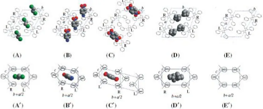
Different classes of co-crystalline forms have been described for s-PS, all exhibiting as a common feature the s(2/1)2 helical polymer conformation, with a repetition period of nearly 0.78 nm 1,2: monoclinic δ-clathrates 1,3, triclinic δ-clathrates 4,5, intercalates (also named δ-intercalates) 6,7 and ε-clathrates 8,9.
Fig. 1.1 Schematic projections along the chain (up) and perpendicular to the chain (down) of s-PS co-crystalline forms: A, A’ monoclinic
δ-clathrate form with 1,2-dichloroethane 10, B, B’ triclinic δ -clathrate
form with p-nitroaniline 4, C, C’ triclinic δ -clathrate form with
1,4-dinitrobenzene 5 D, D’ intercalate form with norbornadiene 11, and E,
E’ monoclinic δ -nanoporous phase 12
Monoclinic δ-clathrates (Fig. 1.1A–A’) 10, triclinic
δ-clathrates (Fig. 1.1B–B’) 4,5, intercalates (Fig. 1.1D–D’) 11 as well as the nanoporous δ form (Fig. 1.1E) 12, are all
characterized by ac layers of close-packed alternated enantiomorphous helices as a common structural feature. The distance between such layers (d010) as well as between the
guest molecules can change largely, mainly depending on the class of the co-crystalline form but also on the molecular volume of the guest molecules. In particular, monoclinic
δ-clathrates are obtained with guests whose molecular volumes are significantly lower than 0.12 nm3. As the bulkiness of the guest increases, there is typically an increase of the spacing (d010 = bsin γ) between the ac layers (in the range 1.06–1.2
nm) 1,3,13. Because in most cases a single guest molecule is present in each cavity, the maximum molar ratio between guest molecules and styrenic units is 1/4. On the other hand, triclinic δ-clathrates are generally obtained with guests whose molecular volumes are higher than 0.12 nm3.4,5 Guests having a volume close to 0.12 nm3, like p-nitro-aniline (NA, Vmol ~
0.122 nm3), can be accommodated only by shifting the dense
ac layers along the chain axis, keeping the d010 value almost
equal to that of the δ form, and thus leading to a triclinic unit cell (Fig. 1.1B–B’) 4. Also, in this case the maximum molar ratio between guest molecules and styrenic units is 1/4. For triclinic δ-clathrates obtained with guests having a higher molecular volume, e.g. 1,4 dinitrobenzene (DNB) (Vmol ~
0.134 nm3) or dibenzofuran (DBF) (V
mol ~ 0.159 nm3), the
close packing of the polymer chains in the ac layers is partially lost and one half of the cavities initially present in the δ phase are lost allowing the volume of the other half to increase and becoming suitable to host bulky guest molecules (Fig. 1.1C–C’) 5. This of course halves the maximum guest/monomer-unit molar ratio that then becomes 1/8.
A further class of s-PS δ co-crystals, defined as δ-intercalates (or simply intercalates) 6,7, is characterized by the same layers of alternated enantiomorphous s(2/1)2 polymer helices (Fig.1.1A, B, D), but the spacing between the ac layers (d010),
for all the known s-PS intercalates, is larger than 1.4 nm and values as high as 1.75 nm have been observed 13. In fact, in these co-crystals the guest molecules are not isolated into host cavities but are contiguous inside layers intercalated with the dense polymer ac layers. Of course, these intercalate structures present higher guest content with respect to the clathrate structures.
Therefore disordered crystalline modifications formed by s(2/1)2 helices of syndiotactic polystyrene (s-PS) can be
prepared by the removal of bulky guest molecules from intercalate as well as from triclinic δ clathrate forms. As a consequence, the obtained disordered crystalline modifications of s-PS can be fully considered disordered nanoporous-crystalline modifications.
Moreover the semicrystalline s-PS samples exhibiting the nanoporous δ 12,14 and ɛ 15,16 crystalline phases can rapidly and
selectively absorb volatile organic compounds as well as gas molecules, even when present at very low concentrations. This active packaging by nanoporous-crystalline films, based on the removal of molecules generated by the vegetables being detrimental for their preservation, could be complemented by the slow-release of antimicrobial molecules. The film not only is able to absorb ethylene17 and carbon dioxide, but also could be complemented by the slow-release of antimicrobial molecules, which could be included as guest of the film crystalline cavities.
In fact, it is well established that the release of many low molecular– mass guest molecules is slower from the s-PS nanoporous-crystalline host phases than for the corresponding amorphous phases.17,18 In addition, guest diffusivity can be further reduced by suitable selection of the uniplanar orientation 19,20 of the host crystalline phase. In particular, in most cases the guest diffusivity is minimized for the δ-form films exhibiting their dense ac layers of closely packed alternated enantiomorphous helices, green ribbons in Fig. 1.2 (A), parallel to the film plane. This orientation brings the a and c axes parallel to the film plane and has been defined as
a
//c
// uniplanar orientation.17,18,21,22Fig 1.2 Schematic projections along the chain axis (c axis) showing the crystal structure of: (A) monoclinic nanoporous δ-form; (B) monoclinic d clathrate form with carvacrol. High density ac layers of closely packed enantiomorphous helices have been pointed out by green ribbons. [Color figure can be viewed in the online issue, which is available at http://wileyonlinelibrary.com.]
1.2 Guest removal from intercalate forms
In Fig.1.3, X-ray diffraction patterns of s-PS films exhibiting a//c// uniplanar orientation of intercalate phases including
1,4-dimethyl-naphthalene (DMN) 13 (A) and 1,3,5-trimethyl- benzene (TMB) 13 (B) are shown. The formation of intercalate
structures is clearly shown by the shift of the 010 diffraction peak of the δ form samples (2θCuKα = 8.4°)12 down to 2θCuKα =
5.8°, and down to 2θCuKα = 6.0° for the intercalate structures
including DMN (A) and TMB (B), respectively.
After extraction of the bulky guests from the intercalates by using acetonitrile (AN), a shift of the 010 diffraction peaks from 2θCuKα = 5.8° (Fig.1.3A), or from 2θCuKα = 6.0°
Fig.1. 3 X-ray diffraction patterns of s-PS films exhibiting a//c// uniplanar
orientation of: A intercalate phase with DMN, B intercalate phase with TMB, C disordered crystalline modification obtained by DMN removal, and D disordered crystalline modification obtained by TMB removal (Fig.1.3B), to a higher 2θ value of 2θCuKα= 9.8° is apparent
(Fig. 1.3C–D); moreover, a decrease of crystallinity is clearly observed. For instance, the degree of crystallinity, as evaluated on powdered samples, changes from χc = 38 % for
s-PS films including TMB to χc = 26 % for the same films
after TMB removal.
Fig. 1.4 X-ray diffraction patterns of s-PS films exhibiting the a//c//
uniplanar orientation of the intercalate phase with TMB, before (A) and after (B) TMB removal. The photographic pattern of a δ form film exhibiting the a//c// uniplanar orientation is also reported for comparison
(C)
In Fig. 1.4 the EDGE photographic X-ray diffraction patterns, i.e. the pattern taken by having the X-ray beam parallel to the
film surface (and by placing the film sample parallel to the axis of the cylindrical camera), for films including TMB before (A) and after (B) guest removal are reported. The EDGE photographic pattern of the δ form film exhibiting the a//c// uniplanar orientation, obtained as described in Ref. 19, is
also included for comparison (Fig. 1.4C).
It is worth noting that for sPS films exhibiting the a//c//
uniplanar orientation, after extraction of bulky guests from the intercalates, a decrease of uniplanar orientation is observed. In particular, the degree of uniplanar orientation of the 010 crystallographic planes (f010), with respect to the film plane,
formalized by using Hermans’ orientation function according to Experimental section (Chapter 5 section 5.2.1 equation 1), is f010 = 0.8 for the s-PS film including TMB (Fig. 1.4A) and
f010 = 0.65 after TMB removal (Fig.1. 3B).
In summary, the guest extraction from the considered intercalates of sPS films can lead to a disordered crystalline modification characterized by a broad diffraction peak at 2ɵCuKα ~ 9.8° and presenting a lower degree of crystallinity as
well as a lower degree of uniplanar orientation with respect to the starting intercalate samples.
1.2.1 Guest removal from the triclinic δ clathrate form with DBF
The X-ray diffraction pattern of an unoriented s-PS film presenting the s-PS/styrene 23 monoclinic δ clathrate form is reported in Fig.1. 4A.
Fig.1.5 X-ray diffraction patterns of unoriented s-PS samples: A monoclinic δ co-crystalline phase with styrene, B triclinic δ-clathrate phase with DBF, C sample A after styrene removal by supercritical CO2
(monoclinic nanoporous-crystalline δ form), D sample B after DBF removal by acetonitrile (disordered nanoporous-crystalline modification), and E fully amorphous film. FTIR spectra (F–H), in range 600–500 cm–1, of the s-PS samples whose X-ray diffraction patterns are shown in (C–E) Styrene was removed by supercritical carbon dioxide, leading to a highly crystalline nanoporous δ phase (Fig. 1.5C, χc ~ 40
%). Analogous highly crystalline δ form samples can be obtained by styrene removal by other volatile guests such as acetone or acetonitrile. Guest desorption processes from the s-PS co-crystalline form, leading to the nanoporous δ phase, have been thoroughly described elsewhere 6,20.
The achievement of the nanoporous δ form by removal of guests from δ co-crystalline forms is rather common but it is not general. In fact, different behaviors can be observed when δ removing bulky guest molecules from triclinic δ-clathrate forms. For instance, the X-ray diffraction pattern of a s-PS film, including the triclinic δ-clathrate form with DBF, is shown in Fig. 1.5B. The DBF clathrate was prepared by immersion of a δ form film in a saturated solution of DBF in acetone 5. The δ spacing (1.06 and 0.91 nm) of the two
diffraction peaks at lower angles (2θ = 8.4° and 9.7°, Fig.1. 4B) clearly indicates the occurrence of this s-PS/DBF triclinic
of Fig. 1.5B, after complete guest removal by AN, only shows two broad peaks centered at 2θCuKα ~ 9.6° and 20.3° and two
shoulders at 16.6° and 23.6° and (Fig. 1.5D).
Moreover, the crystallinity index, going from the co-crystalline (Fig. 1.5B) to the disordered empty co-crystalline form (Fig. 1.5D), is reduced from χc ~ 27 % down to
χc ~ 17 %. For the sake of comparison, the X-ray diffraction
pattern of a fully amorphous s-PS sample (obtained by quenching s-PS samples from the melt) is also shown in Fig. 1.5E.
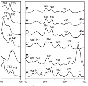
A substantial reduction of crystallinity as a consequence of DBF removal is clearly confirmed by the FTIR spectra reported in Fig.1.4F–H for the spectral range 600–500 cm-1. In fact, the 572 and 501 cm-1 absorbance peaks, corresponding to vibrational modes of the s(2/1)2 helices of the crystalline phases 24,25, present intensities for the sample of Fig. 1.5G that are lower than for the δ form sample (Fig. 1.5F) and higher than for the fully amorphous sample (Fig. 1.5H).
It is worth noting that similar or more crystalline samples, presenting in their X-ray diffraction patterns two broad peaks centered at 2θCuKα ~ 9.6° and 20.3°, analogous to those shown in Fig. 1.5D, can be obtained by crystallization of amorphous samples by using eco-friendly solvents such as ethylacetate 26.
1.2.2 Guest sorption from the disordered crystalline form obtained by guest (DBF-TMB) removal from their clathrate and intercalate forms
The choice of an organic pollutant (1,2-dichloroethane, DCE) was motivated by its presence in contaminated aquifers and by its resistance to remediation techniques based on reactive barriers containing Fe027,28. Moreover, additional information comes from its conformational equilibrium 29, 30, 31, 32, 22, essentially only the trans conformation is included in the crystalline clathrate phase, while both trans and gauche conformers are included in the amorphous phase. Quantitative evaluations of vibrational peaks associated with these conformers allow the evaluation of the amounts of DCE
confined as a guest in the clathrate phase or simply absorbed in the amorphous phase.
The sorption of DCE from aqueous dilute solutions (50 ppm) are compared for s-PS films of similar thickness exhibiting a disordered crystalline modification, obtained by DBF (dist) as
well as TMB (disi) removal from their co-crystalline forms,
and for s-PS films showing the nanoporous δ as well as the dense γ forms.
The FTIR spectra in the range 1400–1140 cm-1 of unoriented films showing different crystalline phases, after DCE equilibrium sorption from 50 ppm aqueous solutions, are shown in Fig. 1.6.
Fig. 1.6 FTIR spectra of sPS films in the range 1,400–1,140 cm–1 showing the A nanoporous δ phase, B disordered nanoporous crystalline modification obtained from the intercalate phase (disi), C disordered
(dist), and D dense δ phase, before (thin line) and after (thick line) DCE
equilibrium sorption from 50 ppm aqueous solutions
As already discussed in previous reports, the DCE uptake, which can be easily quantified by the absorbance of the trans (T) DCE peak at around 1234 cm-1, is nearly negligible for the γ form sample (Fig. 1.6D) while it is high (nearly 5 wt%) for the δ form sample (Fig. 1.6A). The DCE uptake in the sample presenting the disordered crystalline modifications of Fig.1.5D and Fig.1.3D is intermediate (Fig. 1.6B, C), nearly 2.7 wt% as revealed from thermogravimetric measurements (TGA) (Fig. 1.7), lying between that of the δ form and that of the γ form samples, and it is similar to those recently reported for the s-PS films exhibiting the ε nanoporous crystalline form
22.
Fig.1.7 TGA measurements of sPS films in the disordered nanoporous-crystalline modification, obtained from the intercalate phase (disi) and
from the triclinic clathrate phase (dist), after DCE equilibrium sorption
from 50 ppm aqueous solutions
Furthermore, the DCE absorbance peaks are characterized by features similar to those of DCE molecules in the nanoporous δ form samples (Fig. 1.6A), i.e. by a very low intensity of the gauche (G) DCE peak located at 1285 cm-1 and by the location of the trans (T) peak at 1234 cm-1 rather than at 1232
cm-1 as for liquid DCE or for DCE simply absorbed in the amorphous phases of s-PS 22.
Both features clearly indicate that the DCE molecules are essentially absorbed only as guest of a co-crystalline phase
rather than dissolved in the amorphous phase. Hence, these disordered crystalline modifications can be better defined as disordered nanoporous-crystalline modifications.
In this respect, it is worth adding that the mesomorphic helical modification, as obtained by thermal treatments in the temperature range 90–120 °C 33,34, presents a negligible DCE uptake comparable with those of the γ form samples.
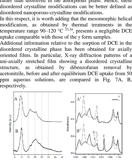
Additional information relative to the sorption of DCE in the disordered crystalline phase has been obtained for axially oriented films. In particular, X-ray diffraction patterns of a uni-axially stretched film showing a disordered crystalline structure, as obtained by dibenzofuran removal by acetonitrile, before and after equilibrium DCE uptake from 50 ppm aqueous solutions, are compared in Fig. 7A, B, respectively.
Fig. 1.8 X-ray diffraction patterns of an axially oriented s-PS film exhibiting the disordered crystalline phase before (A) and after DCE equilibrium sorption from a 50 ppm aqueous solution (B). FTIR spectra, in the range 1400–1140 cm–1, taken with the polarization plane parallel (thin line) and perpendicular (thick line) to the stretching direction of films The intense peak at d = 0.92 nm (2θ ~ 9.6°) indicates the presence of the disordered crystalline phase. Moreover, the decrease of the intensity of this peak and the appearance of a peak at d = 0.88 nm (2θ ~ 10°) suggests formation of the
Even more informative are the polarized FTIR spectra of the films of Fig. 1.8A, B, taken with the polarization plane parallel (thin line) and perpendicular (thick line) to the film stretching direction, which are shown in Fig. 1.8A’, B’, before and after equilibrium DCE uptake from 50 ppm aqueous solutions, for the spectral range 1400–1140 cm-1. In fact, not only the peaks corresponding to the vibrational modes of the s(2/1)2 helices of the crystalline phase (like those at 1277 and 1354 cm-1) 25 are highly dichroic (indicating a high degree of axial orientation, fc = 0.96), but the DCE peak of the trans
conformer (T) is also highly dichroic. In particular, based on the dichroism of this peak, it is possible to evaluate the angle between the transition moment vector of the corresponding vibrational mode with respect to the chain axis of the host crystalline phase (α = 76.5°), which is indistinguishable from that of DCE molecules being the guest of highly crystalline δ form films 31.
Hence, the results of Fig. 7 clearly indicate that the host–guest co-crystalline phases obtained by sorption of DCE molecules (also from dilute aqueous solutions) in the δ form or in the disordered nanoporous-crystalline form are essentially indistinguishable.
So disordered crystalline modifications formed by s(2/1)2 helices of syndiotactic polystyrene (s-PS) can be prepared by the removal of bulky guest molecules from intercalate as well as from triclinic δ clathrate forms. The X-ray diffraction pattern of the disordered crystalline modification is characterized from 2θCuKa<12° by only a broad diffraction
peak whose maximum is located in the 2θCuKa range between
8.7° and 9.8°. Films presenting disordered crystalline modifications have been used for the removal of an organic pollutant from dilute aqueous solutions. The sorption behaviors of the disordered crystalline modifications are compared with that of the nanoporous-crystalline δ form as well as of the dense γ form. The disordered crystalline modifications of s-PS presents pollutant (1,2-dichloroethylene) uptake comparable to those of the
nanoporous δ and ε forms and much higher than those obtained for the dense γ form. Moreover, FTIR data relative to sorption of 1,2-dichloroethane show that the guest sorption occurs essentially only in the crystalline phase. As a consequence, the obtained disordered crystalline modifications of s-PS can be fully considered disordered nanoporous-crystalline modifications.
1.3. sPS\ carvacrol co-crystalline film
Antimicrobials are widely used for food protection, and are also often provided as additives to food packaging.35,36 A
wide variety of essential oils and many of their components, mainly mono- and sesquiterpenes, have been shown to have antimicrobial activity.37,38 In particular, many studies describe the antimicrobial activity of carvacrol,39,40 a well known phenolic monoterpene (Scheme 1) constituent of essential oils produced by various aromatic plants such as oregano, thyme, and marjoram.
SCHEME 1.1 A schematic representation of carvacrol molecular structure.
X-ray diffraction patterns, as taken by an automatic powder diffractometer, of an axially oriented δ-form s-PS film before and after immersion in a 40 wt % carvacrol/chloroform solution (followed by complete chloroform removal) are shown in Figure 1.9 (A,A’).
Fig. 1.9 X-ray diffraction patterns, as taken by an automatic powder diffractometer, of s-PS films exhibiting: (A,B) the δ nanoporous-crystalline form with (A) axial orientation and (B) a//c// uniplanar
orientation; (A’,B’) the sPS/carvacrol δ-clathrate form, with (A’) axial orientation and (B’) a//c// uniplanar orientation.
The formation of a s-PS/carvacrol co-crystalline form is clearly shown by the large increase of the intensity of the 102 reflection at 2θ~10° and by the nearly complete disappearance of the intense 010 reflection, typical of the nanoporous δ-form. 1,12, 13 It is worth adding that, for the s-PS/carvacrol
co-crystalline form, this phenomenon of increase and decrease of intensities of the 210 and 010 reflections, respectively, is more pronounced than generally observed for other δ clathrates. 1,12,,13
Weak 010 reflections can be easily studied by using films exhibiting the a//
c
// uniplanar orientation, because their X-raydiffraction patterns, as taken by an automatic powder diffractometer, maximize the intensity of the 0k0 reflections,
that is, the reflection planes parallel to the ac plane.19-41 X-ray
patterns, by an automatic powder diffractometer, of a δ-form s-PS film with a//
c
//uniplanar orientation (
f
010=0.85),41 beforeand after immersion in a 40 wt % carvacrol/chloroform solution (followed by complete chloroform removal) are shown in Figure 1.9 (B,B’). As usual, the starting nanoporous-crystalline film shows essentially only the 010 and 020 reflections [Fig. 1.9(B)].19-41 The pattern of this film after carvacrol sorption (amplified of 10 times, due to the weakness of the 0k0 reflections) clearly shows that the 010 reflection of the s-PS/carvacrol co-crystalline form is located at 2ɵ ~ 7.2°, corresponding to a Bragg distance d010=1.23 nm.
A comparison with the data of δ clathrates, presenting the d010
values included between 1.02 nm for s-PS/azobenzene
and 1.16 nm for s-PS/indene δ clathrates (Table 1 of ref. 5), show that the s-PS/carvacrol δ clathrate presents the largest distance between the ac layers of closely packed alternated enantiomorphous helices. As discussed in detail in ref. 5, d010 >1.1 nm clearly indicates the formation of a monoclinic
(rather than of a triclinic) δ clathrate form.5
A schematic projection along the polymer chain axes of the crystal structure of the monoclinic δ clathrate form with carvacrol is shown in Figure 1.2 (B). As generally occurs for monoclinic δ clathrates of s-PS, only one isolated guest molecule is enclosed per host crystalline cavity, with a preferential orientation of the guest molecular plane perpendicular to the polymer chain axes.1,12,13,14
1.3.1 FTIR Method to discriminate carvacrol molecules in co-crystalline and amorphous polymer phases
s-PS films exhibiting the δ clathrate form with carvacrol have also been characterized by FTIR measurements. Particularly informative is the spectral region 3650–2800 cm-1, shown in
Figure 1.10. In fact, the broad O-H stretching band of liquid carvacrol, in the range 3630–3100 cm-1 (thick dashed line) is located in a spectral region where the absorbance of the polymer host (dotted line) is negligible. The spectrum of a δ
clathrate film with a carvacrol content close to 40 wt % [obtained by immersion in a 20 wt % carvacrol acetone solution, whose X-ray diffraction pattern is similar to that one of Fig. 1.9 (B’)] presents an intense narrow peak located at 3532 cm-1 (thin full line in Fig. 1.10).
Fig.1.10 FTIR spectra of s-PS films presenting: (thin dotted line) nanoporous δ-form; (thin full line) δ clathrate form with carvacrol and (thick full line) λ-form.The FTIR spectrum of liquid carvacrol is shown as a thick dashed line.
This clearly indicates the presence of OH groups of carvacrol molecules isolated in apolar environment, as expected for isolated guests of δ clathrate forms. In this respect, particularly useful is a comparison with a s-PS γ-form film,
42-43 that is, a film presenting a dense crystalline phase, where
guest molecules (as generally occurs for semicrystalline polymers) can be only located in the amorphous phase. This film containing roughly 20 wt % of carvacrol presents on the broad OH stretching band only a weak peak located at 3541 cm-1. This indicates that most carvacrol molecules, being dissolved in amorphous s-PS phases, are not isolated.
Also highly informative are the polarized FTIR spectra of axially oriented s-PS films, before and after carvacrol uptake, which are represented in Figure 1.11(A,B).
Fig. 1.11 Polarized FTIR spectra, in the wavenumber ranges 3650–3200 and 1550–800 cm-1, taken with polarization plane parallel (thin lines) and
perpendicular (thick lines) to the draw direction, for axially oriented s-PS films presenting different crystalline forms: (A) nanoporous form; (B) δ-clathrate form with carvacrol (~10 wt %). Symbols (//) and (┴), label dichroic guest peaks, whose transition moment vectors are preferentially parallel and perpendicular to the stretching direction (and hence to the chain axis, c, of the co-crystalline phase), respectively. Symbol (*) labels guest peaks, exhibiting poor linear dichroism.
The absorbance peaks of the s(2/1)2 helical polymer chains,
44-25 like for example those at 1354 and 1277 cm-1
[Fig. 1.11(A)], are highly dichroic and a high degree of axial orientation (fc,IR ~ 0.95) can be evaluated. The FTIR spectrum
of the s-PS/carvacrol co-crystalline film [Fig. 1.11(B)], with a carvacrol content roughly equal to 10 wt %, shows dichroism also for the guest peaks labeled by (//) and (┴), that is, peaks that present higher sorption of light with polarization parallel and perpendicular with respect to the film stretching direction, respectively. As for the guest OH stretching band, it is worth noting that the broad band nearly centered at 3400 cm-1 is not dichroic whereas the OH stretching peak of isolated carvacrol molecules at 3532 cm-1 exhibits a high dichroism
(R=A///A┴=2.3). This clearly confirms the hypothesis that the
isolated OH groups belong to molecules being isolated guests of axially oriented δ-clathrate phases whereas the
hydrogen-bonded OH groups belong to molecules dissolved in the essentially unoriented amorphous phase.
The use of the O-H stretching region of FTIR spectra to discriminate between guest molecules of the crystalline phase and molecules dissolved in the amorphous phase, is shown here for the first time for nanoporous-crystalline polymers. It is worth adding that preliminary analyses have shown that this method is applicable to many other substituted phenols, like for example, thymol (2-isopropyl-5-methylphenol) and methylparaben (Benzoic acid, 4-hydroxy-, methyl ester) as well as to alcohols like ethanol and menthol.
1.3.2 Partition of carvacrol molecules between amorphous and crystalline phases of δ-form films
The carvacrol content in s-PS films and its partition between amorphous and nanoporous-crystalline δ phases have been investigated, mainly by FTIR analyses. In particular, we show results relative to films exhibiting a partial filling of the crystalline cavities, as obtained by carvacrol sorption from diluted solutions, as well as to films exhibiting a complete filling of the crystalline cavities, as obtained by carvacrol sorption from concentrated solutions. The FTIR spectra in the wavenumber range 3650–3150 cm-1 of a δ-form s-PS film with a 5-µm thickness, after equilibrium sorption (18 hours) from a 5 wt % carvacrol solution in acetone are compared in Figure 1.12, for different desorption times.
Fig 1.12 FTIR transmission spectra in the wavenumber range 3650–3150 cm-1 of a δ-form s-PS film, after equilibrium sorption from a 5 wt %
carvacrol solution in acetone, as collected at various desorption times: (a) 1 hour; (b) 24 hours; (c) 155 hours; (d) 395 hours.
The variation of the absorbances of the O-H stretching carvacrol peaks at 3532 cm-1 (labeled as cr) and at 3400 cm-1 (labeled as am), as normalized with respect to a s-PS reference peak (1601 cm-1), are compared in Figure 1.12; and they are associated with carvacrol molecules isolated guest of the crystalline phase and dissolved in the amorphous phase, respectively.
Fig. 1.13 Absorbances, normalized with respect to the 1601 cm-1 s-PS
peak, of the carvacrol peaks at 3532 cm-1 associated with the fraction
hosted in the crystalline phase (cr) and at 3400 cm-1 associated with the
fraction dissolved in the amorphous phase (am) as well as of an intense solvent carrier peak (of acetone at 1716 cm-1), versus desorption time at
room temperature. The inset enlarges the observed behavior for the first few hours of desorption.
Figure1.13 also shows the solvent carrier (acetone) desorption by the decrease of the absorbance of the intense carbonyl peak at 1716 cm-1. It is apparent that the complete desorption of the carrier molecules occurs at room temperature after nearly 90 hours.
For the first hours of desorption (inset in Fig.1.13), a large increase in the absorbance of the peak associated with isolated carvacrol molecules (cr) and a corresponding decrease of the absorbance of the broad peak associated with aggregated carvacrol molecules (am) are clearly apparent.
After nearly 20 hours of desorption, both A(cr) and A(am) become constant and their ratio A(cr)/A(am) becomes nearly equal to 10. These data can be easily rationalized by assuming
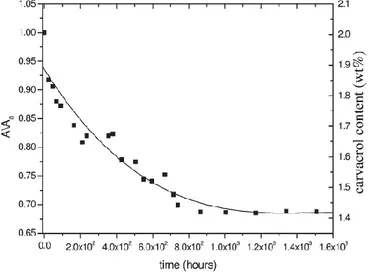
that, with the carrier desorption, the crystalline cavities, initially occupied by the carrier molecules, are progressively filled by the carvacrol molecules, which mostly move from the amorphous toward the crystalline phase. After the preparation procedure described in Figure 1.13, the overall carvacrol content in the film, as determined by TGA measurements, is of 2.1 wt %.
The carvacrol desorption at 35 °C of a film, like that one of Figure 1.12(D) with most molecules being guest of the s-PS crystalline phase, based on the reduction of the absorbance of the 3532 cm-1 FTIR peak, is shown in Figure 1.14.
Fig. 1.14 Carvacrol desorption at 35 °C from a s-PS film with a carvacrol content of 2.1 wt % with most molecules being guest of the s-PS crystalline phase.
A simple approximate calculation, based on the degree of crystallinity (35%) and on the styrene/cavity ratio of the nanoporous crystalline δ-form (4/1) allows establishing that <15% of the crystalline cavities are filled by carvacrol molecules while the other crystalline cavities remain empty. This can be an advantage for packaging films, because a fraction of the crystalline cavities are used for natural antimicrobial release while the other crystalline cavities
remain active for removal of molecules (ethylene and carbon dioxide) generated by vegetables being detrimental for their preservation. The FTIR spectra of a s-PS δ-form film (with a thickness of 62 µm thick and exhibiting a//c// uniplanar
orientation), after immersion in a 40 wt % carvacrol/chloroform solution for 6 hours are shown in Figure 1.15, for different desorption times (up to 104 hours). Differently from the case of sorption from diluted solutions (Fig. 1.13), continuous carvacrols desorption from both crystalline and amorphous phase is apparent.
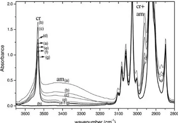
Fig. 1.15 FTIR transmission spectra in the wavenumber range 3650–2800 cm-1 of a s-PS/carvacrol co-crystalline film, with an initial carvacrol
content close to 40 wt %, as collected at various desorption times (up to 104 hours): (a) t50; (b) 2 days; (c) 20 days; (d) 45 days; (e) 63 days; (f) 157 days; (g) 595 days. FTIR spectra s-PS films nanoporous δ-form is also reported (h).
A quantitative evaluation of the guest desorption is shown in Figure 1.16, where the absorbance reduction for several vibrational peaks (A/Ao, where Ao is the peak absorbance in
the spectrum of the freshly prepared co-crystalline film) are reported versus the desorption time.
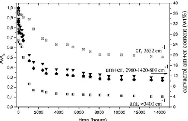
Fig. 1.16 Reduced absorbance of carvacrol infrared bands of guest molecules only in the crystalline (3532 cm-1) or only in the amorphous
(3400 cm-1) or in both phases (2960, 1420, and 809 cm-1), versus
desorption time. On the right scale, the overall carvacrol content in the film, as determined by thermogravimetry
It is clearly apparent that the reduction of the intensity of the broad band at 3400 cm-1, (labeled as am), due to carvacrol molecules in the polymer amorphous phase, is much faster than for the peak at 3532 cm-1, (labeled as cr), due to
carvacrol molecules in the polymer co-crystalline phase. The reduction of the intensity with time of the 2960 (labeled as cr+am), 1420, and 809 cm-1 peaks, due to carvacrol molecules
in both polymer phases, is intermediate.
Thermogravimetric scans indicate that the residual carvacrol content in the film is of 10.5 wt %. Based on this kind ofTGA calibration, the overall carvacrol content in the film is shown in the right scale of Figure 1.16, indicating that the initial overall carvacrol content is close to 40 wt %. The ratio A(cr)/A(am), after long-term desorption (more than 104 hours), is close to 5, indicating that nearly 80% of carvacrol is in the crystalline phase.
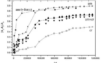
The results of Figure 1.16 are also reported as classical Fick’s plots in Figure 1.17, with (A0-At)/A0 versus the square root
For the sake of comparison, the desorption kinetics of carvacrol being dissolved in the amorphous phase of a γ-form film (maximum carvacrol uptake of nearly 20 wt %, FTIR spectrum in Fig. 1.10), is also shown in Figure 1.17.
Fig. 1.17 Carvacrol desorption isotherms, presenting the FTIR absorbance variations (A0-At)/A0 versus the square root of desorption time divided by
film thickness (√t/L). For comparison, the desorption of carvacrol form the amorphous phase of a γ-form s-PS film is also reported (black squares). The curves of Figure 1.17 confirm that the slowest carvacrol desorption occurs from the crystalline phase (Dcrys ~ 4.3 x
10-14 cm2 s-1) while a definitely faster desorption occurs from the amorphous phase (Dam,δ ~ 3.7 x 10⁻¹² cm2 s-1).
The plot also indicates that a much faster desorption occurs from the amorphous phase of the semicrystalline film exhibiting the dense λ phase (Dam,γ ~ 3.2 x 10211 cm2 s-1).
The slower carvacrol desorption from the amorphous phase of the δ-form film is possibly due to the equilibrium between carvacrol molecules hosted by the amorphous and nanoporous-crystalline phases, which has been clearly shown by the data of Figure 1.13.
So a monoclinic δ-clathrate form of syndiotactic polystyrene (s-PS) with carvacrol (a relevant natural phenolic antimicrobial) has been prepared and characterized by X-ray
diffraction. Very informative are Fourier transform infrared spectra, in particular their OH stretching region that shows a narrow peak and a broad band, corresponding to carvacrol molecules being isolated guest of the co-crystalline phase or dissolved in the amorphous phase, respectively. Analogous spectral features allow discriminating, for many different s-PS guests, between molecules being in crystalline or in amorphous phases. s-PS co-crystalline films with carvacrol molecules being prevailingly (more than 90%) guest of the cocrystalline phase have been prepared, even for high carvacrol content (up to 10–11 wt %). The location of most antimicrobial molecules in the crystalline phase assures a decrease of desorption diffusivity of two to three orders and hence longterm antimicrobial release.
1.4 Conclusion
In section 1.2 we talk abaut the disordered nanoporous crystalline modification and we show that the guest removal from s-PS intercalates or triclinic δ-clathrates can lead to disordered crystalline modifications, still exhibiting s(2/1)2 helices. In particular, the X-ray diffraction pattern of the disordered crystalline modification is characterized, from 2θCuKa<12°, by only a broad diffraction peak whose maximum
is located in the 2 θCuKa range between 8.7° and 9.8°. The
sorption of an organic pollutant (1,2-dichloroethane) from dilute solutions in water, for films presenting these disordered crystalline structures, has been compared with those of analogous films exhibiting the nanoporous δ and the dense γ crystalline phases. The equilibrium DCE uptake from these films is roughly 60 % with respect to that of highly crystalline δ form films and much higher than for γ form films. FTIR spectra have shown that, as previously established for the nanoporous δ phase, for the disordered crystalline modification, the DCE molecules are absorbed essentially only as guest of a co-crystalline phase, rather than dissolved in the amorphous phase.
In summary, the reported results show that these disordered crystalline modifications of s-PS can be considered to be formed of small bundles of s(2/1)2 helices, which can recrystallize as a consequence of sorption of suitable guest molecules, eventually leading to δ co-crystalline phases. This makes the sorption behavior of the disordered crystalline phase high and comparable to that of the nanoporous δ phase, rather than being negligible as for the dense γ phase. As a consequence, these disordered crystalline forms of s-PS can be considered as nanoporous-crystalline.
Moreover in section 1.3 we study the co-crystalline phase of sPS including antimicrobial guest.
A co-crystalline form of s-PS with carvacrol (a relevant natural antimicrobial molecule) has been prepared by carvacrol sorption, from solutions in suitable carrier solvents, in films exhibiting the nanoporous-crystalline δ phase. For s-PS/carvacrol co-crystalline samples, FTIR spectra are very informative and in particular their OH stretching region. In fact, this spectral region shows a narrow peak and a broad band corresponding to carvacrol molecules being isolated guest of the co-crystalline phase or simply dissolved in the corresponding amorphous phase, respectively. This interpretation has been confirmed by polarized FTIR spectra of axially oriented films. It is worth adding that this phenomenon, which allows discriminating between guest molecules being present in crystalline and amorphous phases, is shown here for the first time for polymeric co-crystalline phases and occurs for many other guests exhibiting hydroxy groups. FTIR analyses have allowed investigating the carvacrol content in s-PS films and its partition between amorphous and nanoporous-crystalline δ phases. In particular, analyses of carvacrol sorption from diluted solutions (5 wt %) show that the crystalline cavities, initially occupied by carrier molecules, are progressively filled by carvacrol molecules moving from the amorphous phase. After carrier desorption for a guest content of nearly 2 wt %, more than 90% of the guest molecules are in the crystalline phase, which constitutes
<40 wt % of the film sample. For these films, <15% of the crystalline cavities is filled by carvacrol molecules while the other crystalline cavities remain empty. Analyses of carvacrol sorption from concentrated solutions (40 wt %) show that very large carvacrol uptake is possible (up to 40 wt %), occurring in both crystalline and amorphous phase. The analysis also shows that desorption from the amorphous phase is much faster than desorption from the crystalline phase. The apparent carvacrol diffusivity at room temperature from the co-crystalline phase is nearly 102 times lower than from the corresponding amorphous phase and nearly 103 times lower
than from the amorphous phase of s-PS films exhibiting a dense crystalline phase (γ form). In summary, we have described the preparation of s-PS films with antimicrobial molecules, being prevailingly present as guest of the crystalline phase. This assures slow antimicrobial release and hence long-term antimicrobial properties. These s-PS films, mainly for low guest content (possibly <1 wt %) can be useful for packaging of fruits and vegetables, due to the combination of ethylene and carbon dioxide removal from the empty cavities of the δ nanoporous crystalline phase and the slow release of a scented natural antimicrobial guest from the filled cavities of the same crystalline phase.
Refernces
1. Chatani, Y., Inagaki, Y., Shimane, Y., Ijitsu, T., Yukimori, H., Shikuma, H.:. Polymer 1993,34, 1620
2. Albunia, A.R., D’Aniello, C., Guerra, G. Cryst. Eng. Commun. 2010,12, 3942
3. Tarallo, O., Schiavone, M.M., Petraccone, V. Eur. Polymer J. 2010, 46, 456
4 . Tarallo, O., Petraccone, V., Daniel, C., Guerra, G. Cryst. Eng. Commun. 2009,11, 2381
5. Tarallo, O., Petraccone, V., Albunia, A.R., Daniel, C., Guerra, G. Macromolecules 2010,43, 8549
6. Rastogi, S., Goossens, J.G.P., Lemstra, P.J. Macromolecules 1998, 31, 2983
7. Albunia, A.R., Rizzo, P., Coppola, M., De Pascale, M., Guerra, G. Polymer , 2012, 53, 2727
8. Tarallo, O., Schiavone, M.M., Petraccone, V., Daniel, C., Rizzo, P., Guerra, G. Macromolecules 2010, 43, 1455
9. Albunia, A.R., D’Aniello, C., Guerra, G Cryst. Eng. Commun. 2010,12, 3942
10. De Rosa, C., Rizzo, P., Ruiz de Ballesteros, O., Petraccone, B., Guerra, G Polymer 1999, 40, 2103
11. Petraccone, V., Tarallo, O., Venditto, V., Guerra, G. 2003, 8, 6965
12 De Rosa, C., Guerra, G., Petraccone, V., Pirozzi, B Macromolecules 1997, 30, 4147
13. Tarallo, O., Petraccone, V., Venditto, V., Guerra, G Polymer 2006, 47, 2402
14. Gowd E. B., Shibayama N., Tashiro K., Macromolecules 2006, 39, 8412–8418
15. Rizzo P., Daniel C., De Girolamo Del Mauro A., Guerra G., Chem. Mater. 2007, 19, 3864
16. V. Petraccone, O. Ruiz de Ballesteros, O. Tarallo, P. Rizzo, G.Guerra, Chem. Mater. 2008, 20, 3663
17. Albunia A. R., Minucci T., Guerra G., J. Mater. Chem. 2008, 18, 1046.
18. Annunziata L., Albunia A. R., Venditto V., Guerra G., Macromolecules 2006, 39, 9166.
19. Rizzo P., Lamberti M., Albunia A. R., Ruiz de Ballesteros O., Guerra G., Macromolecules 2002, 35, 5854
20. Albunia A. R., Rizzo P., Guerra G., Macromolecules 2011, 44, 5671.
21. Milano G., Guerra G., Muller-Plathe F., Chem. Mater. 2002, 14, 2977.
22. Albunia A. R., Rizzo P., Guerra G., Polymer 2013, 54, 1671 23. Loffredo, F., Pranzo, A., Venditto, V., Longo, P., Guerra, G. Macromol. Chem. Phys. 2003, 204, 859
24 Albunia, A.R., Rizzo, P., Guerra, G., Torres, F.J., Civalleri, B., Zicovich-Wilson, C.M. Macromolecules 2007 40, 3895
25. Torres, F.J., Civalleri, B., Meyer, A., Musto, P., Albunia, A.R., Rizzo, P., Guerra, G. J. Phys. Chem. B 2009, 113, 5059
26. Albunia, A. R., Bianchi, R., Di Maio, L., Galimberti, M., Guerra, G., Pantani, R., Senatore, S. Disordered nanoporous crystalline form of syndiotactic polystyrene, production method and