Nuovi approcci molecolari nello studio della
fisiopatologia del diabete gestazionale
Facoltà di Medicina e Odontoiatria Dipartimento di Medicina Sperimentale
Corso di Dottorato in Scienze Endocrinologiche
Curriculum Scienze Endocrinologiche, Metaboliche e Andrologiche Direttore: Prof. Andrea Lenzi
Tiziana Filardi
Relatore
Prof. Susanna Morano
A.A. 2019-2020
Nuovi approcci molecolari nello studio della
fisiopatologia del diabete gestazionale
Novel molecular approaches to the study of
gestational diabetes pathophysiology
Facoltà di Medicina e Odontoiatria Dipartimento di Medicina Sperimentale
Corso di Dottorato in Scienze Endocrinologiche
Curriculum Scienze Endocrinologiche, Metaboliche e Andrologiche Direttore: Prof. Andrea Lenzi
Tiziana Filardi
Relatore
Prof. Susanna Morano
A.A. 2019-2020
Abstract research study 1 ... 5
Cross-talk between foetal membranes and visceral adipose tissue involves HMGB1-RAGE and VIP-VPAC2 pathways in human gestational diabetes mellitus ... 5
Abstract research study 2 ... 6
MicroRNA expression profile in circulating exosomes and plasma of patients with GDM and healthy pregnant women ... 6
Chapter 1. Gestational diabetes mellitus: an overview ... 8
Chapter 2. Research study 1 ... 11
Cross-talk between foetal membranes and visceral adipose tissue involves HMGB1-RAGE and VIP-VPAC2 pathways in human ... 11
gestational diabetes mellitus ... 11
INTRODUCTION ... 11
Inflammation and immune response in physiological pregnancy ... 11
The placenta ... 12
Foetal membranes ... 12
Inflammation and immune response in GDM pregnancy ... 13
Novel pro-inflammatory and anti-inflammatory molecules ... 14
High Mobility Group Box 1 (HMGB1) and Receptor for Advanced Glycation Endproduct (RAGE) ... 14
Vasoactive Intestinal Peptide (VIP), VPAC1 and VPAC2 ... 17
AIM ... 18
PATIENTS AND METHODS ... 18
FMs explants and culture ... 18
VAT explants and culture ... 19
Western blotting ... 19
Cytokines detection ... 19
Statistical analysis ... 20
RESULTS ... 20
Characteristics of the study population ... 20
HMGB1-RAGE expression in FMs and VAT ... 22
VPACs and VIP expression in VAT and FMs ... 23
Circulating levels of HMGB1 and VIP ... 25
HMGB1 and VIP in vitro release by FMs and VAT ... 25
DISCUSSION ... 26
CONCLUSIONS ... 28
Chapter 3. Research study 2 ... 29
MicroRNA expression profile in circulating exosomes and plasma of patients with GDM and healthy pregnant women ... 29
INTRODUCTION ... 29
General aspects of microRNAs ... 29
MicroRNAs: role in GDM pathophysiology and complications ... 30
Role of exosomes in pregnancy and pregnancy-related disorders ... 33
MicroRNAs in exosomes and GDM ... 35
AIM ... 35
PATIENTS AND METHODS ... 35
Participants ... 35
Blood Samples Processing ... 36
Exosome isolation ... 36
Western blot ... 36
Transmission electron microscopy ... 37
RNA extraction ... 37
Statistical analysis ... 38
Differential expression analysis ... 38
Target gene identification and bioinformatics analysis ... 39
RESULTS ... 39
Clinical and biochemical characteristics of the whole population and the profiling cohort ... 39
Exosome isolation and characterisation ... 42
MicroRNA expression profile in exosomes and plasma from GDM and NGT ... 42
MicroRNA expression profile in exosomes and plasma from GDM, NGT and NP ... 46
Target gene prediction and network analysis of microRNAs differentially expressed in exosomes ... 46
Target gene prediction and network analysis of microRNAs differentially expressed in plasma ... 48
DISCUSSION ... 50
CONCLUSIONS AND FUTURE PERSPECTIVES ... 55
Chapter 4: Other peer-reviewed papers published during the PhD ... 56
REFERENCES ... 57
Abstract research study 1
Cross-talk between foetal membranes and visceral adipose tissue involves HMGB1-RAGE and VIP-VPAC2 pathways in human gestational diabetes
mellitus
Introduction: Gestational diabetes mellitus (GDM) is defined as glucose intolerance that is
first diagnosed during pregnancy. Foetal membranes (FMs) and maternal visceral adipose tissue (VAT) secrete various molecules that are relevant players in the pathogenesis of GDM.
Aim: This pilot study aimed to comparatively evaluate the expression of high mobility group
box 1 protein (HMGB1) and its receptor for advanced glycation end products (RAGE), and vasoactive intestinal peptide (VIP) and its receptors (VPAC1, VPAC2) in FMs and VAT in GDM and in healthy pregnant women.
Patients and Methods: FMs, omental VAT explants, and serum samples were obtained
from twelve patients with GDM and twelve pregnant women with normal glucose tolerance (NGT) at delivery. The expression of HMGB1, RAGE and VIP, VPAC1 and VPAC2 was detected by Western Blotting in explants; circulating levels and in vitro release of HMGB1 and VIP were measured by ELISA tests.
Results: HMGB1 tissue expression was higher in FMs obtained from GDM patients
(p=0.02) than in FMs from NGT women. VPAC2 (p=0.03) and RAGE (p=0.03) tissue expressions were significantly increased in VAT from GDM patients compared to NGT. Only FMs of NGT released detectable levels of HMGB1, which was not observed in samples obtained from GDM. VAT of GDM released lower levels of VIP (p=0.05) than NGT samples.
Conclusions: This study suggests that a fine tuned regulation exists between FMs and VAT
throughout pregnancy to maintain immune metabolic homeostasis. In GDM a balance between pro-inflammatory and anti-inflammatory mediators has been observed. Further studies are needed to establish their exact role on foetal and maternal outcomes in GDM.
Abstract research study 2
MicroRNA expression profile in circulating exosomes and plasma of patients with GDM and healthy pregnant women
Introduction: MicroRNAs are small non-coding RNAs, playing critical roles in modulating
gene expression. The deregulation of microRNAs has been observed in GDM, highlighting their crucial involvement both in the pathogenic mechanisms of this condition and in the development of its complications. Circulating microRNAs can be packaged into exosomes, and exosome signalling has emerged as a novel mechanism of cell-to-cell communication. Through exosomes, microRNAs are delivered in distant target cells and are able to affect gene expression.
Aim: The aim of this study was to explore microRNA expression in circulating exosomes
and in plasma obtained from patients with GDM and healthy control subjects in the third trimester of gestation, to potentially elucidate some relevant aspects of GDM pathophysiology and individuate novel potential candidate biomarkers for GDM.
Patients and Methods: A profiling cohort of plasma samples collected from GDM (n=3,
age: 34.7 ± 4.9 years; BMI 27.0 ± 3.7 Kg/m2) and NGT women (n=3, age: 34.3 ± 3.1 years; BMI 26.4 ± 1.1 Kg/m2) was recruited. In addition, a profiling cohort of healthy non-pregnant age- and BMI-matched women (NP, n=5) was used as negative control. The microRNA patterns of expression in exosomes and plasma have been assessed with the innovative technology NanoString nCounter microRNA expression (NanoString Technologies inc., Seattle, WA, USA). Target gene identification and bioinformatics analysis of the differentially expressed microRNAs have been performed with Ingenuity Pathway Analysis (IPA, QIAGEN Redwood City, USA).
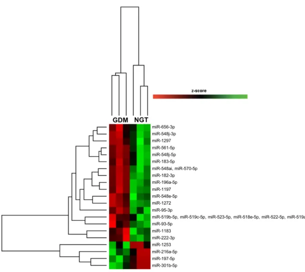
Results: A specific set of microRNAs resulted to be differentially expressed in exosomes
and plasma from GDM patients compared to NGT. Specifically, five exosomal microRNAs were significantly upregulated, while 23 were downregulated in GDM compared to NGT. As for plasma, 4 microRNA were upregulated, while 9 were downregulated in GDM compared to NGT. In addition, two microRNAs, miR-196a-5p and miR-652, resulted to be significantly downregulated in GDM compared both to NGT and NP in exosomes and plasma, respectively, suggesting that their deregulation might hallmark GDM pregnancy. In bioinformatics analysis the major predicted target genes and biological processes of the deregulated microRNAs were associated with insulin resistance, abnormal glucose and lipid metabolism, consistently linked to GDM pathophysiology.
Conclusions: GDM might markedly alter microRNA profile in exosomes and plasma,
conceivably mirroring the metabolic alterations described in GDM pregnancy. In light of this, exploring circulating microRNA expression might help unravel the molecular events leading to the metabolic alterations observed in GDM.
Chapter 1. Gestational diabetes mellitus: an overview
Gestational Diabetes Mellitus (GDM) is a metabolic disease diagnosed in the second or third trimester of pregnancy (1). This condition is increasingly frequent worldwide due to the large spread of obesity (2). The global figures of GDM range from 5.8% to 12.9%, displaying important differences among countries (3). Unless properly diagnosed and treated, GDM can lead to poor pregnancy outcomes. Women with GDM are at increased risk for gestational hypertension and pre-eclampsia (4). Shoulder dystocia, birth trauma and preterm delivery, due to excessive foetal growth, are common birth complications (5). Other short-term adverse outcomes include hypoglycaemia, hypoxia, risk of stillbirth and respiratory distress syndrome (6). Remarkably, the negative impact of GDM is observed even later in life, both in the mother and in the offspring. Importantly, mothers previously diagnosed with GDM display an increased risk of developing type 2 diabetes (T2D) and metabolic syndrome (7). Similarly, a higher risk of metabolic diseases and cardiovascular disease (CVD) has been reported in children born to mothers affected by GDM over adulthood (8,9).
Current diagnostic criteria for GDM have been established by the International Association of Diabetes and Pregnancy Study Groups (IADPSG) and ensued from the results of the Hyperglycaemia and Adverse Pregnancy Outcome (HAPO) study (4). A strong link between high maternal glucose and the occurrence of adverse pregnancy outcomes emerged in this multinational longitudinal study of a large cohort of pregnant women, helping individuate the glycaemic thresholds for GDM diagnosis. According to the IADPSG recommendations, an oral glucose tolerance test (OGTT) with 75 g-glucose is performed at 24-28 weeks of gestation. Fasting, 1-h and 2-h plasma glucose are assessed and the cut-off values of glycaemia at OGTT are 5.1 mmol/l, 10 mmol/l and 8.5 mmol/l, respectively. One or more values exceeding or equalling these thresholds establish the diagnosis of GDM (10). Although the IADPSG does not recommend routinely screening before 24-28 weeks of gestation, detecting GDM as early as possible is crucial to avoid poor pregnancy outcomes. Accordingly, a screening approach that takes into account the main GDM risk factors might anticipate the diagnosis and prevent complications (11). Overweight/obesity, excessive weight gain, history of previous GDM or macrosomia, advanced maternal age, first degree family history of T2D, multi-parity and non-Caucasian ethnicity are well-established risk factors for GDM (12-14). In addition, lifestyle, such as diet, physical activity or emerging environmental factors are likely to have an impact on the risk of developing GDM (15,16). The identification of phenotypic classes associated with adverse outcomes, defined by the
presence of some risk factors, turned out to be potentially useful in customizing and modulating the diagnostic and therapeutic approach in GDM.
Insulin resistance and β-cell dysfunction are the main pathophysiological features of GDM. Insulin sensitivity significantly varies throughout pregnancy, constantly adapting to the energy demands of the mother and the foetus. Overall, insulin sensitivity follows a biphasic trend in healthy pregnancy, experiencing a sharp increase initially and a marked reduction as pregnancy evolves. The metabolic adaptation observed in the initial phase (the first two trimesters) is aimed at storing essential sources of energy, such as glucose and fatty acids, as fat deposits, necessary for the next stages of pregnancy (17). Estrogens and progesterone synthesis progressively increases, together with other molecules of placental origin, such as human placental lactogen and human placental growth hormone, contributing to the progressive fall in insulin sensitivity. Thus, the insulin resistance state observed in physiological pregnancy is an adaptive response that favours the rise in glucose and free fatty acid blood levels, shifting energy sources from the mother to the foetus (18). The pancreatic β-cell largely compensates through an increased insulin release. Hypertrophy and hyperplasia of β-cell mass, explained by enhanced proliferation and reduced apoptosis, have been reported in human pregnancy (19). However, if β-cell dysfunction occurs, the compensative effect is lost and GDM becomes manifest.
Insulin resistance is a consequence of impaired peripheral insulin signalling. Indeed, glucose uptake is almost halved and insulin resistance is enhanced in GDM compared to healthy pregnancy (20). Insulin signalling is affected by altered phosphorylation of the insulin receptor or insulin receptor substrate (IRS)-1, although the number of receptors on the cell surface is preserved (21). Remarkably, pro-inflammatory cytokines contribute to the development of insulin resistance. In pregnancy complicated by GDM, an exacerbated pro-inflammatory state has been reported, compared to healthy pregnancy. Adipose tissue and gestational tissues are able to secrete pro-inflammatory cytokines, such as TNF-α, IL-1-β and IL-6, that impair insulin signalling by inhibiting IRS-1 through serine phosphorylation (22).
As regards β-cell dysfunction, it has been observed that it is reduced by 30–70% in GDM, indicating that β-cells are unable to compensate for the increase in insulin resistance, resulting in the development of GDM (23). However, whether it is a consequence of excessive insulin secretion or a hidden pre-existing condition that arises when the compensatory effect is required remains unclear.
The mechanisms underlying β-cell dysfunction are not fully uncovered, but are likely to overlap those described in T2D. Largely, alterations at each step of insulin synthesis or secretion have been described, and β-cell dysfunction is triggered by hyperglycaemia and hyperlipidemia (24). Oxidative stress, mitochondrial dysfunction, endoplasmic reticulum stress are well-established consequences of glucotoxicity and lipotoxicity, and impair insulin synthesis, secretion and β-cell survival (25). Pro-inflammatory cytokines further promote endoplasmic reticulum stress, and are able to induce β-cell de-differentiation (26).
The treatment of GDM is aimed to achieve normal levels of fasting plasma glucose (FPG) and post-prandial glycaemia (PPG) in order to prevent complications. Indeed, high glucose levels, especially postprandial elevations, are linked to negative pregnancy outcomes (27). The first therapeutic approach consists of dietary and lifestyle modifications. However, insulin therapy is mandatory unless good glycaemic control is achieved (28). Medical nutritional therapy should provide adequate nutrients for normal foetal growth, but it should not induce maternal weight gain or loss. Gestational weight gain is largely due to excessive energy intake and fuel requirements usually increase from 10 to 30 weeks of gestation. Caloric restriction is not recommended during pregnancy, even in GDM, because it may have adverse effects on birth weight. Advice for energy intake, as well as for weight gain, should take into account pre-pregnancy BMI (29).
Chapter 2. Research study 1
Taken from:Cross-talk between foetal membranes and visceral adipose tissue involves HMGB1-RAGE and VIP-VPAC2 pathways in human
gestational diabetes mellitus
Santangelo Carmela, Filardi Tiziana, Perrone Giuseppina, Mariani Marianna, Mari Emanuela, Scazzocchio Beatrice, Masella Roberta, Brunelli Roberto, Lenzi Andrea, Zicari Alessandra, Morano Susanna
In Acta Diabetol. 2019 Jun;56(6):681-689
doi: 10.1007/s00592-019-01304-x. Epub 2019 Feb 28. PMID: 30820673
INTRODUCTION
Inflammation and immune response in physiological pregnancy
Physiological pregnancy is hallmarked by an altered inflammatory profile compared to the non-pregnant state. In particular, in the earliest stages of pregnancy, the local inflammatory response is crucial for the embryo implantation after conception. By contrast, in the post-implantation phase, the innate immune response is finely regulated and the adaptive immune response is relatively suppressed, in order to prevent foetal rejection and to establish maternal-foetal tolerance (22). The result of the complex regulation of the immune response in pregnancy is a systemic low-grade inflammatory state, which derives from the fine balance between and anti-inflammatory cytokine production (30). The main pro-inflammatory cytokines produced in pregnancy are Tumor Necrosis Factor-α (TNF-α), interleukin-1ß (IL-1ß) and interleukin-6 (IL-6). On the contrary, interleukin-10 (IL-10) and the Transforming Growth Factor-ß (TGF-ß) have anti-inflammatory actions.
As regards adaptive immunity, in physiological pregnancy the T-helper (Th) cells mainly polarize towards a Th2 profile, hallmarked by the production of anti-inflammatory cytokines (IL-10, IL-4, IL-5, IL-13). Differently, the production of pro-inflammatory cytokines, such as interleukin-2 (IL-2), interferon-γ (IFN-γ) and TNF-α is sustained by the Th1 profile. The
In particular, it has been observed that high circulating levels of cytokines of the Th1 line are associated with spontaneous abortion in the early stage of pregnancy (31-33).
During pregnancy, gestational tissues (placenta and foetal membranes) and maternal adipose tissue are important sites of cytokine production, which play a wide variety of autocrine, paracrine and endocrine functions.
The placenta
The placenta is a tissue of both maternal and foetal origin, consisting of a maternal side and a foetal side. The maternal side is the basal decidua, which is the pregnancy endometrium, while the foetal side, also named villous chorion, contains the chorionic villi anchored to the basal decidua. The placenta is both a target tissue for systemic cytokines and a site of cytokine production. Indeed, from the earliest stages of its development, the placenta contributes to the synthesis and the release of a wide range of cytokines, with local and systemic actions (34). Specifically, the Hofbauer cells (the resident placental macrophages), the cytotrophoblast, the syncytiotrophoblast and the vascular endothelial cells, are able to release all the currently known cytokines (35). Remarkably, the contribution of each cell type to the pregnancy cytokine network considerably varies between the different stages of pregnancy and is not yet completely clear. At the maternal-foetal interface, the placenta is exposed to cytokines deriving from both maternal and foetal circulation. While some authors have reported that maternal pro-inflammatory cytokines do not cross the foetal-placental barrier (36), other studies have observed a minimal cross-placental passage of cytokines from the mother to foetus. Therefore, the cytokines identified in the foetal circulation seem to originate from the mother, the placenta and the foetus itself (37).
Foetal membranes
Foetal membranes (FMs) are tissues of foetal origin, located at the interface between the mother and the foetus. They include the amnion, which delimits the amniotic cavity, and the
smooth chorion, connected to the maternal decidua.
The amnion and the smooth chorion are considered as a single functional unit: the
amnio-chorionic membrane. Not only are FMs essential for mechanical support to the foetus, but
they have also multiple immune-endocrine functions. In particular, the isolated amnio-chorionic tissue is able to produce pro-inflammatory cytokines (IL-1α/ß, IL-6, TNF-α), lymphocytic (2)/macrophage (15) cytokines , anti-inflammatory cytokines (4,
IL-10, IL1RA, TGF-ß), interferons, chemokines [Monocyte Chemoattractant Protein (MCP)-1/2/3/4, Regulated on Activation, Normal T Cell Expressed and Secreted (RANTES), Macrophage Inflammatory Proteins (MIP-1α/ß)], chemokines (mainly IL-8), hematopoietic growth factors [Macrophage Colony-Stimulating Factor (M-CSF)]. The expression of these molecules can be constitutive or induced by various pro-inflammatory triggers (physical stress, prostaglandin E2, TNF-α, products of bacterial and viral origin). Pro-inflammatory cytokines and chemokines produced by FMs play a central role in the defence against pathogens, in the maintenance of maternal-foetal tolerance and in the physiological events during labour and delivery (38).
Inflammation and immune response in GDM pregnancy
In GDM pregnancy the inflammatory response is amplified. Prospective studies have shown an increase in the expression of pro-inflammatory cytokines and a reduction in the expression of anti-inflammatory cytokines in patients with GDM (39,40).
Immune cells, adipose tissue and gestational tissues contribute to the amplification of the systemic inflammatory state in GDM, and the altered inflammatory profile influences glucose metabolism: pro-inflammatory signals can activate signal transduction pathways, such as the Jun-N-terminal Kinase (JNK) and Ikß kinase (IKK)/NF-kB pathways, impairing insulin signalling in peripheral tissues by inhibiting IRS-1 through serine phosphorylation (30,41).
An increased expression of the pro-inflammatory cytokine TNF-α in GDM has been reported in observational studies. Several cross-sectional studies have shown an association between higher levels of circulating TNF-α in the second and third trimester of pregnancy and the presence of GDM (42,43). A meta-analysis of 10 observational studies showed elevated TNF-α serum levels in patients with GDM, compared to healthy controls (44). A prospective study revealed a significant association between TNF-α, Homeostatic Model Assessment - Insulin Resistance (HOMA-IR) and Matsuda insulin sensitivity index, in the second trimester, even after adjustment for age, BMI, hypertriglyceridemia and serum adiponectin levels (45).
Circulating levels of the pro-inflammatory cytokine IL-6 also increase in pregnancy, mainly due to an increased placental production (46). In cross-sectional studies, circulating levels of IL-6 positively correlated with BMI and FPG during pregnancy and after delivery
(47-compared to healthy control subjects, even after adjustment for age and BMI, both at GDM screening and 2 months after delivery (48). Furthermore, a correlation between the values of this cytokine and the Matsuda insulin sensitivity index has been observed. However, no prospective studies are currently available to confirm these data (50).
Leptin is produced by the adipose tissue and seems to have a role in the development of inflammation in pregnancy and in the pathophysiology of GDM (51). Leptin and its receptors are also expressed in the placenta (46). Increased leptin levels might amplify the low-grade inflammatory response in GDM, by stimulating the production of IL-6 and TNF-𝛼, which in turn increase leptinemia (52). Cross-sectional studies have shown higher circulating levels of leptin in patients with GDM compared to healthy subjects (53-55). A meta-analysis of 18 observational studies have confirmed this result, even after adjustment for BMI (44). In a prospective study, leptin levels before the 16th week of pregnancy were found to be predictive of GDM, independently of pre-pregnancy BMI (56). Further studies are needed to confirm these findings.
Adiponectin is an insulin-sensitizing, anti-inflammatory and anti-atherogenic adipokine, exclusively secreted by the adipose tissue (57). In physiological pregnancy, maternal adiponectin decreases progressively, and its circulating levels are inversely correlated with BMI (58). Overall, there is consistent evidence in literature about the role of adiponectin as independent predictor of GDM. Low adiponectin exacerbates insulin resistance and correlates with pancreatic ß-cell dysfunction (59). Low adiponectin levels are predictive of GDM several months before diagnosis, regardless of BMI. This data has been confirmed in prospective studies (60).
Novel pro-inflammatory and anti-inflammatory molecules
High Mobility Group Box 1 (HMGB1) and Receptor for Advanced Glycation Endproduct (RAGE)
HMGB1 is a nuclear protein belonging to the superfamily of HMG proteins, first extracted and identified in bovine thymus in 1973 by Goodwin and Johns (61). The family name refers to the marked mobility of these molecules at polyacrylamide gel electrophoresis. On the basis of their molecular weight and structure, three families have been identified: HMGA, HMGB and HMGN. The HMGB family has four components: HMGB1, HMGB2, HMGB3, HMGB4. HMGB1 is the most highly expressed of all the HMG family members in
mammals. It is a non-histone chromatin protein, expressed by almost all tissues, with essential functions. Indeed, HMGB1 knockout mice die immediately after birth, due to a reduced expression of the gene encoding for the glucocorticoid receptor and the inability to mobilize hepatic glycogen reserve. Glucose administration can extend survival, but HMGB1 -/- mice generally die before reaching sexual maturity (62). The main structure of HMGB1 consists of a single polypeptide chain of 215 amino acids (AA), with three distinct structural domains: the A-box (AA 1-79), the B-box (AA 89-162) and the C-terminal acidic tail (AA 186-215). Both the A-box and the B-box are DNA binding domains. The B-box is the functional region for inflammation and contains the binding sites for Toll Like Receptor (TLR)-4 and for RAGE (63).
HMGB1 is involved in several nuclear activities, including DNA replication and repair, recombination, regulation of gene transcription, genome stabilization, nucleosome maintenance (64,65). HMGB1 has long been known as a nuclear protein only. In 1999, Wang et al. reported on its extracellular role as a late inflammatory mediator in the pathogenesis of sepsis (66). Specifically, it is known that HMGB1 can be actively secreted by multiple cell lines, mainly immune cells (activated monocytes, macrophages, dendritic cells, natural killer cells) and endothelial cells, or passively released by necrotic and damaged cells. Once released into the extracellular compartment, HMGB1 is able to promote immune responses, cell migration, differentiation, proliferation and tissue regeneration. HMGB1 is a crucial player in several acute inflammation models and is therefore involved in the pathogenesis of multiple diseases, including cancer, trauma, ischemia/reperfusion injury, sepsis, cardiogenic shock, diabetes, autoimmune diseases, pre-eclampsia, neurodegenerative diseases (63).
The pro-inflammatory signal of HMGB1 is mainly transduced by RAGE, a multiligand (AGE, ß-amyloid, S-100 protein) trans-membrane receptor, expressed by numerous cell lines (monocytes, macrophages, neurons, endothelial cells, cancer cells). Besides RAGE, HMGB1 binds to TLR 2/4/9 (64-66). HMGB1 induces the activation of nuclear factor kappa-light-chain-enhancer of activated B cells (NF-kB) and the production of pro-inflammatory cytokines, such as IL-6, IL-1ß and Tα. Interestingly, the activation of NF-kB, in turn, induces HMGB1 expression, creating a vicious circle that amplifies inflammation (63). Besides NF-kB, other transduction pathways can be activated (i.e. JNK, ERK, PI3K/AKT, JAK). The active secretion of HMGB1 by immune cells can be induced by various pro-inflammatory triggers, including bacterial endotoxin, pro-inflammatory
intervenes in other biological processes as well, such as cell differentiation and migration, tissue regeneration, angiogenesis, proliferation, cell death and senescence (63).
The human placenta and FMs express both HMGB1 and RAGE. In the latest stages of pregnancy, the increase in HMGB1 production in FMs plays a key role in promoting inflammation and inducing labour (67). High levels of HMGB1 have been found in spontaneous preterm birth (PTB) indicating its involvement in sterile inflammation (68). As a late mediator of inflammation, HMGB1 might be involved in the pathogenesis of T2D and diabetes-related complications. Increasing evidence indicates a role of the HMGB1 pathway in the regulation of metabolic processes. Obesity alters HMGB1 expression and secretion in adipose tissue (69). RAGE overexpression induces adipocyte hypertrophy in 3T3-L1 cells, in association with reduced levels of glucose transporter type 4 (GLUT- 4), and attenuation of insulin sensitivity (70). HMGB1 might promote inflammation in adipose tissue, inducing the M1 pro-inflammatory state in macrophages, amplifying inflammation (71).
Remarkably, circulating levels of HMGB1were found to be increased both in animal models of diabetes and in patients with T2D compared to healthy controls (72). In addition, it has been observed that glucose administration is able to increase serum levels of HMGB1 in rats (73). The mechanisms underlying the association between HMGB1 and T2D are still unknown. However, not differently from other pro-inflammatory cytokines, HMGB1 might be involved in the development of insulin resistance through the activation of the JNK and IKK/NF-kB transduction pathways (74). Furthermore, IL-1 and HMGB1 seem to play a crucial role in pancreatic islets inflammation and ß-cell apoptosis (75). Of note, it has been shown that the activation of HMGB1-RAGE complex is able to induce ß-cell apoptosis in the diabetic rat (76). A possible role of HMGB1 in the pathogenesis of diabetic complications has been speculated as well. In particular, a high expression of this protein has been observed in animal models of diabetic retinopathy (77). Recently, in a cross-sectional study of 75 women with GDM and 48 healthy controls, significantly higher serum levels of HMGB1 have been reported in the GDM group compared to control group, in the third trimester of pregnancy, after adjustment for age and BMI. Furthermore, in the same population, HMGB1 was an independent predictor of GDM (78).
Vasoactive Intestinal Peptide (VIP), VPAC1 and VPAC2
VIP is a 28 amino acid peptide belonging to the VIP/glucagon family. VIP structure is similar to that of other gastrointestinal hormones of the same family, such as glucagon, secretin, gastric inhibitor peptide (GIP). VIP exerts its biological functions by binding to two receptor subtypes, VPAC1 and VPAC2, mainly expressed by immune cells. VPAC1 and VPAC2 are G protein-coupled receptors (GPCR) which increase the intracellular concentration of adenylate cyclase (79). VIP is produced by immune cells and is secreted in response to multiple immunological triggers. Furthermore, similarly to other neuropeptides, it is also released by parasympathetic nerve fibres in lymphoid organs. Its expression can be induced in neutrophils, basophils, eosinophils, monocytes, T-lymphocytes and likely in macrophages by multiple pro-inflammatory stimuli (lipopolysaccharide, IL-1ß, IL-6, TNF-α) (79). Importantly, VIP is exclusively secreted by Th2 lymphocytes, not by Th1 lymphocytes, and promotes Th2 anti-inflammatory responses in vivo (74). In light of this, it is fully considered a Th2 cytokine. Indeed, VIP is able to modulate macrophage activity inhibiting pro-inflammatory cytokine (TNF-α, IL-6, IL-12 and NO) and chemokines production both in vitro and in vivo, and stimulating the production of the anti-inflammatory cytokines IL-10 and IL-1Ra (79). The inhibitory effect on the monocyte-macrophage line is mainly mediated by the VPAC1 receptor. Overall, VIP exerts an inhibitory effect on the innate immune response (80). As regards the adaptive immune response, T lymphocytes are the main target of VIP anti-inflammatory action. In particular, it has been observed that VIP is able to down-regulate the production of IL-2 in CD4+ T lymphocytes, inhibiting their proliferation (81). Recent studies have suggested that VIP promotes Th2 differentiation, and this effect is mainly mediated by the VPAC2 receptor, expressed in the CD4+ T cells (79). In light of this, VIP is a potential therapeutic agent for a wide range of diseases (i.e. septic shock, rheumatoid arthritis, asthma, neoplasms, neurodegenerative diseases, erectile dysfunction, inflammatory bowel diseases) (79). In addition, VIP contributes to the control of feeding behaviour (82), and agonists of VPCA2 stimulate glucose-dependent insulin secretion (83). Recent data showed that VIP participates in maintaining the immune homeostasis at the early stages of pregnancy, with anti-inflammatory and tolerogenic effects (84). There has been growing interest in the possible association between VIP and T2D. In particular, the role of VIP as promoter of post-prandial insulin secretion in the pancreatic islets is well documented (85). In transgenic mice, the overexpression of VIP in ß-cells led to a reduction in plasma glucose levels. Accordingly, VIP knockout mice exhibit reduced
glucose tolerance (86). Besides its insulinotropic effect, VIP is an anti-inflammatory peptide and ß-cell inflammation is thought to play a crucial role in the pathogenesis and progression of T2D (87). Furthermore, it has been observed that the SNP rs9677 polymorphism of the VPAC1 gene is associated with T2D status in women and that the CC genotype is related to a worse glycaemic and lipid control in the same population (88,89).
AIM
The aim of this study was to comparatively evaluate HMGB1/RAGE and VIP/VPAC1-VPAC2 protein expression in FMs and in omental VAT in patients with GDM and in healthy pregnant women.
PATIENTS AND METHODS
Twenty-four (n. 24) pregnant women scheduled for elective Caesarean section, n.12 with GDM and n.12 with normal glucose tolerance (NGT), were recruited in the outpatient clinics of Policlinico Umberto I, “Sapienza” University Hospital of Rome. Diagnosis of GDM was performed in accordance with current recommendations (11). The protocol was approved by the hospital ethics committee and written informed consent was obtained from all participants. All subjects were enrolled at third trimester of pregnancy. Women with non-Caucasian ethnicity; pre-pregnancy BMI ≥35 Kg/m2; pre-pregnancy impaired fasting glucose (FPG 100-125 mg/dl); multiple or induced pregnancy; infectious, inflammatory and autoimmune diseases; polycystic ovarian syndrome; psychiatric diseases; alcohol and drug abuse and steroid therapy were excluded. Anthropometric/vital (weight, BMI, blood pressure and heart rate) and laboratory parameters were obtained. Information about therapy for GDM (diet or insulin) and other therapies was also collected (antihypertensive, other drugs). Foetal ultrasound parameters at third trimester and delivery data were obtained. Maternal omental VAT, FMs (amnion and chorion) and a maternal venous blood sample were obtained at delivery.
FMs explants and culture
Samples of reflected FMs were collected immediately after placenta delivery. The surgical blade was quite distant from both the free edge and the placental plate. Tissue samples were washed several times with saline tamponed solution to remove blood and clots, then put in
Petri multi plates containing RPMI 1640 culture medium (Gibco, Grand Island, NY) 2 ml, penicillin (100 U/ml) and streptomycin (100 mg/ml) and incubated for 24h in atmosphere of 95% air and 5% CO2. At the end of the incubation time, the culture media and tissue samples were collected and stored at -80°C. Representative samples of FMs were examined histologically and were found to consist of amnion, chorion and, in a minor part, of decidual cells.
VAT explants and culture
Pieces of maternal omental VAT were obtained. Tissue biopsies were washed in 0.9% NaCl, then cut into ~ 200 mg tissue samples and incubated at 37°C in 2 ml of low glucose, serum-free DMEM containing 1% BSA for 18h. After incubation both tissue and medium were collected and stored separately at -80°C for further analysis.
Western blotting
Tissue lysates and Western blotting were prepared as previously described [19]. Immunoblotting analysis was carried out using specific antibodies for HMGB1 (R&D Systems, Minneapolis, MN, USA), RAGE (clone DD/A11 Millipore), VIP (M-19, SC7841, Santa Cruz Biotechnology, Santa Cruz, CA), VPAC1(H-130; SC-30019 Santa Cruz), VPAC2 (H-50, SC-30020, Santa Cruz). Blots were treated with appropriate secondary antibodies, conjugated with horseradish peroxidase followed by ECL detection (Amersham Bio-Sciences, Buckinghamshire, UK). Equal loading of proteins was verified by immunoblotting with goat anti-cyclophilin antibody. Densitometric analysis was performed using a molecular imager FX (Bio-Rad, Hercules, CA, USA).
Cytokines detection
Sera were collected before delivery and stored at -80°C until assayed. HMGB1 and VIP concentrations were analysed in both serum and tissue culture supernatants by using specific ELISA kits for HMGB1 (Aviva Systems Biology, San Diego, CA) and VIP (BioSite, Taby, Sweden), according to the manufacturer’s instructions. Limits of detection were 0.156 ng/ml for HMGB1 and 7.81 pg/ml for VIP.
Statistical analysis
Continuous variables are expressed as mean ± standard deviation (SD) or as median (interquartile range). Categorical variables are reported as percentage. Continuous variables were tested for normality with Kolmogorov-Smirnov test. Differences between the two groups were evaluated with independent sample t-test for normally distributed continuous variables and with Mann-Whitney U test for not normally distributed continuous variables. Categorical variables between groups were compared using Fisher’s exact test. A p-value <0.05 was considered statistically significant. Statistical analysis was performed with IBM SPSS Statistics software version 23 (Chicago, IL, USA).
RESULTS
Characteristics of the study population
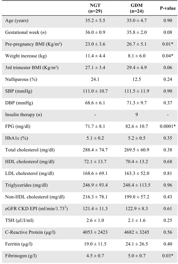
Clinical and biochemical parameters of GDM and NGT are reported in Table 1. Foetal ultrasound parameters at third trimester and neonatal parameters are reported in Table 2. Mothers in both groups were comparable for maternal age, pre-pregnancy BMI, third trimester BMI, weight gain in pregnancy, gestational age and FPG at third trimester. Six patients with GDM were treated with diet only, while six patients required insulin therapy. Foetal and neonatal parameters did not differ between the two groups.
Table 1. Clinical and biochemical parameters of the enrolled subjects NGT (n=12) GDM (n=12) P-value Age (years) 33.0 ± 6.2 34.3 ± 4.0 0.30 Gestational week (n) 37.0 ± 1.1 36.4 ± 1.2 0.23 Pre-pregnancy BMI (Kg/m²) 24.5 ± 4.2 27.3 ± 3.4 0.09 Weight increase (kg) 11.2 ± 4.4 7.6 ± 7.2 0.16 III trimester BMI (Kg/m²) 28.0 ± 4.0 30.1 ± 2.5 0.12
Nulliparity (n) 3 0 -
Gestational hypertension (n) 0 3 -
Diastolic blood pressure (mmHg) 69.6 ± 5.4 71.3 ± 10.0 0.61 Insulin therapy (n) - 6 - Total cholesterol (mg/dl) 300.1 ± 71.1 254.3 ± 68.1 0.17 HDL cholesterol (mg/dl) 75.1 ± 12.1 64.2 ± 12.2 0.07 LDL cholesterol (mg/dl) 182.6 ± 81.1 152.4 ± 56.0 0.43 Triglycerides (mg/dl) 252.1 ± 116.8 275.1 ± 117.4 0.68 Non-HDL cholesterol (mg/dl) 225.0 ± 79.5 190.0 ± 64.7 0.31 HbA1c (%) 5.0 ± 0.1 5.1 ± 0.6 0.86 eGFR MDRD (ml/min/1.732) 130.0 ± 16.3 122.6 ± 21.7 0.39 eGFR CKD EPI (ml/min/1.732) 125.2 ± 5.2 121.8 ± 9.3 0.29
TSH (µUI/ml) 2.6 ± 1.4 1.7 ± 1.7 0.17
C-Reactive Protein (µg/l) 4643 ± 2306 4875 ± 3399 0.88 Ferritin (µg/l) 18.2 ± 11.1 26.2 ± 28.6 0.42 Fibrinogen (g/l) 4.6 ± 0.8 4.9 ± 0.7 0.34 Leukocytes (109/l) 8.8 ± 1.7 8.9 ± 2.6 0.72
LDL: low-density lipoprotein; HbA1c: glycated haemoglobin; eGFR: estimated glomerular filtration rate; CKD EPI: chronic kidney disease epidemiology collaboration; TSH: thyroid-stimulating hormone; BMI: body mass index; GDM: gestational diabetes mellitus; HDL: high-density lipoprotein. *p < 0.05. Data are expressed as mean value ± standard deviation or frequency
Table 2. Foetal ultrasound and neonatal parameters NGT (n=12) GDM (n=12) P-value FOETAL US PARAMETERS Gestational week (n) 33.9 ± 2.0 34.3 ± 2.2 0.63 AC (mm) 298.1 ± 25.0 307.8 ± 27.1 0.42 HC (mm) 308.6 ± 18.2 293.7 ± 48.9 0.46 HC/AC ratio 1.05 ± 0.06 0.97 ± 0.18 0.30 Bi-parietal diameter (mm) 85.8 ± 4.5 86.0 ± 4.6 0.93
Femur length (mm) 64.9 ± 4.7 67.8 ± 4.2 0.14 Humerus length (mm) 58.0 ± 4.1 60.8 ± 5.3 0.20 Estimated foetal weight (g) 2308 ± 509 2535 ± 473 0.27 NEONATAL PARAMETERS
Weight (g) 3292 ± 356 3182 ± 420 0.50 Head circumference (cm) 34.8 ± 1.2 34.5 ± 1.3 0.58 Length (cm) 49.1 ± 1.4 49.0 ± 1.6 0.82 Gestational week - delivery (n) 38.8 ± 0.8 38.5 ± 1.0 0.39
Apgar 5’ 9.3 ± 0.6 9.6 ± 0.5 0.17 Male/Female (n/n) 6/6 7/5 1.00 Foetal complications 1 2 - Neonatal jaundice (n) ARDS (n) 1 0 1 1
US: ultrasound; HC: head circumference; AC: abdominal circumference; ARDS: acute respiratory distress syndrome; *p < 0.05. Data are expressed as mean ± standard deviation or frequency
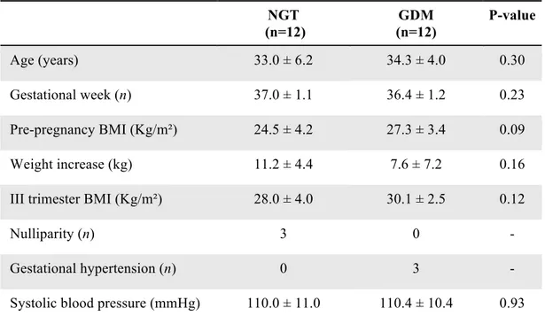
HMGB1-RAGE expression in FMs and VAT
The Western blotting analyses for HMGB1 and RAGE expression in FMs are shown in
Figure 1a-b. The expression of HMGB1 was significantly higher in FMs from GDM
patients, compared to NGT subjects (GDM 1.10 ± 0.38 vs NGT 0.58 ± 0.36, p=0.02); RAGE expression was not significantly different between the study groups (GDM 0.93 ± 0.40 vs NGT 0.82 ± 0.50, p=0.62). HMGB1 protein expression was comparable in VAT between the two groups (GDM 1.74 ± 1.37 vs NGT 1.90 ± 0.94, p=0.76) (Figure 1c), whereas the expression of HMGB1 receptor RAGE was significantly higher in GDM compared with NGT (GDM 2.10 ± 1.32 vs NGT 1.15 ± 0.59, p = 0.03) (Figure 1d).
Figure 1. High mobility group box 1 protein (HMGB1) and receptor for advanced glycation end products (RAGE) protein expression in foetal membranes (a, b) and visceral adipose tissue (c, d) in normal glucose tolerance subjects (NGT) and gestational diabetes mellitus (GDM) patients.
*p < 0.05. Data are expressed as mean value ± standard deviation. Representative blots are shown
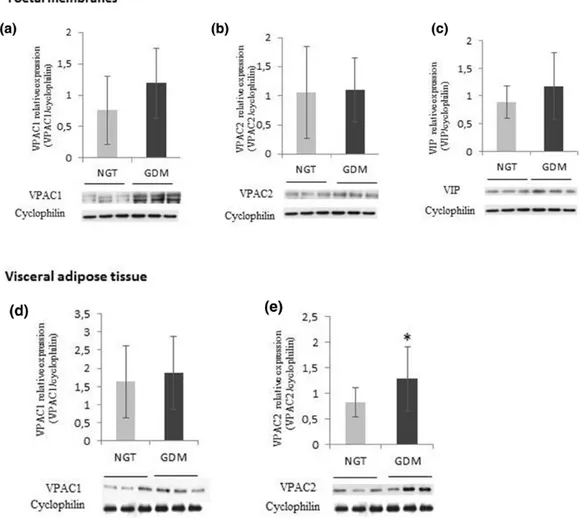
VPACs and VIP expression in VAT and FMs
VPAC2 protein expression was significantly higher in VAT obtained from women with GDM (GDM 1.28 ± 0.63 vs NGT 0.83 ± 0.29, p=0.03) compared to NGT (Figure 2b). There was no effect of GDM on VPAC1 expression (GDM 1.87 ± 1.05 vs NGT 1.63 ± 1.47, p=0.70) (Figure 2a). VIP protein expression was not detectable in VAT from both GDM and NGT women.
The expression of VPAC1, VPAC2 and VIP in FMs was not significantly different in GDM compared to NGT [VPAC1 (GDM 1.19 ± 0.56 vs NGT 0.76 ± 0.54, p=0.17), VPAC2 (GDM 1.10 ± 0.55 vs NGT 1.06 ± 0.79, p= 0.97), VIP (GDM 1.18 ± 0.60 vs NGT 0.89 ± 0.29, p=0.28)] (Figure 2c-e).
Figure 2. Vasoactive intestinal peptide (VIP), VPAC-1 and VPAC-2 protein expression in tissues collected from normal glucose tolerance (NGT) and gestational diabetes mellitus (GDM) women. VPAC1 (a), VPAC2 (b) and VIP (c) in foetal membranes; and VPAC1 (d) and VPAC2 (e) in visceral adipose tissue
*p < 0.05. Data are expressed as mean value ± standard deviation. Representative blots are shown.
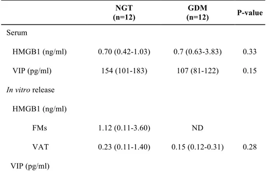
Circulating levels of HMGB1 and VIP
Serum levels of HMGB1 were not different between GDM and NGT [GDM 0.7 (0.63-3.82) vs NGT 0.7 (0.42-1.03) ng/ml; p=0.33)] (Table 3). VIP levels tended to be lower in women with GDM than in control subjects [NGT 154 (101-183) vs GDM 107 (81-122) pg/mL, p=0.15] (Table 3), although the result did not reach statistical significance.
HMGB1 and VIP in vitro release by FMs and VAT
As for HMGB1 in vitro release, high variability of HMGB1 levels was observed in FM foetal culture supernatant from NGT women [NGT 1.12 (0.11-3.6) ng/mL], whereas no detectable levels were obtained in culture supernatants from GDM FMs (Table 3).
HMGB1 release was detected in VAT culture supernatant from both NGT and GDM [NGT 0.23 (0.11-1.4) vs GDM 0.15 (0.12-0.31) ng/mL p=0.28]. However, no significant differences were observed between the two groups.
Although VIP protein expression was not detectable by Western blot analysis in VAT samples, VIP secretion was evaluated in VAT culture supernatant. VIP concentration tended to be lower in women with GDM than in control subjects [NGT 35 (24-72.5) vs GDM 19 (11.25-28) pg/mL, p=0.05]. VIP levels in FMs supernatant did not differ between GDM and controls [NGT 39 (20.5-69.5) vs GDM 22 (17.25-41.25) pg/mL, p=0.43].
Table 3. Circulating HMGB1 and VIP and tissue release in vitro NGT (n=12) GDM (n=12) P-value Serum HMGB1 (ng/ml) 0.70 (0.42-1.03) 0.7 (0.63-3.83) 0.33 VIP (pg/ml) 154 (101-183) 107 (81-122) 0.15 In vitro release HMGB1 (ng/ml) FMs 1.12 (0.11-3.60) ND VAT 0.23 (0.11-1.40) 0.15 (0.12-0.31) 0.28 VIP (pg/ml)
FMs 39.0 (20.5-69.5) 22.0 (17.25-41.25) 0.43 VAT 35.0 (24.0-72.5) 19.0 (11.25-28.0) 0.05
HMGB1: High Mobility Group Box 1; VIP: vasoactive intestinal peptide; FMs: foetal membranes; VAT visceral adipose tissue . *p < 0.05. Results are expressed as median (Interquartile range). ND: not detected
DISCUSSION
In this study, the expression of HMGB1/RAGE and VIP/VAPC1-2 has been evaluated for the first time in human FMs and VAT obtained from women with GDM. GDM was found to be associated with an increased protein expression of HMGB1 in FMs, and a high RAGE (HMGB1 receptor) and VPAC2 (VIP receptor) expression in VAT.
There has been emerging evidence about the pathophysiological relationship between HMGB1 and T2D. GDM, as well as T2D, is characterized by a low-grade inflammatory state, and the excessive production of pro-inflammatory mediators seems to play a crucial role in GDM pathophysiology (22). Over the last decades, a large number of studies have focused on the association between different inflammatory mediators and GDM (90). However, only one study has evaluated circulating levels of HMGB1 in patients with GDM (78). To date, the expression of this protein had never been evaluated at a tissue level in patients with GDM. The choice to explore the expression of pro- and anti-inflammatory mediators in FMs is related to the growing interest in these tissues’ functions, which are not yet fully known. Indeed, FMs have long been considered as dead tissues, exerting mechanical functions for foetal protection only. On the contrary, FMs are able to produce a wide spectrum of molecules at different gestational stages and have a central role both in physiological and in pathological pregnancy. Specifically, the amnio-chorionic membrane plays a pivotal role in the physiology of labour and delivery, which are pro-inflammatory events (38). In light of this, to avoid the possible effect of confounding factors, in this study FMs were collected after elective Caesarean section. Furthermore, FMs are known to be involved in the pathophysiology of premature rupture of membranes and preterm birth (91). The higher expression of HMGB1 in FMs in patients with GDM are in line with previous findings, reporting higher levels of circulating HMGB1 in the same population, compared to control subjects, at third trimester of pregnancy (78). Overall, these data suggest that
inflammation in GDM is not only observed at a systemic level, but might also directly involve gestational tissues at the maternal-foetal interface, likely contributing to an unfavourable foetal environment with conceivable short- and long-term complications. Other studies have observed an overexpression of HMGB1 in FMs in the context of human spontaneous preterm birth (68), in placental tissue obtained from pregnancies complicated by pre-eclampsia (92) and in damaged FMs from women with intra-amniotic inflammation (93), supporting a central role of this mediator in maintaining the inflammatory milieu in pregnancy complications.
In line with other authors’ findings, in this study, RAGE expression in FMs was not different between the two study groups (94). However, HMGB1 exerts its pro-inflammatory action also by binding to other receptors, such as TLR 2/4/9 (64-66).
Furthermore, RAGE expression in VAT in GDM patients was significantly higher than in control subjects, possibly playing a role in maintaining the chronic pro-inflammatory state in obesity and insulin resistance (69). Specifically, in human SW872 pre-adipocyte cell line, RAGE activation induced the secretion of pro-inflammatory cytokines (93). Although in this study HMGB1 expression in VAT did not significantly differ between GDM and NGT, the increased expression of RAGE in GDM might reflect an amplified inflammatory activity as well. This finding is in accordance with the results of a previous in vitro study which showed that the overexpression of RAGE, via NFkB activation, induced the transcription of inflammatory cytokines and was involved in adipocyte hypertrophy and in the development of insulin-resistance (70). It can be therefore speculated that other ligands might activate RAGE in VAT. Indeed, several exogenous and endogenous ligands, including AGE, ß-amyloid, S-100 protein, advanced oxidation protein products, and lipopolysaccharide, are well known ligands for RAGE and are involved in inflammation (95).
To date, data on the role of VIP–VPAC pathways at third trimester of pregnancy are lacking. In this study, VAT showed an increased expression of VPAC2 in GDM compared to NGT. Physiological pregnancy is characterized by a fine balance between pro- and anti-inflammatory cytokines (33). Giving the anti-anti-inflammatory activity of VIP/VPAC2 axis (79), a possible autocrine role of this pathway might be speculated. VPAC2 overexpression might therefore exert anti-inflammatory effects, counterbalancing the pro-inflammatory effects of other mediators. Furthermore, VIP and VPAC receptors can activate several signalling pathways, modulating adipose tissue metabolism and regulating appetite and body composition (96). Remarkable changes in maternal and foetal lipid metabolism are observed
vitro study has shown that VIP induced lipolysis through VPAC2 activation in primary rat
adipocytes (98). Remarkably, the lipolytic action of VIP via VPAC2 might potentiates insulin secretion by increasing free fatty acid levels, contributing to the maintenance of energy homeostasis both in conditions of energy deprivation and after food intake. Thus, it is conceivable that the increased expression of VPAC2 observed in VAT of GDM patients might be an attempt to manage hyperglycaemia.
CONCLUSIONS
FMs and VAT are likely to be involved in the immune metabolic processes observed in pregnancy affected by GDM. Remarkably, the increased protein expression of HMGB1 in FMs, together with the higher expression of VPAC2 (VIP receptor) and RAGE (HMGB1 receptor) in VAT in GDM, suggest a possible role of these pro-, anti-inflammatory mediators and pathways in the regulation of FM and VAT function in pregnancy. These findings suggest a complex cross-talk between FMs and VAT throughout pregnancy complicated by GDM. Further studies in larger populations are needed to confirm these data and to evaluate the impact of tissue inflammation on the possible short- and long-term maternal-foetal outcomes of pregnancy affected by GDM.
Chapter 3. Research study 2
MicroRNA expression profile in circulating exosomes and plasma of patients with GDM and healthy pregnant women
INTRODUCTION
General aspects of microRNAs
MicroRNAs are small (19-25 nucleotides) non-coding single stranded RNAs that function destabilizing or depleting target mRNAs (99). Since their discovery in Caenorhabditis
elegans in 1993 (100,101), they have been described in many other species (102). To date,
2654 human mature microRNA sequences have been identified (miRbase version 22.1 released in October 2018) (103). Their dysregulation has been described in the context of many pathological processes, spanning from cancer to neurological and metabolic diseases. Although their functions are still not completely defined, the large number of the identified microRNAs and their wide distribution among different species suggest their crucial role in gene regulation (104,105).
MicroRNA biogenesis, secretion and function involve several complex molecular events, not completely understood yet. MicroRNA genes are usually transcribed by RNA polymerase II and, to a lesser extent, by RNA polymerase III. The pri-miRNA sequence is capped at the 5’, polyadenylated at the 3’ end, and recognized by the Microprocessor complex, composed by the Di George Syndrome Critical Region 8 (DGCR8) nuclear protein and the RNAse III Drosha. This first nuclear maturation step releases a precursor microRNA, called pre-miRNA. The protein Exportin-5 is then responsible for the translocation from the nucleus to the cytoplasm, where the pre-miRNA is cleaved by the RNAse III Dicer with the production of a double strand microRNA of about 22 nucleotides in length. After loading onto Ago2, a member of the Argonaute (Ago) protein family, the microRNA is included into the RNA-induced silencing complex (RISC), a big ribonucleoprotein effector complex. The binding with Ago2 favours the most stable strand, while the passenger strand is degraded to produce a mature RISC. The main mechanisms responsible for the target mRNA silencing are mRNA degradation and translational repression. Specifically, if the sequence homology between the microRNA and its target mRNA is complete, Ago proteins degrade the target
mRNA. Otherwise, if the sequence homology is only partial, there is only translational repression (106,107).
Although the cellular compartment is the site of microRNA production and action, recent evidence demonstrated that microRNAs can also act into the extracellular compartment after secretion. Indeed, extracellular microRNAs are involved in cell-to-cell communication (108,109). Circulating microRNAs have been detected in different biological fluids, such as serum, plasma and urine. It has been demonstrated that microRNAs can be packaged into shedding vesicles and exosomes, or coupled with high-density lipoproteins (HDL), low-density lipoproteins (LDL) or Ago proteins, and actively secreted by cells. In addition, microRNAs can be passively secreted in apoptotic bodies (110).
Importantly, circulating microRNAs associated with protein complexes and/or packed into extracellular vesicles (EVs) are highly stable in the extracellular environment, therefore becoming potentially reliable diagnostic, prognostic and therapeutic biomarkers (111-113).
MicroRNAs: role in GDM pathophysiology and complications
The pathophysiological features of GDM have been elucidated and mainly include maternal insulin resistance, β-cell dysfunction, placental dysfunction, endothelial dysfunction and inflammation. However, the molecular mechanisms involved in the pathophysiology of this condition, as well as in the development of its complications, are not yet fully uncovered. There has been increasing interest in the potential roles of microRNAs as regulators of biological processes, mediators of tissue cross-talk and biomarkers of disease. Hence, a growing number of studies have characterized microRNA expression in biological fluids and in gestational tissues, highlighting their crucial involvement both in the pathogenic mechanisms of GDM, such as insulin resistance and β-cell dysfunction, and in the development of GDM-related short- and long-term complications (114).
Placenta and placenta-derived molecules play essential roles both in physiological pregnancy and in the pathogenesis of GDM. Indeed, the placenta has been widely demonstrated to promote the major metabolic adaptations in pregnancy. Many studies addressed the role of placenta-derived microRNAs in healthy and GDM pregnancies (41,115), identifying a considerable number of differentially expressed tissue microRNAs, critically involved in energy control, lipid homeostasis and inflammation, playing putative roles in the development of insulin-resistance (116-119). Less is known about the role of
VAT-derived microRNAs in the pathogenesis of GDM. To date, there is only one report that evaluated microRNA expression profile in omental VAT in GDM (120).
As for β-cell dysfunction, recent studies have indicated a conceivable microRNA-mediated cross-talk between placenta and maternal β-cells, which enriches the complex pathophysiological picture in GDM (121-124).
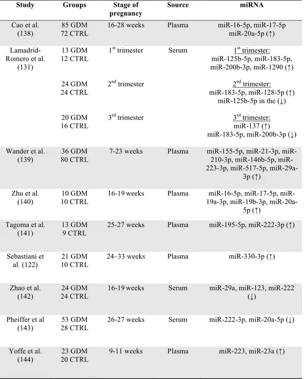
Numerous studies have revealed a complex link between several dysregulated microRNAs and impairments in placenta development and functions in GDM, possibly contributing to the development of adverse outcomes (125-127). Indeed, microRNAs seem to play crucial roles in regulating foetal growth (128,129). Since one of the main complications of GDM is foetal macrosomia, the identification of dysregulated microRNAs that may be involved in this phenomenon could be of utmost importance (130,131). Mounting evidence suggests that gene expression in the offspring might be altered by an adverse intrauterine environment, even in the absence of underlying changes in DNA sequences, and the modifications in microRNA expression are believed to mediate this process (132). Besides short-term complications, microRNA deregulation in pregnancy might contribute to the intrauterine foetal programming for the development of long-term GDM-related complications, such as metabolic diseases and CVD, both in the mother and in the offspring, later in life (133-137). It is well established that early diagnosis and appropriate treatment are essential elements to prevent poor pregnancy outcomes in GDM. According to current guidelines, GDM diagnosis is generally performed in the late second trimester, when the metabolic alterations have already developed and emerge at the OGTT. The identification of circulating biomarkers that might effectively predict GDM at an earlier stage of pregnancy is therefore crucial to prevent mother and foetal complications through prompt lifestyle and diet intervention. Evidence suggests a potential function of several circulating microRNAs as early predictors of GDM. Several studies have investigated the putative role of microRNAs as circulating plasma/serum biomarkers for GDM (Table 4.).
Table 4. Studies that evaluated circulating microRNAs in GDM
Study Groups Stage of
pregnancy Source miRNA Cao et al. (138) 85 GDM 72 CTRL
16-28 weeks Plasma miR-16-5p, miR-17-5p miR-20a-5p (↑) Lamadrid-Romero et al. (131) 13 GDM 12 CTRL 24 GDM 24 CTRL 20 GDM 16 CTRL 1st trimester 2nd trimester 3rd trimester Serum 1st trimester: miR-125b-5p, miR-183-5p, miR-200b-3p, miR-1290 (↑) 2nd trimester: miR-183-5p, miR-128-5p (↑) miR-125b-5p in the (↓) 3rd trimester: miR-137 (↑) miR-183-5p, miR-200b-3p (↓) Wander et al. (139) 36 GDM 80 CTRL
7-23 weeks Plasma 155-5p, 21-3p, 210-3p, 146b-5p, miR-223-3p, miR-517-5p, miR-29a-3p (↑) Zhu et al. (140) 10 GDM 10 CTRL
16-19weeks Plasma 16-5p, 17-5p, miR-19a-3p, miR-19b-3p,
miR-20a-5p (↑) Tagoma et al.
(141)
13 GDM 9 CTRL
25-27 weeks Plasma miR-195-5p, miR-222-3p (↑)
Sebastiani et al. (122)
21 GDM 10 CTRL
24–33 weeks Plasma miR-330-3p (↑)
Zhao et al. (142)
24 GDM 24 CTRL
16-19weeks Serum miR-29a, miR-123, miR-222 (↓)
Pheiffer et al (143)
53 GDM 28 CTRL
26-27 weeks Serum miR-222-3p, miR-20a-5p (↓)
Yoffe et al. (144)
23 GDM 20 CTRL
9-11 weeks Plasma miR-223, miR-23a (↑)
The identification of tissue and/or cell origin of circulating microRNAs is another crucial point for further research. Over the last decades, growing interest has emerged in characterizing microRNAs in tissue-specific vesicles. Advances in this field might significantly contribute to dissect the cross-talk between tissues and biological fluids both in physiological and in pathological contexts.
Role of exosomes in pregnancy and pregnancy-related disorders
The term EVs refers to membrane vesicles containing cytosol from the secreting cells, enclosed in a lipid bilayer. The release of EVs into the extracellular environment is a highly conserved process throughout evolution (145). In pluricellular organisms, EVs have been isolated from different body fluids, such as blood, urine, saliva, breast milk, amniotic fluid, ascites, cerebrospinal fluid, bile, and semen. According to various features, such as size, cell or tissue of origin and function, EVs are classified into at least three subgroups, including microvesicles (MVs), exosomes and apoptotic bodies (146). Vesicles ranging from 100 to 1000 nm, released by budding from the plasma membrane, are named MVs. Exosomes (~30-150 nm) are vesicles of endosomal origin, released during reticulocyte differentiation as a consequence of the fusion of multivesicular endosomes or multivesicular bodies with the plasma membrane (147). Vesicles formed during apoptosis are larger than 1000 nm and are named apoptotic bodies (148). However, as it is not possible to discern between exosomes and MVs on the basis of their intrinsic properties, discriminating between them is challenging and there is increasing interest in seeking novel methods of isolation and purification. Another major ongoing challenge is to understand the origin of the different populations of vesicles and to unravel their physiological relevance. Exosomes express specific late endosomal markers, such as CD63, and carry a wide variety of molecules, including proteins, lipids, nucleic acids. They are critically involved in cell-to-cell communication, playing key roles in biological processes both in physiologic and in pathologic conditions, such as cancer and metabolic diseases. Exosomes are released via exocytosis into the extracellular compartment. Their secretion is regulated by local factors, including glucose and free fatty acid concentration (149,150). The activity of both proximal and distal target cells is regulated by various interactions, including the modification of the extracellular milieu of the cellular target, the activation of cell membrane receptors, and the endocytosis by the target cell (151). Exosomes have been reported to regulate a wide range
of cellular activities in target cells (152), such as translational function, proliferation, apoptosis, angiogenesis, and metabolic functions. Exosomes contain small RNA molecules, including mRNAs and microRNAs, which are therefore protected from RNAse digestion (111). Thus, through exosomes, microRNAs are delivered in distant target cells and are able to affect gene expression.
Placental cells, including syncytiotrophoblast and extravillous trophoblast, are able to secrete exosomes into the maternal circulation. A complex network of cell-to-cell communication between the placenta and other organs is therefore thought to be sustained by circulating exosomes, delivering their cargos into target cells, likely contributing to the metabolic adaptations observed in pregnancy (153).
Interestingly, it has been observed that plasma concentration of placenta-derived circulating exosomes is significantly higher in pregnant women compared to non-pregnant women (154,155). Specifically, placenta-derived exosomes are detectable in maternal plasma at the very early stages of gestation (149) and progressively increase with gestational age, positively correlating with placental weight in the third trimester (156,157).
Furthermore, it has been reported that plasma exosome concentration is significantly increased in GDM pregnancies, compared to healthy pregnancies, and that high glucose concentration is able to induce exosome delivery by trophoblast cells in the first trimester (111,158). In particular, Salomon et al. observed a two-fold higher plasma exosome concentration throughout GDM pregnancies compared with normal pregnancies, speculating a conceivable role of plasma exosome profiling in predicting GDM at 11-14 weeks, long before the time of the actually recommended screening (24-28 weeks) (159). Other authors have observed a strong correlation between the total number of circulating exosomes and maternal BMI, a major risk factor for GDM (160).
Remarkably, non-placental-derived exosomes seem to contribute more than placenta-derived exosomes to the total exosome concentration in GDM pregnancies, although the significance of this phenomenon has not yet been defined (159).
It is well established that GDM is characterised by an imbalance between circulating pro-and anti-inflammatory cytokines (161). Evidence from in vitro studies suggests that exosomes are involved in the modulation of the inflammatory response. In particular, exosomes isolated from both healthy subjects and GDM patients can be internalised by endothelial cells and increase pro- and anti-inflammatory cytokine release (159,162).
MicroRNAs in exosomes and GDM
Exosome signalling has emerged as a novel mechanism of cell-to-cell communication both in physiological and pathological contexts. Exosomes are enriched in microRNAs, protecting themfrom exogenous ribonucleases. In light of this, microRNAs in exosomes are highly stable mediators that deliver key information and affect cellular function in their target tissues. Since microRNAs in exosomes can be isolated from body fluids, they are candidate biomarkers for several pathological conditions, including GDM (111).
AIM
The aim of this study was to comparatively evaluate microRNA expression profile in circulating exosomes and in plasma of patients with GDM and healthy control subjects in the third trimester of gestation, to potentially help elucidate the complex mechanisms underlying GDM pathophysiology and individuate novel potential candidate biomarkers for GDM.
PATIENTS AND METHODS
Participants
Fifty-three pregnant women, n.24 with GDM (mean age 35.0 ± 4.7 years) and n.29 with normal glucose tolerance (NGT) (mean age 35.2 ± 4.7 years), were recruited in the Diabetes out-patient Unit and in the Obstetrics and Gynaecology out-patient Unit, respectively, of Policlinico Umberto I, “Sapienza” University Hospital of Rome. Diagnosis of GDM was performed or excluded in accordance with current recommendations (11). The protocol was approved by the hospital ethics committee and written informed consent was obtained from all the participants. All subjects were enrolled during the third trimester of pregnancy. Women with non-Caucasian ethnicity; pre-pregnancy BMI ≥35 Kg/m2; pre-pregnancy impaired fasting glucose (FPG 100-125 mg/dl); multiple or induced pregnancy; infectious, inflammatory and autoimmune diseases; polycystic ovarian syndrome; psychiatric diseases; alcohol and drug abuse and steroid therapy were excluded. At enrolment, a detailed medical history was obtained and anthropometric/vital (weight, BMI, blood pressure and heart rate) parameters were assessed. Foetal ultrasound parameters at third trimester were obtained. Peripheral blood samples were collected to evaluate laboratory parameters and microRNA