Facoltà di Scienze Matematiche, Fisiche e Naturali
Dipartimento di Biologia
Scuola Dottorale in Biologia
Sezione di Biologia Applicata alla Salute dell’Uomo
XXII ciclo
“The role of mitotic spindle alterations in the
induction of apoptosis”
“Ruolo delle alterazioni del fuso mitotico
nell’induzione di apoptosi”
PhD student Supervisor Dr. C. Cenciarelli Dr. A. Antoccia
SUMMARY ... 1 1. Italian ... 1 2. English ... 3
INTRODUCTION
... 6 1. MITOTIC SPINDLE ... 6 1.1 MICROTUBULES ... 6 1.2 CENTROSOME ... 9 1.3 MITOTIC SPINDLE ... 112. MICROTUBULES AND CANCER THERAPY ... 12
2.1 MICROTUBULES DAMAGING AGENTS (MDA) ... 13
2.2 COMBRETASTATIN CA-4 ... 15
3. APOPTOSIS ... 16
3.1 CASPASE-INDEPENDENT CELL DEATH (CICD) ... 18
3.2 MITOTIC CATASTROPHE ... 19
4. BCL-2 PROTEINS ... 19
4.1 BH3-ONLY protein: BIM ... 22
5. MECHANISMS OF CELL DEATH INDUCED BY MDA ... 24
6. P53 and its INDEPENDENT TRANSCRIPTION ACTIVITY ... 25
RESULT
... 301. Interfering with spindle microtubule-dynamics arrests cells at metaphase stage with misaligned chromosomes ... 30
2. Combretastatin A-4 affects spindle organization giving rise to “star-like” mitosis. ... 31
3. Pericentriolar matrix components are present in CA-4-induced “star-like” structures ... 32
4. Combretastatin induces pericentriolar matrix dismantling and caspase-dependent mitotic catastrophe ... 35
5. Inhibition of Bim strongly reduced caspase-3 cleavage and apoptosis .. 37
6. Bim bound physically to microtubules by interaction with dynein, is released after CA-4 treatment and relocalizes to mitochondria ... 40
7. p53 is linked to dynein and localized on mitochondria after CA-4 treatment ... 42
8. Bim coimmunprecipitated with p53 in the mitochondrial protein fraction ... 42
DISCUSSION
... 43CONCLUSION
... 481 SUMMARY
1. Italiano
I microtubuli, insieme con l’actina e i filamenti intermedi sono i principali componenti del citoscheletro delle cellule eucariotiche e svolgono un ruolo fondamentale in molti processi cellulari: divisione, motilità, “trafficking” e mantenimento della forma cellulare. Il principale Centro Organizzatore dei Microtubuli (MTOC) nelle cellule di mammifero è il centrosoma che è composto da una coppia di centrioli circondata da una matrice elettron-densa chiamata materiale pericentriolare (PCM). Essendo i microtubuli fortemente coinvolti nei processi di divisione cellulare, rappresentano un target preferenziale nella terapia del cancro. Gli agenti che danneggiano i microtubuli (MDA) rappresentano una importante classe di farmaci antitumorali e benchè alcune di queste sostanze promuovano la stabilizzazione dei microtubuli e altre inducano la depolimerizzazione, tutti gli MDA presentano la caratteristica comune di sopprimere la dinamica e quindi la funzione dei microtubuli. In questo modo gli MDA causano la distruzione del fuso mitotico e un blocco nella progressione del ciclo cellulare nella transizione metafase-anafase che puo’ portare all’attivazione dei pathways apoptotici e alla catastrofe mitotica. Quest’ultima è stata descritta recentemente come una morte cellulare che avviene durante la mitosi in seguito all’utilizzo di agenti che causano alterazioni del fuso mitotico. Recentemente abbiamo focalizzato la nostra attenzione sulla Combetastatina (CA-4), un MDA isolato dal Combretum Caffrum, un albero sud Africano che ha la caratteristica di non essere riconosciuta dalla pompa Multi Drug Resistance, una pompa cellulare che elimina rapidamente sostanze estranee inclusi molti farmaci antitumorali. Gli MDA agiscono inoltre anche sul centrosoma causando alterazioni strutturali, frammentazione e inappropiata duplicazione del centrosoma. La CA-4 è in grado di arrestare le cellule in mitosi causando, in maniera dipendente dalla concentrazione, un aumento della disorganizzazione dell’arrangiamento cromosomico e la formazione di particolari strutture chiamate “star-like”.
2
L’analisi in microscopia a fluorescenza e confocale ha evidenziato che le “star-like” sono strutture di forma sferica formate da piccoli fasci di microtubuli connessi al cinetocore. Al centro di queste strutture sono presenti alcuni componenti della matrice pericentriolare come la -tubulina, la pericentrina e la nineina. Il trattamento con la CA-4, oltre a determinare alterazioni di tipo strutturale, causa morte cellulare per catastrofe mitotica caratterizzata dall’attivazione delle caspasi-3/-9 e dalla frammentazione del DNA. Il meccanismo che porta dalla depolimerizzazione dei microtubuli alla morte cellulare non è ancora completamente chiarito. Recentemente è stato dimostrato che Bim, una proteina pro-apoptotica appartenente alla famiglia Bcl-2, è associata con il motore proteico dineina e sequestrata quindi dai microtubuli. Gli MDA, attraverso l’alterazione della struttura dei microtubuli, potrebbero causare il rilascio di Bim dal citoscheletro e la sua traslocazione sul mitocondrio. Inoltre dati recenti mostrano che esistono altri meccanismi che regolano Bim come la regolazione trascrizionale. Allo stesso modo, p53, una proteina pro-apoptotica molto conosciuta è associata con i microtubuli ed utilizza il motore proteico dineina per essere trasportata nel nucleo ed agire come fattore di trascrizione. Basandoci su questi dati abbiamo valutato il coinvolgimento di Bim e di p53 nell’apoptosi indotta in seguito al trattamento con CA-4. Il trattamento con la CA-4 determina sia il rilascio dai microtubuli sia un aumento del livello trascrizionale di Bim. Inoltre il silenziamento di Bim riduce fortemente l’attivazione della caspasi-3, una delle caspasi effettrici del pathway apoptotico mitocondriale. Inoltre, recentemente è stato dimostrato che p53 puo’ indurre apoptosi non solo agendo come fattore di trascrizione ma anche attraverso una azione indipendente dalla trascrizione grazie alla capacità di interagire direttamente con proteine della famiglia Bcl-2 presenti sul mitocondrio come Bcl-2 e Bcl-Xl. Comunque l’importanza dell’attività indipendente dalla trascrizione di p53 dopo utilizzo degli MDA su cellule tumorali rimane poco compresa e studiata. Nei nostri esperimenti abbiamo verificato che il trattamento con la combretastatina causa una variazione nel legame di p53 con la dineina e parallelamente determina la presenza di tale proteina sul mitocondrio. Inoltre esperimenti di coimmunoprecipitazione rivelano che Bim e p53
3
interagiscono sul mitocondrio e sono probabilmente responsabili dell’attivazione del pathway mitocondriale apoptotico. I nostri dati indicano quindi che cellule H460 trattate con la combretastatina sono uccise attraverso il meccanimo della catastrofe mitotica dipendente da un prolungato arresto mitotico, attivazione delle caspasi e traslocazione mitocondriale di Bim e p53.
2. English
Microtubules, together with actin and intermediate filaments are the major components of the cytoskeleton of eukaryotic cells and play a critical role in many cellular processes, including cell division, cell motility, intracellular trafficking, and cell shape maintenance. The principal Microtubule Organizing Center (MTOC) in mammalian cells is the centrosome that is composed of a pair of centrioles surrounded by an osmiophilic matrix termed pericentriolar material (PCM). As the microtubule network is highly involved in cell proliferation, it appeared to be a preferential target for cancer therapy. Microtubule Damaging Agents (MDA) represent an important class of anticancer drugs. Although some of this agents promote microtubules stabilization and other induce microtubules disruption, all MDA share the common property of suppressing microtubules dynamics, and thereby microtubules function, leading to the disruption of the mitotic spindle and the blockade of cell cycle progression at the transition from metaphase to anaphase that leads to apoptosis or mitotic catastrophe commitment. Mitotic catastrophe has been described as cell death that occurs from metaphase of mitosis in response to agents that cause mitotic-spindle damage. Recently we focused our attention on the antineoplastic spindle poison agent combretastatin-A4 (CA-4). CA-4 is an MDA isolated from the South African tree Combretum Caffrum that is not recognized by the multi drug resistance pump, a cellular pump which rapidly ejects foreing molecules including many anticancer drugs. MDAs also promote several
4
effects on the centrosome including abnormal centrioles structure, centrosome fragmentation and inappropriate centrosome duplication. CA-4 is able to arrest cells at mitosis in a dose depending manner accompanied by an increasing disorganization of chromosomal arrangement, led to formation of particular structure named “star-like”. This structures contain pericentrosomal matrix components like -tubulin, pericentrin and ninein. Confocal and immunofluorescence microscopy revealed that “star like” structure are spherical structure formed by residual fragments of microtubules connected to kinetochore. At the centre of this structure are present some component of the pericentriolar matrix like -tubulin, pericentrin and ninein. Treatment with CA-4, which produced high frequency of “star-like” mitosis, was accompanied by mitotic catastrophe commitment characterized by activation of caspases-3/-9 and DNA fragmentation. The mechanisms that lead from microtubules depolymerization to cell death are still not completely clear. Recently it has been demonstrated that Bim, a proapoptotic Bcl-2 protein, is associated to the dynein motor complex and sequestered by microtubules. MDA, through the alteration of microtubules structure, might lead to the release of Bim from cytoskeleton and to its translocation to the mitochondria. Moreover, recent data show that further mechanisms might be at the base of Bim-induced apoptosis such as regulation of Bim transcriptional levels. In a similar way, p53 another well known pro-apoptotic molecule, is associated with cellular microtubules and is transported to the nucleus by dynein. Based on these data, we have investigated whether upon tubulin depolymerization, Bim and p53 could be involved, and eventually in which manner, in the first steps of apoptosis. CA-4 treatment induce the release of Bim from microtubules and its increase at transcriptional level. In addition, Bim silencing strongly reduced apoptotic commitment. It has recently reported that p53 can also induce apoptosis independently of new protein synthesis however the functional importance of p53 in MDA-induced apoptosis of human cancer cells remain poorly understood. Concerning the role of p53 in CA-4-induced apoptosis, we found that treatment determined
5
its release from cytoskeleton and re-localization to mitochondria. Data obtained from this study showed that Combretastatin-treated H460 cells are killed via a mitotic catastrophe mechanism strictly dependent on the achievement of mitotic block, caspases activation and Bim and p53 translocation to mitochondria.
6 INTRODUCTION
1. MITOTIC SPINDLE 1.1 MICROTUBULES
Microtubules (MT), together with actin and intermediate filaments are the major components of the cytoskeleton of eukaryotic cells. Microtubules are long, stiff and cylindrical tubes with a diameter of about 25 nm consisting of α- and β-tubulin heterodimers that polymerize into 13 protofilaments, which form the wall of the microtubules (Fig.1)(Cassimeris, 1993;Schmidt and Bastians, 2007). Microtubules play a critical role in many cellular processes, including cell division, cell motility, intracellular trafficking, and cell shape maintenance (Bhat and Setaluri, 2007).
MTs play a particularly important role in cell organization: they pull the chromosomes apart at mitosis, act as a ‘railroad system’ for intracellular transport, and define the localization and structure of internal membrane systems (Rogers and Gelfand, 2000;Kline-Smith and Walczak, 2004;Gregoretti et al, 2006). They are highly dynamic polymers and their polymerization dynamics are tightly regulated both spatially and temporally (Jordan and Wilson, 2004). Two characteristics of MTs are particularly significant for these functions. First, MT nucleation is regulated, and the limitation of nucleation to an organelle near the nucleus (the centrosome) endows most cell types with a radial organization. Second, and perhaps more importantly, the cytoskeleton is dynamic: individual MTs in the same cell constantly change in length, either growing or shrinking with random transitions between these phases (Gregoretti et al, 2006). This dynamic polymerization behavior is driven by the hydrolysis of GTP, which is bound at tubulin subunits. The two ends of microtubules are distinct: one end, the plus end, is much more dynamic and can show a net growth while the other, the minus end, is anchored in the centrosome or Microtubule Organizing Center (MTOC), and is less dynamic and can show net shrinkage. Thus, at a given time, microtubules can show no change in their polymer mass, yet exhibit very high dynamics. The second important dynamic behavior of microtubules is termed treadmilling, which is a net growth at the plus end and a balanced net shortening at the minus end. Consequently, treadmilling results in a flow of tubulin subunits from the plus to the minus end of
7
microtubules (WatermanStorer and Salmon, 1997). Both, dynamic instability and treadmilling are important features for the function of microtubules, especially during mitosis (Schmidt and Bastians, 2007).
Fig.1 Polymerization of microtubules. Heterodymers of α- and β-tubulin assemble to
form a microtubules. (From (Jordan and Wilson, 2004d)
The functional diversity of microtubules is achieved in several ways: through the binding of various regulatory proteins, including Microtubule Associated Proteins (MAPs), to soluble tubulin and to the microtubule surfaces and ends; by expression of different tubulin isotypes, which have different functions; and through several post-translational modifications of tubulin (Jordan and Wilson, 2004c). For example, human tubulin isotypes (6 forms of α-tubulin and 7 forms of β–tubulin) are expressed at varying levels in different cells and tissues. These tubulins can be further modified
8
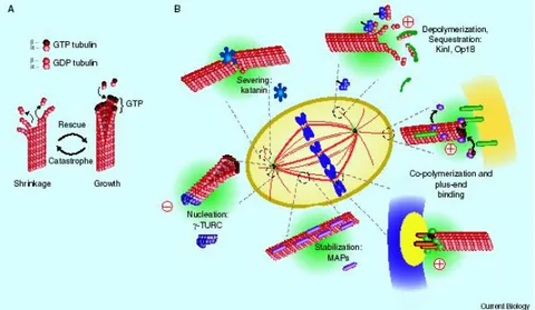
post-translationally by polyglutamylation, polyglycylation, phosphorylation, acetylation, detyrosination/tyrosination and removal of the penultimate glutamic-acid residue of α-tubulins. There are many different MAPs, including the dynein and kinesin motor proteins, as well as many microtubule-regulatory proteins, such as survivin, stathmin, TOG, MCAK, MAP4, EB1, dynactin 1 (Fig.2)(also known as p150Glued), RAC1 and FHIT3–8 (Jordan and Wilson, 2004b). Some of these proteins move along microtubules mediating intracellular cargo transport, others have specific functions in mitosis in centrosome positioning, chromosome congression and segregation (Cassimeris and Spittle, 2001). Some of these differences predominate in certain cancer cells and some are associated with the development of drug resistance (Jordan and Wilson, 2004a).
Fig. 2 Regulation of microtubules dynamic. (From(Gadde and Heald, 2004).
9 1.2 CENTROSOME
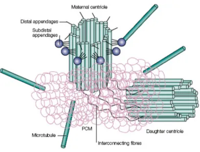
The centrosome of animal cells is a small non membranous organelle, and it is often associated with the nuclear membrane. It is composed of a pair of centrioles and a surrounding electron dense matrix of protein aggregates referred to as the pericentriolar material (PCM) (Fukasawa, 2002;Dutertre et al, 2002)). Each centrioles has a cylindrical shape and it is build with nine sets of triplet microtubules (Fig.3). Centrioles of the pair are different: one of them is named mother centrioles and unlike the daughter centrioles bears additional structures. It is worth noting that MT triplets of centriolar cylinders are very stable and unlike the cytoplasmic MT are not disassembled after the action of Microtubules Damaging Agents (MDA) and cold (Osborn and Weber, 1976;Alieva and Uzbekov, 2008). Many different proteins are present in the centrosome and they can be divided into three main group: MT triplet and centriolar matrix proteins, proteins of the PCM and proteins whose associations with centrosome is labile (Alieva and Uzbekov, 2008). Between proteins of centriolar matrix and PCM are centrin, pericentrin, ninein and γ-tubulin (Salisbury, 1995) that are involved in anchoring and nucleation of MT. The centrosome function as a Microtubule Organizing Centre (MTOC), which estabilishes polarity and orientation of MT during interphase, and direct the assembly of bipolar spindles during mitosis (Fukasawa, 2002). MT nucleated by centrosome are polarized with their rapidly growing (plus) ends in the cytoplasm and their slow-growing (minus) ends anchored at the centrosome (Doxsey, 2001). MT nucleations is due to the presence in the PCM of γ-tubulin Ring Complex (γ-TuRC)(Oakley and Oakley, 1989;Zheng et al, 1991;Moritz et al, 1995). The main subunity of the complex is a tetramer composed by two γ-tubulin molecules and two other proteins GPC2 and GPC3 (Knop and Schiebel, 1997;Oegema et al, 1999). The ring structure, observed by electronic microscopy, reveals the presence of six or seven tetramers togheter with at least other four proteins. Every daugther cell inherits only one centrosome and so the cell must duplicate the centrosome prior to the next mitosis. Duplication of centrosomes must proceed in coordination with other cell cycle events indeed initiation of centrosome duplication occurs near the G1/S boundary, while elongation of daughter centrioles and maturation of centrosome continue during S and G2 phase (Blagden and Glover, 2003;Doxsey et al, 2005). The presence of more than two centrosomes in a cell result in the formation of defective mitotic spindles
10
which increase the chromosomes segregation errors and it is responsible of chromosomes instability. This role of centrosomes was initially proposed by Theodor Bovery in 1914 and it was confirmed only many years later with the discovery that centrosome abnormality is a common marker in human cancer (Pihan et al, 1998;Lingle et al, 1998;Brinkley and Goepfert, 1998;Fukasawa, 2002)
Fig. 3 The centrosome is composed by a pair of centrioles surrounded by pericentriolar material that nucleates microtubules around the ends closest to one another. Only the maternal centriol has two sets of extra appendages, distal and subdistal interconnetting fibres links the closest ends of the two centrioles. (From (Doxsey, 2001)
11 1.3 MITOTIC SPINDLE
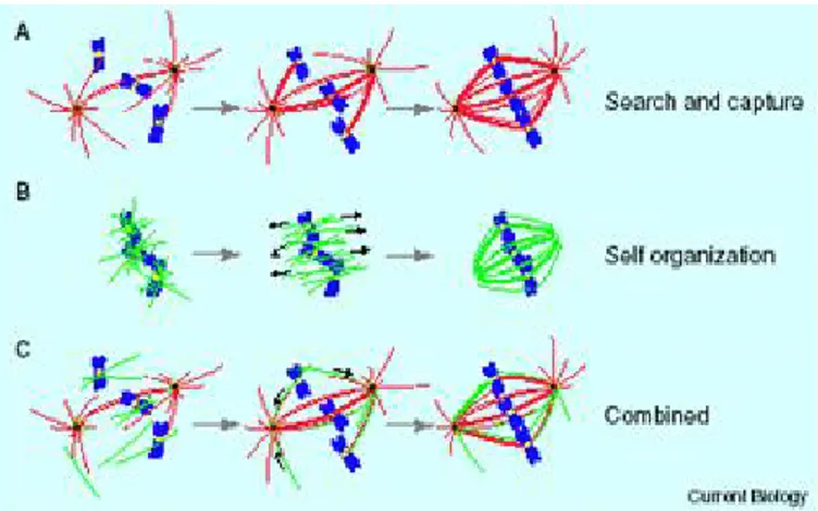
The predominant model of spindle assembly in the presence of centrosomes is based on microtubules dynamic instability and is known as the “search and capture” model (Fig.4)(Kirschner and Mitchison, 1986;Gadde and Heald, 2004). In this scenario microtubules emanating from a centrosome go under cycles of growth and shrinkage, randomly probing the cytoplasm until running into a kinetochore, with which they form a stable attachment (Kirschner and Mitchison, 1986;Gadde and Heald, 2004). This lead to bipolar spindle formation. “Search and capture” mechanism is not relevant for spindle assembly in cell that lack centrosomes like plants and oocyte of animal cells. In this cells, acentrosomal spindle assembly (self organization model) start with microtubules nucleation near chromosomes, in a process that involves Ran/RCC1 activity (Karsenti and Vernos, 2001;Li et al, 2003;Kalab et al, 2006). This two mechanism have been viewed mutually exclusive for many years, however, several lines of evidence have changed this view. When the centrosome is physically removed in vertebrate somatic cells using a laser beam or microsurgery, functional bipolar spindle form nevertheless (Khodjakov et al, 2000;Hinchcliffe et al, 2001;Mahoney et al, 2006). Besides, mutations in Drosophila that inactivate centrosomes didn’t inactivate functional spindle formation (Bonaccorsi et al, 1998;Megraw et al, 2001). The effect on mitotic progression in the absence of centrosomes is that spindle are more misoriented. These observations highlight the idea that multiple mechanisms promote bipolar spindle formation. It remains to estabilish if the acentrosomal mechanism is activated only when cell are not able to use the “search and capture” model. Besides, recent observation suggested that kinetochore could promote microtubules polymerization (K fibres)(Khodjakov et al, 2003).
12
Fig. 4 Models of spindle assembly: search and capture (a); self organization (b);
combined (c). (From (Gadde and Heald, 2004)
2. MICROTUBULES AND CANCER THERAPY
Most chemotherapeutic anti-cancer drugs used in the clinic today include agents that target the cell cycle in order to inhibit the hyperproliferation state of tumor cells and, subsequently, to induce apoptosis, which is the desired outcome of chemotherapy (Lee and Schmitt, 2003). Based on their mode of action these chemotherapeutic drugs can be subdivided into distinct groups: (i) drugs that interfere with DNA synthesis, (ii) drugs that introduce DNA damage and (iii) drugs that inhibit the function of the mitotic spindle. The latter have been proven to be exceptionally successful in the clinic and are classically represented by microtubule binding drugs frequently referred to as spindle poisons (Mollinedo and Gajate, 2003;Jordan and Wilson, 2004f;Pasquier et al, 2006). As the microtubule network is highly involved in cell proliferation, it appeared to be a preferential target for cancer therapy.
13
These drugs bind to and inhibit the function of microtubules of the mitotic spindle apparatus. However, since microtubules fulfill important functions in resting and differentiated cells these drugs exhibit a plethora of unwanted side effects. Therefore, novel drug targets that spare microtubules, but inhibit the progression of mitosis are highly desired and already exploited for the development of novel anti-mitotic drugs (Schmidt and Bastians, 2007).
The interest in MTAs is accompanied with advances in the fundamental understanding of their mechanism of action, that may provide alternative or additional approaches in the treatment of cancers (Esteve et al, 2007a). Their importance in mitosis and cell division makes microtubules an important target for anticancer drugs.
For that matter, great efforts have been devoted to discover drugs that affect microtubules.
2.1 MICROTUBULES DAMAGING AGENTS (MDA)
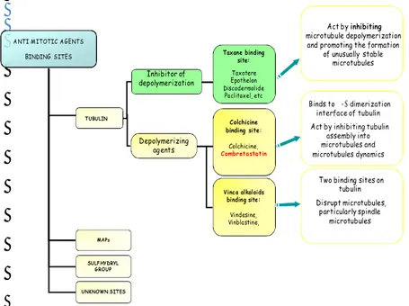
MDA constitute a class of anti-cancer drugs largely used in the clinic for the treatment of various cancers. MDA have been demonstrated to exert a high level of clinical activity, represented by clinical remissions in advanced ovarian, breast and the upper aerodigestive tract cancers (Rowinsky and Donehower, 1995;Miller and Sledge, 1999;Jordan and Wilson, 2004e). Microtubule damaging agents are usually classified into two main groups (Fig.5). One group, known as the microtubule-destabilizing agents, inhibits microtubule polymerization at high concentrations and includes several compounds — such as the Vinca alkaloids (vinblastine, vincristine, vinorelbine, vindesine and vinflunine), cryptophycins, halichondrins, estramustine, colchicine and combretastatins — that are used clinically or are under clinical investigation for treatment of cancer. The second main group is known as the microtubule-stabilizing agents. These agents stimulate microtubule polymerization and include paclitaxel (the first agent to be identified in this class), docetaxel (taxotere) and the epothilones (Jordan and Wilson, 2004g). The most important action of these drugs is the suppression of spindle-microtubules dynamics, which result in the blocking of mitosis at the metaphase-anaphase transition and induction of apoptotic cell death.
14
Fig. 5 A representative pattern of MDA and their binding sites.
ANTI MITOTIC AGENTS BINDING SITES TUBULIN MAPs SULFHYDRYL GROUP Inhibitor of depolymerization Depolymerizing agents UNKNOWN SITES Colchicine binding site: Colchicine, Combretastatin Vinca alkaloids binding site: Vindesine, Vinblastine, Taxane binding site: Taxotere Epothelon Discodermolide Paclitaxel, etc
Two binding sites on tubulin Disrupt microtubules, particularly spindle microtubules Binds to α-β dimerization interface of tubulin Act by inhibiting tubulin
assembly into microtubules and microtubules dynamics
Act by inhibiting microtubule depolymerization
and promoting the formation of unusually stable
15 2.2 COMBRETASTATIN CA-4
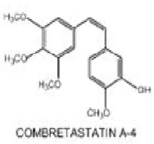
Combretastatin A-4 (CA-4) is a natural stilbenoid isolated from the South African bush of Combretum caffrum that is structurally related to colchicines but have potent anti-vascular properties at well-tolerated doses (Fig.6)(Tozer et al, 2002). CA-4 is the most active of 17 compounds isolated from the natural source (Uppuluri et al, 1993;Pettit et al, 1995;Jordan et al, 1998). CA-4 is not recognized by the Multi Drug Resistance (MDR) pump which rapidly ejects foreing molecules including many anticancer drugs (Patocka and Strunecka, 1999;Islam and Iskander, 2004).
16 3. APOPTOSIS
Cell death is an essential strategy for the control of the dynamic balance in living systems. Briefly there are two different forms of cell death, apoptosis and necrosis. Necrosis is an accidental passive process resulting in an early disruption of the cell membrane and in the breakdown of cell structures in response to violent environmental perturbations. In contrast, apoptosis is tightly regulated and involves the activation of an energy-requiring intracellular machinery (Yuan, 1996;Vermeulen et al, 2005). This form of cell death affects single cells and it is able to eliminate cells exposing the organism to danger (Shibata et al, 1994;Green and Martin, 1995;Liebermann et al, 1995;Vermeulen et al, 2005).
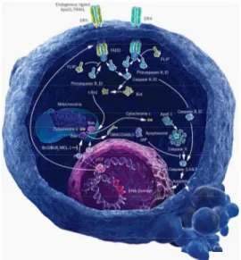
Apoptosis is characterized by many morphological changes like cell shrinkage, nuclear pyknosis (chromatin condensation), karyorhexis (nuclear fragmentation), blebbing and phosphatidylserine exposure on the surface of the plasma membrane (Kerr et al, 1972;Wyllie et al, 1984;Martin et al, 1995;Zamzami et al, 1996;Kroemer et al, 2007). Apoptosis is a genetically predetermined mechanism characterized by two major biochemical events, namely the activation of caspases and the permeabilization of the outer mitochondrial membrane, with the consequent release of many death effectors from the mitochondrial intermembrane space (Kerr et al, 1972;Brenner and Kroemer, 2000;Ferri and Kroemer, 2000;Ravagnan et al, 2002). Apoptosis may be elicited by different pathways of which the best characterized are called the extrinsic and intrinsic (Fig.7).
In the extrinsic pathway apoptosis is triggered by activation of death receptors at the cell surface. This causes the recruitment and oligomerization of the adapter molecule FADD (Fas-Associating Death Domain-containing protein) within the Death-Inducing Signaling Complex (DISC). Finally this oligomerization led to activation of caspase-8 and 10
(Debatin and Krammer, 2004;Kroemer et al, 2007). The intrinsic or mitochondrial pathway is the predominant form of cell
death in vertebrates. The critical event in this pathway is the mitochondrial membrane permeabilization (MMP) that is responsible for caspases activation. Normally cytochrome C (cytC) resides in the inner mitochondrial space where it functions as an electron shuttle in the respiratory chain (Bernardi and Azzone, 1981). During the intrinsic
17
pathway of apoptosis cytC is released into the cytosol where it can bind to APAF-1 (Apoptosis Protease Activating Factor-1). APAF-1 exist in the cytosol as a monomer and its activation depend on the presence of citochrome C and ATP/ADP (Cain et al, 2002). The multimerization of APAF-1 led to activation of caspase-9 and to initiation of mitochondrial apoptosis pathway through activation of effector caspases. Effectors caspases act on a variety of substrates resulting in proteolysis of cellular proteins and death by apoptosis. One of the best characterized and the earliest substrate of caspase is Poly-(ADP-Ribose) Polymerase (PARP), a nuclear protein implicated in DNA repair (Duriez and Shah, 1997). Caspase cleavage and inactivation of ICAD (Inhibitor of Caspase-Activated DNAse) allow CAD (DNA fragmentation factor) to translocate the nucleus where it is responsible for internucleosomal DNA cleavage, generating DNA fragments (Liu et al, 1997;Sakahira et al, 1998;Vermeulen et al, 2005). Besides caspase cleavage of lamins and cytoskeletal proteins leads to nuclear shrinkage and cytosolic reorganization (Martin et al, 1995;Mashima et al, 1995;Orth et al, 1996;Liu et al, 1997;Vermeulen et al, 2005).
Fig. 7. Extrinsic and intrinsic pathways of apoptosis. A representative image from
18
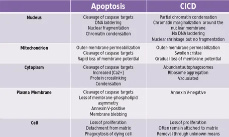
3.1 CASPASE-INDEPENDENT CELL DEATH (CICD)
Many studies have shown that apoptosis is linked to caspases activity and that caspases are required to induce cell death (Taylor et al, 2008;Tait and Green, 2008). Recently it has been described a different type of cell death that occurs when a signal that normally induced apoptosis fails to activate caspases (Taylor et al, 2008;Tait and Green, 2008). This type of cell death often shares common characteristic with apoptosis (Table 1).
Table 1. Characteristics of apoptotic and caspase-independent cell death. (From
(Tait and Green, 2008) modified).
Many studies in both cell culture models and utilizing mice deficient in apoptotic signaling supported the existence of CICD tipically by inducing apoptosis. In the presence of chemical caspase inhibitors such as zVAD-fmk (Hirsch et al, 1997;Ohta et al, 1997;Sarin et al, 1997;Tait and Green, 2008). CICD has also been observed in cell expressing genetically encoded caspase inhibitors such as XIAP, CrmA or p35 following induction of apoptosis (Okuno et al, 1998;Denmeade et al, 1999;Wilkinson et al,
Apoptosis CICD
Nucleus Cleavage of caspase targets
DNA laddering Nuclear fragmentation Chromatin condensation
Partial chromatin condensation Chromatin marginalization around the
nuclear membrane No DNA laddering Nuclear shrinkage but no fragmentation
Mitochondrion Outer-membrane permeabilization
Cleavage of caspase targets Rapid loss of membrane potential
Outer-membrane permeabilization Swollen cristae Gradual loss of membrane potential
Cytoplasm Cleavage of caspase targets
Increased [Ca2+] Protein crosslinking Condensation Abundant autophagosomes Ribosome aggregation Vacuolated
Plasma Membrane Cleavage of caspase targets
Loss of membrane-phospholipid asymmetry Annexin V-positive Membrane blebbing
Annexin V-negative
Cell Loss of proliferation Detachment from matrix Phagocytosis of dying cell
Loss of proliferation Often remain attached to matrix Removal through unknown means
19
2004;Tait and Green, 2008). Besides cells that lack components of the intrinsic apoptotic pathway such as Apaf-1, cytochrome c or caspase-9 demonstrated the existence of an alternative type of cell death (Hakem et al, 1998;Kuida et al, 1998;Li et al, 2000;Tait and Green, 2008).
3.2 MITOTIC CATASTROPHE
Mitotic catastrophe has been first described in Schizosaccharomyces pombe as a temperature-sensitive lethal phenotype, linked to gross abnormalities of chromosome segregation, that was observed in some mutant strains (Ayscough et al, 1992;Castedo et al, 2004). Some authors view mitotic catastrophe of mammalian cells as the failure to undergo complete mitosis which usually ends in the formation of large cells with multiple micronuclei and decondensed chromatin (Swanson et al, 1995;Ianzini and Mackey, 1997;Roninson et al, 2001;Castedo et al, 2004). However, reports describing mitotic catastrophe frequently show cells with some phenotypic characteristic of apoptosis (Castedo et al, 2004). Mitotic catastrophe would be a type of cell death occurring during mitosis, as a result of DNA damage or deranged spindle formation coupled to the debilitation of different checkpoint mechanism that would normally arrest progression into mitosis and hence suppress catastrophic events until repair has been achieved. (Castedo et al, 2004;Tait and Green, 2008). Damage leading to mitotic catastrophe can be induced, in particular, by microtubules damaging agents. So, mitotic catastrophe can be considered as a type of cell death occurring during mitosis or resulting from mitotic failure.
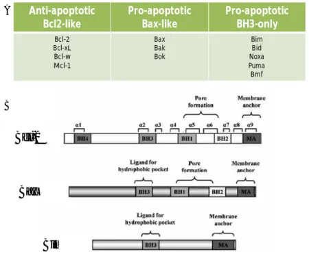
4. BCL-2 PROTEINS
The major regulators of apoptosis belong to Bcl-2 family that is composed of up to 30 proteins (Strasser et al, 2000a;Esteve et al, 2007b). Bcl-2 was the first human oncoprotein described to promote cell survival (Vaux et al, 1988). Classification of other members has then been based on their Bcl-2 homology (BH) domains (Fig. 8)(Yin et al, 1994;Chittenden et al, 1995a;Zha et al, 1996;Gibson et al, 1996;O'Connor et al, 1998). Bcl-2 and
20
its closest homologs (Bcl-xl, Bcl-w, Mcl-1) (Boise et al, 1993;Gibson et al, 1996) share three or four regions of homology (BH1-BH4 regions) and are able to inhibit apoptosis (Vaux et al, 1988;Strasser et al, 2000a). Other members of this family have the capability to promote apoptosis and for this reason they are called pro-apoptotic proteins. These proteins can be divided into the Bax subfamily (Bax, Bak, and Bok multidomain proteins) (Oltvai et al, 1993;Farrow et al, 1995;Kiefer et al, 1995;Chittenden et al, 1995b;O'Connor et al, 1998) or the BH3-only subfamily (Bid, Bim and PUMA). The N-terminal BH4 domain is present only in some anti-apoptotic proteins, while BH1, BH2 and BH3 can be found in both sub-families. Some members of the Bcl-2 family have a hydrophobic C-terminal region that allows their localization on intracellular membranes (Schinzel et al, 2004;Lucken-Ardjomande and Martinou, 2005). Homotypic and heterotypic interaction between individual members of the family are important mechanism that controls their activity (Cheng et al, 2001;Cory et al, 2003;Lucken-Ardjomande and Martinou, 2005;Chen et al, 2005).
The relative level of anti- and pro-apoptotic Bcl-2 proteins arbitrate cell life or death decision (Oltvai et al, 1993;Oltvai and Korsmeyer, 1994). Bcl-2 like members are mainly localized to mitochondrial membranes and prevent permeabilization of the mitochondrial outer membrane. There are two models that explain how Bcl-2 proteins can induce mitochondrial membrane permeabilization: indirect and direct model. The indirect model proposes that anti-apoptotic proteins must continually inhibit the function of Bax and Bak to ensure mitochondrial integrity and survival. So in this scenario, mitochondrial membrane permeabilization arise when all anti-apoptotic proteins are functionally neutralized by activated BH3-only proteins. At the contrary, in the direct model BH3-only proteins (Bid and Bim) directly interact with Bax or Bak leading to mitochondrial membrane permeabilization (Chipuk et al, 2004). Data present today in literature do not allow to clarify which is the correct model.
Association of an anti-apoptotic with a pro-apoptotic protein, such as Bcl-2 or Bcl-xL) with Bax (or Bak), requires the BH1 and BH2 domains of the former (Yin et al, 1994;Hanada et al, 1995;Sedlak et al, 1995) and the BH3 domain (Chittenden et al, 1995a;Zha et al, 1996;Simonian et al, 1996). In the tertiary structure of Bcl-xL,the BH1, BH2 and BH3 domains form an elongated hydrophobic cleft (Muchmore et al, 1996) along which the amphipathic helix formed by BH3 domains of the pro-apoptotic proteins
21
can bind (Sattler et al, 1997). The importance of the BH3 region for facilitating apoptosis has been underscored by the discovery of several BH3-containing proteins: Bik/Nbk (Boyd et al, 1995;Han et al, 1996) Bid (Wang et al, 1996) and Hrk/DP5 (Imaizumi et al, 1997;Inohara et al, 1997), which are otherwise potent activators of apoptosis when overexpressed.
Fig. 8 The Bcl-2 family members classified in three groups (A). Linear structure of
Bcl-2, Bax and Bim. (From (Esteve et al, 2007a)modified) Anti-apoptotic Bcl2-like Pro-apoptotic Bax-like Pro-apoptotic BH3-only Bcl-2 Bcl-xL Bcl-w Mcl-1 Bax Bak Bok Bim Bid Noxa Puma Bmf A B Bcl-2 Bax Bim
22 4.1 BH3-ONLY protein: BIM
The BH3-only proteins require only their BH3 domain for binding to and neutralizing the activity of antiapoptotic Bcl-2 family members, and it appears that this is the only mechanism by which they induce cell death. (Strasser et al, 2000b).
At least nine mammalian BH3-only family members (Bad, Bik, Blk, Hrk, Bid, Bim, Noxa, PUMA, Bmf) have been identified. Bim was initially isolated by screening a cDNA expression library with a Bcl-2 protein probe (O'Connor et al, 1998;Miyashita et al, 2001). Three Bim isoforms have been reported and designated as BimEL, BimL and BimS, BimS being the most potent. These isoforms all have a BH3 domain, but vary in a region toward the N-terminus (O'Connor et al, 1998;Strasser et al, 2000b). Whether the Bim isoforms are expressed differentially in different tissues or developmental stages or in response to particular apoptotic stimuli is currently under study. Bim can interact with Bcl-2, Bcl-xL, Bcl-w and also with two more distantly related family members that promote cell survival, Mcl-1 (Kozopas et al, 1993;O'Connor et al, 1998) and A1 (Lin et al, 1993;O'Connor et al, 1998).
The pro-apoptotic activity of Bim is controlled by several mechanisms (Puthalakath and Strasser, 2002;Willis and Adams, 2005;Ley et al, 2005;Czernick et al, 2009):
transcriptional regulation: the Bim promoter contains two putative FOXO3a binding sites and FOXO3a has been implicated in regulating Bim expression in several cell types (Gilley et al, 2003;Essafi et al, 2005;Urbich et al, 2005;Cai and Xia, 2008). FOXO3a directly binds to the Bim promoter and activate Bim transcription. Furthermore, members of the MAP kinase family, such as JNK and ERK5 have been reported to directly or indirectly regulate FOXO3a expression. Also ERK1/2 phosphorylates FOXO3a leading to the degradation and subsequent inhibition of FOXO3a (Yang et al, 2008). JNK regulates FOXO3a nuclear translocation in breast cancer cells after paclitaxel treatment (Sunters et al, 2006). Recently it has been demonstrated that inhibition of p38 increases FOXO3a phosphorylation at T32: an inhibitory phosphorylation site that is phosphorylated by the pro-survival kinase Akt (Brunet et al, 1999;Cai and Xia, 2008).
23
post translational modifications: Bim’s pro-apoptotic activity is situmulated by phosphorylation through pro-apoptotic JNK and p38 MAP kinase and inhibited by phosphorylation through pro-survival ERK. (phosphorylation of Bim-EL by Erk1/2 on S69 is necessary and sufficient to promote its rapid degradation by the proteasome and to protect cells from apoptosis (Luciano et al, 2003).
intracellular localization: BimL and BimEL, but not BimS, bound to LC8 dynein light chain both in yeast and mammalian cells (Puthalakath et al, 1999). Single amino acid substitutions in BimL that disrupt its interaction with LC8 increased its killing potency to that of BimS, proving that interaction with LC8 is a physiological regulator of Bim proapoptotic activity (Puthalakath et al, 1999).
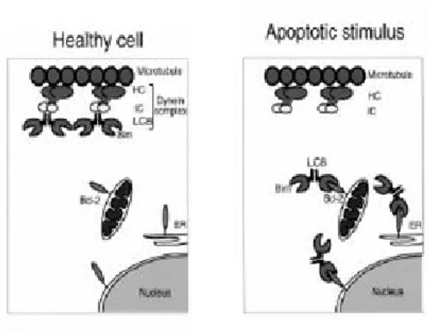
In healthy cells, most endogenous BimL and BimEL molecules were bound via LC8 to the microtubule-associated dynein motor complex, and thereby sequestered from Bcl-2 and its homologs. Certain apoptotic stimuli, disrupted the interaction between LC8 and the dynein motor complex. This freed Bim to translocate together with LC8 to Bcl-2 and neutralize its antiapoptotic activity. This process did not require caspase activity, and therefore constitutes an initiating event in apoptosis signaling (Puthalakath et al, 1999;Strasser et al, 2000b)
Recently it has been demonstrated that in cells exposed to UV radiation, activated JNK can phosphorylates Bim on T56 leading to its release from dynen (Lei and Davis, 2003).
24
Fig. 9. Model for the regulation of Bim. (From (Puthalakath and Strasser, 2002).
5. MECHANISMS OF CELL DEATH INDUCED BY MDA
MDAs act on microtubule network integrity in different ways and they induce diverse signals responsible for cell death initiation and execution. MDA effectiveness is largely accepted as a consequence of caspase activation through the intrinsic apoptotic pathway (Andre et al, 2002;Pourroy et al, 2004;Wu et al, 2005) that is mainly regulated by members of the Bcl-2 family (Walensky, 2006). Up-regulation of the pro-apoptotic proteins Bad, PUMA, Bax and/or Bak has been observed after treatment with paclitaxel, epothilone B as well as Vinca alkaloids (Jones et al, 1998;Tudor et al, 2000;Yamaguchi et al, 2004;Pourroy et al, 2004). Likewise, Bcl-XS, Bax or Bad overexpression sensitizes cancer cells to paclitaxel and vincristine (Sumantran et al, 1995;Sawa et al, 2000). Paclitaxel, epothilones and vinblastine trigger Bax activation through its conformational change (Makin et al, 2001;Yamaguchi et al, 2004). It allows N-terminal domain exhibition and Bax stable insertion into the outer mitochondrial membrane (Antonsson et al, 2001;Cartron et al, 2003).
25
MDAs may also activate Bim translocation from microtubules to mitochondria, as recently proposed for paclitaxel (Li et al, 2005). Specifically, the BH3-only protein Bim, has been shown to play a major role in paclitaxel-induced cell death (Sunters et al, 2003;Li et al, 2005;Li et al, 2007). Direct depletion of Bim or indirect depletion of Bim through siRNA repression of Bim transcriptional activators, delays paclitaxel- mediated apoptosis in cell based models (Sunters et al, 2003;Li et al, 2005;Li et al, 2007). As well, in vivo mouse models of bim-/- cells confirmed the importance of Bim expression for paclitaxel cytotoxicity (Tan et al, 2005).
Modulating the activity and the expression levels of Bcl-2 family proteins, MDAs increase the amounts of active proapoptotic members at mitochondrial membranes while they decrease those of active anti-apoptotic members. The ratio among Bcl-2 family members thus turns in favor of mitochondrial permeability and caspases activation.
6. P53 and its INDEPENDENT TRANSCRIPTION ACTIVITY
The p53 tumor suppressor protein is stabilized and activated by different genotoxic and non-genotoxic stress, playing a central role in the chemosensitivity and radiosensitivity of cancer cells by inducing cell cycle arrest and apoptosis (Vogelstein et al, 2000;Fei and El Deiry, 2003;Yamaguchi et al, 2004). p53 triggers apoptosis by inducing mitochondrial membrane permeabilization through transcription dependent mechanism: many pro-apoptotic proteins of the Bcl-2 family such as Bax, PUMA, Noxa and Bid are transcriptional target of p53 (Miyashita and Reed, 1995;Oda et al, 2000;Nakano and Vousden, 2001;Yu et al, 2001;Sax et al, 2002). Interestingly it has been recently demonstrated that upon DNA damage p53 translocate to mitochondria where it directly binds to Bcl-XL/Bcl-2 or Bak and induces Bax/Bak activation and cytochrome C release (Marchenko et al, 2000;Mihara et al, 2003;Dumont et al, 2003;Chipuk et al, 2004;Deng et al, 2006) and in the recent years have been accumulating evidences for transcription-independent p53-mediated apoptosis. Moreover the trascriptionally inactive p53 mutants (1-214) and p53 22/23QS act as potent death inducers in tumor cells (Haupt et al, 1995;Chen et al,
26
1996;Mihara et al, 2003). Also in cell-free cytoplasts, activation of cytosolic p53 can induce mitochondrial cytochrome C release (Schuler and Green, 2001). Targeting p53 to mitochondria by fusing it with mitochondrial address peptides, thereby bypassing the nucleus, is sufficient to launch apoptosis (Marchenko et al, 2000;Sansome et al, 2001;Moll et al, 2005). Pro-apoptotic protein and p53 rapidly translocate to mitochondria after a death stimulus, bind and inhibit the protective Bcl-XL and Bcl-2 proteins. The complex formed by p53 and BH3-only proteins with Bcl-XL/Bcl-2 is critical for theirs apoptogenicity: interference with their binding, blocks the mitochondrial killing activity (Oda et al, 2000;Cheng et al, 2001;Nakano and Vousden, 2001;Mihara et al, 2003). thus, tumor-derived p53 mutations may represent “double hits”, eliminating the trascriptional as well as the direct mitochondrial functions of p53 (Moll et al, 2005). it was determined that p53 DNA interface and the BH4 domain of Bcl-XL are involved in the formation of the complex : this result was confirmed by NMR studies (Petros et al, 2004;Moll et al, 2005). Besides p53-Bcl-2 binding appears to disrupt Bcl-2-Bax binding and thereby promotes free Bax to be potentially activated by a BH3-only proapoptotic protein (Deng et al, 2006) infact p53 can directly disupt the Bcl-2/Bax complex by binding to a novel regulatory region of Bcl-2’s FLD (Flexible Loop Regulatory Domain)(aa 32 to 68) that has been identified as regulatory domain in Bcl-2 (Deng et al, 2006). Phosphorylation in the FLD can positively regulate Bcl-2’s antiapoptotic function, inhibiting p53 binding and its apoptogenic role at mitochondria (Deng et al, 2006) but the mechanism is not clear. It has been proposed that localization of p53 on mitochondria could represent a rapid and direct way to induce cell death, amplifying its transcription-based apoptotic action that requires more time to take effect (Mihara et al, 2003;Moll et al, 2005). However the role of p53 in apoptosis induction varies between cell types and death signals, the role of p53 in MDA-induced apoptosis of human cancer cells remain controversial. Besides it has been recently demonstrated that p53 is associated with cellular microtubules through its interaction with dynein and that only in cells with a functional microtubules network it accumulated in the nucleus after DNA damage (Moll et al, 1996;Giannakakou et al, 2000). Treatment with MDA, disrupting microtubules network and dynamics can impair nuclear accumulation of p53 after DNA damage. Result obtained after microinjection of antibody
27
against the dynein intermediate chain, further supporting the idea that dynein functions in p53 transport (Giannakakou et al, 2000).
28
RATIONALE AND AIM OF THE PROJECT
Microtubules, together with actin and intermediate filaments are the major components of the cytoskeleton of eukaryotic cells and play a critical role in many cellular processes, including cell division, cell motility, intracellular trafficking, and cell shape maintenance. The principal Microtubule Organizing Center (MTOC) in mammalian cells is the centrosome that is composed of a pair of centrioles surrounded by an osmiophilic matrix termed pericentriolar material (PCM). As the microtubule network is highly involved in cell proliferation, it appeared to be a preferential target for cancer therapy. Microtubule-Damaging Agents represent an important class of anticancer drugs. Although some of this agents promote microtubule stabilization and other induce microtubules disruption, all MDA share the common property of suppressing microtubules dynamics, and thereby microtubules function, leading to the disruption of the mitotic spindle and the blockade of cell cycle progression at the transition from metaphase to anaphase that leads to apoptosis or mitotic catastrophe commitment. Mitotic catastrophe has been described as cell death that occurs from metaphase of mitosis in response to agents that cause mitotic-spindle damage. Recently we focused our attention on the antineoplastic spindle poison agent CA-4. CA-4 is an MDA isolated from the South African tree Combretum Caffrum that is not recognized by the multi drug resistance pump, a cellular pump which rapidly ejects foreing molecules including many anticancer drugs. MDAs also promote several effects on the centrosome including abnormal centrioles structure, centrosome fragmentation and inappropriate centrosome duplication. Aim of this project was to clarify the relationship between microtubular dynamics, dismantling of pericentriolar components and induction of apoptosis after exposure of H460 non-small lung cancer cells to this antimitotic drug. CA-4 is able to arrest cells at mitosis in a dose depending manner accompanied by an increasing disorganization of chromosomal arrangement, led to formation of particular structure named “star-like”. This structures contain pericentrosomal matrix components like -tubulin, pericentrin and ninein. Confocal and immunofluorescence microscopy revealed that “star like” structure are spherical structure formed by residual fragments of microtubules connected to kinetochore. At the centre of this
29
structure are present some component of the pericentriolar matrix like -tubulin, pericentrin and ninein.
After the analysis of structural alteration induced by Combretastatin we have focused our attention on the capability of this drug to induce cell death. Treatment with CA-4, which produced high frequency of “star-like” mitosis, was accompanied by mitotic catastrophe commitment characterized by activation of caspases-3/-9 and DNA fragmentation. The mechanisms that lead from microtubules depolymerization to cell death are still not completely clear. Recently it has been demonstrated that Bim, a proapoptotic Bcl-2 protein, is associated to the dynein motor complex and sequestered by microtubules. MDA, through the alteration of microtubules structure, might lead to the release of Bim from cytoskeleton and to its translocation to the mitochondria. Moreover, recent data show that further mechanisms might be at the base of the Bim-induced apoptosis such as regulation of Bim transcriptional levels. In a similar way, p53 another well known pro-apoptotic molecule, is associated with cellular microtubules and is transported to the nucleus by dynein. Based on these data, we have investigated whether upon tubulin depolymerization, Bim and p53 could be involved, and eventually in which manner, in the first steps of apoptosis. CA-4 treatment induce the release of Bim from microtubules and its increase at transcriptional level. In addition, Bim silencing strongly reduced apoptotic commitment. It has recently reported that p53 can also induce apoptosis independently of new protein synthesis however the functional importance of p53 in MDA-induced apoptosis of human cancer cells remain poorly understood. Concerning the role of p53 in CA-4-induced apoptosis, we found that treatment determined its release from cytoskeleton and re-localization to mitochondria. It remains to evaluate which are the Bcl-2 proteins involved in independent-trascription p53-induced apoptosis. For this purpose we are investigating the interaction of p53 with some members of the Bcl-2 family protein like Bcl-2, Bcl-XL, Bak and Bax.
30 RESULT
1. Interfering with spindle microtubule-dynamics arrests cells at metaphase stage with misaligned chromosomes
H460 (non-small-cell lung cancer) cells were incubated for 8 h with the “tubulin depolymerising agent” CA-4 in a dose-range of 1 nM to 100 nM. Treatments caused a dose-dependent block of the cell cycle in mitosis (Fig. 1a). Cells were arrested mainly at metaphase stage as evaluated by Giemsa staining (Fig. 1b), with mitotic index values of 35% and 47% and with ana-telophase ratio close to 0 at concentrations ≥5nM for combretastatin (Fig. 1a). After low concentrations of Ca-4 a high degree of metaphases showed misaligned chromosomes (approximately 20% at 2.5 nM) (Fig. 1c). With increasing concentrations the vast majority of combretastatin-treated cells presented collapsed chromatin (98% at 10 nM).
Figure 1. CA-4 caused mitotic arrest and generation of abnormal metaphases in H460 cells. (a, b). Mitotic index (M.I.) and ana-telophase ratio (A-T) of H460 cells left untreated or treated for 8h with different concentrations of CA-4 (a). Data are mean of three independent experiments SE. Metaphase organization in cells treated with CA-4 or (c). Mitoses were classified into four categories based on the GIEMSA staining profiles: normal metaphases (normal), metaphases with displaced chromosomes (displaced, thin black arrow) or collapsed chromatin (thick black arrows), C-mitoses. Images are representative of three independent experiments. Panel c reports quantitative data recorded from three independent experiments (mean SE).
nM - 1.2 2.5 5 10 20 100 %
a
b
1.2 nM 2.5 nM 5 nM 20 nM 0 10 20 30 40 50 60 MI A-T 0 20 40 60 80 100 Collapsed C-mitoses Normal Displaced 1.2 2.5 5 10 20 % nMc
Untreated31
2. Combretastatin A-4 affects spindle organization giving rise to “star-like” mitosis.
In both control and treated cells, the mitotic spindle arrangements were analyzed by immunostaining with antibodies directed against microtubules (-, -acetylated-tubulin) and centrosomes (-tubulin). Mitotic spindles of cells blocked in metaphase by low concentrations of combretastatin (0.1 nM) were bipolar with few chromosomes displaced from the metaphase plate and positioned in the vicinity of the spindle poles (spindle type stage I) (Fig. 2a). With increasing concentrations of combretastatin, spindle morphology became more abnormal and increasing numbers of chromosomes were located near the poles of a shorter bipolar spindle rather than in the metaphase plate (spindle type stage II) (not shown). After 7.5 nM CA-4 treatment many spindles (95%) had a multipolar organization with ball-shaped aggregations of condensed chromosomes containing multiple asters of microtubules (spindle types stage III and IV); this structure is called “star-like” (Fig. 2a). These small spindles contained residual kinetochore microtubules that were acetylated, as shown in immunostaining using an antibody that recognizes the acetylated form of -tubulin (Fig. 2b). Acetylated microtubules are more stable and commonly resist to drug-induced disassembly (32). The “star-like” structures were characterized by the presence of multiple signals of -tubulin (Fig. 2c), and occurred only in mitosis as observed after co-staining with phospho-histone H3 (not shown). Following prolonged exposure or high concentrations of drug (≥20 nM) causing complete microtubules depolymerisation, non-centrosomal gamma-tubulin “star-like” structures did not formed; whereas centrosomes retained γ-tubulin signals (spindle type stage V) (Figs. 2a, b, c). Immunoblot analysis aimed at quantitating the amount of -tubulin after combretastatin-treatment showed no variation due to drug treatment (not shown)
32
3. Pericentriolar matrix components are present in CA-4-induced “star-like” structures
To better understand the spatial organization and the reciprocal localization of the components of the “star-like” structure, 7.5 nM combretastatin-treated cells were analyzed by transmission electron microscopy and by confocal microscopy, after immunolabeling with antibodies against -tubulin, -tubulin and CREST. After combretastatin treatment, -tubulin was located in close proximity to the chromosomes, at the center of a reduced monopolar spindle of -tubulin that was connected to kinetochores and chromosomes (Fig. 3). The localization of centrioles and of other centrosome or PCM components after CA-4 treatments was evaluated by fluorescence microscopy using specific antibodies against centrin, pericentrine and ninein (Fig. 3c, d, e). In control cells, two distinct dots of centrin that, represent a pair of centrioles, colocalized with -tubulin, pericentrin and ninein on each spindle pole (Figs. 3c, d, e). On the other hand, in combretastatin-treated cells, the two dots of centrin colocalized only with one -tubulin pole of the multiple “star-like structure” (Fig. 3c); this means that the structural integrity of centrioles was not affected by treatment. In some cases four spots of centrine were visible in close proximity to each other, indicating that combretastatin also interfered with the centriole-pairs separation that occurred during early mitosis. Drug-induced “star-like” arrangements were also observed for pericentrin (Fig. 3d) and ninein (Fig. 3e).
33
Figure 2. Interferences in microtubular dynamics induce spindle abnormalities. (a-c). Organization of mitotic spindles following treatment for 8 h with the indicated concentrations of CA-4 (a-c). Cells were stained with antibodies that specifically recognize tubulin (a), α-acetylated tubulin (b) or γ-tubulin (c) (all emitting in green). DNA was counterstained with DAPI. Representative examples of three independent experiments are shown. White arrows show chromosomes displaced from the metaphase plate. See the text for a detailed description of abnormal spindles.
a
b
c
0.1 nM 7.5 nM 20 nM Untreated34
Figure 3. Spatial arrangement of mitotic spindle and centrosome components upon CA-4 treatment. H460 cells were left untreated or treated for 8 h with 7.5 nM Ca-4 prior to immunostaining with different combinations of antibodies and observation by fluorescence microscopy. The following coimmunostaining were performed: (a). CREST serum (green signal) and α-tubulin (in red). (b) CREST serum (in green) and γ-tubulin (in red). (c). γ-tubulin (in red) and centrin (in green) (d). α-tubulin (in red) and pericentrin (green). (e). α-tubulin (in red) and ninein (in green). In all cases DAPI was employed to counterstain DNA (in blue). Please note that the structural integrity of centrioles is not affected by the treatments (centrin, c), whereas the microtubular interference induced by CA-4 led to the generation of the “star-like” structures accompanied by the dismantling of the PCM components (γ-tubulin, b; pericentrin, d; and ninein, e). (f). Proposed model of the arrangement of the “star-like” structure induced by CA-4 treatment (I). As observed by ultra structural assessment by TEM and by confocal microscopy co-immunostaining with antibodies specific for α-tubulin (in green) and γ-tubulin or pericentrin (red) (II and III, respectively), γ-tubulin and pericentrin are located at the centre of each monopolar spindle, in turn composed of small and stable α-tubulin microtubules connected to kinetochores. Representative examples of three independent experiments are shown. See text for a detailed description.
a
b
c
d
e
C A -4 7 .5 n M U n tr ea te d -tubulin -tubulinf
I
II
III
pericentrin -tubulin35
4. Combretastatin induces pericentriolar matrix dismantling and caspase-dependent mitotic catastrophe
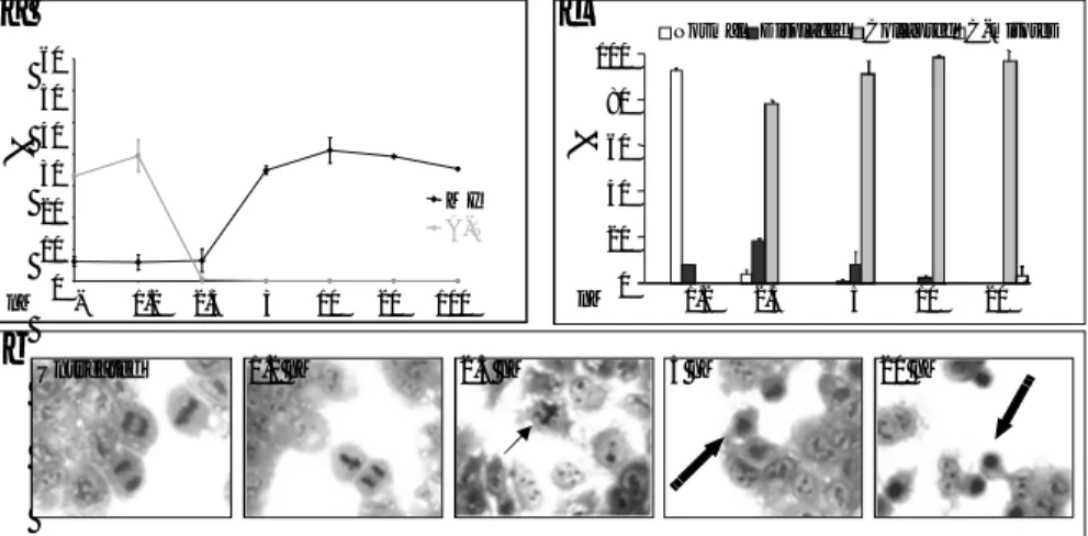
To evaluate the relationship between redistribution of the pericentriolar matrix in the form of “star-like” structures and the apoptotic process, biochemical markers like caspase activity and DNA fragmentation, were analyzed. Caspase-involvement in CA-4-induced cell death, was analyzed at whole cellular level, in fluorogenic assays. In fact, a 2-3 fold increase of caspase-9 and –3 expression was detected in cells exposed for 8 h to 7.5 nM CA-4. Such increase was even higher at 24 h with values ranging between 3- and 4- fold, and very close to values of caspase activation obtained after treatment with 2 µM staurosporine for 6 h (Fig. 4a). After treatment with 7.5 nM CA-4 for 8 h, Caspase 3-7 activation was also monitored at single cell level in time-lapse experiments by using the Magic Red substrate. Positive staining was observed in mitotic arrested cells as a result of a prolonged arrest which was not followed by resumption of cell cycle or attempt to divide, giving rise to a multinucleated cell (mitotic catastrophe) (Fig. 4b). Apoptosis was further confirmed by DNA fragmentation, evaluated by cytofluorimetric analysis after Fluorescent-FragELstaining, showing about 40 and 90% apoptotic cells, at 8 and 24 h, respectively (Fig. 4c).
36
Fig. 4 PCM dismantling induced by CA-4 is followed by apoptosis induction. (a) Caspase-9 and -3 activities in protein extracts from cells left untreated or treated for 8h or 24h with CA-4, as assessed by fluorogenic assays after incubation with caspase-9 (Ac-LEHD-AMC) or caspase-3 (Ac-DEVD-AMC) substrate. As a positive control, cells were treated for 4h with 2µM staurosporine, a known activator of the caspases. Columns represent the fold increase in the activity of caspases as compared to untreated cells. Results are expressed as the mean of 3 independent experiments ±SE. (b) Activation of caspase3-7 has been analyzed in untreated and treated cells using the magic red fluorescent substrate. Magic Red/BF or DIC images were collected at regular intervals throughout experiments; selected images are shown in the panel. (c) Cells were treated or not for the indicated time with CA-4, then analyzed for apoptosis-associated DNA fragmentation using Fluorescent-FragELTM DNA fragmentation kit by
flow-cytometry. The percentage of cells with cleaved DNA is reported. The results reported are representative of 3 independent experiments.
0 1 2 3 4 5 0 1 2 3 4 5
a
2.2% 70.0% 38.5% 91.4% Ctrl Sts CA-4 8h 24hc
CA-4 2.5nM 8h CA-4 7.5nM 8h Sts Ctrlb
Ctrl Sts 8h 24h CA-4 Ctrl Sts 8h 24h CA-437
5. Inhibition of Bim strongly reduced caspase-3 cleavage and apoptosis To investigate whether Bim protein mediates CA-4-induced cell death by apoptosis, siRNA were used to inhibit endogenous Bim expression and then we evaluated the cleavage and hence the activation of caspase-3. Infact Real Time analysis clearly showed that BimEL mRNA is induced by CA-4 treatment in a time-dependent manner in H460 cells (Fig. 5a), as also confirmed at protein level (Fig. 5b). As a positive control, taxol, known to up-regulate Bim was included in the experiment (not shown). In parallel experiments, cells were exposed to Bim siRNA or non specific siRNA for 24h then, treated with CA-4 for 0, 8, 24 and 48h. All cells have been collected after 72h trasfection for preparation of whole cell lysates and for isolation of RNA. The level of cellular mRNA and Bim proteins were substantially suppressed after 72h treatment with Bim siRNA (Fig. 5c) in cells treated with CA-4. In contrast, non specific scrambled siRNA had no significant effect on expression of endogenous Bim. Based on our previous results, showing the capability of CA-4 to induce apotosis in H460 cells in a caspase-3 dependent manner (Cenciarelli et al, 2008) we evaluated whether the inhibition of endogenous Bim following CA-4 induction may suppress cell death. Such analysis was carried out using the activation of pro-caspase-3 to caspase-3 as readout in both immunoblot assay (Fig. 5d) and in time-lapse analysis using the Magic red substrate (Fig. 5e). In both assays, the activation of caspase-3 was strongly reduced when CA-4 treatment was performed in the presence of Bim siRNA, thus indicating the active role played by Bim in CA-4-induced apoptosis.
38
Bim EL -20KDa -48KDa Tubulin
Whole cell lysates
0,0 1,0 2,0 3,0 4,0 0,0 0,5 1,0 1,5 2,0 2,5 3,0 3,5 CA-4 Ctrl siRNA - + 8h siRNA - + 48h siRNA - + 24h siRNA - + Ctrl 8h 24h CA-4 48h Bim EL Cleaved Caspase-3 -48KDa Tubulin -20KDa 8h 24h Ctrl siRNA - + 48h siRNA - + siRNA - + siRNA - + -19KDa -17KDa
Ctrl CA-4 siRNA BIM CA-4
Magic Red TM
b
c
d
39
Fig. 5 Real Time and Western Blot analysis showed clearly that Bim mRNA and protein were induced by CA-4 treatment (a, b). H460 cells treated with Bim siRNA or non specific siRNA were also treated with CA-4 for 0, 8, 24 and 48h. All cells have been collected after trasfection for preparation of whole cell lysates and for isolation of RNA. The level of cellular BimEL proteins and mRNA were substantially suppressed after 48h treatment with Bim siRNA (c, d) in cells treated with CA-4. In contrast, non specific siRNA had no significant effect on expression of endogenous Bim. In the presence of Bim siRNA and so during the inhibition of endogenous Bim there’s no cleveage of caspase-3 as evaluated by Western Blot and Time Lapse analysis using Magic RedTM(d, e).
40
6. Bim bound physically to microtubules by interaction with dynein, is released after CA-4 treatment and relocalizes to mitochondria
By means of co-immunoprecipitation assay with a dynein antibody, it was analysed whether CA-4 could induce Bim release from microtubules as a response to tubulin depolymerization. For this purpose, whole cells lysate were prepared after treatment with CA-4 for up to 48h. As reported in Fig. 6a, we found that Bim interacts in a physical manner with dynein. Such interaction was partly retained for the 8h treatment, whereas at longer incubation times of 24 and 48h, the physical interaction between Bim and dynein was markedly impaired, indicating that with increasing in the rate of microtubules depolymerization Bim was released from the microtubular network. Once assessed that CA-4 caused Bim release from microtubule, we analyzed its relocalization on mitochondria. Western Blot analysis of both cytosolic and mitochondrial fraction proteins of cells treated with 7.5nM CA-4 for 0-48h shows a detectable enrichment of Bim in mitochondria (Fig. 6b). In untreated cells the amount of Bim at mitochondria was very low, contrastingly to that observed in T cells (Zhu et al. 2004). Interestingly an increase of Bim was detectable even in the cytosolic extract indicating that beside relocalization, CA-4 promoted also a transcriptional up-regulation of Bim as also suggested by RT-PCR experiments. The occurrence of Bim localization at mitochondia was also confirmed by means of confocal analysis in immunofluorescence experiments using the specific mitotracker staining (Fig. 6c). Taxol was used as internal control (not shown).