CONTENTS
SOMMARIO i
PREFACE
- Cardiac morpho-functional remodelling:
the power of natural animal models pag. 1
- Fish as experimental model to study physiological
and patho-physiological cardiac remodelling pag. 3
AIM pag. 7
CHAPTER 1. Humoral influences: role of Angiotensin II 1.1. Introduction
1.1.1. Teleost cardiac plasticity in response to humoral factors pag. 9
1.1.2. Angiotensin II pag. 10
1.1.3. AngII-dependent short-term modulation of cardiac activity pag. 12 1.1.4. AngII-dependent long-term cardiac readjustment pag. 12
- Section A: AngII-dependent cardiac remodelling in the eel A. Anguilla
1.2A. Materials and methods
1.2A.1. Animals pag. 16
1.2A.2. Chemicals pag. 16
1.2A.3. Experimental protocols
1.2A.3.1. Morphological analysis pag. 16
1.2A.3.2. Western blotting and densitometric analysis pag. 18
1.2A.3.3. Nitrite measurements pag. 19
1.2A.4. Statistics pag. 19
1.3A. Results
1.3A.1. Cardiac morphometry pag. 21
1.3A.4. NOS/NO system pag. 24
1.4A. Discussions pag. 28
- Section B: AngII-dependent cardiac remodelling in the zebrafish D. rerio
1.2B. Materials and Methods
1.2B.1. Animals pag. 34
1.2B.2. Chemicals pag. 34
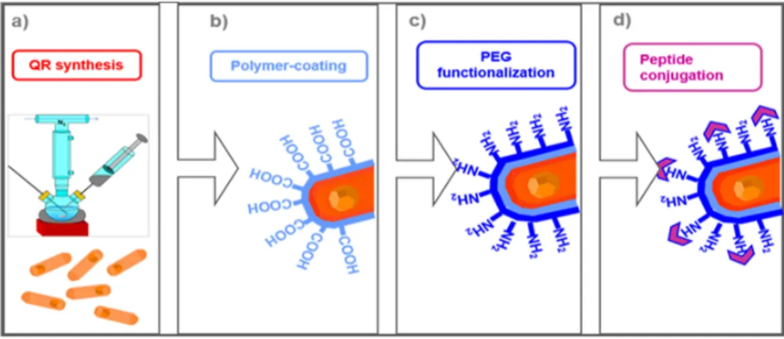
1.2B.2.1. AngII octapeptide conjugation with
Quantum Rods (QRs) pag. 34
1.2B.3. Experimental protocols
1.2B.3.1. Morphometric analysis pag. 35
1.2B.3.2. Heart rate pag. 36
1.2B.3.3. Morphological analysis pag. 36
1.2B.3.4. Western blotting and densitometric analysis pag. 36
1.2B.4. Statistics pag. 37
1.3B. Results
1.3B.1. Cardiac morphometry and morphology pag. 38
1.3B.2. Heart rate pag. 40
1.3B.3. AngII receptors pag. 40
1.3B.4. Work in progress pag. 41
1.5. Conclusions Chapter 1 pag. 43
CHAPTER 2: Hypoxia influences: the role of the NOS/NO system 2.1. Introduction
2.1.1. Hypoxia tolerance in fish pag. 45
2.1.2. The NOS/NO system pag. 46
2.2. Materials and Methods
2.2.1. Animals pag. 49
2.2.2. Nitrate reduction and determination of NO metabolites pag. 50
2.2.3. Muscle Mb concentration pag. 51
2.3. Results
2.3.1. NO metabolites and Mb level in tissues exposed to deep hypoxia pag. 52
2.3.2. Nitrate reductase activity pag. 55
2.3.3. Ethanol production pag. 56
2.4. Discussion pag. 60
2.5. Conclusions Chapter 2 pag. 63
GENERAL CONCLUSIONS pag. 64
REFERENCES pag. 65
SOMMARIO
Il rimodellamento cardiaco è un fenomeno complesso che permette un’adeguata attività d’organo in risposta a cambiamenti fisiologici e patologici e coinvolge modificazioni a livello tissutale, cellulare e molecolare, secondo meccanismi ancora poco noti. Il cuore dei vertebrati mostra una notevole capacità di adattarsi funzionalmente alle richieste emodinamiche dell’organismo. Questa plasticità è esemplificata dalla modulazione “beat-to-beat” della contrattilità in risposta a cambiamenti del carico pressorio e volumetrico (risposta di Starling) e dal riarrangiamento morfo-funzionale adattativo. I maggiori determinanti della risposta plastica del cuore sono gli stimoli fisici come lo stress meccanico e lo stiramento dei miociti, o chimici, come ad esempio quelli esercitati da cardiomodulatori, inclusi i Peptidi Natriuretici (NPs), l’endotelina-1 (ET-1), l’Ossido Nitrico (NO) e l’Angiotensina II (AngII). Queste molecole attivano specifici circuiti endocrini/paracrini/autocrini responsabili del controllo omeostatico della funzione e della crescita cardiaca. Nei mammiferi, il cuore adulto è considerato un organo terminalmente differenziato nel quale la crescita e il rimodellamento avvengono per ipertrofia. Questo paradigma è stato recentemente confutato da evidenze che mostrano come, durante la normale crescita cardiaca, i miocardiociti adulti proliferano e muoiono. L’identificazione di stimoli e meccanismi che, attivando programmi genetici e/o metabolici silenti, possano permettere ai cardiomiociti di mammifero adulto di proliferare, è al momento tra gli scopi più importanti della ricerca cardiovascolare moderna. A tal riguardo un possibile approccio è quello di identificare esempi di plasticità cardiaca fra i vertebrati.
Negli ultimi anni il cuore dei pesci ha rappresentato un importante strumento di ricerca per analizzare aspetti molecolari, cellulari e tissutali della plasticità cardiaca. Nei pesci, il cuore adulto conserva la capacità di crescita iperplastica in risposta a cambiamenti delle condizioni ambientali, all’esercizio fisico e alla maturità sessuale. Esempio estremo di questa plasticità è rappresentato dalla completa rigenerazione del cuore di zebrafish in seguito a rimozione chirurgica di una porzione del ventricolo. In una prospettiva traslazionale, i risultati di studi effettuati nei pesci potrebbero rappresentare uno strumento importante per decifrare i determinanti e i
Partendo da queste premesse, questo progetto di tesi ha voluto valutare se e in che misura, fattori umorali e ambientali influenzano la plasticità cardiaca dei pesci. In particolare, in questa tesi di dottorato sono riportati e discussi i risultati ottenuti da studi relativi al controllo umorale del rimodellamento morfo-funzionale cardiaco mediato dall’esposizione cronica all’AngII nel cuore dell’anguilla europea Anguilla anguilla e dello zebrafish Danio rerio, e i meccanismi fisiologici e molecolari attivati nel cuore della carpa, Carassius carassius, in risposta all’ipossia.
L’AngII, il prodotto bioattivo del Sistema Renina Angiotensina (RAS) è un potente ormone le cui azioni biologiche sono state ampiamente studiate nei mammiferi. A livello cardiaco, legandosi a recettori di tipo AT1 e AT2, l’AngII influenza positivamente la contrattilità miocardica e regola la crescita miocitaria. Un RAS omologo a quello dei mammiferi è presente anche nei pesci. In particolare, nei teleostei, l’AngII può indurre modificazioni cardiache a breve (modulazione della contrattilità) e lungo termine (rimodellamento morfo-funzionale). Nel 2013, Imbrogno e collaboratori hanno dimostrato che il cuore di anguille esposte per 4 settimane ad iniezioni intraperitoneali di AngII mostrava una migliore capacità di rispondere ad incrementi di post-carico. Quest’effetto era mediato dall’attivazione dei recettori AT2 e accompagnato da una differente espressione e localizzazione di proteine coinvolte nella regolazione della crescita e dell’apoptosi. Partendo da queste premesse, questo studio ha voluto meglio investigare le modificazioni strutturali e molecolari attivate nel cuore di anguille esposte per 8 settimane ad iniezioni intraperitoneali di AngII. Analisi morfologiche hanno evidenziato che i cuori di animali trattati mostravano un incremento della muscolarità ventricolare associato ad un aumento dello spessore del compatto e ad una riduzione degli spazi lacunari nello spugnoso. Queste modificazioni erano accompagnate da un’incrementata vascolarizzazione del compatto. Analisi di western blotting e immunofluorescenza hanno evidenziato un’aumentata espressione del recettore AT2 nei cuori di animali trattati, associata ad una diversa localizzazione: nei controlli AT2 localizza principalmente a livello miocitario mentre in seguito a trattamento, un segnale fluorescente si osserva a livello dell’endotelio endocardico. In relazione al ruolo cruciale svolto dallo NO nei meccanismi di regolazione del rimodellamento cardiaco nei pesci, lo studio ha voluto anche analizzare il cross-talk tra AngII e il sistema NO
immediatamente metabolizzato a nitriti o nitrati. Per questa ragione, il dosaggio dei nitriti può essere utilizzato per misurare i livelli di NO prodotti dalla NOS e di conseguenza la funzionalità stessa dell’enzima. Il dosaggio dei nitriti, effettuato su omogenati di cuori di animali trattati con AngII ha evidenziato una riduzione significativa della concentrazione di nitriti rispetto ai controlli, indice di una ridotta attività della NOS. Analisi di western blotting non hanno evidenziato variazioni significative nei livelli di espressione della forma fosforilata (e quindi attiva) della NOS. Tuttavia, il trattamento ha influenzato la localizzazione dell’enzima fosforilato. Infatti, mentre nei controlli peNOS localizza sia nei miociti che a livello dell’endotelio endocardico (EE), nei trattati si osserva una maggiore localizzazione a livello miocitario e solo un debole segnale a livello endocardico. Questo dato è accompagnato da una maggiore espressione, a livello dell’EE, di NOSTRIN, un modulatore negativo della NOS, responsabile della traslocazione dell’enzima dalla membrana plasmatica all’interno di vescicole citoplasmatiche. Analisi di western blotting hanno anche evidenziato una riduzione nei livelli di espressione della chinasi Akt e dello chaperone HSP90, due tra i principali modulatori positivi della NOS. Nel complesso questi risultati dimostrano che il cuore di anguilla cronicamente esposto all’azione dell’AngII va incontro ad un riarrangiamento strutturale e ad una modulazione dei livelli di NO e delle proteine che regolano l’attività della NOS. Gli effetti cronici dell’AngII sono stati investigati anche nello zebrafish. Sebbene questo studio è ancora in corso, i primi risultati sperimentali dimostrano che anche il cuore di zebrafish va incontro ad un rimodellamento strutturale se esposto per 8 settimane all’azione dell’AngII. In particolare, i cuori trattati con AngII mostrano un incremento del compatto e una riduzione degli spazi lacunari, indicativi di un aumento della muscolarità ventricolare. Questo è associato ad un aumento del collagene e ad un significativo aumento del peso del cuore e dell’indice cardiosomatico (rapporto in percentuale tra il peso del cuore e il peso dell’animale). Analisi di western blotting hanno evidenziato inoltre un’aumentata espressione di entrambi i recettori AT1 e AT2 in omogenati cardiaci di animali trattati con AngII. La seconda parte della tesi ha analizzato i meccanismi fisiologici e molecolari attivati nella carpa in risposta all’ipossia.
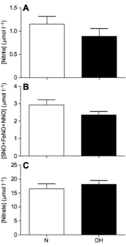
tempo se esposti a condizioni di ipossia o anossia. Questa capacità è legata alla loro abilità di mantenere o aumentare la performance cardiaca in risposta ad una riduzione di ossigeno ambientale, favorendo così gli scambi tra i tessuti. Inoltre, questi pesci sono in grado di convertire il lattato (prodotto ultimo della glicolisi) in etanolo, che è poi eliminato attraverso le branchie. Una caratteristica dei pesci è la loro capacità di captare, attraverso le branchie, nitriti e nitrati dall’acqua in cui si trovano; in condizioni di ipossia, quando l’attività della NOS è compromessa, i nitriti possono rappresentare un’importante fonte di NO che, a sua volta, interviene nei processi di citoprotezione. Sulla base di queste osservazioni, in collaborazione con il Prof. Jensen (Università di Odense) e la Prof.ssa Fago (Università di Aarhus), è stato avviato uno studio volto a valutare il ruolo dei nitrati e della mioglobina come fonti alternative di nitriti, e quindi di NO, nella carpa esposta per un giorno ad ipossia profonda (1<PO2<3 mmHg). Omogenati cardiaci e di muscolo rosso e bianco hanno
evidenziato un aumento di nitriti e di altri metaboliti dello NO, quali Fe-nitrosile (FeNO) S-nitroso (SNO) e N-nitroso (NNO), sia nel cuore che nel muscolo rosso di carpe esposte ad ipossia. Questo incremento non era correlato a differenze nelle concentrazioni di mioglobina. Il dosaggio dell’attività nitrato reduttasica tissutale in omogenati di cuore, muscolo bianco e fegato, ha inoltre evidenziato che in condizioni di ipossia profonda, il muscolo e il fegato sono in grado di convertire il nitrato esterno in nitrito. Questa attività non è stata riscontrata nel cuore, dove risulta invece evidente una riduzione nelle concentrazioni di nitriti in risposta all’ipossia. Questi risultati supportano il ruolo dei nitriti come fonte alternativa per la generazione di NO durante l’ipossia; questo risulta maggiormente evidente in tessuti ricchi in mioglobina e mitocondri come il cuore, dove la conversione di nitriti a NO potrebbe rappresentare un importante meccanismo di cardioprotezione. Inoltre, in condizioni ipossiche, l’attività nitrato reduttasica riscontrata nel muscolo e nel fegato degli animali esposti ad ipossia potrebbe essa stessa supplementare le riserve intracellulari di nitriti contribuendo alla citoprotezione.
In conclusione, i risultati riportati in questa tesi suggeriscono che se esposto a influenze umorali o ambientali il cuore dei pesci va incontro ad un significativo rimodellamento molecolare, strutturale e fisiologico. Sebbene molto resti ancora da chiarire sui meccanismi molecolari attivati, questi risultati indicano chiaramente un
di cascate molecolari che controllano le risposte cardiache adattative nei pesci. Ricerche future contribuiranno a meglio decifrare i complessi networks molecolari coinvolti nella plasticità cardiaca di questi vertebrati. In questo contesto, la scelta di modelli sperimentali appropriati, caratterizzati da una marcata adattabilità morfo-funzionale ad una varietà di stimoli ambientali (temperatura, pH, pressione parziale di ossigeno, etc.) potrà essere di grande aiuto.
Preface
PREFACE
Cardiac morpho-functional remodelling: the power of natural animal models Cardiac remodelling may be defined as genome expression, molecular, cellular and interstitial changes that are manifested as changes in size, shape and function of the heart in response to physiological and/or pathological conditions (Cohn et al., 2000). In mammals, exercise, pregnancy, and postnatal growth promote physiologic growth of the heart, whereas neurohumoral activation, pressure overload (aortic stenosis, hypertension), inflammatory heart muscle diseases (myocarditis) and myocardial injury could be causes of pathologic hypertrophic growth. Likewise, protracted bed rest, mechanical unloading with a ventricular assist device and prolonged weightlessness during space travel, are causes of cardiac atrophy (Hill and Olson, 2008) (Fig. 1). Although the etiologies of these diseases are different, they share several pathways in terms of molecular, biochemical and mechanical events. Physiological cardiac hypertrophy can lead to an increase in heart size characterized by normal cardiac morphology with a normal and/or enhanced cardiac function (Ooi et al., 2014). By contrast, pathological hypertrophy is characterized by thickening of ventricular wall, decrease in the size of the cardiac chambers and reduced capacity of the heart to pump blood to the tissues and organs around the body. This is associated with a shift toward glycolytic metabolism, alterations in calcium handling contractility, disorganization of the sarcomere, mutations in genes coding for contractile proteins, loss of myocytes with fibrotic replacement, systolic or diastolic dysfunction, and “electrical remodelling" (i.e. alterations in the expression or function of ion-transporting proteins, or both) (see for references Hill and Olson, 2008). From a morphological point of view, cardiac growth may be classified in:
- concentric remodelling: increase in relative wall thickness with normal cardiac mass;
- concentric hypertrophy: increase in relative wall thickness and cardiac mass with little or no change in chamber volume;
- eccentric hypertrophy: increase in cardiac mass with increased chamber volume; relative wall thickness may be normal, decreased or increased.
Preface Myocytes represent cardiac cells mostly involved in the remodelling process; other components include the interstitium, fibroblasts, collagen and coronary vasculature (Cohn et al., 2000). During cardiac hypetrophy, myocytes increase in size with changes in sarcomere organization, protein expression and activation of pro-growth signalling cascades. Cardiomyocytes plasticity is often accompanied by a re-induction of the “fetal gene program”, in which patterns of gene expression mimic those seen during embryonic development (for references see Hill and Olson, 2008). Moreover, recent findings suggested the presence in the adult mammalian myocardium of a cell population with the behaviour and potential of cardiac stem cells (CSC): their presence has identified myocyte death and myocyte renewal as the two sides of the proverbial coin of cardiac homeostasis (Nadal-Ginard et al., 2003). Myocyte renewal depends on the differentiation of the CSCs into immature myocytes that divide two to four times before becoming terminally differentiated and permanently withdrawn from the cell cycle (Nadal-Ginard et al., 2003). Adult epicardial-derived (EDC) cells are also supposed to retain their stem cell capacity, being able to divide and differentiate into adult cardiac cells (fibroblasts, coronary vascular smooth muscle cells) (Winter and Gittenberger-de-Groot, 2007). In the presence of cardiac injury, increased workload or a number of bioactive molecules, this proliferation significantly contributes to pathological organ remodelling. These findings have opened an unexpected frontier toward the possibility to manipulate mammalian adult heart growth also for therapeutic benefit in human diseases. Identifying both stimuli and mechanisms which may switch on silent genetic and/or metabolic programs to allow adult mammalian cardiac cells to proliferate, is now the goal for modern cardiovascular research. A possible approach in this regard is to identify examples of cardiac plasticity among vertebrates. Cardiac flexibility is remarkably elevated in several non mammalian vertebrates like amphibians and fish. Accordingly, in last years fish and amphibian hearts have represented fundamental research tools to analyse molecular, cellular and tissue aspects of cardiac development, epigenetic remodelling and adult heart regeneration. In amphibians, adult ventricular myocytes retain their proliferative potential (Bettencourt-Dias et al., 2003). In fish, while myocytes hypertrophy significantly contributes to normal cardiac growth, the adult heart retains the ability for hyperplastic growth with age,
Preface in response to changes in environmental conditions, increased demand associated with training/exercise activity and sexual maturation (Gamperl and Farrell 2004, and references therein; Cerra, et al., 2004). Extreme examples of this plasticity are provided by the complete epimorphic regeneration of newt (Bettencourt-Dias et al., 2003) and zebrafish (Poss et al., 2002) injuried ventricle. The increasing body of information on fish (i.e. zebrafish) cardiac plasticity suggests that molecular mechanisms, which are silent in the adult mammalian heart, are instead continuously active in these species, so that they drive, in response to appropriate stimuli, heart remodelling towards hyperplasia and/or hypertrophy.
Fish as experimental model to study physiological and patho-physiological cardiac plasticity
In recent years, fish have become widely used animal models to study at molecular, tissue, and organ level, morphological and functional cardiac modifications and their determinants. Although fish diverged from humans more than 400-million years ago, in a translational perspective, results obtained in fish may represent a significant tool to decipher the mechanisms involved in the mammalian cardiac morpho-functional remodelling. Fish provide a number of exceptional advantages. For example, laboratory fish models are small and can be bred and maintained in large numbers easily and at low cost. They offer the opportunity to combine the analytical clarity of developmental biology with the power of genetics. Moreover, transgenic lines can be easily and quickly produced. Fish are also amenable to high-throughput approaches such as whole-genome mutagenesis or chemical library drug screens (Schartl, 2014). Among fish, the zebrafish (Danio rerio) represents the non-mammalian vertebrate model largely used to investigate molecular aspects of cardiac development and remodelling. Zebrafish shows all the advantages described above, e.g. small size, short generation time, facility in breeding in large numbers in the laboratory. Interestingly, it produces transparent eggs, which can be easily manipulated to study the embryonic development. Its genome is sequenced (Howe et al., 2013) and large genomic resources have been built up: transgenic fluorescent marker lines that allow these fish model to be used for bioimaging, downregulation of gene expression during early development (achieved by morpholinos) and gene knockout mediated
Preface by genome-editing technologies. Zebrafish embryos are particular well suited for studying gene function, protein expression and cellular aspects of cardiovascular development (see for references Bakkers, 2011). Adult zebrafish represent a useful model to study heart regeneration. They are able to completely regenerate cardiac tissue after that ∼20% of the heart is surgically removed from the apex (Poss et al., 2002). The site of injury initially clots off; this is followed by the replacement of red blood cells with fibrin. However, within the first month after injury, the fibrin is quickly replaced by cardiac myofibers, and by 2 months postinjury, the cardiac tissue results indistinguishable from the hearts of sham-operated controls (Poss et al., 2002). Three major regeneration events are proposed in the zebrafish heart: i) the epicardium is activated by re-expressing embryonic markers, contributing to new vascularization of the regenerating myocardial cells; ii) cardiomyocytes start to proliferate to replace lost myocardium; iii) the endocardium is also activated and starts retinoic acid synthesis, which supports cardiomyocyte proliferation (Bournele and Beis, 2016). Although several developmental signaling pathways have been functionally associated with zebrafish heart regeneration, molecular mechanisms involved are still unknown.
Besides zebrafish, other fish are now emerging as new experimental models useful as “evolutionary mutant models” for complex human diseases. This concept was first introduced by Craig Albertson and colleagues (Albertson et al., 2009), and it is based on the awareness that evolution by adaptation to a specific environment has resulted in species or populations of many animal groups, including fish, that exhibit phenotypes closely resembling or mimicking human diseases. Among others, specimens of cyprinidae (Carassius carassius, Carassius auratus), which routinely experience hypoxia during their lifecycle, represent useful models to study hypoxia tolerance and preconditioning mechanisms in mammals. Both the goldfish (Carassius auratus) and the crucian carp (Carassius carassius) are able to tolerate prolonged and severe hypoxic conditions and remain active when overwintering in ice-covered ponds (Bickler and Buck, 2007). This capacity has been correlated with their ability to generate ethanol as an anaerobic end product, which is acid–base neutral, in contrast with the normal glycolytic end product lactic acid. Ethanol is produced in the myotomal musculature and is released to the outside water through the gills (Bickler
Preface and Buck, 2007). A remarkable feature of these species is their ability to maintain or increase cardiac performance and autonomic cardiovascular regulation in response to oxygen deprivation (Stecyk et al., 2004). In the anoxic crucian carp, the sustained cardiac output, the reduced peripheral vascular resistance and the conserved autonomic control ensure the rapid distribution of glucose to metabolically active tissues and the transport of waste lactate to the site of ethanol production (the muscle), thereby allowing for efficient ethanol shuttling to the gills for excretion (Stecyk et al., 2004). Similarly, the surprising capability of the goldfish heart to enhance its performance during acute hypoxia, as well as its hypoxia-enhanced sensitivity to the heterometric (i.e. Frank-Starling mechanism) regulation may be crucial for maintaining the functional and metabolic interactions between organs and tissues that are required for the hypoxia tolerance of the organism (Imbrogno et al., 2014).
Many fish species experience remarkable changes in cardiac mechanical performance associated, for instance, with thermal acclimation, or with changes in locomotive habits and/or body growth, or both (see for references Cerra et al., 2004; Imbrogno 2013), thus they represent appropriate models to evaluate the cardiac morpho-functional remodelling associated with body growth and changes in life style. This is exemplified by the European eel Anguilla anguilla. The eel has a complex life cycle which, following metamorphosis, includes a spawning migration requiring high levels of swimming performance and elevated metabolic demands (see for references Imbrogno, 2013). The relevant metabolic and cardio-respiratory capacities to satisfy tissue demands are associated to its great ability to regulate cardiac aerobic metabolism during hypoxia (McKenzie et al., 2003) and to the growth-related cardiac morphodinamic remodelling (Cerra et al., 2004). Indeed, by comparing small juvenile fish with their large adult counterparts, Cerra and colleagues (Cerra et al., 2004) demonstrated that, during ontogenetic growth, the eel heart undergoes morphodynamic changes. With respect to the small counterpart, the growing eel ventricle is characterized by enhanced hemodynamic performance expressed as a better capacity of the heart to sustain increased output pressure. These changes in mechanical behaviour positively correlate with structural modifications of the heart ventricle, consisting in an increase in compacta thickness and in the diameter of the
Preface trabeculae in the spongiosa, together with a reduction of the lacunary spaces (Cerra et al. 2004). Therefore, because of these growth-related morphodynamic changes, the cardiac ventricle of small eels, with its limited response to pressure overload and large lacunary spaces, appears better suited to produce volume work; contrarily, the heart of the large eels is better adapted to produce pressure work (see for references Icardo et al., 2005).
AIM
AIM
In recent years, the research group of the Laboratory of Organ and System Physiology, where I spent my PhD period, gave a significant contribute in deciphering physiological, structural and molecular aspects of cardiac plasticity in fish. Taking advantages of their experience in the field of fish cardiac physiology, the aim of this PhD research project was to analyse the influence of humoral and environmental factors on cardiac plasticity in fish. In particular, this thesis will provide an overview of results obtained by studies regarding the humoral control of cardiac remodelling mediated by chronic exposure to AngII in the heart of the European eel Anguilla anguilla and in the zebrafish Danio rerio, and the physiological and molecular mechanisms activated in the heart and in the muscle of the common carp Carassius carassius in response to hypoxia exposure. These two aspects will be separately analysed in Chapter 1 and Chapter 2, respectively. For each topic, a brief introduction on the state of art will be furnished.
During my PhD in Life Science, my research activity also contributed to obtain information about the role of neuropeptides and neuroendocrine modulators, such as Nesfatin-1, Chromogranin A-derived serpinine peptide, and Selenoprotein T, on the cardiac performance of the goldfish C. auratus. Published results will be not described in the thesis but attached as appendix in the pdf format.
CHAPTER 1. Introduction
CHAPTER 1
Humoral influences:
role of AngiotensinII
CHAPTER 1. Introduction
1.1. INTRODUCTION
1.1.1. Teleost cardiac plasticity in response to humoral factors
To support the varying physiological requirements of the animal, the fish heart adjust its metabolic and hemodynamic functions in response to both extrinsic and intrinsic signalling pathways. The former represented by circulating and intracardiac hormones, the latter consisting of humoral autocoids (i.e. locally generated signalling substances). Both endocrine and autocoid signalings are responsible for beat-to-beat (e.g. Starling’s law of the heart), short term (E-C coupling, myocardial contractility) and long-term (modified gene expression) cardiac modulation under both normal and stress-induced conditions.
Most of the knowledge on the morpho-functional heterogeneity and flexibility of the fish heart is based on studies on teleost fish. This group of fishes has been largely used to illustrate how endocrine circuits contribute to organ integration, in relation to the role of both classic [(i.e., catecholamines (CAT), angiotensin II (AngII) and natriuretic peptides (NPs)], and novel cardiac modulators [i.e., chromogranin-A (CgA) and its derived peptides]. Moreover, the cardiac role of locally released autacoids [i.e., carbon monoxide (CO), nitric oxide (NO) and hydrogen sulphide (H2S)], which evolved as regulatory molecules before the establishment of hormonal
networks typical of complex animal body plans, has been also deeply investigated (Gattuso et al., 2018).
A major integrative mechanism that contributes to teleost heart modulation by coordinating both humoral signals and the cross-talk between the endocardial endothelium (EE) and the myocardium, is represented by the nitric oxide synthase (NOS)/NO system. The comparatively greater amount of EE covering the internal cavity of the teleost heart represents an important source of NO that by acting as a freely diffusible messenger modulates the performance of the subjacent myocardium. This EE-NOS signalling, located at the crossroads of many extrinsic and intrinsic neuro-endocrine pathways, coordinates many chemically activated cascades. For example, endoluminal chemical stimuli such as acetilcholine (ACh), Ang II, vasostatin-1 (VS-1), catestatins (Cts), as well as β3 adrenoceptor activation, all converge in their contractile effects via NO signalling, which requires the obligatory
CHAPTER 1. Introduction involvement of the EE (Imbrogno et al., 2001; Imbrogno et al., 2003; Imbrogno et al., 2004; Imbrogno et al., 2006; Imbrogno et al., 2010). Thus, the intracardiac NOS/NO system, located downstream and at the crossroads of a large number of regulatory endocrine pathways represents a “knot” for convergent molecular signalling. The result is the modulation of intracellular target, such as membrane ion channels, transporters and contractile proteins, and a fine control of myocardial contractility, relaxation and stretch induced modulation. As hereafter illustrated, this occurs also in the eel heart in which, results reported in this thesis documented the involvement of the NOS/NO system in mediating the cardiac effects of AngII.
1.1.2. Angiotensin II
Angiotensin II (AngII), the bioactive component of the Renin-Angiotensin System (RAS), is a pluripotent hormone whose biological actions are extensively studied in mammals. The RAS cascade starts with the renin-mediated cleavage of the decapeptide angiotensin I (AngI) from angiotensinogen. Then, a dipeptidyl-carboxypeptidase, the angiotensin converting enzyme (ACE), removes a C-terminal dipeptide from AngI, generating the octapeptide AngII. In mammals, the heart itself expresses all RAS components in both myocytes and fibroblasts (Dostal 2000 and references therein). In addition, in cardiac cells, the enzyme chymase contributes to AngI/II conversion.
A RAS, analogous to that found in mammals, is present in teleost fish (see Kobayashi and Takei, 1996). It was mainly studied in relation to ion and osmotic regulation, in particular under dehydrating conditions, such as seawater acclimation (references in Imbrogno and Cerra 2017). The bioactive peptide AngII has been identified and sequenced in various teleost species, including the chum salmon Oncorhynchus keta (Takemoto et al., 1983), the Japanese goosefish Lophius litulon (Hayashi et al., 1978) and the American eel Anguilla rostrata (Khosla et al., 1985).
The teleost heart possesses an intracardiac RAS. ACE activity is present in the ventricle of a variety of species (e.g. Heteropneustes fossilis, Clarias batrachus, Channa gachua, Anabas testudineus, Notopterus chitala, Monopterus cuchia: Olson et al., 1987), immunoreactive AngII-like material was described in the heart of the Antarctic teleost Champsocephalus gunnari (Masini et al., 1997), cardiac AngII binding sites were
CHAPTER 1. Introduction detected in the trout Oncorhynchus mykiss (Cobb and Brown, 1992), and in the eel A. anguilla (Imbrogno et al., 2013). This suggests also in the teleost heart, the presence of an AngII-dependent intracardiac circuit of regulation.
In mammals, AngII-dependent cardiac effects are mediated by plasma membrane AT1 and AT2 receptors, classified according to the affinity for different antagonists (see references in De Gasparo, 2002, and Cerra et al., 2001). AT1 is mainly responsible for AngII-dependent positive myocardial inotropy and myocyte growth. It stimulates, via G-protein phospholipase C, Ca2+ mobilization, inositol triphosphate,
diacylglycerol and protein kinase C (see Dostal, 2000, for references). It also activates the JAK-STAT pathway, thus inducing cardiac growth (Booz et al., 2002). In contrast, cardiac AT2 receptors, whose expression is reduced after birth, antagonizes AT1 growth promoting effects via a number of phosphatases. This receptor is also coupled with the NO/cGMP signalling, either directly or indirectly through enhanced bradykinin or endothelial NOS expression (references in Dostal 2000).
Several studies attempted to obtain the molecular and biochemical characterization of teleost AngII receptors (see for example Marsigliante et al., 1996; Tran van Chuoi et al., 1998). Studies were mainly performed by using commercial drugs and analogues with often negative or inconsistent results, possibly attributed to the lack of specificity of the drugs and/or intrinsic biochemical traits of the receptors (see Russell et al., 2001 for references). It was found that two AngII receptor types are present in the eel. One, characterized in the European eel A. anguilla, shows a cDNA sequence [GenBank accession number AJ05132 (Tran van Chuoi et al., 1998)] with 60% homology with the mammalian AT1 receptor (Russell et al., 2001); the other, characterized in the Japanese eel Anguilla japonica (Wong and Takei, 2013), shares the origin with mammalian AT2, as demonstrated by molecular phylogenetic and synteny analysis (Wong and Takei, 2013). Only AT receptors able to bind mammalian anti-AT2 antibody were detected by Western Blotting in the cardiac tissue of A. anguilla and were associated to the long-term effects of the peptide (Imbrogno et al., 2013).
CHAPTER 1. Introduction
1.1.3. AngII-dependent short-term modulation of cardiac activity
The heart of the American eel A. rostrata and the trout O. mykiss is either directly or indirectly stimulated by homologous [Asn1, Val5]-AngII (Oudit and Butler, 1995). Indirect effects are supposed to be mediated either by CAT or by adrenergic tone modulation. In fact, the dose-dependent increments in cardiac output (CO) and stroke volume (SV) observed in O. mykiss after the exposure to teleost AngII are inverted by α-adrenergic blockage (see for references Imbrogno and Cerra 2017). In contrast to these cardio-stimulatory effects, AngII administered to the isolated and in vitro perfused working eel (A. anguilla) heart reduces both inotropism and chronotropism. This direct cardio-inhibition involves AT1-like receptors, Gi/o proteins, the cholinergic system, and an EE–NO–cGMP–protein kinase G (PKG) cascade (Imbrogno et al., 2003). Although the reasons of this divergence are unclear, authors hypothesized a role of species-related differences and/or the organization level under study. This may be the case of results obtained on intact cardiovascular system vs those observed on in situ heart or on isolated and denervated working cardiac preparation. For example, in the in vitro heart of A. anguilla the AngII-dependent cardio-suppressive effect is suggested to function as a local mechanism of cardio-inhibitory protection that balances/opposes systemic cascades of convergent excitatory stimuli (Imbrogno et al., 2003). This effect, in vivo could be overridden by cardiovascular excitatory stimulation due to the synergism of adrenergic and RAS pathways, both activated under stress and emergency conditions (Hazon et al., 1995). 1.1.4. AngII-dependent long-term cardiac readjustment
In mammals, AngII is an important mediator of cardiac growth. By directly and indirectly cross-talking with growth-promoting and growth-inhibiting factors (i.e. NO, IGF-1, ET-1, NPs), and with angiogenic molecules (i.e. VEGF, FGF-1), AngII induces cardiac growth and remodelling, particularly under pathologic conditions (references in Imbrogno and Cerra, 2017).
Of note, also the teleost heart appears a target for AngII-induced long-term effects. In A. anguilla, chronic (4 weeks) administration of AngII elicits cardiac morpho-functional readjustment. AngII treated animals show an enhanced cardiac hemodynamic performance in response to afterload increases and this positively
CHAPTER 1. Introduction correlates with structural modifications of the heart ventricle and/or with an incremented expression of proteins involved in the regulation of cell growth and apoptosis (Imbrogno et al., 2013). Of note, the application of CGP42112, a selective AT2 antagonist which cross-reacts with fish cardiac AngII receptors (Cerra et al., 2001; Imbrogno et al., 2003), revealed the involvement of this receptor subtype in the above-mentioned effects. This is of relevance since in mammals, AT2 generally offsets or opposes the AT1-induced actions on cell growth, blood pressure, and fluid intake, and mediates anti-growth and apoptotic actions (Gallinat et al., 2000). Contrarily, in teleost, this receptor appears to be associated to growth-promoting actions (Imbrogno et al., 2013). Moreover, AngII-treated hearts show an increased expression of a NOS recognised by mammalian anti eNOS isoform (eNOS-like). Since in mammals, a reduced eNOS-derived NO generation is responsible for hypertrophy and Reactive Oxygen Species (ROS) formation (Wenzel et al., 2007), it is possible that in teleost an AngII-dependent increase of NO bioavailability counter-balances the growth promoting signalling and contributes to coordinate cardio-protective programs (Imbrogno et al., 2013). The significance of the AngII-induced growth-promoting action on the eel in relation to cardiac and animal performance is unknown. However, this long-term effect must be considered in the context of the proliferative myocardial response of the piscine heart, which is opposite to the situation encountered in mammals. As exemplified by the complete regeneration of the zebrafish ventricle following cardiac injury (Poss et al., 2002), and the ontogenetic morpho-functional remodelling described in the eel (Cerra et al., 2004), in teleost, hypertrophy and hyperplasia represent the mechanisms of cardiac growth. It is possible that in fish the capacity to rapidly replace myocardial tissue has been retained during evolution as a function of the need for rapid cardiac growth during the adult phase. In fish, prolonged stimuli, e.g. those elicited by cold acclimation or chronic exercise, trigger proliferative signalling and hyperplastic and/or hypertrophic cardiac growth (Vornanen et al., 2005, and references therein). Thus in fish, differently from mammals, the compensatory proliferative signalling is not detrimental. Within this scenario, the long-term effects induced by neurohumoral agents, including AngII, may be fundamental components of the stress defence of the cardio-circulatory system of cold-blooded vertebrates (Imbrogno et al., 2013).
CHAPTER 1. Introduction The influences of chronic (two months) AngII exposure on cardiac morpho-functional remodelling in fish has been investigated in the European eel A. anguilla and in the zebrafish D. rerio. As previously described, this two species show an elevated cardiac plasticity in response to physiological and pathophysiological stimuli, thus representing interesting animal models to evaluate structural and molecular heart remodelling associated to humoral influences.
Cardiac effects of AngII on the eel and the zebrafish heart will be described in the Sections A dependent cardiac remodelling in the eel A. anguilla) and B (AngII-dependent cardiac remodelling in the zebrafish D. rerio), respectively. For each section, methods will be reported and results will be analysed and discussed.
CHAPTER 1. Section A
SECTION A:
AngII-dependent cardiac remodelling
CHAPTER 1. Section A: Materials and Methods
1.2A. MATERIALS AND METHODS 1.2A.1. Animals
Specimens of freshwater European eel (A. anguilla) weighing 83,31 ± 5,39 g (mean ± SD; n= 27), provided by a local hatchery, were kept in aerated freshwater at room temperature (18-20 °C) for 8 weeks and fed twice a week with commercial fish food. Animals were divided into two groups: the control and the AngII-treated group. According to Imbrogno and colleagues (2013), each animal has been subjected to intraperitoneal injections on alternate days (for 8 weeks) with:
- Control group (n=13): 1 ml of physiological saline.
- AngII-treated group (n=14): 1 nmol AngII gBW-1 in 1 ml of physiological saline.
Animal treatment with saline or AngII and heart samples collection were performed during 2013, i.e. before the new Italian law about the care and use of Laboratory Animals, effective from March 2014, in which the European eel is considered an endangered species.
1.2A.2. Chemicals
The homologue teleost octapeptide AngII (Oudit and Butler, 1995) was purchased from SIGMA. Solution was prepared in double-distilled water; dilutions were made in physiological saline immediately before use.
1.2A.3. Experimental protocols 1.2A.3.1. Morphological analysis
The hearts, removed from the pericardial cavity and flushed with phosphate buffered saline (PBS; pH: 7.6), were processed according to the following procedures: Semithin sections. The hearts from control and treated eels (n=3 for each condition) were fixed in 2.5% glutaraldehyde. Small cubes of tissue were taken from the middle anterior wall of the hearts. The pieces were dehydrated, embedded in EPON 812 (Shell Chemical Co., San Francisco, CA) and cutted (0.70 mm) with an ultramicrotome (Ultra Cut, Leica). Sections were stained with methylene blue, and observed with LEICA optical microscope (LEICA DM5000B). Images were
CHAPTER 1. Section A: Materials and Methods digitalized by using Adobe Photoshop 7.0 and the morphometrical evaluations (compacta thickness, percentage of muscular and vascular compartment) were carried out on images (10X magnification) using ImageJ 1.49v. Geometrical scaling was performed prior to start measurements. Compacta thickness was quantified by measuring the distance, from the border, between the epicardium and endocardium. The percentage of surface area occupied by empty spaces was calculated by thresholding on random images of different transverse and longitudinal ventricular sections. The resulting area (in pixels) was subtracted from the total area (in pixels) of the section, thus obtaining the overall area of myocardium and vascular components. The myocardial surface was obtained by subtracting from the overall area (myocardium plus vascular components) the vascularized area (percentage of area occupied by vessels), quantified by measuring each blood vessel (Adobe Photoshop CC15.2Portable).
Light microscopy. The hearts from control and AngII treated animals (eel: n=3 for each condition) were fixed in MAW (methanol:acetone:water, 2:2:1) and then dehydrated in graded ethanol, embedded in paraplast (Sherwood, St. Louis, MO, USA), and serially sectioned at 8 µm. The sections were placed onto Superfrost Plus slides (Menzel-Gläser, Braunschweig, Germany). Several sections were stained with either hematoxylin and eosin for a general assessment of tissue structure and with Sirius red for detection of collagen fibers. Slides were observed under a light microscope (Zeiss Axioscope); images were digitalized by Axiocam 105 color (Zeiss).
Immunofluorescence. The hearts fixed in MAW were dehydrated in graded ethanol, embedded in paraplast, and serially sectioned at 8 µm. Sections were rinsed in Tris-buffered saline (TBS) and incubated with 1.5% BSA in TBS for 1 h. They were then incubated overnight at 4 °C with rabbit polyclonal antibodies directed against NOSTRIN (cat# Sc-134803), AT2 receptor (cat# Sc-48452), and goat polyclonal anti pNOS3-Ser1177 (cat# Sc-12972), diluted 1:100 in TBS. All antibodies were from Santa Cruz Biotechnology, Inc., Heidelberg, German. For signal detection, after washing in TBS (3X10 min), slides were incubated with FITC-conjugated anti rabbit and anti-goat IgG (SIGMA, 1:100) and mounted with mounting medium (Vectashield, Vector Laboratories Burlingame, CA, USA). Slides were observed under a fluorescence microscope (Axioscope, Zeiss), and the images were digitalized by Axiocam 105
CHAPTER 1. Section A: Materials and Methods color (Zeiss). Negative controls were obtained on parallel sections treated in the same manner, excluding primary antibody. For nuclear counterstaining, sections were incubated with Propidium iodide (SIGMA; 1:10.000) for 5 min.
1.2A.3.2. Western blotting and densitometric analysis
To evaluate whether chronic AngII treatment affects the expression pattern of protein involved in cellular growth and NO metabolism, western blotting analysis was performed on heart extracts. The hearts of control and AngII-treated groups (n=3 for each condition) were rapidly immersed in liquid nitrogen and stored at -80 °C. The ventricle, separated from the atrium and the bulbus arteriosus, was prepared according to Amelio et al., 2006. Amounts of 60 µg of proteins were separated on 8% SDS-PAGE gel (for eNOS/peNOS detection) or 12% SDS-PAGE gels (for ERK1-2/ pERK1-2, NOSTRIN, Hsp90, Akt/pAkt, AT1 and AT2), and electro-blotted on to a nitrocellulose membrane. For immunodetection, blots were incubated overnight at 4°C with either rabbit polyclonal antibodies directed against NOSTRIN, Akt1/2/3 (cat# Sc-8312), pAkt1/2/3-Ser473 (cat# Sc-7985-R), ERK2 (cat# Sc-154), eNOS (cat# N3893), or mouse monoclonal antibodies directed against AT1 receptor (cat# Sc-57036), pERK1-2 (cat# Sc-7383), or goat polyclonal antibody directed against pNOS3-Ser1177, Hsp90 (cat# Sc-1055), AT2 receptor. eNOS was purchased from SIGMA; all other antibodies were from Santa Cruz Biotecnology.
Peroxidase linked secondary antibodies (anti-rabbit and anti- goat) (Amersham) were diluted 1:2000 in TBS-T containing 5% non-fat dry milk. Immunodetection was performed by using an enhanced chemiluminescence kit (ECL PLUS, Amersham). Autoradiographs were obtained by exposure to X-ray films (Hyperfilm ECL, Amersham).
Proteins were separated on 12% SDS-PAGE gels and electro-blotted on to a nitrocellulose membrane. For immunodetection, blots were incubated overnight at 4°C with mouse monoclonal antibodies directed against AT1 receptor or goat polyclonal antibody directed against AT2 receptor.
Immunoblots were digitalized and the densitometric analysis of the bands was carried out using WCIF ImageJ software based on 256 grey values (0=white; 256=black). Quantification of the bands was obtained by measuring (eight times on
CHAPTER 1. Section A: Materials and Methods each band) the mean optical density of a square area, after the background has been subtracted.
1.2A.3.3. Nitrite measurements
For nitrite measurements, hearts from control and AngII-treated eels (n=4 for each condition) were washed in a phosphate-buffered saline [50 mM phosphate buffer; pH 7.8; 85 mM NaCl; 2.4 mM KCl; 10 mM N-ethylmaleimide (NEM); 0.1 mM dieth- ylenetriaminepentaacetic acid (DTPA)], and then dried on a paper towel, weighed and frozen in liquid N2. Each heart was homogenized in 50 mM phosphate buffer (4
µl mg-1 tissue; pH 7.3), containing 10 mM NEM (N-ethylmaleimide) and 0.1 mM
DTPA (diethyletriaminepentaacetic acid) to stabilize S-nitrosothiols. Samples were centrifuged (2 min, 16,000 g, 4°C) and the supernatant was immediately measured. Nitrite was measured by reductive chemiluminescence, using a Sievers (Boulder, CO, USA) NO Analyzer (NOA, model 280i) according procedures described in Hansen and Jensen 2010. Sample was injected into the NOA purge vessel, which contained a reducing agent that converted NO metabolites to NO. The NO was transported with a carrier gas (N2) to the NOA reaction cell, where NO reacted with ozone to form O2
and electronically excited nitrogen dioxide (NO2*), which (on decay to its ground
state) emitted light in the near-infrared region. The emitted light was detected by a photomultiplier, and the amplified signal was sent to a computer. An injected sample resulted in a peak, and the area under the peak was determined by integration. By comparing the area with areas produced by known nitrite standards, the amount of NO produced from the injected sample could be calculated.
The tri-iodide (I3–, made by adding NaI and I2 to acetic acid) assay at 25°C was used
to release NO from nitrite, SNO, FeNO and NNO compounds. 1.2A.4. Statistics
Differences in morphometric indices were expressed as means ± SEM of absolute values; statistic was assessed by unpaired t-test (*p < 0.05).
Compacta thickness and vascularization rate were calculated on 5 images for each group; values represent the means ± S.E.M. of 6 measurements for each image.
CHAPTER 1. Section A: Materials and Methods Statistical significance of differences was assessed using the Student's t-test (*p < 0.05; **p < 0.005).
For densitometric analyses, values were expressed as means ± S.E.M. of absolute values from individual experiments; statistic was assessed by unpaired t-test (**p < 0.005; ***p < 0.0005). GraphPad Prism software, version 4 (GraphPad Software, San Diego, CA) was used for all the statistical analysis.
CHAPTER 1. Section A: Results
1.3A. RESULTS
1.3A.1. Cardiac morphometry
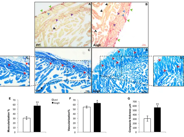
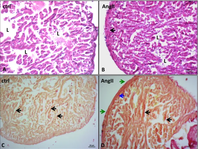
Ventricular sections stained with Sirius red (Fig. 1A and B) showed collagen fibers localized both in the compacta and spongiosa. An increase of collagen fibers located around the sub-epicardial vessels, in coronary vessels wall, at the border between compacta and spongiosa, and in the interstitium of the compacta characterized the hearts of AngII-treated animals. Increased collagen fibers are also detected in the spongiosa layer at the level of subendocardium and within interstitial spaces (Fig. 1B). Observations carried out on semithin sections (Fig. 1C and D) shown a significant increase of ventricle muscularity in treated eels, associated with an increased thickness of the compacta and trabecular size in the spongiosa. AngII-treated animals also showed an increased compacta vascular compartment (Fig. 1E, G, F).
1.3A.2. AngII receptors
Cardiac expression of AngII receptors, AT1 and AT2, has been evaluated by western blotting and immunofluorescence analysis by using mammalian anti-AT1 and AT2 antibodies.
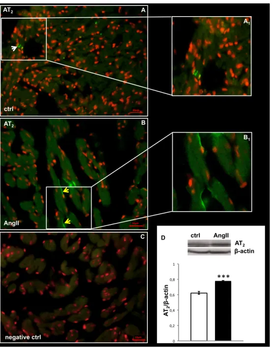
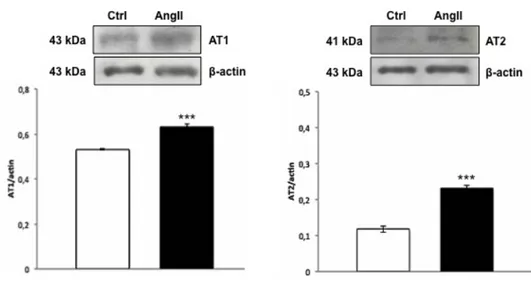
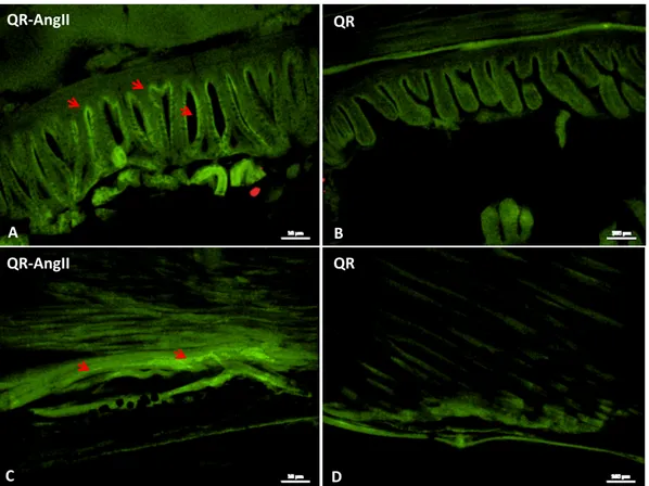
AT1 receptor was undetectable by both immunoblotting and immunofluorescence. In contrast, densitometric analysis of the blot with anti-AT2 antibody revealed an increased expression of this receptor in AngII-treated hearts, with respect to the control (Fig. 2D). Immunofluorescence showed a different localization of AT2 in the hearts of control and treated animals. In particular, AT2 signal was revealed in the myocardiocytes of control hearts (Fig. 2A), and at the EE of AngII-treated hearts (Fig. 2B).
1.3A.3. ERK1-2
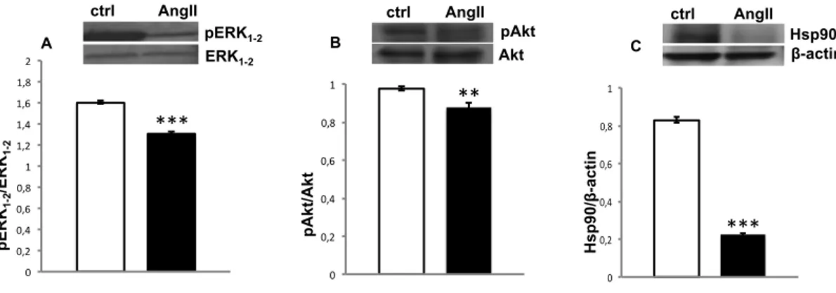
The ERK1-2 signalling pathway is involved in the regulation of myocardial proliferation and fibrosis (references in Mehta and Griendling, 2007). As revealed by western blotting analysis performed on cardiac extracts, prolonged exposition to AngII elicits a significant decrease of ERK1-2 phosphorylation in the hearts of
AngII-CHAPTER 1. Section A: Results treated animals (Fig. 3A). This suggests a negative modulation of the ERK1-2 signalling in the chronic effects of AngII in the eel heart.
Fig. 1. Representative images showing morphological changes occurring in the eel ventricle after AngII treatment.
(A, B) Sirius red staining of control (A) and AngII (B) treated hearts. Collagen fibers localize at subepicardial level (green arrows), in the vessels wall (blue arrows), at subendocardial level (black arrows) and in the interstitium (violet arrows). (C, D) Methylene Blue stained ventricular cardiac semithin sections of control (C) and AngII (D) treated eels. After treatment, the increment of compacta thickness, and of muscular and vascular (red arrows) components is evident. Inset C1 and D1 represent higher magnification of the vascularized area of control and AngII treated hearts, respectively. Statistical significances are reported in the corresponding histograms (E, F, G) (*p < 0.05; **p < 0.005). Values represent the means ± SEM of 6 measurements for each image (N=5).
C D AngII ctrl A B AngII ctrl C1 D1 10µm 10µm 0 10 20 30 40 50 60 70 ctrl AngII 0 100 200 300 400 500 600 700 ctrl AngII 0 10 20 30 40 50 60 70 ctrl AngII ** * ** Mu sc u la ri za ti o n % V as cu la ri za ti o n % C o m p ac ta ti ck n es s µ m E F G
CHAPTER 1. Section A: Results
Fig. 2. AT2 immunolocalization and expression
(A, B) Representative images of AT2 immunolocalization in the ventricle of control (A) and AngII treated (B) eels. White arrow: myocardiocytes. Yellow arrows: endocardial endothelium. Nuclei counterstaining: propidium iodide. Inset A1 and B1 represent a higher magnification of AT2 positive signal. (C) Negative control. (D) Western blotting and densitometric analysis of AT2 in cardiac extracts of ctrl and AngII treated eels. In D, loaded protein amount was verified using anti-β-actin antibody. Statistical differences were evaluated by unpaired t-test (***p < 0.0005). Data are the means ± SEM of 3 determinations for each group.
***
D β-actin AT2 AT 2 /β -a cti n ctrl AngII 0 0,2 0,4 0,6 0,8 1 10µm AT2 ctrl A A1 10µm AT2 AngII B B1 C negative ctrl 10µmCHAPTER 1. Section A: Results
Fig. 3. Western blotting on cardiac extracts
Western blotting and densitometric analysis of pERK1-2/ERK1-2 (A), pAkt/Akt (B) and Hsp90 (C) in cardiac extracts of control and AngII-treated eels. In C, loaded protein amount was verified using anti-β-actin antibody. Statistical differences were evaluated by unpaired t-test (**p < 0.005; ***p < 0.0005). Data are the means ± SEM of 3 determinations for each group.
1.3A.4. NOS/NO system
To evaluate the influence of AngII treatment on the cardiac NO production, the concentration of nitrite [used as an index of constitutive NOS activity (Hansen and Jensen 2010)] was determined in heart homogenates of both control and AngII-treated eels. Results showed a significant decrease of nitrite concentration in hearts from treated animals compared with the control group (control: 1.33 ± 0.13 mmol L-1;
AngII-treated: 0.60 ± 0.04 mmol L-1).
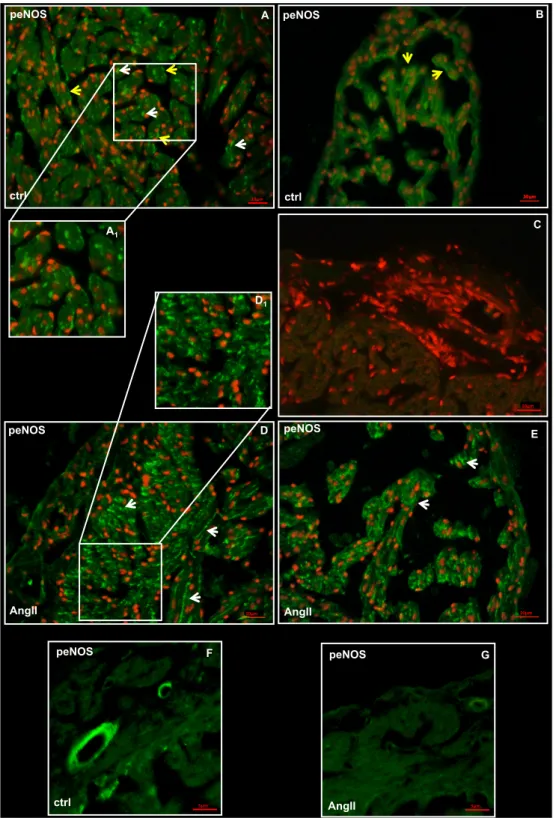
Western blotting of ventricular extracts revealed no significant differences in eNOS phosphorylation between control and treated hearts (data not shown) accompanied by a slight decrease of pAkt expression (a positive modulator of NOS) in treated animals (Fig. 3B). However, immunofluorescence analysis showed a different localization of peNOS in the heart of control and treated animals. In particular, while in the control hearts, a peNOS signal was captured at the EE level and in the cardiomyocytes of ventricle (Fig. 4A) and atrium (Fig. 4B), in treated hearts the enzyme prevalently localized in the myocardiocytes (Fig. 4D and E); only a weak
*** 0 0,2 0,4 0,6 0,8 1 1,2 1,4 1,6 1,8 2 C *** * β-actin Hsp90 ctrl AngII 0 0,2 0,4 0,6 0,8 1 Akt pAkt ctrl AngII ** B 0 0,2 0,4 0,6 0,8 1 pAKT /AKT pAkt /Akt Hsp90/ β -a cti n pERK1-2 ERK1-2 ctrl AngII p ER K1-2 /ER K1-2 A
CHAPTER 1. Section A: Results signal appeared on the EE. In addition, at vascular level, a reduction of the peNOS signal (Fig. 4G) was observed in the vascular endothelium of treated animals, with respect to the control (Fig. 4F).
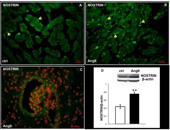
In line with the reduction of pAkt expression, western blotting analysis revealed a strong decrease in the expression levels of Hsp90, a positive modulator of NOS, after AngII treatment (Fig. 3C). These results are accompanied by a significant increase in the expression of the eNOS disabling protein NOSTRIN (Fig. 5D), a negative modulators of eNOS activity which promotes the translocation of the enzyme from the plasma membrane to intracellular vesicles (Michel and Vanhoutte 2010, and references therein). Although in both untreated (Fig. 5A) and AngII-treated (Fig. 5B) hearts, immunofluorescence localized NOSTRIN on the vascular endothelium of the greatest vessels (Fig. 5C) and on the ventricular EE, a strongest signal was detected at the endocardial level in AngII treated hearts (Fig. 5B).
CHAPTER 1. Section A: Results
Fig. 4. peNOS immunolocalization
Representative images showing peNOS immunolocalization on cardiac sections of control (A, B, F) and AngII (D, E, G) treated eels. (A, D) ventricle, (B, E) atrium, (F,G) vessels. Inset A1 and D1 represent a higher magnification of peNOS positive signal. (C) Negative control. White arrows: myocardiocytes. Yellow arrows: endocardial endothelium. Nuclei counterstaining: propidium iodide.
negative ctrl 10µm 10µm peNOS AngII E 10µm A peNOS ctrl 10µm D peNOS AngII A1 C D1 peNOS F ctrl peNOS G 5µm AngII B peNOS ctrl 10µm 10µm 5µm
CHAPTER 1. Section A: Results
Fig. 5. NOSTRIN immunolocalization and expression
Representative images of NOSTRIN immunolocalization in ventricular sections of control (A) and AngII-treated (B, C) eels. NOSTRIN localized in endocardial (yellow arrows) and vascular endothelium (red arrows). C: nuclear counterstaining with propidium iodide. (D) Western blotting of NOSTRIN in cardiac extracts of control and AngII treated eels. Loaded protein amount was verified using anti-β-actin antibody. Statistical differences were evaluated by unpaired t-test (**p < 0.005). Data are the means ± SEM of 3 determinations for each group. A B NOSTRIN NOSTRIN AngII ctrl NOSTRIN ** * D ctrl AngII N O ST R IN /β -a cti n 0 0,2 0,4 0,6 0,8 1 β-actin 5 µm NOSTRIN C AngII
CHAPTER 1. Section A: Discussion
1.4A. DISCUSSION
The results reported in this section showed, for the first time, that chronic (two months) treatment with AngII induces structural modifications of eel ventricular wall and myocardial vessels. This remodelling was paralleled by modification of NO production, and by changes in the expression and localization of molecules which regulate NOS activity, such as the protein kinase B (Akt), the eNOS disabling protein NOSTRIN, and the heat shock protein 90 (Hsp90).
With respect to the compact type of ventricular myoarchitecture typical of homeotherm hearts, eel shows a mixed type ventricle myoarchitecture with an outer compacta layer that enclose an inner spongiosa made up of a crisscrossed array of myocardial bundles (trabeculae) (see Cerra et al., 2004, for references). As revealed by morphological analyses, the cardiac ventricle of eels chronically exposed to AngII undergoes structural modifications, becoming more “muscularized” than untreated animals. This remodelling occurs at the level of both compacta and spongiosa. In particular, when compared to control animals, AngII-treated eels showed an increased compacta thickness, and a larger diameter of the trabeculae that form the spongiosa. These changes are accompanied by higher collagen deposition in the compact layer, shown by an intense Sirius red staining, particularly localized in the wall of subepicardial and coronary vessels. Enhancement of the compact myocardium is a strategy of cardiac growth that occurs in many fish species [Ciprinus carpio: (Bass et al., 1973); Salmo salar: (Poupa et al., 1974); Thunnus thynnus: (Poupa et al., 1981); Salmo gairdneri: (Farrell et al., 1988)]. It was described in A. anguilla during ontogenetic growth (Cerra et al., 2004) and in response to one month exposure to AngII (Imbrogno et al., 2013). Of note, we observed in eels treated for two months with AngII that the increment of the compact layer is accompanied by an increased vascularisation. In non-mammalian vertebrates, the growing heart increases capillarization of the compact myocardium through tightly controlled local mechanisms, which include mechanical (e.g. myocardial stretch), metabolic and growth factors such as vascular endothelial growth factor (VEGF) and basic fibroblast growth factor (bFGF) (Tomanek and Ratajska 1977, and references therein). It is conceivable that the enlarged vascular supply, which occurs in the compacta of
CHAPTER 1. Section A: Discussion the AngII-treated hearts, may allow the heart to cope with the metabolic and energetic demands of the deeper myocardial cells.
In mammals, AngII-dependent effects on myocyte growth correlate with the activation of the AT1 receptor (see Booz et al 2002, for references). Contrarily, the AT2 receptor, is generally reported to mediate anti-growth and apoptotic actions (Gallinat et al., 2000). However, a role for AT2 receptors in AngII-dependent cardiomyocyte hypertrophy and cardiac fibrosis was described in mice from Ichihara and co-workers (Ichihara et al., 2001). In A. anguilla, the few available data correlate the growth-promoting actions of AngII to the activation of a receptor able to bind mammalian anti-AT2 antibody (Imbrogno et al., 2013). AT1 receptors were not detected in control and AngII-treated eels. Conversely, western blotting and immunofluorescence showed, in the heart of the two groups of animals, a different expression and spatial localization of AT2 receptors. In fact, while in control hearts AT2 receptors are expressed in few myocardiocytes, after AngII treatment, they localize more widely in the EE. This suggests that, contrarily to mammals, in the eel heart AT2 receptors participate to the mechanisms of remodelling induced by AngII. No data about a putative AngII-dependent modulation of ATs expression are available in fish. Also in mammals the molecular mechanisms responsible for the expression of the AT2-R gene are not fully defined, although an upregulation of the AT2 expression has been observed in different experimental models, also in response to AngII exposition, and in relation to various physio-pathological conditions (references in Lemarie and Schiffrin, 2010). It is interesting to underline that, although eel AT2 shares its origin with mammalian AT2, it underwent a peculiar evolution after divergence of its lineage (Wong and Takey, 2013). This may explain the contrasting features showed by this receptor in mammals vs teleost, as for example the activation of different transduction pathways (Wong and Takey, 2013), and thus the different elicited responses.
Several studies in mammals reported that AngII exerts its actions on the heart by recruiting ERK1-2 (see for references Mehta and Griendling, 2007). This kinase is known for its stimulatory role in the mechanisms of myocardial proliferation and
CHAPTER 1. Section A: Discussion fibrosis (Aoky et al., 2006; Bueno et al., 2000). However, more recent experiments performed on in vivo knock-out mice showed that ERK1-2 signalling is not required for mediating cardiac hypertrophy (Purcell et al., 2007). In mammalian cardiomyocytes, by acting on AT1 and/or AT2-receptor, AngII exerts distinct effects on ERK1-2 activity. Through AT1 receptor it may induce ERK1-2 activation, while it may inhibit ERK1-2-dependent pathway through AT2 receptor (Wei et al., 2000). Consistent with these observations, the significant reduction of the activated (phosphorylated) form of ERK1-2 observed in AngII-treated animals, is accompanied by an increased expression of AT2 receptors, suggesting that ERK1-2 down-regulation is mediated by the AngII-AT2 binding.
An important aspect of the molecular network guiding the AngII-dependent cardiac morpho-functional modulation is the cross-talk with the NO signalling. In mammals, in addition to the AT2-mediated control of NO production (references in Dostal, 2000), AngII activates a number of signal-transduction pathways that result in reduced NO levels (Morawietz et al., 2006). In turn, NO can counteract AngII-dependent effects by down-regulating the synthesis of both ACE and AT1 receptors (Wiemer et al., 2001; Takemoto et al., 1997). Thus, NO generation is both downstream and upstream the AngII-dependent cascade. NO is a vital signalling molecule that exerts its physiological effects by reversible binding/reacting with hemes, thiols or amines, forming iron-nitrosyl (FeNO), S-nitroso (SNO) and N-nitroso (NNO) compounds (Hill et al., 2010). Furthermore, NO is short-lived and excess NO is rapidly oxidized to nitrite and nitrate. Tissue levels of nitrite represent a marker of constitutive NOS activity and consequently of NO generation (Hansen and Jensen, 2010). By measuring total nitrite concentration in endothelial cell cultures, Li and co-workers observed an AngII-dependent reduction of eNOS-dependent NO production (Li et al., 2016). In this study, we found a significant decrease in the ventricle nitrite concentration from 1.33 mmol L-1 in control animals (which
compares with other fish species (Sandvik et al., 2012; Jensen et al., 2015) to 0.6 mmol L-1 in AngII treated eels. This suggests that chronic exposure to AngII is accompanied
by a reduced NO generation, possibly mediated by a blunted NOS functionality. To analyse this possibility, the molecular modulation of NOS-dependent NO production