Table of contents
ABSTRACT ... 1
INTRODUCTION ... 2
Breast cancer ... 2
Tumor microenvironment ... 2
• Cancer Associated Fibroblasts ... 3
• Tumor Associated Macrophages ... 5
• SDF-1α/CXCR4 axis ... 7
PPARγ ... 9
AIM OF THE THESIS ... 13
Reagents ... 14
Plasmids ... 14
Cell cultures ... 14
CAFs isolation ... 15
Conditioned medium systems ... 15
Coculture THP1 and breast cancer cells conditioned media ... 16
Cytotoxicity Assays ... 16
Cell viability assay ... 16
Immunoblot analysis ... 17
RT-PCR/qRT-PCR ... 17
Transient transfection assay ... 19
Immunofluorescence ... 19
Chromatin immunoprecipitation assay ... 19
DNA affinity precipitation assay ... 20
RNA silencing ... 20
Wound-healing assays ... 20
Transmigration assays... 21
Invasion assays ... 21
Enzyme-linked immunosorbent assay ... 21
SDF-1α -immunodepleted conditioned media ... 22
Statistical analysis ... 22
Abbreviations ... 22
Ligand-activated PPARγ downregulates CXCR4 expression and its gene promoter activity in breast cancer cells .... 23
Identification of a functional PPAR responsive element (PPRE) within the CXCR4 promoter ... 25
BRL inhibits motility in breast cancer cells ... 30
Ligand-activated PPARγ counteracts stroma-mediated breast cancer cell migration ... 33
ABSTRACT
Stromal Derived Factor-1α (SDF-1α) and its cognate receptor CXCR4 play a key role in mediating breast cancer cell invasion and metastasis. Therefore, drugs able to inhibit CXCR4 activation may add critical tools to reduce tumor progression, especially in the most aggressive form of the breast cancer disease. Peroxisome Proliferator-Activated Receptor (PPAR) γ, a member of the nuclear receptor superfamily, has been found to downregulate CXCR4 gene expression in different cancer cells, however the molecular mechanism underlying this effect is not fully understood. Here, we identified a novel PPARγ-mediated mechanism that negatively regulates CXCR4 expression in both epithelial and stromal breast cancer cells. We found that ligand-activated PPARγ downregulated CXCR4 transcriptional activity through the recruitment of the silencing mediator of retinoid and thyroid hormone receptor (SMRT) corepressor onto a newly identified PPAR response element (PPRE) within the CXCR4 promoter in breast cancer cell lines. As a consequence, the PPARγ agonist rosiglitazone (BRL) significantly inhibited cell migration and invasion and this effect was PPARγ-mediated, since it was reversed in the presence of the PPARγ antagonist GW9662. According to the ability of Cancer-Associated Fibroblasts (CAFs), the most abundant component of breast cancer stroma, to secrete high levels of SDF-1 α, BRL reduced migratory promoting activities induced by conditioned media (CM) derived from CAFs and affected CXCR4 downstream signaling pathways activated by CAF-CM. In addition, CAFs exposed to BRL showed a decreased expression of CXCR4, a reduced motility and invasion along with a phenotype characterized by an altered morphology. A further component of the tumor microenvironment, that contributes to breast cancer progression and metastasis, is represented by Tumor Associated-Macrophages (TAMs), which phenotype is shaped by complex interactions with breast cancer cells. We found that the PPARγ ligand BRL, as well as DHA conjugates to ethanolamine and serotonin DHEA and DHA-5-HT respectively, were able to counteract the effects of CM derived from breast cancer cells on macrophage polarization. Collectively, our findings provide novel insights into the role of PPARγ in inhibiting breast cancer progression and further highlight the utility of PPARγ ligands for future therapies aimed at targeting both cancer and surrounding stromal cells in breast cancer patients.
INTRODUCTION
Breast cancer
Breast cancer is a genetic disease caused by the accumulation of genetic mutations and epigenetic modifications in genes that control the proliferation, differentiation, death and integrity of the cellular genetic heritage. It is a multistep process during which transformed tumor cells escape normal cellular growth control mechanisms, multiply and lead to an epithelial hyperplasia (Barrett C.J., 1993; Baxter E. et al., 2014).
Advanced stages of the disease are characterized by the invasion and colonization of tissues and organs distant from the site of origin of the tumor. This process is defined as metastasis and, to date, represents the main cause of recurrence of the tumor, in fact about 30% of patients affected by breast cancer still relapse and die of metastatic disease within five years (Ferlay J. et al., 2012). Although considerable progress has been made in understanding the cancer genetic alterations and the consequent signaling abnormalities that drive tumor initiation and progression, breast cancer remains the leading cause of female cancer related deaths in developed countries. Breast cancer displays a defined molecular profile based on the expression of hormone receptors such as ER estrogen receptor, PR progesterone receptor and/or ERBB2/HER2 receptor, thus most therapies have been designed to oppose hormone receptors action. However, the most aggressive breast tumors, like triple negative breast tumors, lack of effective treatments since they are resistant to hormone therapies. For long periods the anticancer therapeutic strategies have focused only on the tumor cells. Numerous studies have shown that both the neoplastic cells and the surrounding microenvironment strongly contribute to the growth and the tumor progression (Lorusso G. and Rüegg C., 2008; Mbeunkui F. and Johann D.J., 2009). It is therefore fundamental to study not only the biology of the tumor cells, but also the surrounding microenvironment and their mutual interactions, in order to identify new therapeutic approaches.
Tumor microenvironment
In physiological conditions epithelial and stromal cells communicate with each other to assure the normal development and differentiation of the mammary gland counteracting the uncontrolled cell growth and neoplastic transformation (Folgueira M.A. et al., 2013; Barsky
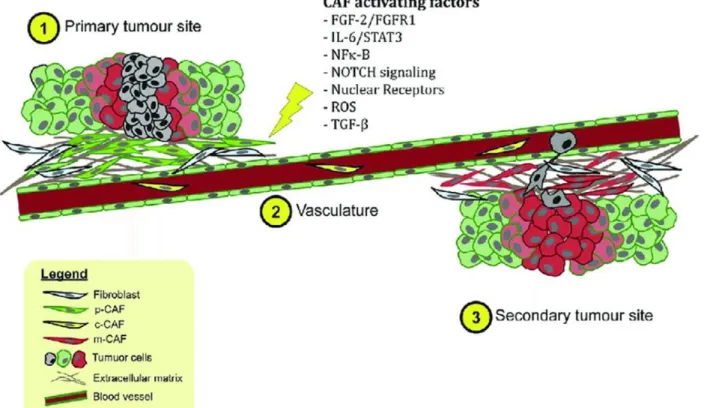
S.H. et al., 2005). In fact, studies have shown that normal myoepithelial cells can suppress growth, invasion and angiogenesis of breast cancer cells acting as natural tumor suppressor. When cancer occurs, myofibroblasts and fibroblasts acquire protumor properties and through paracrine signaling promote tumorigenesis and metastatic spread (Hu M. et al., 2008). Hence, tumor cells can create a tumoral microenvironment ensuring favorable conditions to their own development and progression. Breast tumor microenvironment encompasses stromal cells like fibroblasts, immune cells, pericytes, adipocytes, but also cancer stem cells (CSC) and signaling molecules including cytokines, chemokines, growth factors and extracellular matrix proteins which establish an autocrine and paracrine crosstalk that allows microenvironment and tumor cells to support each other (Figure. 1).
Figure 1. Cancer cells and tumor microenvironment interplay. Elevated levels of produced cytokines and growth
factor by tumor cells recruit tumor-associated macrophages, neutrophils, and mast cells, which secrete additional growth factors, forming a positive feedback loop that promotes tumor cell invasion and metastasis. CSC, cancer stem cells.
• Cancer Associated Fibroblasts
Fibroblasts are the most abundant cell types in the stroma. They play an important role in supporting the architecture of tissues and organs and their interaction with the neighboring cells, through the secretion of different signaling molecules, is fundamental to regulate
tissue development, repair and homeostasis processes (Parsonage G., 2005; McGettrick, H.M., 2012). In physiological conditions fibroblasts are quiescent cells; whereas during pathophysiologic processes they are stimulated and activated by various soluble factors. Indeed, different extrinsic signals from cancer cells can activate normal fibroblasts, giving rise to the majority of cancer associated fibroblasts (CAFs) resident in the tumor (Rasanen, K. and Vaheri, A., 2010; Shiga, K. et al., 2015; Alexander J. and Cukierman E., 2016) (Figure.2). The activated fibroblasts acquire an increased capacity for protein synthesis and contraction functions, their shape change from fusiform and elongated to a wide-cruciform structure.
Figure 2. CAFs and their activating factors. Upon activation by appropriate signals or mediators, normal fibroblasts
at the primary site are activated to CAFs (p-CAFs) and can contribute to chemoresistance, metastasis, and invasion. Circulating CAFs (c-CAFs) are detected in the vasculature. CAFs associated with secondary tumors are also known as m-CAFs.
Both normal fibroblasts and cancer-associated fibroblasts appear to have a very similar phenotype, but several studies indicate that CAFs have different mRNA and protein expression profiles respect fibroblasts in normal breast tissue (Allen M. and Jones J., 2011; Folgueira M.A. et al., 2013). CAFs have been shown to express a number of markers like α-SMA (alpha-smooth muscle actin), FAP (fibroblast activation protein), MMPs (metalloproteinases), PDGFRα/β (platelet-derived growth factor) but none of them is
specific for CAFs which, for this reason, are difficult to identify within the tumor (Buchsbaum R.J. and OH S.Y., 2016).
CAFs strongly regulate tumor proliferation, invasiveness, angiogenesis and direct tumor growth through the secretion of several soluble factors such as growth factors and chemokines that modulate the tumor stroma and induce cancer cells to support protumoral processes (Orimo A. et al., 2005; Barone I. et al., 2012; Hugo H.J. et al., 2012). This bidirectional interplay promotes a positive feedback loop in which both cancer cells and CAF facilitate their own survival and proliferation.
• Tumor Associated Macrophages
Immune population in the mammary gland encompasses different immune cells among which macrophages have an important role in maintaining the balance between destruction and restoration of the tissue, pathogen elimination and homeostasis maintenance (Lavin Y. et al., 2015).
Several molecules released in the tumor microenvironment by tumor and stromal cells are responsible of macrophages functional and phenotypic diversity and plasticity. Macrophages educated by the tumor microenvironment are called tumor associated macrophages (TAM); they include resident macrophages or they can derive from blood monocytes and therefore myeloid-derived suppressor cells (MDSCs) which arise from bone marrow-derived immature myeloid cells in response to a variety of chemokines and cytokines among which CSF1 is one of the main chemoattractants (Lin E.Y. et al., 2001; Franklin R.A. and Li M.O., 2016).
Generally, macrophages can be distinguished in classically (M1) and alternatively activated (M2) type which can also be sub grouped in M2a, M2b, and M2c depending on the activating stimuli (Mantovani A., 2004). The two macrophage subtypes differ in cytokine and chemokines secretion, metabolism and receptor expression and presentation on their surface and response to different stimuli (Mantovani A. and Allavena P., 2015). Therefore, pro-inflammatory ligands such as TNFα, IFNγ, lipopolysaccharide and GM-CSF stimulate M1 macrophages that facilitate T helper 1 (Th1) response including antigen presentation and tumoricidal immunity, whereas M2 macrophages are polarized by TGFβ, IL10, IL4, IL13 and glucocorticoids and participate in Th2 activities, inflammation resolution and
tumorigenic activities (Murray P.J., 2014; Mills C.D., 2012; Ostuni R. et al., 2015) (Figure. 3).
Figure 3. TAMs polarization and functions. Macrophages can be schematically classified into two main classes
depending on their phenotypic polarization: macrophages differentiate into M1 in response to M-CSF, IFNα, LPS and other microbial products, whereas they differentiate into M2 in the presence of M-CSF, IL-4, IL-10, IL-13 and other molecules. M1 and M2 display different functions: M1 macrophages are able to trigger Th1 immune response and exert antitumor activity, M2 macrophages activate Th2 immune response and promote tumor progression, angiogenesis, tissue remodeling and metastasis.
Breast cancer macrophages exhibit an undetermined phenotype and adapt their phenotype to distinct stimuli: in early stage of tumorogenesis, characterized by an enhanced inflammatory state, M1 cells secrete pro-inflammatory cytokines including IL1β, IL6, and TNFα; whereas, once malignancy has been established, most TAM belong to the M2 protumor phenotype, they secrete anti-inflammatory ligands such as IL10, CCL2, TGFβ, prostaglandin E2, and IL1 receptor antagonist (Grugan K.D. et al., 2012; Mantovani A. et al., 2017). TAM play a pivotal role in supporting tumor progression by promoting angiogenesis, suppressing adaptive immunity, supporting cancer stem cells; they also
facilitate tumor cell systemic dissemination and spread via secreting different matrix-degrading enzymes (Joyce J.A. and Pollard J.W., 2009; Farmer P et al., 2009; Nouh M.A. et al., 2011). High density of TAMs are linked to a worse prognosis in breast cancer patients (Williams C.B. et al., 2016). Therefore, reprogramming or inhibiting tumor-protecting properties of TAMs could represent a viable therapeutic strategy.
• SDF-1α/CXCR4 axis
Among the various signaling pathways resulting from the interactions between tumor cells and microenvironment components, the activation of SDF-1α/CXCR4 play a significant role in breast cancer migration and metastasis (Müller A. et al., 2001; Hassan S. et al., 2009). The C-X-C chemokine receptor 4 (CXCR4), a member of the G protein-coupled cell surface receptors (GPCRs) displaying 7 transmembrane-spanning domains, is the physiological receptor for the CXC chemokine stromal-derived-factor-1 (SDF1-α or CXCL12) which binding promotes the interaction with different effector proteins and initiate intracellular signaling cascades, thus regulating cell survival, proliferation, chemotaxis, migration and adhesion, contributing tumorigenesis and cancer progression (Dewan M.Z. et al., 2006). CXCR4 is constitutively expressed by many tissues like brain, thymus, spleen, stomach, lymphatic tissue and small intestine (Nagasawa T. et al., 1994). It is highly expressed in various types of cancer including breast cancer (Billadeau D.D., 2006; Dewan M.Z. et al., 2006; De Falco V. et al., 2007; Gangadhar T. et a., 2010). In primary and metastatic breast cancer cells, CXCR4 is highly expressed, while it is present at low level or even absent in normal breast tissue (Müller A. et al., 2001). Indeed, CXCR4 directs different steps of breast cancer metastasis; in fact, it can drive the epithelial-mesenchymal transition (ETM), a process by which ephitelial cells display reduced intracellular adhesion and, consequently, an increased motility (Larue L. and Bellacosa A., 2005). Moreover, it is involved in chemotaxis process since large amounts of its ligand SDF-1α are released by breast cancer metastatic cells in the bone, lung and liver, tissues commonly affected by metastatic breast cancer (Figure 4). Furthermore, CXCR4 can increase the expression of other chemokine receptors and cytokines, leading to cell migration, lymphatic invasion and thus tumor metastasis (Sobolik T. et al., 2014).
Figure 4. Potential role of CXCR4 in breast cancer. Stromal cell derived factor (SDF-1) bound-CXCR4 receptor,
expressed by breast tumor cells. Tumor expressed CXCR4 directs metastasis to sites such as bone, liver, lung, brain, lymph node, and kidney. In addition, SDF-1/CXCR4 interacts locally in autocrine and paracrine manner to increase primary tumor growth.
SDF-1α/CXCR4 axis is correlated with breast cancer progression; apart from tumoral cells also tumoral stromal cells contribute to the amount of SDF1-α in the tumor microenvironment; for instance, CAFs secrete high levels of SDF1-α that, through activation of CXCR4 signaling, promote tumor cell proliferation, motility and invasion (Orimo A. et al., 2005). Therefore, drugs able to inhibit CXCR4 expression and/or activity may represent critical tools against breast cancer disease.
Recent studies have reported that activated PPARγ reduces invasion and motility of colon, lung and prostate cancer cells, through CXCR4 downregulation (Richard and Blay, 2007; Tai C.J. et al., 2010; Qin L. et al.,2014;). However, despite these studies, either the
regulatory mechanism by which PPARγ may regulate CXCR4 expression in breast cancer cells or how PPARγ works in the context of breast tumor microenvironment remain largely unknown.
PPARγ
Peroxisome proliferator-activated receptors (PPARs) belong to the family of nuclear hormone receptors (NHRs) that include estrogen, thyroid hormone receptors, retinoic acid and Vitamin D3 receptors as well as retinoid X receptors (RXRs). NHRs superfamily is a class of ligand-activated transcription factors, their activation through binding to small lipophilic molecules regulate the transcription of target genes involved in adipogenesis, cell growth, tissue homeostasis and energy metabolism, proliferation and tumor progression (Michalik L. et al., 2004; Germain P. et al., 2006).
PPAR subfamily include three subtypes PPARα, PPAR β/δ and PPARγ each encoded by different genes that share 60-80% homology in their ligand-binding and DNA-binding domains and display a different tissue distribution (Desvergne B. and Wahli W., 1999; Papadaki I. et al., 2005). PPARα is mainly expressed in liver, heart, kidney and intestinal cells, PPAR β/δ is widely expressed in organism tissues and PPARγ is expressed in endothelial and immune system cells, it is highly expressed in adipose tissue and in tumors originated from various organs including breast cancer (Wahli W. et al., 1995; Grommes C. et al., 2004). Like the other members of nuclear receptor superfamily, PPARγ displays a characteristic structure consisting in three general function domains: the NH2-terminal domain that contains important phosphorylation sites, the DNA-binding domain (DBD) that targets the receptor to specific DNA sequences and the ligand-binding domain (LBD) that encompasses specific sites for ligand binding (Desvergne B. and Wahli, W., 1999). PPARγ activation occurs in the cytoplasm where the ligand binding to the LBD site causes a conformational change following which the receptor heterodimerizes with the Retinoid-X Receptor (RXR). The PPARγ/RXR translocates to the nucleus where it binds to the DBD and precisely to the Peroxisome Proliferator Response Elements (PPREs) located within the promoter regions of target genes (Figure. 5). The PPRE consists of a direct repetition of the consensus AGGTCA nucleotide sequence spaced by one or two nucleotides (Berger J. et al., 2002). Transcriptional activity of PPARγ is controlled by the recruitment of accessory
proteins called “co-activators” and “co-repressors” that bind to the N-terminal part of the LBD in a ligand-dependent manner. PPARγ co-activators are essential for its transcriptional function acting by remodeling chromatin structure and/or linking the complex to key transcriptional machinery, they include histone acetyltransferase p300, CREB-binding protein (CBP), steroid receptor coactivator (SRC)-1, Krueppel-like factor (KLF)-2, mediator of RNA polymerase II transcription (MED)-1 and PPARγ coactivator (PGC)-1 (Qi C. et al., 2000; Leader J.E. et., 2006). The nuclear receptor corepressor (N-CoR) and the silencing mediator for retinoid and thyroid hormone receptor (SMRT) are corepressor proteins that repress the transcriptional process by binding to the promoter to which the complex binds.
Figure 5. Structure and molecular mechanism of action of peroxisome proliferator-activated receptor alpha (PPARγ). PPARγ has different functional domains: the N-terminal ligand-independent transactivation domain; DNA
binding domain (DBD), including an activation function-1 (AF-1); and C-terminal domain including a ligand binding domain (LBD) and an activation function-2 (AF-2). PPARγ and retinoid X receptor (RXR) heterodimer, which can recruit diverse coactivators and corepressors that modulate the transcriptional activity of PPARγ, binds to PPAR-response elements (PPRE) to activate target gene transcription.
In addition to the main role in regulating adipogenesis, PPARγ also regulate insulin sensibilization, lipid metabolism, atherosclerosis and inflammation (Wahli W. et al., 1995; Chinetti G. et al., 2000). Several studies reveled that PPARγ is also implicated in carcinogenesis in a wide range of tumors such as liposarcoma, colon cancer, prostate
carcinoma, gastric carcinoma, pancreatic carcinoma, myeloid leukemia and breast cancer (Koeffler H.P., 2003; Liu H. et al., 2003).
It has been widely demonstrated how the activation of PPARγ upon binding to its synthetic and/or natural ligands, inhibits the proliferation and induces apoptosis and autophagy processes in different in vitro and in vivo models of breast cancer (Grommes et al., 2004; Bonofiglio et al., 2011; Catalano S. et al., 2011; Rovito D. et al., 2015). PPARγ ligands include several synthetic and naturally compounds. Synthetic ligands include non-steroidal anti-inflammatory drugs (NSAIDs) and anti-diabetic thiazolidinedione (TZD) class of drugs like Troglitazone (TGZ), Pioglitazone and Ciglitazone (CIG) and Rosiglitazone (BRL49653, BRL). A previous metanalysis of randomized clinical trials has reported that Rosiglitazone was not associated with a significant modification of the risk of cancer, while the incidence of malignancies was significantly lower in Rosiglitazone-treated patients than in control groups (Monami M. et al., 2008). Moreover, a recent study showed that in female patients with type 2 diabetes mellitus treatment with both Rosiglitazone and metformin exhibited the lowest breast cancer risk (Tseng C.H., 2017).
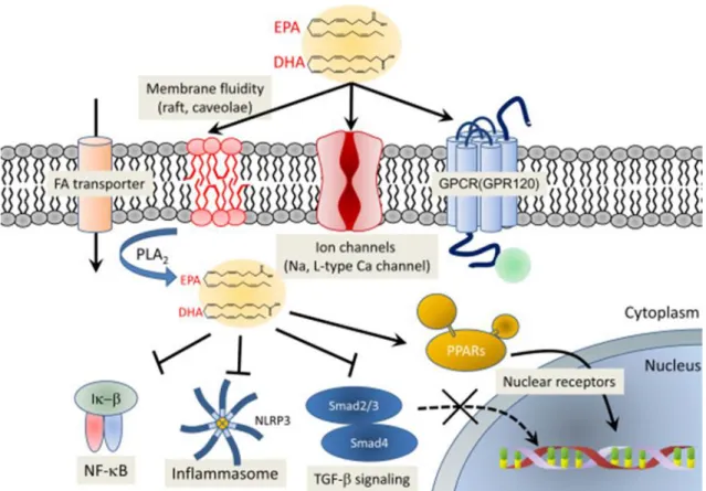
Natural ligands are small lipophilic molecules such as 15-deoxy-Δ12,14-Prostaglandin J2 (15-PGJ2), prostanoids and long chain polyunsaturated fatty acids (PUFAs). (Willson, T.M. et al., 2000; Grygiel-Górniak B.,2014). PUFAs are endogenous mediators that can be synthesized in the human body except essential fatty acids which are required for biological processes and must be obtained from dietary sources. They display different roles: acting as transcription factors, they modulate the protein synthesis; they can be involved in signal transduction, or they can constitute membrane components and be able to regulate the fluidity, permeability, and dynamics of cell membranes (Chapkin R.S. et al., 2008) (Figure. 6).
Epidemiological studies have shown a correlation between diets rich in polyunsaturated acids and a lower risk of occurrence of some forms of cancer, including breast cancer (MacLean C.H. et al., 2006; Brennan S.F. et al., 2010). The two main n−3 PUFAs are the eicosapentaenoic acid (EPA) and docosahexaenoic acid (DHA), that in breast cancer cells, can be directly converted to N-acylethanolamines, DHEA, and EPEA, respectively (Brown et al., 2011); other conjugates of n−3 PUFAs with serotonin, L-alanine, L-serine, histidine, GABA, glutamic acid or dopamine have been found in mammals where they exert
anti-inflammatory activities (Brown I. et al., 2011; Meijerink J. et al., 2013). Omega-3 fatty acids have been shown to decrease cell viability, proliferation, invasion, and increasing chemosensitivity in breast cancer (Evans L.M. and Hardy R.W., 2010). The anti-cancer activities exerted by EPA and DHA are also due to their ability to bind Peroxisome Proliferator-Activated Receptor gamma (PPARγ) (Gani O.A., 2008). Recently, our research group showed how the ethanolamine and dopamine conjugates of omega 3 fatty acids exert antiproliferative effects on several lines of breast cancer by activating PPARγ, since they are its natural ligands (Rovito D. et al., 2013; Rovito D. et al., 2015). Omega-3 polyunsaturated fatty acids and their conjugates show a biological relevance which can suggest them as new pharmacological tools to be implemented in the adjuvant therapy for breast cancer treatment.
Figure 6. Potential molecular mechanism exerted by omega-3 PUFAs. Omega-3 PUFAs modulate cell membrane
property when incorporated into the phospholipid bilayer and are involved in signal pathways that regulate different biological processes among which inflammation and carcinogenesis.
AIM OF THE THESIS
The overall aim of this project was to investigate the role of the nuclear receptor PPARγ in inhibiting breast cancer progression focusing on the complex interplay between breast cancer and stromal cells. First, we studied the molecular mechanism by which PPARγ activated by its synthetic and specific ligand BRL, through CXCR4 downregulation, reduces motility and invasiveness in different breast cancer cell lines. Next, we investigated the role of ligand activated PPARγ in contrasting migratory promoting activities of CAFs. Finally, we extended our results in the context of heterotypic signaling working in tumor-stroma interactions examining the ability of a panel of natural and synthetic PPARγ ligands to counteract the effects of breast tumor cells on macrophage polarization and their cytokine secretion pattern, that in turn, may negatively impact breast cancer progression.
MATERIALS AND METHODS
Reagents
Rosiglitazone (BRL49653, BRL) was obtained from Alexis (San Diego, CA), GW9662 (GW) and 15-deoxy- Delta 12,14-prostaglandin J2 (PGJ2) from Sigma Aldrich (Milan, Italy) and Stromal-cell Derived Factor-1alpha (SDF-1α) from Prospec (Rome, Italy). 2-(5-Bromo-1H-indol-1-yl)-N´-(pyrazin-2-yl) benzohydrazide (FIL2) was kindly provided by Dr. Grande. LPS was obtained from Sigma-Aldrich (Schnelldorf, Germany). IL-4 was obtained from R&D system (Abingdon, U.K.). Docosahexaenoyl serotonin (HT, DHA-5-HT) and docosahexaenoyl ethanolamide were purchased from Cayman Chemical (Ann Arbor, MI, USA). ELISAs (IL-6, IL1Ra and IL10) were performed using R&D Systems kits (Abingdon, U.K.).
Plasmids
The human CXCR4 gene promoter constructs (p-2300, p-2144, p-1507) were a gift from Prof. M. Z. Ratajczak (Stem Cell Institute at James Graham Brown Cancer Center, University of Louisville, Louisville, KY).
Cell cultures
Human ERα-positive MCF-7, the triple-negative (ER-, PR- and Her2-negative) MDA-MB-231 breast cancer epithelial cells and human monocytic cell line THP1 were acquired from American Type Culture Collection where they were authenticated, stored according to supplier’s instructions, and used within 4 months after frozen aliquots recovery. Every 4 months, cells were authenticated by single tandem repeat analysis at our Sequencing Core; morphology, doubling times, estrogen sensitivity, and mycoplasma negativity were tested (MycoAlert, Lonza). MCF-7 cells were cultured in DMEM (Life Technologies, Carlsbad, CA, USA) supplemented with 10% fetal bovine serum (FBS) (Life Technologies), 1 mg/ml penicillin-streptomycin (Life Technologies) and 0.01 mg/ml insulin (Sigma Aldrich) at 37 °C with 5% CO2 air. MDA- MB-231 cells were cultured in DMEM/F-12 plus glutamax (Life Technologies) containing 10% FBS and 1 mg/ml penicillin-streptomycin. MCF-10A non tumorigenic breast epithelial cells were grown in DMEM-F12 plus glutamax containing
hydrocortisone (Sigma Aldrich), and 10 mg/ml insulin. THP1 cells were cultured in Roswell Park Memorial Institute 1640 (RPMI-1640, Lonza,Verviers SPRL, Belgium) medium, supplemented with 10% fetal calf serum (FCS, Lonza, Verviers SPRL, Belgium), 1% penicillin−streptomycin (Corning), at 37°C in a 5% humified incubator. For experimental purposes, cells were grown in phenol red-free media containing 5% charcoal-treated FBS (CT-FBS) for 24 h and then treated as described.
CAFs isolation
Human breast cancer specimens were collected in 2013–2014 from primary tumors of patients who signed informed consent in accordance with approved Human Subject’s guidelines at Annunziata Hospital (Cosenza, Italy), following the procedures previously described (Barone I. et al., 2012). Briefly, small pieces of fresh tumor excision were digested (500 IU collagenase in Hank’s balanced salt centrifugation (90 g for 2 min), the supernatant containing CAFs was centrifuged (500 g for 8 min), resuspended, and cultured in MEDIUM 199 (Life Technologies)/F-12 (Sigma Aldrich) (1:1) supplemented with 15% FBS and antibiotics. The fibroblastic nature of the isolated cells was confirmed by microscopic determination of morphology, and characterization by α-SMA, vimentin. CAFs between 4 and 10 passages were used.
Conditioned medium systems
CAFs were incubated with regular full media (48 h). Conditioned media (CM) were collected, centrifuged to remove cellular debris, and used in respective experiments. Breast cancer cells were plated in complete media, and when cultures reached 80–90% confluence, the medium was replaced with fresh serum-free medium for 48 hours. The obtained conditioned media was centrifugated at 2,000 g at 4°C for 10 min to remove cell debris and preserved at -80°C for further study.
Differentiation of THP1 monocytes to macrophages
To obtain the macrophage-like state (M0), 1 million monocytic THP1 cells were seeded in 6-well plates in 2-mL RPMI media plus 61.7 ng/mL (100nM) or 10 ng/mL (16nM) phorbol 12-myristate 13-acetate (PMA; Sigma) for 24 hours of treatment. Differentiated,
plastic-adherent cells were washed twice with culture medium and rested for another 24 hours in the culture medium (RPMI 1640 medium without PMA but containing 10% FBS and 1% P/S). To obtain the M1 polarization state, M0 macrophages were stimulated for 6 hours with lipopolysaccharide (LPS; Sigma-Aldrich Schnelldorf, Germany) at different concentrations (10 pg/mL, 10 ng/mL and 1 µg/mL); to obtain M2 macrophages, M0 cells were treated with 20 ng/mL interleukin-4 (IL4; R&D system Abingdon, U.K) at different time of incubations (24, 48 and 72 hours). Based on the evaluation of typical markers for the characterization of M1 and M2 phenotypes, the experimental conditions used for differentiation and polarization of THP1 cells were the following: PMA 100 nM for 24 hours, LPS 10 ng/mL for 6 hours and IL4 20 ng/mL for 72 hours.
Coculture THP1 and breast cancer cells conditioned media
1 million THP1 cells were seeded in 6-well plates and differentiated in M0 macrophages as previously described. After the resting period the medium was replaced with breast cancer cells conditioned medium in a 1:1 ratio with fresh RPMI medium. Cocultures were maintained for 72 hours, then the cells were washed and the medium was replaced with serum-free medium for another 24 h. Supernatants were collected, centrifuged for 5 minutes at 2,800 g, aliquoted and stored at −20 °C until further analysis.
Cytotoxicity Assays
Cytotoxicity of the samples was evaluated through an LDH Cytotoxicity Detection Kit (Roche Applied Science, Almere, The Netherlands) according to the manufacturer’s protocol. Briefly, M0 macrophages (1x10⁶ cells/well) were seeded in 6-well plates and incubated with MCF7 and MDA-MB-231 conditioned media with the test compounds for 72 hours. Successively, supernatants were carefully removed and mixed with enzyme reagents (diaphorase/NAD mixture) and dye solutions (iodotetrazolium chloride and sodium lactate). After 30 min of incubation at 25°C, the absorbance was measured at 492 nm.
Cell viability assay
Cell viability was determined with the 3-(4,5-dimethylthiazol-2-yl)-2,5-diphenyltetrazolium (MTT) assay. Cells (40,000 cells/well) were grown in 24- well plates and exposed to
treatments as indicated. MTT (2 mg/ml, Sigma Aldrich) was added to each well, and the plates were incubated for 2 h at 37°C followed by medium removal and solubilization in 500 μl DMSO (Sigma Aldrich). The absorbance was measured at 570 nm.
Immunoblot analysis
Cells were treated as indicated before lysis for total protein extraction (Bonofiglio D. et al., 2011). Equal amounts of cell extract proteins were resolved on 8–11% SDS-polyacrylamide gels, transferred to nitrocellulose membranes, and probed with anti-CXCR4 (NB100, dil 1:500, BD Biosciences, San Jose, CA, USA), -PPARγ (H-100, dil 1:1000), -pFAK (Tyr576/577, dil 1:1000), -FAK (A-17, dil 1:1000), -pAKT (Ser473, D9E, dil 1:500), -AKT (5C10, dil 1:500), -GAPDH (FL335, dil 1:5000 ) (Santa Cruz Biotechnology, Santa Cruz, CA, USA), and -pERK 1/2 (Thy202/Tyr204, dil 1:1000), -ERK 1/2 (dil 1:1000) (Cell Signalling Technology, Danvers, MA, USA) antibodies. The antigen-antibody complex was detected as previously described (Bonofiglio D. et al., 2011).
RT-PCR/qRT-PCR
Analysis of gene expression was performed using qRT-PCR. Total cellular RNA was extracted using TRIZOL reagent (Life Technologies) as suggested by the manufacturer. The purity and integrity were checked spectroscopically and by gel electrophoresis before carrying out the analytical procedures. Two micrograms of total RNA were reverse transcribed in a final volume of 20 μL using a RETROscript kit (Applied Biosystems, Monza, Italy) as suggested by the manufacturer. cDNA was diluted 1:3 in nuclease-free water and 5 μl were analyzed in triplicates by qRT-PCR in a iCycler iQ Detection System (Bio-Rad, Milan, Italy) as previously described (Rovito D. et al., 2013). Negative control contained water instead of first strand cDNA was used. Each sample was normalized on its GAPDH mRNA content. The primers set used were:
5ʹ-AATCTTCCTGCCCACCATCT-3ʹ (CXCR4-forward), 5'-GACGCCAACATAGACCACCT-3ʹ (CXCR4-reverse), 5ʹ-TTACCCGCAAAAGACAAGT-3ʹ (SDF1-α forward), 5ʹ-AGGCAATCACAAAACCCAGT-3ʹ (SDF1-α reverse), 5ʹ-CACCCGGCAGTATCATGAGA-3ʹ (SMRT-forward),
5ʹ-CGAGCGTGATTCCTCCTCTT-3ʹ (SMRT-reverse), 5ʹ-GGCTTCATGACAAGGGAGTTTC-3ʹ (PPARγ-forward), 5ʹ-AACTCAAACTTGGGCTCCATAA AG -3ʹ(PPARγ-reverse), 5ʹ-CCCACTCCTCCACCTTTG AC-3ʹ (GAPDH-forward), 5ʹ-TGTTGCTGTAGCCAAATT CGTT-3ʹ (GAPDH-reverse).
Referred to coculture experiments total RNA was extracted using TrizolR (Invitrogen, Breda, The Netherlands). RNA (1 μg per sample) was reverse transcribed to give complementary DNA (cDNA) using the reverse-transcription system from Promega (Leiden, The Netherlands). cDNA was amplified by PCR using the master-mix Sensimix SYBR (Bioline Reagents Ltd., London, U.K.) on a CFX Real Time System apparatus (Bio-Rad, Veenendaal, The Netherlands). Samples were analyzed in duplicate and mRNA expression levels of the different genes were normalized to RPS27A2. The following primer pairs were used for amplification:
5′-AACCTGAACCTTCCAAAGATGG -3′ (IL6-forward), 5′-TCTGGCTTGTTCCTCACTACT-3′ (IL6-reverse), 5′-CACGATGCACCTGTACGATCA-3′ (IL1β-forward), 5′-GTTGCTCCATATCCTGTCCCT-3′ (IL1β-reverse), 5′-CCCCAGTCACCTGCTGTTAT-3′ (MCP1-forward), 5′-AGATCTCCTTGGCCACAATG-3′ (MCP1-reverse), 5′-ATGAGCACTGAAAGCATGATCC-3′ (TNFα-forward), 5′-GAGGGCTGATTAGAGAGAGGTC-3′ (TNFα-reverse), 5′-GGGTTGCTATCACTCTCTATGC-3′ (CD206-forward), 5′-TTTCTTGTCTGTTGCCGTAGTT-3′ (CD206-reverse), 5′-ACTTGAAGACTCTGGATCTGCT-3′ (CD163-forward), 5′-CTGGTGACAAAACAGGCACTG-3′ (CD163-reverse), 5′-GCCTCCGCAGTCACCTAAT-3′ (IL1Ra-forward), 5′-TCCCAGATTCTGAAGGCTTG-3′ (IL1Ra-reverse), 5′-ACTTTAAGGGTTACCTGGGTTGC-3′ (IL10-forward), 5′-TCACATGCGCCTTGATGTCTG -3′ (IL10-reverse). 5′- GTTAAGCTGGCTGTCCTGAAA-3′ (RPS27A2-forward), 5′-CATCAGAAGGGCACTCTCG-3′ (RPS27A2-reverse).
Transient transfection assay
Breast cancer cells were plated into 24-well plates with 500 ml regular growth medium the day before transfection. The medium was replaced with phenol red-free media, containing 1% cs-FBS the day of transfection, which was performed using X-TREME reagent (Roche, Indianapolis, IN, USA), as recommended by the manufacturer, with a mixture containing 0.5 mg of a vector containing the CXCR4 promoter-luciferase or its deleted constructs, kindly provided by Prof. M. Z. Ratajczak and 20 ng of TK Renilla luciferase plasmid. After 6 h of transfection, the medium was changed and the cells were treated as described for 12 h and then lysed them in 50ml passive lysis buffer. Firefly and Renilla luciferase activities were measured by Dual Luciferase kit (Promega, Madison, WI). The firefly luciferase data for each sample were normalized based on the transfection efficiency measured by Renilla luciferase activity and data were reported as fold induction.
Immunofluorescence
Cells were fixed with 4% paraformaldehyde, permeabilized with PBS 0.2% Triton X-100 followed by blocking with 5% bovine serum albumin, and incubated with anti-CXCR4 (BD Biosciences), anti-vimentin (Santa Cruz Biotechnology) and anti-α-SMA (Sigma Aldrich) antibodies and with fluorescein isothiocyanate-conjugated secondary antibodies. IgG primary antibody was used as negative control. 4’,6-Diamidino-2-phenylindole (DAPI; Sigma Aldrich) staining was used for nuclei detection. Fluorescence was photographed with OLYMPUS BX51 microscope, 100× objective.
Chromatin immunoprecipitation assay
Cells were treated with BRL for 1 h and then DNA/ protein complexes were extracted as described (Rovito D. et al., 2015). The immuno-cleared chromatin was precipitated with specific anti- PPARγ and anti-Polymerase II (POLII) (Santa Cruz Biotechnology) antibodies. The anti-PPARγ immunoprecipitated samples were re-immunoprecipitated (Re-ChIP) with an anti-NCoR and anti-SMRT antibodies (Santa Cruz Biotechnology). A 5 ml of each sample and input were used for real-time-PCR. The primers flanking the PPRE sequence present in the CXCR4 promoter region were the following: 5ʹ-CCACTACCAGGCTTTGTGAA- 3ʹ and 5ʹ-CGTAATGCAAGGCCTGTGAG-3ʹ. Final
results were calculated using the ΔΔCt method using input Ct values instead of the GAPDH. The basal sample was used as calibrator.
DNA affinity precipitation assay
DNA affinity precipitation assay was performed as previously described (Zhu Y. et al., 2002). The DNA motif probes were prepared by annealing a biotinylated sense
oligonucleotide (for CXCR4-PPRE, 5ʹ-[Bio]-
TTATAAAGGATACAGATGAAGAGATACG-3ʹ; for CXCR4-mutated PPRE, 5ʹ-[Bio]-TTATAACTTATACAGACTCAGAGATACG-3ʹ) with the respective unbiotinylated
complementary oligonucleotide (for CXCR4-PPRE,
5ʹ-CGTATCTCTTCATCTGTATCCTTTATAA-3ʹ; for CXCR4- mutated PPRE,
5ʹ-CGTATCTCTGAGTCTG TATAAGTTATAA-3ʹ.
RNA silencing
Cells were transfected with RNA duplex of stealth siRNA targeted for the human PPARγ mRNA sequence 5ʹ-AGA AUA AUA AGG UGG AGA UGC AGG C-3ʹ (Life Technologies), human SMRT mRNA sequence (Ambion, ID:s74031) or with a control siRNA used as a control for non-sequence-specific effects to a final concentration of 100 nM using Lipofectamine 2000 (Life Technologies) as recommended by the manufacturer. After 6 h the transfection medium was changed 5% CT-FBS for 48 h and then the cells were exposed to treatments.
Wound-healing assays
For the measurement of cell migration during wound healing, confluent cell cultures were incubated in phenol-red and serum-free medium for 24 h before the beginning of the experiment. Cell monolayers were then scraped, washed to remove debris and treated as indicated in the respective experiments. Wound closure was monitored over 24 h. Cells were then fixed, stained with Comassie Brillant Blue and photographed after wounding under phase contrast microscopy at 10× magnification. The rate of wound healing was quantified from the images using Image J and standard deviations along with associated P
values for the biological replicates were determined by using GraphPad- Prism5 software (GraphPad Inc., San Diego, CA). Pictures represent one of three-independent experiments.
Transmigration assays
Cells under the various experimental conditions were placed in upper compartments of Boyden-chambers (8 μm-membranes, Corning). Bottom well contained regular-growth media. After 24 h, migrated cells were fixed and stained with DAPI. Migration was quantified by viewing five-separate fields/membrane (OLYMPUS-BX51 microscope, 10×-magnification) and expressed as mean numbers of migrated cells. Data represent three-independent experiments, assayed in triplicate.
Invasion assays
Matrigel-based invasion assay was performed in Boyden-chambers (8 μm-membranes) coated with Matrigel (BD Bioscences, 0.4 μg/ml), as described (Catalano S. et al., 2016). After 24 h, invaded cells were quantified as reported for transmigration assays.
Enzyme-linked immunosorbent assay
SDF1-α was measured in CM from MCF-7 and MDA-MB-231 cells using a commercially available ELISA Kit in accordance with the instructions by the manufacturer (Human CXCL12/SDF-1 alpha Quantikine ELISA Kit, R&D Systems, Inc. Minneapolis, USA). For binding assay, breast cancer cells were untreated (-) or treated with BRL 10 μM in phenol red-free media containing 5% CT-FBS for 24 h. Then, cells were harvested with versene reagent, washed twice in PBS and 103 cells/ well were incubated with CAF-CM in a final volume of 100 μl binding buffer (50 mM HEPES, pH 7.4, 1 mM CaCl2, 150 mM NaCl, 5 mM MgCl2, 5% bovine serum albumin). Samples were incubated for 60 min at 4°C with rotation. After incubation, cells were centrifuged and washed twice with 300 μl wash buffer (50 mM HEPES, pH 7.4, 1 mM CaCl2, 500 mM NaCl, 5 mM MgCl2) and freezed to -20°C and thawed to room temperature 3 times and then centrifuged at 1500×g for 10 minutes at 2 − 8°C to remove cellular debris. The supernatants were collected for assaying human SDF-1α levels (R&D Systems). The optical density of each well was determined using a microplate reader at 450 nm (Bio-Rad Model 3550 microplate reader, Richmond,
CA) and normalized for cell number. At least three independent experiments were performed.
Referred to coculture experiments culture medium from macrophages was collected and centrifugated at 2000 rpm, 4°C for 10 minutes to remove cell debris. Levels of IL6 and IL10 were determined using ELISA R&D Systems kits (Abingdon, U.K.) according to manufacturer’s instructions. Each experiment was performed in duplicate and repeated twice to assess the consistency of the results.
SDF-1α -immunodepleted conditioned media
Protein G-agarose beads were incubated with anti- SDF1-α (Cell Signalling Technology) or IgG antibodies. Antibody-beads complexes were incubated with CAF-derived CM and centrifuged. SDF1-α immunodepletion was verified by ELISA.
Statistical analysis
Each datum point represents the mean ± SD of three different experiments. Experimental data were analyzed for statistical significance by one-way ANOVA test using the GraphPad Prism5 software program. *P < 0.05 was considered as statistically significant.
Abbreviations
Peroxisome Activated Receptor gamma, PPARγ; Peroxisome Proliferator-Activated Receptor Response Element, PPRE; Stromal Derived- Factor-1a, SDF1-α; Cancer-Associated Fibroblast, CAF; Silencing Mediator of Retinoid and Thyroid hormone receptor, SMRT; Tumor-Associated Macrophage, TAM; Docosahexaenoyl Ethanolamide (DHEA); Docosahexaenoyl Serotonin (DHA-5-HT).
RESULTS
Ligand-activated PPARγ downregulates CXCR4 expression and its gene
promoter activity in breast cancer cells
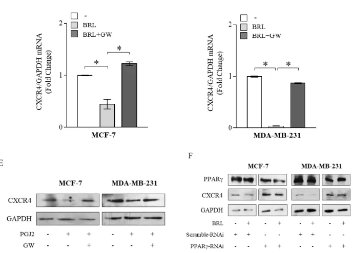
Previous evidences have indicated that tumor cells express distinct, tumor type-specific, nonrandom patterns of chemokine receptors and that signaling through these receptors is crucial for chemotactic migration, invasion and cancer metastasis (Scotton C.J. et al., 2001; Balkwill F., 2004). CXCR4 is one of the most common chemokine receptors that has been demonstrated to be over expressed in human cancers, while its expression is low or absent in many normal tissues, including breast (Yagi H. et al., 2011), emphasizing a critical role for this chemokine receptor in modulating cancer cell behavior. Thus, we first aimed to evaluate protein and mRNA expression levels of CXCR4 in non-tumorigenic breast epithelial cells, MCF- 10A, and in two different human breast cancer cell lines by immunoblotting and qRT-PCR analyses. As shown in Figure 1A, CXCR4 expression was detected at very low levels in MCF-10A cells in respect with ERα-positive MCF-7 breast cancer cells, while higher CXCR4 levels were observed in ER-negative MDA-MB-231 breast cancer cells, which are well-characterized in terms of their metastatic potential and properties (Zhang R.D. et al., 1991). Rosiglitazone (BRL), a PPARγ agonist used in type 2 diabetes treatment, has been shown to inhibit CXCR4 expression and to reduce the malignancy in colon, lung and prostate cancer cells (Richard C.L. et al., 2007; Tai C.J. et al., 2010; Qin L. et al., 2014). Therefore, we evaluated PPARγ expression in MCF-7 and MDA-MB-231 breast cancer cells (Figure 1B) and assessed the effects of BRL on CXCR4 expression at both protein and mRNA levels in both cell lines. We found that BRL at 10 μM significantly reduced CXCR4 expression as evaluated by immunoblotting as well as immunofluorescence (Figure 1C) and qRT-PCR (Figure 1D) analyses in both cells. Treatment with the natural PPARγ ligand 15-Deoxy-delta12,14-prostaglandin J2 (PGJ2) at 10 μM also significantly reduced CXCR4 expression in MCF-7 and MDA-MB-231 cells (Figure 1E). To investigate the direct involvement of PPARγ in the downregulation of CXCR4 induced by BRL, cells were treated with the PPARγ antagonist, GW9662 (GW). We found that the reduction of CXCR4 levels induced by PPARγ ligands was completely abrogated in the presence of GW treatment (Figure 1C, 1D, 1E), addressing that these effects on CXCR4 expression were mediated by PPARγ. Using siRNA technology, we
confirmed the specific role of PPARγ in regulating CXCR4 expression in both cell lines (Figure 1F).
Figure 1: Ligand-activated PPARγ downregulates CXCR4 expression in breast cancer cells. (A) Immunoblots
(upper panel) and real-time RT-PCR (lower panel) of CXCR4 expression in MCF-10A non tumorigenic breast epithelial cells, MCF-7 and MDA-MB-231 breast cancer cells. GAPDH was used as loading control. Each sample was normalized on its GAPDH mRNA content. The results are expressed as fold change compared to breast epithelial cells. (B) Immunoblots (upper panel) and real-time RT-PCR (lower panel) of PPARγ expression in MCF-7 and MDA-MB-231 breast cancer cells. GAPDH was used as loading control. Each sample was normalized on its GAPDH mRNA content. The results are expressed as fold change compared to MCF7 cells. (C) Immunoblots (upper panels) and immunofluorescence (middle panels) of CXCR4 protein expression in MCF-7 and MDA-MB-231 cells treated with vehicle (−), BRL 10 μM with or without GW 10 μM for 24 h. GAPDH was used as loading control. Numbers below the blots represent the average fold change between CXCR4 and GAPDH protein expression vs vehicle-treated cells. 4,6-Diamidino-2-phenylindole (DAPI) was used for the determination of the nuclei. Small squares, negative controls. Scale bar, 10 μm. (D) Real-time RT-PCR of CXCR4 expression in MCF-7 and MDA-MB-231 cells treated with vehicle (−), BRL 10 μM with or without GW 10 μM for 12 h. Each sample was normalized on its GAPDH mRNA content. (E) Immunoblots of CXCR4 protein expression in MCF-7 and MDA-MB-231 cells treated with vehicle (-), PGJ2 at 10 µM with or without GW 10 μM for 24h. GAPDH was used as loading control. (F) Immunoblots of CXCR4 protein expression in MCF-7 and MDA-MB-231 cells transfected with scramble RNA interference (RNAi) or with PPAR RNAi as reported in Materials and Methods Section and treated with vehicle (-) or with BRL 10 µM for 24h. GAPDH was used as loading control. The results are expressed as fold change compared to vehicle-treated cells. The values represent the mean ± SD of three different experiments, each performed with triplicate samples. *P < 0.05. GAPDH, glyceraldehyde-3-phosphate dehydrogenase.
Identification of a functional PPAR responsive element (PPRE) within the
CXCR4 promoter
The results obtained prompted us to determine whether the human CXCR4 gene may be a target of ligand-activated PPARγ. To this aim, transient transfection experiments were
performed in MCF-7 cells using a luciferase reporter plasmid containing the human CXCR4 promoter region spanning from –2237 bp to +62 bp relative to the start of the transcription, named p-2300 (Figure 2A). BRL administration induced a significant reduction of CXCR4 promoter activity, which was reversed by the addition of GW, indicating that it was mediated by PPARγ activation (Figure 2A). The CXCR4 promoter region presents multiple transcription factor binding motifs, including c/EBP, Oct-1, NFkB and Sp1 that may represent potential PPARγ binding sequences (Bruemmer D. et al., 2003; Bonofiglio D. et al., 2006; Bonofiglio D. et al., 2008; Siersbaek R. et al., 2010;). To evaluate which elements in the CXCR4 promoter can mediate the above described effects, CXCR4 promoter deleted constructs were tested in transient transfection experiments (Schematically reported in Figure 2A). By using p-2144 (−2144/+62) construct, the reduced luciferase activity upon BRL treatment was still present, whereas when we used the construct p-1507 (−1507/+62) the downregulatory effects were no longer noticeable (Figure 2A). This addresses that the region between −2144 and −1507 bp is required for the BRL-induced repression of CXCR4 promoter and may contain putative PPARγ responsive region(s).
Figure 2: PPARγ modulates the transcriptional activity of CXCR4 gene promoter containing a putative PPAR response element (PPRE). (A) Schematic representation of the CXCR4 promoter constructs used in this study (left
panel). MCF-7 cells were transiently transfected with luciferase plasmids containing the CXCR4 promoter (p-2300) and
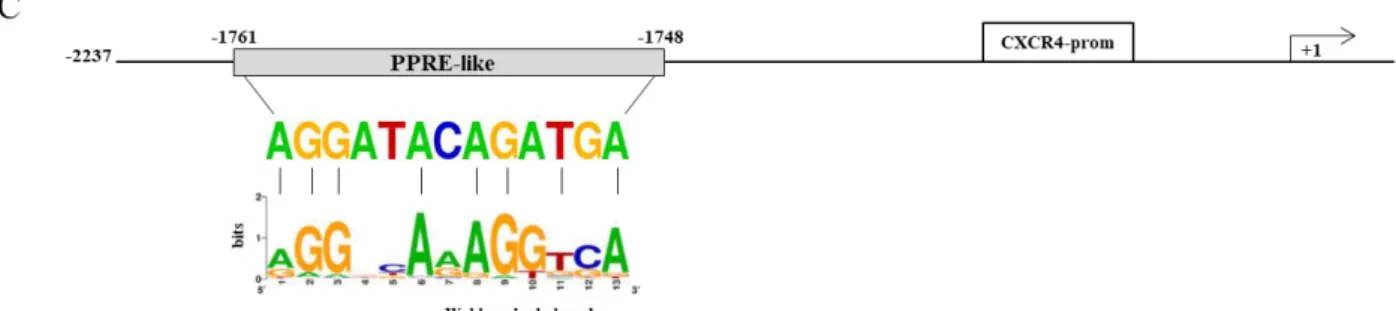
its deleted constructs (p-2144 and p-1507) and then treated with vehicle (−), BRL 10 μM with or without GW 10 μM for 12 h (right panel). The results are expressed as fold change respect to the vehicle-treated cells (−). The results are mean ± SD of three different experiments, each performed with triplicate samples. *P < 0.05. n.s. = not significant. (B) Chromosomal localization of the human cxcr4 gene at chromosome 2 (left panel). A shot from NCBI genome browser to illustrate the localization of cxcr4 gene. The location of Peroxisome proliferator response element (PPRE)-like is highlighted by vertical line and zoomed-in to view the genomic sequence spanning from 136119907 to 136119895 base pair in the negative strand (right panel) (C) The genomic sequence of the PPRE-like motif within CXCR4 promoter is aligned to a logo graphic representation of PPRE sequence generated using a PPRE collection with WebLogo (Lemay D.G. and Hwang D.H. et al., 2006).
Our subsequent studies were directed to identify the putative sequence responsive to PPARγ within the promoter region of the CXCR4 gene. Interestingly, nucleotide sequence analysis revealed that CXCR4 promoter contains the sequence AGGATAcAGATGA located at position -1761 upstream of the translation initiation codon, spanning from 136119895 bp to 136119907 bp on chromosome 2 (Figure 2B), that displays a high sequence homology with the canonical PPAR response elements (PPRE). We then compared our putative PPRE sequence with a consensus one generated using a PPRE collection from the literature (Lemay D.G. and Hwang D.H. et al., 2006) and visualized as a ‘sequence logo’. As shown in Figure 2C, we observed that the two motif profiles exhibited many similarities, particularly in the first hexad sequence bound to PPARγ, the nucleotides AGG located at position 1–3 as well as the nucleotide A located at position 6 are present in the putative PPRE sequence, suggesting the existence, within the CXCR4 promoter, of a novel PPRE-like region. To further investigate the functional importance of the identified PPRE sequence, we tested the hypothesis that PPARγ could effectively bind to it. To this aim, DNA affinity precipitation assay (DAPA) was performed in MCF-7 cells by using a biotinylated-double-stranded oligonucleotide containing the putative PPRE sequence (Figure 3A). Endogenous PPARγ was found to be associated with the putative consensus oligonucleotide following BRL treatment. Co-treatment with GW markedly decreased the BRL-induced DNA-binding complex demonstrating the direct involvement of PPARγ. A
mutant oligonucleotide abolished PPARγ binding, indicating that the in vitro DNA-PPARγ binding is sequence-specific. Next, to assess whether the endogenous PPARγ, after BRL treatment, localizes to the native CXCR4-promoter, chromatin immunoprecipitation (ChIP) assay was performed by using primers flanking the PPRE sequence present in the CXCR4 promoter region. PPARγ occupancy of this region was significantly enhanced upon BRL treatment. This event was concomitant with the inhibition of RNA POL II recruitment onto the CXCR4 promoter (Figure 3B). Transcriptional control by PPARγ requires interaction with co-regulator complexes, either a coactivator for stimulation or a corepressor for inhibition of target gene expression (Glass C.K. et al., 2000; Cohen R.N., 2006; Ricote M. et al., 2007). To determine if the negative regulation of the CXCR4 transcriptional activity induced by BRL might be caused by the cooperative interaction between PPARγ and negative transcriptional regulators, we investigated the involvement of N-CoR and SMRT, which interact with and function as negative coregulators of PPARγ. Re-ChIP assay demonstrated a significant increase of PPARγ/SMRT complex occupancy of the PPRE containing region of CXCR4 promoter after BRL exposure. No interaction of N-CoR was observed under the same experimental conditions (Figure 3C). Finally, to better define the role of SMRT in the PPARγ-dependent modulation of the CXCR4 levels, RNA silencing technologies were used to knockdown the expression of endogenous SMRT in MCF-7 cells. SMRT expression was effectively silenced as revealed by real-time PCR analysis after 48 h of siRNA transfection (Figure 3D). As expected, silencing of SMRT completely abrogated the down-regulation of CXCR4 mRNA levels induced by the activated PPARγ (Figure 3D), highlighting a crucial role of SMRT corepressor in regulating CXCR4 expression upon BRL treatment. All these BRL-induced effects were reversed in presence of combined treatment with GW (Figure 3B–3D). Overall, these findings clearly demonstrated that ligand-activated PPARγ by binding to a newly identified PPRE motif within the CXCR4 promoter downregulates CXCR4 expression levels in human breast cancer cells.
Figure 3: Ligand-activated PPARγ binds to a PPRE-like site within CXCR4 promoter. (A) DAPA on nuclear
extracts from MCF-7 cells treated with vehicle (−), BRL 10 μM with or without GW 10 μM for 3 h. PPRE-like (CXCR4-PPRE, 5ʹ-[Bio]- TTATAAAGGATACAGATGAAGAGATACG-3ʹ) or mutated (Mut-PPRE, CXCR4-mutated PPRE, 5ʹ-[Bio]-TTATAACTTATACAGACTCAGAGATACG-3ʹ) biotinylated oligonucleotides were used. Nuclear Extracts, positive control. (B) Schematic representation (upper panel) of PPRE-like site in CXCR4 promoter region. Chromatin Immunoprecipitation (ChIP) assay (lower panel) with anti-PPARγ and anti-POL II antibodies in MCF-7 cells treated with vehicle (−), BRL 10 μM with or without GW 10 μM for 1 h. (C) ChIP with the anti-PPARγ antibody was re-immunoprecipitated (Re-ChIP) with the anti-SMRT or anti-NCOR antibodies. The CXCR4 promoter sequence including the putative PPRE site was detected by Real-time-PCR with specific primers (see Material and Method section). (D) mRNA levels of SMRT (upper panel) and CXCR4 (lower panel) evaluated by Real-time RT-PCR in MCF-7 cells transfected with control RNAi (Scramble RNAi) or SMRT RNAi for 24 h and then treated with vehicle (−), BRL 10 μM with or without GW 10 μM for 24 h has indicated. Each sample was normalized on its GAPDH mRNA
content. The results are expressed as fold change respect to the vehicle-treated cells. The values represent the mean ± SD of three different experiments, each performed with triplicate samples. *P < 0.05. n.s. = not significant.
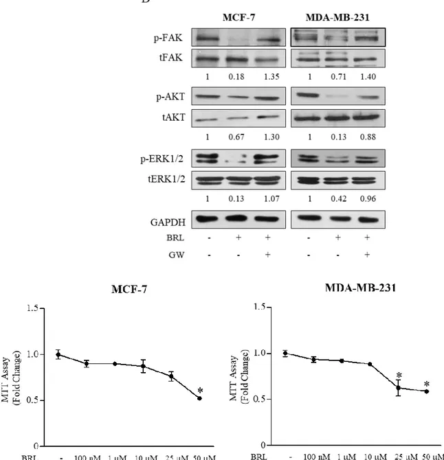
BRL inhibits motility in breast cancer cells
Given the largely documented role of SDF-1α/ CXCR4 axis in modulating cancer cell migration (Taichman R.S. et al., 2002; Burger M. et al., 2003; Fernandis A.Z. et al.,2004), we next assessed the ability of PPARγ agonist to influence cell migration and invasion of both breast cancer cells. First, ELISA measurement in breast cancer cell media showed that SDF-1α levels were 171,6 ± 24,5 pg/mL and 143,35 ± 52,9 pg/mL in MCF7 and MDA-MB-231 cell-derived conditioned media (CM), respectively. Thus, we tested the capacity of cells to migrate in wound-healing scratch assays as well as to across uncoated membrane in transmigration assays and to invade an artificial basement membrane Matrigel in invasion assays upon treatment with BRL at 10 μM of concentration for 24 h (Figure4A–4C). Our data clearly showed that BRL treatment significantly reduced motility and invasion in MCF7 and MDA-MB-231 cells, interfering with the autocrine effects of SDF-1α/CXCR4 system in these cells. These effects were abrogated when cells were exposed to GW co-treatment (Figure 4A–4C). Moreover, we observed, as expected, that ligand-activated PPARγ reduced breast cancer cell migration induced by SDF-1α (data not shown). We also tested the effects of ligand-activated PPARγ on CXCR4 downstream signaling pathways and we found decreased levels of phosphorylated FAK, AKT and ERK1/2 upon BRL treatment which was reversed in presence of GW, confirming that BRL reduces the CXCR4 signaling in a PPARγ-dependent manner in both breast cancer cell lines (Figure 4D). Moreover, we ascertained that the inhibited migratory capability mediated by BRL was not due to a decrease in cell viability, since when MCF-7 and MDA-MB-231 cells were incubated with 10 μM BRL for 24 h ~90% of breast cancer cells were still viable (Figure 4E).
Figure 4: Effects of BRL on motility and invasion of MCF-7 and MDA-MB-231 breast cancer cells.
Wound-healing (A), transmigration (B) and invasion (C) assays in breast cancer cells treated with vehicle (−), BRL 10 μM with or without GW 10 μM for 24 h. Small squares: time 0. Histograms in A represent the mean ± SD of three separate experiments in which migrated cells were calculated by image analysis using Image J software and expressed as fold change compared to vehicle-treated cells. Migration and invasion were quantified by viewing five-separate fields/membrane (10×-magnification) and expressed as mean numbers of migrated cells. Data represent the mean ± SD of three-independent experiments, assayed in triplicate. *P < 0.05. (D) Immunoblots of phosphorylated levels (p) of FAK, AKT and ERK1/2 and total proteins from cells treated with vehicle (−), BRL 10 μM with or without GW 10 μM for 24 h. Numbers below the blots represent the average fold change between phosphorylated and total protein and GAPDH protein expression vs vehicle-treated cells. GAPDH, glyceraldehyde-3-phosphate dehydrogenase. (E) Cell viability was determined by 3-(4,5-dimethylthiazol-2-yl)-2,5-diphenyltetrazolium (MTT) assays in MCF-7 and MDA-MB-231 breast cancer cells treated with vehicle (-) or with increasing concentrations (100 nM, 1, 10, 25, 50 μM) of BRL for 24h. The results are expressed as fold change respect to vehicle-treated cells. The values represent the mean ± SD of three different experiments, each performed with triplicate samples. *P<0.05 vs vehicle-treated cells.
Ligand-activated PPARγ counteracts stroma-mediated breast cancer cell
migration
There is increasing evidence that breast cancer behavior reflects an interconnection between the malignant epithelial compartment and the surrounding microenvironment. Cancer Associated Fibroblasts (CAFs) represent the most abundant stromal cell type populating the tumor microenvironment and play a pivotal role in the development and progression of breast cancer via production of hormones, extracellular matrix remodeling enzymes and cytokines such as SDF-1α (Cabioglu N. et al., 2007). To investigate the role of activated PPARγ in the context of heterotypic signaling working in tumor-stroma interactions, we examined the ability of BRL to reduce CAF-induced effects through CXCR4 axis inhibition in breast cancer cells. To this aim, two different types of CAFs, named CAF #1 and CAF #2, isolated from biopsies of primary breast tumors, were used in co-culture systems. First, MCF-7 and MDA-MB-231 cells were pretreated with BRL 10 μM for 24 h and then incubated with CAF-derived CM to assess stromal SDF-1α ligand binding to breast cancer cells. In line with BRL-induced CXCR4 downregulation, we observed a significantly decreased SDF-1α/CXCR4 binding in cells pretreated with BRL compared to vehicle-treated cells (Figure 5A). Accordingly, treatment with BRL attenuated migration-promoting activities of CM from CAF #1 and CAF #2 (Figure 5B and 5C). SDF-1α was then immunodepleted from CAF-derived CM by a specific antibody, and resulting media were tested in cells treated with BRL for the ability to reduce migration of breast cancer cells. As expected, SDF-1α-depletion (CAF-CM + SDF-1α-Ab) significantly reduced the migratory effects of CAF-CM, particularly in the presence of BRL treatment (Figure 5D). CM treated with a nonspecificrabbit IgG had no effects, suggesting the specificity of SDF-1α antibody. In addition, as shown in Figure 5E, BRL in combination with the CXCR4 antagonist FIL2, a newly benzohydrazide compound synthesized in our laboratory (Grande F et al., 2016), strongly decreased cell motility induced by CAF-CM. Moreover, we demonstrated that BRL was also able to counteract the increased activation of FAK, AKT and MAPK signaling pathways induced by CM from CAFs in both breast cancer cells (Figure 5F). The PPARγ antagonist GW abolished the effects of BRL on migratory promoting activities induced by CAF-CM (Figure 5B, 5C and 5F).
Figure 5: BRL antagonizes motility and signaling activation induced by cancer-associated fibroblasts -derived conditioned media in breast cancer cells. (A) CAF-secreted SDF-1α ligand binding to breast cancer cells was
analyzed by ELISA at 450 nm of absorbance (Abs) as described in Material and Methods. The results are expressed as percentage of optical density (OD) respect to vehicle-treated cells. The values represent the mean ± SD of three different experiments, each performed with triplicate samples. (B and C) Wound-healing assays in MCF-7 and in MDA-MB-231 cells treated with phenol-red and serum-free medium (−), conditioned media derived from cancer-associated fibroblasts (CAF-CM), BRL 10 μM with or without GW 10 μM for 24 h. Small squares, time 0. Histograms represent the mean ± SD of three separate experiments in which migrated cells were calculated by image analysis using
Image J software and expressed as fold change compared to vehicle-treated cells. *P < 0.05. (D) Wound-healing assays in MCF-7 and in MDA-MB-231 cells treated with phenol-red and serum-free medium (−), CAF #1-CM and/or SDF-1α-depleted conditioned media (SDF-1α-Ab) with or without BRL 10 μM for 24 h. Conditioned media treated with a nonspecific IgG as a control (IgG-Ab). Small squares, time 0. Histograms represent the mean ± SD of three separate experiments in which migrated cells were calculated by image analysis using Image J software and expressed as fold change compared to (−) treated cells. *P < 0.05. (E) Wound-healing assays in MCF-7 and in MDA-MB-231 cells treated with phenol-red and serum-free medium (−), CAF #2-CM, FIL2 1 μM with or without BRL 10 μM for 24 h. Small squares, time 0. Histograms represent the mean ± SD of three separate experiments in which migrated cells were calculated by image analysis using Image J software and expressed as fold change compared to (−) treated cells. *P < 0.05. (F) Immunoblots of phosphorylated (p) FAK, AKT and ERK1/2 and total proteins from cells treated as in C. Numbers below the blots represent the average fold change between phosphorylated, total and GAPDH protein expression vs vehicle-treated cells. GAPDH, glyceraldehyde-3-phosphate dehydrogenase. CAFs: Cancer-associated fibroblasts; CM: Conditioned media.
BRL affects phenotypic characteristics of CAFs
As a final step of this study, we wondered whether PPARγ ligands by influencing CXCR4 expression may also impact biological features of CAFs. As previously reported (Knower K.C. et al., 2013), we found that CAFs showed a detectable mRNA and protein levels of PPARγ which was significantly increased upon 10 μM BRL exposure and reversed by GW co-treatment (Figure 6A). In addition, we observed that exposure to BRL reduced, in a PPARγ- dependent manner, CXCR4 expression evaluated at both mRNA and protein levels (Figure 6B). As a consequence, BRL treatment reduced CAF motility assessed by wound healing and trans-migration assays (Figure 6C and 6D). The ability of GW to completely abrogate this effect addressed a direct involvement of PPARγ. It was observed that incubation with 10 μM BRL for 24 h did not affect cell viability of CAFs (Figure 6E), while interestingly BRL elicited a dramatic alteration in the shape of CAFs in vitro (data not shown), accompanied by a reduced expression of α-SMA and vimentin in both types of CAFs (Figure 6F). Taken together our results indicate that CAFs exposed to BRL acquired a phenotype characterized by an altered morphology, a decreased expression of CXCR4 and inhibited migratory capabilities, all features that may negatively impact breast tumor progression.