Department of Pharmacy, Health and Nutritional Sciences
Ph.D. program in Translational MedicineXXX Cycle CHIM/09
Advanced Materials for Pharmaceutical
and Biomedical Purposes
Coordinator:
Prof. Sebastiano ANDÒ
Supervisor: Candidate:
Dott.ssa Roberta CASSANO Dott.ssa Silvia MELLACE
Author’s e-mail: [email protected]
Author’s address: Department of Pharmacy, Health and Nutritional Sciences University of Calabria Edificio Polifunzionale 87036 Arcavacata di Rende, Cosenza, Italy.
ph.: +39 0984 493227 ph.: +39 0961 915533 cell.: +39 3299320290
The one who follows the crowd will usually go no further than the crowd. Those
who walk alone are likely to find themselves in places no one has
ever been before.
(Albert Einstein)
I
Table of contents
Preface: General Introduction, Aims and Organization of the Thesis ... 9
1. General Introduction ... 9
2. Aims of the thesis ... 10
3. Organization of the Thesis ... 11
References ... 13
Italian abstract ... 14
SECTION 1: HYDROGELS AND MICROSPHERES FOR
ACTIVE MOLECULES RELEASE
16 PART A: NEW GALLIC ACID BASED HYDROGEL FOR PHLORETIN INTESTINAL RELEASE ... 17Abstract ... 17
1. Introduction ... 18
2. Materials and Methods ... 20
2.1 Chemicals ... 20
2.2 Instruments ... 20
2.3 Acrylation of 3,4,5-trihydroxybenzoic acid with 2-propenoic Acid ... 21
2.4 Hydrogel based on diacrylate gallic acid preparation ... 21
2.5 Phloretin incorporation into the performed hydrogel ... 21
2.6 Antioxidant activity evaluation ... 22
2.7 Hydrogel swelling studies ... 22
2.8 Drug release studies ... .23
2.9 Statistical analysis ... 23
3. Results and Discussions ... 24
3.1 Acrylation of 3,4,5-trihydroxybenzoic acid with 2-propenoic Acid ... 24
3.2 Preparation of the hydrogel based on gallic acid diacrylate ... 25
3.3 Hydrogel impregnation with phloretin ... 25
3.4 Antioxidant activity evaluation ... 25
II
3.6 Hydrogel release studies ... 27
4. Conclusions ... 27
References ... 29
PART B: NOVEL MICROSPHERES BASED ON TRITERPENE SAPONINS FROM THE ROOTS OF PHYSOSPERMUM VERTICILLATUM (WALDST & KIT) (APIACEAE) FOR THE IMPROVEMENT OF GEMCITABINE RELEASE ... 32
Abstract ... 32
1. Introduction ... 33
2. Materials and Methods ... 35
2.1 Chemicals ... 35
2.2 Plant materials ... 36
2.3 Extraction procedure ... 36
2.4 Measurements ... 36
2.5 Esterification of triterpene saponins with acrylic acid ... 37
2.6 Preparation of microspheres based on triterpene saponins ... 37
2.7 Microspheres characterization ... 38
2.7.1 Dimensional analysis ... 38
2.7.2 Swelling studies ... 39
2.8 Impregnation of the microspheres with gemcitabine ... 39
2.9 In vitro release studies of gemcitabine from microparticles ... 40
2.10 Statistical analysis ... 40
3. Results and discussion ... 40
3.1 Esterification of triterpene saponins with acrylic acid ... 41
3.2 Preparation of microspheres ... 41
3.3 Characterization of microspheres ... 43
3.4 Studies of swelling ... 43
3.5 Impregnation of the microspheres with gemcitabine ... 44
3.6 In vitro gemcitabine release studies from hydrogel ... 44
4. Conclusions ... 45
III
PART C: LIQUID CRYSTALLINE MICROSPHERES FOR
5-FLUOROURACIL SPECIFIC RELEASE ... 51
Abstract ... 51
1. Introduction ... 52
2. Materials and Methods ... 54
2.1 Chemicals ... 54
2.2 Measurements ... 55
2.3 Fenoprofen preparation ... 55
2.4 Esterification of poloxamer with fenoprofen ... 56
2.5 Derivatization of the hydroxyl group of 1 with acryloyl chloride 56 2.6 Microspheres preparation ... 56
2.7 Swelling studies ... 57
2.8 Microspheres preparation with 5-FU ... 58
2.9 Release studies ... 58
2.10 Statistical analysis ... 58
3. Results and Discussion ... 59
3.1 Synthesis of acrylate 2 ... 59
3.2 Microspheres characterization ... 60
3.3 Swelling studies ... 63
3.4 Microspheres loading with 5-FU ... 63
3.5 Release studies ... 64
4. Conclusions ... 66
References ... 67
SECTION 2: CHEMICAL MODIFICATION OF FATTY
ACIDS
FOR
THE
IMPLEMENTATION
OF
DRUG
DELIVERY SYSTEMS
... 72PART A:
α
-TOCOPHERYL LINOLENATE SOLID LIPID NANOPARTICLES FOR THE ENCAPSULATION, PROTECTION, AND RELEASE OF OMEGA-3 POLYUNSATURATED FATTY ACID: IN VITRO ANTI-MELANOMA ACTIVITY EVALUATION ... 73IV
1. Introduction ... 74
2. Materials and Methods ... 76
2.1 Chemicals ... 76
2.2 Measurements ... 76
2.3 Synthesis of cis, cis, cis-9,12,15-octadecatrienoate of (2R) -2,5,7,8-tetramethyl-2- (4R, 8R) - (4,8,12-trimetiltridecil) -6-chromanol (TL) ... 77
2.4 Preparation of Solid Lipid Nanoparticles (SLNs) ... 77
2.5 Encapsulation efficiency determination ... 78
2.6 SLNs antioxidant activity evaluation ... 79
2.7 Cell effect – MTT reduction assay ... 79
2.8 Statistical analysis ... 80
3. Results and Discussion ... 80
3.1. Synthesis of cis, cis, cis-9,12,15-octadecatrienoato of (2R)-2,5,7,8-tetramethyl-2-(4R,8R)-(4,8,12-trimetiltridecil)-6-chromanol (TL) ... 80
3.2. Preparation and characterization of SLNs ... 81
3.3. Evaluation of SLNs antioxidant activity ... 83
3.3. Evaluation of cytotoxic activity ... 84
4.Conclusions ... 86
References ... 87
PART B: SOLID LIPID NANOPARTICLES FOR CYCLOSPORIN A TOPIC RELEASE ... 90
Abstract ... 90
1. Introduction ... 91
2. Materials and Methods ... 92
2.1 Chemicals ... 92
2.2 Instruments ... 92
2.3 Esterification of the oleic acid with trehalose ... 93
2.4 Preparation of Solid Lipid Nanoparticles (SLNs) ... 93
2.5 SLNs Characterisation ... 94
2.5.1. Particle Size ... 94
2.5.2 Entrapment Efficiency determination ... 94
V
2.6. Skin Permeation Experiments ... 95
2.6.1. In Vitro Skin Permeation Studies ...95
2.6.2. Quantification of Drug in Skin Using Tape Stripping ...95
2.6.3. Localization of Nanoparticles in Skin (CLSM study) ... 96
2.7. Statistical analysis ...96
3. Results and Discussion ...97
3.1. Esterification of trehalose with oleic acid ...97
3.2. Preparation and characterization of SLNs ...98
3.2.1. Size, Entrapment Efficiency and Size Distribution ...98
3.2.2. Differential Scanning Calorimetry (DSC) Analysis ...99
3.3. Skin Permeation Experiments ...100
3.3.1 In Vitro Permeation Studies ...100
3.3.2 Tape Stripping Test ...101
3.2.2. CLSM Studies ...102
4. Conclusions ...103
References ...104
SECTION 3: NATURAL AND SYNTHETIC POLYMERS
FOR THE PREPARATION OF BIOMEDICAL MATERIALS
...108PART A: HEMOSTATIC GAUZE BASED ON CHITOSAN AND HYDROQUINONE: PREPARATION, CHARACTERIZATION AND BLOOD COAGULATION EVALUATION ...109
Abstract ...109
1. Introduction ...110
2. Materials and Methods ...111
2.1 Chemicals ...111
2.2 Instruments ...112
2.3 Derivatization of chitosan with hydroquinone ...112
2.4 Test for the determination of total polyphenols ...112
2.5 Gauze carboxylation ...113
2.6 Determination of carboxylic groups content ...113
2.7 Derivatization of carboxylated gauze with chitosan-hydroquinone by Mitsunobu reaction ...114
VI
2.9 Release studies ... 115
2.10 In vitro whole blood clotting test ... 115
2.11 Swelling test ... 116 2.12 Statistical analysis ... 117 3. Results ... 117 4. Discussions ... 123 5. Conclusions ... 125 References ... 126
PART B: APPLICATION OF IN VITRO AND IN SILICO METHODS FOR THE ACCURATE AND EFFICIENT PREDICTION OF HUMAN PHARMACOKINETICS FOLLOWING TRANSDERMAL ADMINISTRATION ... 130
Abstract ... 130
1. Introduction ... 131
2. Materials and Methods ... 133
2.1 Chemicals ... 133
2.2 Fabrication of Dissolvable Microneedle Arrays ... 133
2.3 Characterization of Dissolvable Microneedle Arrays ... 134
2.3.1 DMN Morphological Properties ... 134
2.3.2 DMN Mechanical Performance ... 134
2.3.3 Drug Loading Amount ... 135
2.3.4 DMN Penetration Study ... 135
2.3.5 Short-term Stability Studies ... 135
2.4. Drug Release Study ... 136
2.5 Skin Permeation Studies ... 136
2.6 Drug Analyses ... 137
2.7 In Silico Modelling ... 138
2.7.1 SKIN-CAD® Model Strategy for in vitro/ in vivo correlation ... 138
2.7.2 Simcyp® Model Strategy for in vitro observed /predicted parameters ... 141
2.8 Statistical analysis ... 141
3. Results ... 141
VII
3.2 Characterization of Dissolvable Microneedle Arrays ...142
3.2.1. DMN Morphological Properties ...142
3.2.2. DMN Mechanical Performance ...143
3.2.3. Drug Loading Amount ...145
3.2.4. Skin Penetration Study ...145
3.2.5. Short-term Stability Studies ...146
3.3 Drug Release Studies ...148
3.4 Skin Permeation Studies ...150
3.5 Drug Analyses ...152
3.6 In Silico Models ...152
3.6.1 SKIN-CAD® for in vitro/ in vivo correlation ...152
3.6.2 Simcyp® for in vitro observed /predicted parameters ...155
4. Discussion ...156
5. Conclusions ...158
References ...159
9
Preface
General Introduction, Aims and Organization of this Thesis
1. General Introduction
The XX century has witnessed more than doubling in global life expectancy thanks to the historical evolution of clinical medicine, therapeutics and pharmacy. In this context, design and synthesis of advanced materials may influence the future of drug delivery systems, that are of vital importance for healthcare and medicine. Materials innovation and nanotechnology have synergistically helped the advancement of drug delivery. Novelty in material chemistry allows the generation of biodegradable, biocompatible, environment-responsive, and targeted delivery systems (1). Nanotechnology permits control over size, shape and multi-functionality of particulate drug delivery systems. Since the first FDA approval of drug delivery systems (DDS), several DDS are commercially available to treat varied diseases from cancer to fungal infection and to muscular degeneration (2). In improving therapeutic efficacy, DDS has benefited tens of millions of patients by relieving suffering and prolonging life (3). Innovations in materials chemistry have initially fuelled the development of DDS, creating carriers that are several implementations from numerous point of view, these carriers may protect a drug from degradation, enhance drug absorption, modify drug tissue distribution profile, and/or improve intracellular penetration and distribution. The comprehension that size and shape of nanoparticles (NPs) can help pilot biological carriers has stimulated the application of nanofabrication technologies to develop more effective particulate DDS. For example, the size of NPs determines their biodistribution. While particles smaller than 20nm will be cleared from circulation via reticuloendothelial system (RES) within a few hours when injected intravenously, larger ones will be trapped in the liver and the spleen within minutes (4, 5). Moreover, innovations in design and fabrication of materials could help to alter and optimize the pharmacokinetics of therapeutic agents (6). Since the size of polymer-drug conjugates could be controlled by adjusting the molecular weight of the polymer, it could be optimized to maximize the benefits of enhanced permeability effect at leaky tumour vasculature (7). Furthermore,
10
conjugating drugs to a polymeric carrier can enhance solubility of hydrophobic drugs, extend drug circulation in vivo and enhance uptake by addition of targeting motifs to the polymer (8). They possess low toxicity and potentially favourable pharmacokinetics in the circulation. For these reasons among numerous biomaterials based on inorganic or organic matter, polymers are considered to be widely used in formulation of drug delivery systems for controlled and targeted drug delivery. In particular, the use of natural polymers such as polysaccharides (9) as drug carriers has a long history of significant clinical benefits (10). DDS were also found useful to improve the performance of imaging techniques applied for the in vivo diagnosis of tumours and other diseases (11). Additionally, the applications of nanotechnology in the food sector to improve the nutritional value, shelf-life and traceability of food products has the potential to subvert agriculture and food systems. In fact, the nanoscale level of foods can affect the safety, efficiency, bioavailability, and nutritional value properties of food, as well as the molecular synthesis of new products and ingredients. The development of new functional materials in food production are only new emergent, but it is already predicted to grow rapidly in the coming years (12). In summary, intensive efforts for the last three decades enabled the development of functional devices, most of which belong to lipids, peptides and other polymers. There is great potential in the combination of bioactive macromolecules with polymers, either covalently through conjugation or non-covalently using hydrogels or higher-order self-assembled structures, to address challenging in biomedical problems. Currently, the areas of drug delivery and nanomedicine is rapidly expanding. A prerequisite for further development is the design and synthesis of novel materials that are biocompatible and biologically active, are biodegradable with a controlled degradation rate, and have tenable mechanical properties. In conclusion, novel materials synthesis, introduction of environmental-sensitive polymers, successful adoption of natural polymers as carrier and improved understanding of the structure-function relationship have together transformed DDS development.
11 2. Aims of this Thesis
The present thesis was realized in the Pharmaceutical Technology group, Department of Pharmacy, Health and Nutritionals Sciences (University of Calabria) and deals about the development of advanced materials for biomedical and pharmaceutical purposes. Moreover, part of the PhD was focused on the application of in vitro and in silico methods for the effective and efficient prediction of human pharmacokinetics following transdermal administration of dissolvable microneedles array. This last work was carried out during a stimulating and productive scientific visiting period at the School of Pharmacy of the UCC (University College of Cork, Ireland), under the supervision of Dr. Sonja Vucen.
3. Organization of this thesis
Due to the multitude of independent projects, the present thesis is divided into three self-contained sections. The first section, named “Microspheres and Hydrogels for Active Molecules Release”, is itself divided into three self-explained parts. The first part of this section concerns the preparation of a gallate hydrogel for oral administration of phloretin, a flavonoid of natural origin, with proved antioxidant activity. The results suggest a possible use of this hydrogel for intestinal release of phloretin for the maintenance of an appropriate blood glucose level in the diabetic patient. The second part of this first section concerns the preparation and characterization of microspheres based on a mixture of three natural triterpene saponins (saikosaponina a, songarosaponina D, buddlejasaponina IV) useful as a carrier for the specific release of gemcitabine to lung cells. The aim of the third part of this first section was the synthesis and characterization of swellable liquid crystalline microspheres for a specific release of 5-fluorouracil to cancer cells. The second section named “Chemical Modification of Fatty Acids for the Implementation of Drug Delivery Systems”, is itself divided into two self-explained parts. Both are aimed to develop modified lipids for the preparation of new solid lipid nanoparticles. Particularly, in the first part α-linolenic acid was successfully derivatized with α-tocopherol, the results indicated that these lipid nanoparticles could provide the delivery and the protection of unstable molecules, such as
α-12
linolenic acid, and could be a good agent in the treatment of melanoma. Cyclosporin A, in the second part of this second section, was efficiently encapsulated in trehalose oleate SLNs. Results indicate the possibility of using the nanoparticles prepared for topical treatment of psoriasis, indeed, the risk of side effects due to systemic absorption of cyclosporin A is reduced whereas the drug concentration at injury level is increased. The third section named “Natural and Synthetic Polymers for the Preparation of Biomedical Materials” is itself divided into two self-explained parts, both addressed to the preparation of innovative materials for topical approach. The first part was aimed at the preparation of a functional gauze linking coagulant substances, chitosan and hydroquinone useful as a topical haemostatic agent for the treatment of bleeding wounds. The gauze showed the ability to short the blood clotting time and to induce the adhesion and activation of platelets. The second part of this third session contains all the experiments performed during my scientific visiting period at UCC School of Pharmacy. The work aimed to fabricate and characterize ketoprofen loaded dissolvable microneedle arrays using in vitro and in silico methods. The presented delivery strategy demonstrates potential for enhanced delivery of ketoprofen. In silico results showed that the simulation was in agreement with the in vitro experimental results. Overall, dissolving microneedles may be useful as a method for patients to self-administer drugs without the pain or hazards of hypodermic needles.
13 References
1. Zhang Y, Chan HF, Leong KW. Advanced materials and processing for drug delivery: the past and the future. Advanced drug delivery reviews. 2013;65(1):104-20.
2. Allen TM, Cullis PR. Drug delivery systems: entering the mainstream. Science. 2004;303(5665):1818-22.
3. Verma RK, Garg S. Drug delivery technologies and future directions. Pharmaceutical Technology. 2001;25(2):1-14.
4. Schipper ML, Iyer G, Koh AL, Cheng Z, Ebenstein Y, Aharoni A, et al. Particle size, surface coating, and PEGylation influence the biodistribution of quantum dots in living mice. Small. 2009;5(1):126-34. 5. Carrstensen H, Mueller RH, Müller B. Particle size, surface hydrophobicity and interaction with serum of parenteral fat emulsions and model drug carriers as parameters related to RES uptake. Clinical nutrition. 1992;11(5):289-97.
6. Jain RA. The manufacturing techniques of various drug loaded biodegradable poly (lactide-co-glycolide)(PLGA) devices. Biomaterials. 2000;21(23):2475-90.
7. Li C, Wallace S. Polymer-drug conjugates: recent development in clinical oncology. Advanced drug delivery reviews. 2008;60(8):886-98. 8. Vasconcelos T, Sarmento B, Costa P. Solid dispersions as strategy to improve oral bioavailability of poor water soluble drugs. Drug discovery today. 2007;12(23):1068-75.
9. Liu Z, Jiao Y, Wang Y, Zhou C, Zhang Z. Polysaccharides-based nanoparticles as drug delivery systems. Advanced drug delivery reviews. 2008;60(15):1650-62.
10. Felt O, Buri P, Gurny R. Chitosan: a unique polysaccharide for drug delivery. Drug development and industrial pharmacy. 1998;24(11):979-93. 11. Sun C, Lee JS, Zhang M. Magnetic nanoparticles in MR imaging and drug delivery. Advanced drug delivery reviews. 2008;60(11):1252-65. 12. Cassano C, Mellace S, Trombino S. Nano- and Micro-Technologies for the Management of Food Ingredients. Book chapter, Ebook Bentham, (Accepted Manuscript).
14
L'innovazione nella chimica dei materiali e nella nanotecnologia hanno sinergicamente contribuito all'avanzamento e allo sviluppo dei Drug Delivery Systems (DDS) creando devices in grado di proteggere farmaci e sostanze biologicamente attive, aumentarne l'assorbimento, modificarne e migliorarne la penetrazione intracellulare e la distribuzione. Il presente lavoro ha avuto come obiettivo la progettazione, la preparazione e la caratterizzazione di materiali che potrebbero contribuire a trasformare e ottimizzare le performances di vari agenti terapeutici. In sintesi, sono stati preparati diversi dispositivi medici e DDS, utilizzando perlopiù polimeri, poiché esiste un grande potenziale nella combinazione di molecole bioattive con i polimeri, per affrontare sfide in campo farmaceutico e biomedico. Nella progettazione di questi nuovi materiali che sono biocompatibili e biologicamente attivi l’attenzione è stata anche focalizzata sull'applicazione di metodi in vitro e in silico per una previsione efficace ed efficiente dell’attività in vivo. In conclusione, la sintesi di nuovi materiali, l'adozione di polimeri naturali come carriers e una migliore comprensione del rapporto struttura-funzione hanno permesso lo sviluppo di DDS e devices con incoraggianti risultati sperimentali che suggeriscono un possibile utilizzo per una futura sperimentazione in ambito clinico.
16
SECTION 1
“HYDROGELS AND MICROSPHERES FOR ACTIVE
MOLECULES RELEASE”
Part A: New gallic acid based hydrogel for phloretin intestinal release.
Part B: Novel microspheres based on triterpene saponins from the roots of Physospermum verticillatum (Waldst & Kit) (Apiaceae) for the
improvement of gemcitabine release.
17 PART A
“New Gallic Acid Based Hydrogel for Phloretin Intestinal
Release” (1)
Abstract
The present work aims to realize a hydrogel based on gallic acid, a molecule with antioxidant and antidiabetic activity. The compound has in its structure two different functional groups, carboxyl and hydroxyl, susceptible to derivatization. Due to these characteristics, it was developed a hydrogel, potentially useful for oral administration of phloretin, a flavonoid of natural origin, found in apples and pears, with glucose transporter (GLUT) inhibitory activity. The obtained gallate hydrogel was characterized by Fourier Transform Infrared spectroscopy (FT-IR). Its equilibrium swelling degree (α %) and the phloretin release were evaluated in simulating gastric and intestinal fluids. The antioxidant activity in inhibiting lipid peroxidation, induced in vitro by a source of free radicals, was also assessed, after exposure of gallate hydrogel containing phloretin to gastrointestinal environmental conditions. The results showed that the gallate hydrogel could be successfully used in pharmaceutical field for phloretin intestinal release and suggest a possible use for the maintenance of an appropriate blood glucose level in the diabetic patient.
18
1. Introduction
Diabetes mellitus is a group of metabolic diseases characterized by a congenital (type I insulin-dependent) or an acquired (type II non-insulin-dependent) inability to transport glucose from the blood to the cells. The chronic hyperglycemia, due to a deficiency of insulin secretion or insulin resistance (2), leads to glucose toxicity and is associated with long-term damage, dysfunction, and failure of various organs, especially the eyes, kidneys, nerves, heart, and blood vessels (3-5). Consequently it is necessary, in particular, in diabetic patient, the maintenance of an appropriate blood glucose level in postprandial state (6). Carbohydrates in the diet are hydrolyzed by digestive enzymes and then cleaved into monosaccharides, these can be absorbed from the small intestine via influx hexose transporters (7, 8). There are two type of transporter: Na+-dependent glucose transporter (SGLT1), located at the brush border membrane (BBM), that mediates the uptake of glucose into the cell against its concentration gradient. The other type of transporter GLUT2, transports sugars across the basolateral membrane to the blood (9). Literature data suggest that GLUT2 can also be found onto the BBM in the presence of glucose in the lumen (10) and can contribute at the glucose absorption process (11). Thus, these glucose transporters might be an attractive therapeutic target for diabetes (12). In this work, was developed a hydrogel for intestinal release of phloretin. A hydrogel is a three-dimensional, water-swollen structure composed of hydrophilic polymers. This network attains physical integrity and is made insoluble due to the presence of chemical and/or physical crosslinks (13-15). In addition, it is offers excellent potential as oral therapeutic systems due to intrinsic biocompatibility, diversity of both natural and synthetic material options and tunable properties. In particular, stimuli-responsive hydrogels exploit physiological changes along the intestinal tract to achieve site-specific and controlled release of protein, peptide and chemotherapeutic molecules for local and systemic treatment applications (16). In this context, was obtained a hydrogel based on gallic acid, a polyphenolic compound commonly distributed in plant-derived foods, such as cereals, legumes, nuts, vegetables, fruits, and in beverages such as green or black tea, wine, fruit juice, beer and etc (17-19). Literature data proved antidiabetic and antioxidant properties of gallic acid (20). So the polyphenolic compound
19
was chosen for hydrogel development, both for its properties and its chemical structure: in fact, it possesses two different functional groups, carboxyl and hydroxyl, susceptible to derivatization and so able to provide polymerizable derivatives (20). The compound entrapped in our gallate hydrogel is the phloretin (Figure 1).
Figure 1: Graphical representation of the gallic acid based hydrogel loaded with phloretin.
This is a dihydrochalcone flavonoid, found in apples and other fruits, that displays also a potent antioxidant activity in peroxynitrite scavenging and the inhibition of lipid peroxidation (21). It is known to inhibit the protein kinase C (22-24) and human leukemia cell growth (25) . Literature data suggest that phloretin decreases the glucose absorption impeding its intestinal transport mediated by GLUT2 (26, 27). On the other hand, the phloretin, as well as other flavonoids, can be easily modified by environmental factors such as temperature, pH and light (28). These compounds, in fact, are poorly adsorbed in the intestine because are, probably, degraded by intestinal microorganism and/or enzymes, producing different metabolites. For these reasons flavonoids, when administered through delivery systems, show an improved stability and absorption profile and, consequently, their activity becomes enhanced, more detectable and prolonged (28). Therefore, our gallate hydrogel has been formulated
20
specifically to facilitate the phloretin intestinal release, protect it from chemical degradation in gastrointestinal tract and enhancing its bioavailability (29). The obtained hydrogel was characterized by Fourier Transform Infrared spectroscopy (FT-IR). Its equilibrium swelling degree (α%) was also evaluated in simulating gastric and intestinal fluids. The phloretin release from hydrogel was carried out in the same condition of swelling studies. The antioxidant activity in inhibiting lipid peroxidation, in rat-liver microsomal membranes, induced in vitro by a source of free radicals, such as tert-butyl hydroperoxide (t-BOOH), was assessed after exposure of gallate hydrogel, containing phloretin, to gastrointestinal environmental conditions.
2. Materials and Methods
2.1 ChemicalsAcetone, hydrochloric acid, chloroform, diethyl ether, ethanol, isopropanol, methanol, n-hexane, tetrahydrofuran (THF), allyl alcohol and sodium sulfate were purchased from Carlo Erba Reagents (Milan, Italy). Gallic acid (MW = 170.12), phloretin (MW = 274.27), acrylic acid (MW = 72.06, d = 1051 g / ml), dicyclohexylcarbodiimide (DCC), N,N-dimethylaminopyridine (DMAP), potassium tert-butoxide, N,N-dimethylacrylamide (DMAA), ammonium persulfate ((NH4)2S2O8), N,N'-methylene-bis-acrylamide, sorbitan trioleate (Span85), polyoxymethylene sorbitan trioleate (Tween 85), N,N,N',N'-tetramethyl-ethylenediamine (TMEDA), tert-butylhydroperoxide (t-BOOH), trichloroacetic acid (TCA) acid, 2-thiobarbituric acid (TBA), butylated hydroxytoluene (BHT) were purchased from Sigma-Aldrich (Sigma Chemical Co, St Louis, MO, USA).
2.2 Instrument
The FT-IR spectra were performed on KBr pellets using a spectrometer Perkin Elmer 1720, in the range 4000-400 cm-1 (16 scans). The 1H-NMR spectra were obtained by the use of a spectrophotometer Burker VM30; the chemical shifts were expressed as δ and referring to the solvent. The UV-VIS spectra have been realized by means of JASCO-530 UV-
21
spectrophotometer. Samples were freeze-dried using a freeze-drying “Micro Modulyo Edwards apparatus”.
2.3 Acrylation of 3,4,5-trihydroxybenzoic acid with 2-propenoic acid Acrylic acid (0.14 ml, 2 ∙ 10-3 mole) was dissolved in dry THF. Then DCC (0.42 g, 2 10-3 mol) and DMAP (0.05 g, 410-4 mol) were added and the reaction mixture was kept under stirring at 50 °C for 1h. After that, gallic acid (1g, 5.810-3 moles) was added to the solution. The reaction was kept under magnetic stirring at 50 °C for other 72 h and monitored by thin layer chromatography (TLC/silica gel, eluent mixture: chloroform-methanol 7:3). The dicyclohexylurea (DCU), formed during the reaction, was eliminated by filtration. The solvent reaction was removed by evaporation at reduced pressure. The obtained product, with a gelatinous consistency and yellow color, was purified through silica gel column chromatography (eluent: chloroform). The purified product has been characterized by FT-IR, and 1 H-NMR.
2.4 Hydrogel based on diacrylate gallic acid preparation
In a two-neck flask fitted with a reflux condenser, magnetic stirring, thoroughly flamed and maintained under nitrogen bubbling, the acrylated derivative of 3,4,5-trihydroxybenzoic acid was solubilized in an aqueous solution of NH3/urea. Subsequently, DMAA (0.035 g, 0.037 ml, 3.510-4 mol) and ((NH4)2S2O8) (0.8 g, 3.510- 3 moles) were added. The reaction mixture was heated to 60 °C and left under magnetic stirring until the formation of the hydrogel. The obtained hydrogel was subsequently washed with diethyl ether in a porous filter, dried under vacuum and characterized by FT-IR (30).
2.5 Phloretin incorporation into the performed hydrogel
The hydrogel was loaded with phloretin using a drug solution water/ethanol 8/2. At first it was left under constant stirring at 37 ° C in a water bath for 72 h. The solution was than filtrated and analyzed by UV-VIS (λ = 288nm). The drug loading efficiency (LE%) of hydrogel was calculated by the following equation (Eq. 1):
22 𝐿𝐸% =𝑀𝑖−𝑀0
𝑀𝑖 × 100 (Eq.1)
were Mi and M0 are respectively the amount of the drug in solution before and after impregnation. The loading efficiency was measured spectrophotometrically.
2.6 Antioxidant activity evaluation
The ability of the prepared hydrogel, loaded and not with phloretin, to protect against lipid peroxidation induced by tert-BOOH, was examined in rat liver microsomal membranes during 120 min of incubation and after hydrogel exposure to gastrointestinal environmental conditions. Aliquots of both phloretin-loaded and not loaded hydrogel were added to the microsomal suspension. The suspension was then incubated at 37°C in a shaking bath in the dark. After incubation, the thiobarbituric acid– malondialdehyde complex (TBA–MDA) formation was monitored by the use of UV–Vis spectrophotometry at 535 nm (31). The experiment was repeated in triplicate (n= 3).
2.7 Hydrogel swelling studies
The swelling behavior of the hydrogel, was assessed in accordance with a procedure reported in literature (31), in order to check its hydrophilic affinity. This study was performed at two different pHs, which simulated the typical conditions of the gastro-intestinal tract. In particular aliquots of dried hydrogel of 0.05 g were placed in glass filters (
ᴓ
10mm, porosity, G3), previously weighed, and immersed in beakers containing buffer solutions of the two different pHs (1.2 to mimic the acid environment of the stomach and phosphate buffer at 7.4 to mimic the small intestine ones). At predetermined time intervals (1h, 2h, 3h, 6h, 12h, 24h), the water excess has been removed from the filters by percolation at atmospheric pressure. Subsequently, the filters were placed in a properly sized centrifuge test tube by fixing them with the help of a bored silicone stopper, then centrifuged at 8000 rpm for 5 minutes and weighed. This operation was repeated at each time point. The23
weights, measured at each time intervals, were averaged and used to give the water content percent by the following equation:
𝛼(%) =(𝑊𝑠−𝑊𝑑)
𝑊𝑠 × 100 (Eq. 2)
Where Ws and Wd are weights of swollen and dried hydrogels, respectively. The swelling was evaluated for the first two hours at pH 1.2 and then from the third hour onwards the pH was adjusted to 7.4. This was done to simulate the natural transit time in the gastro-intestinal tract.
2.8 Drug release studies
Dried and loaded hydrogel (0.031 g) was placed in the of swelling media. The test tubes were put in a water bath at 37 °C under stirring. At predetermined time intervals (1h, 2h, 3h, 6h, 12h, 24h), the samples were centrifuged, the supernatant were removed and the medium was replaced with fresh solution to maintain the same total volume throughout the study. The phloretin concentration in the different solutions was monitored by the use of UV-Vis spectrophotometry at 288 nm. As well as for swelling studies, the release was evaluated at pH 1.2 only for the first two hours, while the release at 7.4 was performed by the third hour onwards. The experiment was repeated in triplicate (n=3). Data were calculated in terms of drug release percentage.
2.9 Statistical analysis
All data present the mean values ±SD (n ≥ 3) and were analysed using GraphPad version 5 for Windows (GraphPad Software Inc, La Jolla, CA, USA). The significance of the difference between groups was evaluated by Student’s t-test or one-way analysis of variance, as appropriate according to the number of groups analysed. Mean differences with P ≤ 0.05 were considered statistically significant.
24
3. Results and Discussions
Many literature data, concerning polymers as drug delivery systems, show an increasing interest respect of biocompatible and site-specific materials exploiting the possibility of combine the controlled release of the drug with it protection from possible degradation causes like light, pH temperature etc. This work fits in this context and had as its purpose the design and synthesis of a gallate hydrogel useful for phloretin intestinal release and maintenance of an appropriate blood glucose level in the diabetic patient. The starting material is gallic acid, organic acid widely present in the plant kingdom, which presents in its structure a carboxyl group and three hydroxyl groups, all susceptible to derivatization and therefore able to provide various polymerizable derivatives. Therefore, through an acrylation reaction has been possible to obtain a diacrylate derivative of gallic acid which was used for the preparation of a hydrogel.
3.1 Acrylation of 3,4,5-trihydroxybenzoic acid with 2-propenoic acid Objective of this reaction has been to synthesize the diacrylate derivative of gallic acid. This reaction provides for the use of DCC (dicyclohexylcarbodiimide) condensing agent, that reacting with the carbonyl group of acrylic acid, allowed the formation of an adduct which subsequently underwent nucleophilic attack by the hydroxyl groups of gallic acid. The DMAP (N,N-dimethylaminopyridine), has also been used as nucleophilic activator because, deprotonating the hydroxyl group of gallic acid, permitted the alcoholate formation that reacted with the electrophile carbonyl of acrylic acid. In addition, the DMAP, acting as a base, preventing the pH lowering (Scheme 1). The dicyclohexylurea (DCU) that was formed during the reaction was eliminated by treating the product with hot methanol and filtering it. The product was dried under reduced pressure, purified by column chromatography and characterized by FT-IR and 1H-NMR. FT-IR (KBr) ν (cm-1): 3324 (-OH), 3261 (-OH), 3034 (-CH), 1780 (-C = O), 1739 (-C = O), 1710 (- C = O), 1626 (-C = C), 1261 (-CO), 985 (-CH), 905 (-CH). M / Z: 205 (100%), 277 (4%). 1H-NMR (CD3OD) δ (ppm): 5.916 (2H, dd), 6.378 (2H, dd), 6.675 (2H, dd), 7.651 (2H, d). Yield 70%.
25
Scheme 1: Graphical representation of gallic acid acrylation-reaction.
3.2 Preparation of the hydrogel based on gallic acid diacrylate
The gallic acid diacrylated was dissolved in an aqueous solution of NH3/urea. The comonomer N,N-dimethylacrylamide and the radical initiator ammonium persulfate were added. The obtained solution was kept under stirring and warm up until the formation of the hydrogel. This latter, washed with distilled water, frozen and freeze-dried and has been thoroughly characterized by FT-IR that showed the complete disappearance of the bands typical of the acrylic group at 985 cm-1 and 905 cm-1 and the appearance of a new band of stretching carbonyl at 1640 cm-1.
3.3 Hydrogel impregnation with phloretin
The hydrogel was loaded with phloretin using a drug solution water/ethanol 8/2. The solution was analyzed, after filtration, by UV-VIS (λ = 288nm, ε = 3201.4 mol • dm-1 • cm-1). Experiment was repeated in triplicate and results showed that the loading efficiency was 80% ± 1,2.
3.4 Antioxidant activity evaluation
The ability of the gallate hydrogel, loaded and not loaded with phloretin, to inhibit lipid peroxidation induced by the t-BOOH, a source of free radicals, was examined in microsomal membranes of rat liver over a period of 120 minutes of incubation. Both hydrogels were previously exposed for two hours to intestinal environmental conditions (pH 7.4) to verify the preservation of the antioxidant activity of gallic acid-based hydrogel. Results revealed (Figure 2) that the hydrogels antioxidant activity was preserved in the time.
26
Figure 2: Comparative graphs of malondialdehyde formation (MDA) vs time
(min)in the control vs loaded and not gallate hydrogel. Means value ± SD, n = 3.
3.5 Swelling studies
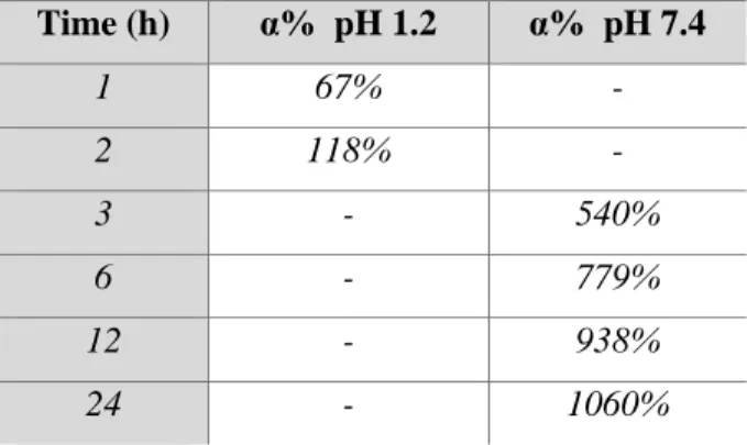
The swelling behavior of the gallate hydrogel has been evaluated at to two different pHs (1.2 and 7.4) and at predetermined time intervals (1h, 2h, 3h, 6h, 12h, 24h). Each experiment was carried out in triplicate and the results were in agreement within ± 4% standard error. The values of swelling degree (α%) for the prepared material at pHs 1.2 and 7.4 and different time intervals are reported in Table 1.
Table 1: Swelling behavior of the gallate hydrogel at pHs 1.2 and 7.4. Time (h) α% pH 1.2 α% pH 7.4 1 67% - 2 118% - 3 - 540% 6 - 779% 12 - 938% 24 - 1060%
27
The obtained data showed the actual ability of this hydrogel for phloretin intestinal specific release.
3.6 Hydrogel release studies
The release studies of phoretin were conducted on fixed aliquots of hydrogel at two different pH (1.2, and 7.4), which mimic the conditions of the gastro-intestinal tract, and at different time intervals (1h, 2h, 3h, 6h, 12h, 24h) through the use of a water bath at 37 ° C and under stirring. The results of release tests revealed that the hydrogel effectively released the drug at higher percentages at pH 7.4 (Figure 3).
Figure 3: Graphic representation of phloretin release from gallate hydrogel, plotting time (h) vs percentage of drug released, at two different pHs. Means
value ± SD, n = 3.
Therefore, these data allow to assume an oral administration of the gallate hydrogel and the use of this material as a vehicle of phloretin in the intestine.
4. Conclusions
The aim of this work was the inclusion of phloretin, a natural dihydrochalcone flavonoid with multiple properties including antidiabetic and antioxidant, in a hydrogel based on gallic acid formulated specifically to facilitate the phloretin intestinal release and protect it from chemical
0 10 20 30 40 50 60 70 80 90 100 0 1 2 3 4 6 12 P hlor etin (% ) Time (h) pH 1.2
28
degradation in gastrointestinal tract, enhancing its bioavailability. The swelling degree of the hydrogel not containing the phloretin, was carried out at two different pHs and predetermined time intervals. In particular, acidic solutions of HCl at pH 1.2 to mimic the pH of the stomach and 7.4 to mimic the pH of the small intestine, have been used. The obtained results revealed that the gallate hydrogel swells more at pH 7.4. Furthermore, the hydrogel was subjected to release studies in the same conditions of swelling tests. The results have shown the ability of hydrogel to release the phloretin at pH 7.4. Finally, the hydrogel was also submitted to evaluation of the antioxidant activity. The obtained data show that the hydrogel based on gallic acid could be used for the controlled release of phloretin through the gastro-intestinal tract, suggesting also a possible use in the treatment of diabetes.
29 References
1. Trombino S, Cassano R, Mellace S, Picci N. New Gallic Acid Based Hydrogel for Phloretin Intestinal Release, IJPRD, 2015;7(5):1-9.
2. Ikumi Y, Kida T, Sakuma S, Yamashita S, Akashi M. Polymer– phloridzin conjugates as an anti-diabetic drug that Inhibits glucose absorption through the Na+/glucose cotransporter (SGLT1) in the small intestine. Journal of controlled release. 2008;125(1):42-9.
3. Schwartz MW. Diabetes complications: why is glucose potentially toxic? Science. 1996;272(5262):699.
4. Bonadonna RC. Alterations of glucose metabolism in type 2 diabetes mellitus. An overview. Reviews in endocrine & metabolic disorders. 2004;5(2):89-97.
5. Mertes G. Safety and efficacy of acarbose in the treatment of type 2 diabetes: data from a 5-year surveillance study. Diabetes research and clinical practice. 2001;52(3):193-204.
6. Hediger MA, Coady MJ, Ikeda TS, Wright EM. Expression cloning and cDNA sequencing of the Na+/glucose co-transporter. Nature. 1987;330(6146):379-81.
7. Wood IS, Trayhurn P. Glucose transporters (GLUT and SGLT): expanded families of sugar transport proteins. British Journal of Nutrition. 2003;89(1):3-9.
8. Drozdowski LA, Thomson AB. Intestinal sugar transport. World journal of gastroenterology: WJG. 2006;12(11):1657.
9. Kellett GL, Helliwell PA. The diffusive component of intestinal glucose absorption is mediated by the glucose-induced recruitment of GLUT2 to the brush-border membrane. Biochemical Journal. 2000;350(1):155-62.
10. Kellett GL, Brot-Laroche E, Mace OJ, Leturque A. Sugar absorption in the intestine: the role of GLUT2. Annu Rev Nutr. 2008;28:35-54.
11. Goto T, Horita M, Nagai H, Nagatomo A, Nishida N, Matsuura Y, et al. Tiliroside, a glycosidic flavonoid, inhibits carbohydrate digestion and glucose absorption in the gastrointestinal tract. Molecular nutrition & food research. 2012;56(3):435-45.
30
12. Peppas NA, Wood KM, Blanchette JO. Hydrogels for oral delivery of therapeutic proteins. Expert Opinion on Biological Therapy. 2004;4(6):881-7.
13. Kim B, Peppas NA. Poly (ethylene glycol)-containing hydrogels for oral protein delivery applications. Biomedical Microdevices. 2003;5(4):333-41.
14. Ichikawa H, Peppas NA. Novel complexation hydrogels for oral peptide delivery: In vitro evaluation of their cytocompatibility and insulin‐ transport enhancing effects using Caco‐2 cell monolayers. Journal of Biomedical Materials Research Part A. 2003;67(2):609-17.
15. Sharpe LA, Daily AM, Horava SD, Peppas NA. Therapeutic applications of hydrogels in oral drug delivery. Expert opinion on drug delivery. 2014;11(6):901-15.
16. Polyfenols BL. Chemistry, dietary sources, metabolism and nutritional significanve. Nutr Rev. 1998;56:317-33.
17. King A, Young G. Characteristics and occurrence of phenolic phytochemicals. Journal of the American Dietetic Association. 1999;99(2):213-8.
18. Trombino S, Cassano R. Preparation And In Vitro Activities Evaluation Of Gallic Acid-Based Microspheres For Phloretin Transdermal Delivery. Int. J Pharm. Res. Dev.2014; 6:1-11.
19. Al-Salih R. Clinical experimental evidence: synergistic effect of gallic acid and tannic acid as antidiabetic and antioxidant agents. Med J. 2010;4:109-19.
20. Rezk BM, Haenen GR, van der Vijgh WJ, Bast A. The antioxidant activity of phloretin: the disclosure of a new antioxidant pharmacophore in flavonoids. Biochemical and biophysical research communications. 2002;295(1):9-13.
21. Wu CH, Ho YS, Tsai CY, Wang YJ, Tseng H, Wei PL, et al. In vitro and in vivo study of phloretin‐induced apoptosis in human liver cancer cells involving inhibition of type II glucose transporter. International journal of cancer. 2009;124(9):2210-9.
22. He X, Liu RH. Triterpenoids isolated from apple peels have potent antiproliferative activity and may be partially responsible for apple's
31
anticancer activity. Journal of agricultural and food chemistry. 2007;55(11):4366-70.
23. Yoon H, Liu RH. Effect of selected phytochemicals and apple extracts on NF-κB activation in human breast cancer MCF-7 cells. Journal of agricultural and food chemistry. 2007;55(8):3167-73.
24. Devi MA, Das N. In vitro effects of natural plant polyphenols on the proliferation of normal and abnormal human lymphocytes and their secretions of interleukin-2. Cancer letters. 1993;69(3):191-6.
25. Baldea LAN, Martineau LC, Benhaddou-Andaloussi A, Arnason JT, Lévy É, Haddad PS. Inhibition of intestinal glucose absorption by anti-diabetic medicinal plants derived from the James Bay Cree traditional pharmacopeia. Journal of ethnopharmacology. 2010;132(2):473-82.
26. Newey H, Parsons B, Smyth D. The site of action of phlorrhizin in inhibiting intestinal absorption of glucose. The Journal of physiology. 1959;148(1):83-92.
27. Bilia AR, Isacchi B, Righeschi C, Guccione C, Bergonzi MC. Flavonoids loaded in nanocarriers: an opportunity to increase oral bioavailability and bioefficacy. Food and Nutrition Sciences. 2014;5(13):1212.
28. Bartoli GM, Giannattasio B, Palozza P, Cittadini A. Superoxide dismutase depletion and lipid peroxidation in rat liver microsomal membranes: correlation with liver carcinogenesis. Biochimica et Biophysica Acta (BBA)-General Subjects. 1988;966(2):214-21.
29. Karakaya S. Bioavailability of phenolic compounds. Critical reviews in food science and nutrition. 2004;44(6):453-64.
30. Cassano R, Trombino S, Muzzalupo R, Tavano L, Picci N. A novel dextran hydrogel linking trans-ferulic acid for the stabilization and transdermal delivery of vitamin E. European Journal of Pharmaceutics and Biopharmaceutics. 2009;72(1):232-8.
31. Trombino S, Cassano R, Bloise E, Muzzalupo R, Tavano L, Picci N. Synthesis and antioxidant activity evaluation of a novel cellulose hydrogel containing trans-ferulic acid. Carbohydrate polymers. 2009;75(1):184-8.
32 PART B
“Novel Microspheres Based on Triterpene Saponins from the roots of
Physospermum Verticillatum (Waldst & Kit) (Apiaceae) for the
Improvement of Gemcitabine Release” (1)
Abstract
Objectives The present work concerns the preparation and characterization of microspheres based on a mixture of triterpene saponins, from
Physospermum verticillatum (Waldst & Kit), as a carrier for the specific
release of gemcitabine.
Methods Triterpene saponins were derivatized with acrylic acid. The obtained polymerizable product was characterized by Fourier Transform Infrared to confirm the ester linkage. Then, spherical microparticles were prepared by suspension radical copolymerization and impregnated with gemcitabine.
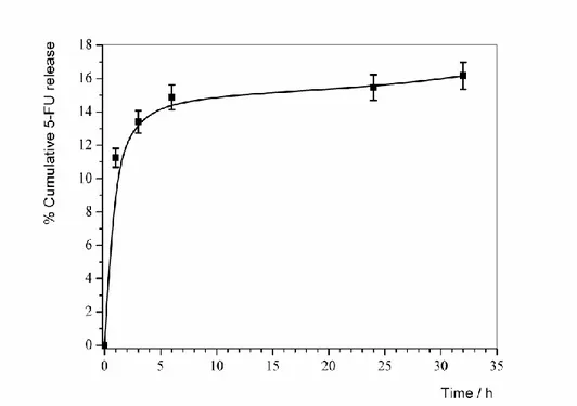
Key findings Microspheres exhibited a mean diameter of 2.7 µ. The swelling studies showed that particles swell most at pH 6.2, typical of the tumor pathology, then at pH 7.4, miming physiological conditions. The microspheres were loaded with gemcitabine (LE 72.2%). Their release profile showed an initial dot of around 24% and a further release for 24 h. Conclusions This carrier could be potentially release the drug in a in the lung, as a function of different pHs between tumor cells and healthy, reducing the systemic drug toxicity, allowing the reduction of the doses number, increasing the drug half-life, and eliminating the problems related to the fast clearance of gemcitabine administration.
Keywords: Triterpene saponins,microspheres, lung cancer, gemcitabine, release.
33
1. Introduction
Over the last years the interest towards pulmonary drug delivery systems, suitable for cancer therapy, is increasing. Lung cancer is one of the most frequently occurring malignancies and the leading cause of death worldwide (2-4). The systemic drug administration has hardly successful for the treatment of this pathology because only a partial amount of the drug reaches the lungs, even when used at high dose. In addition, the majority of these agents acts on normal cells, inhibiting their growth, weakening the patient that undergoes the antitumor therapy causing, in some cases, even the death (5). Therefore, the novel therapeutic strategies aim to improve the delivery of drugs that play a fundamental role in the treatment of lung cancer reducing also their toxicity (5). For this reason, there is an extensive interest in encapsulating the drugs in carrier systems micrometer and nanometer-sized. In particular, liposomes, polymer conjugates, polymeric micelles, microparticles, and nanoparticles have been investigated to selectively deliver various bioactive substances at the tumor site (6-9). Gemcitabine (GEM) is one of this kind of drugs.
It is a difluoro analogue of deoxycytidine (dFdC) and an antineoplastic drug belonging to the class of anti-metabolites that find indications in the treatment of large cell lung cancer, non-squamous cell lung cancer, metastatic pancreatic cancer, ovarian cancer and bladder cancer (10).This drug has a short plasma half-life of about 15 minutes (11),and a slower clearance in women and in the elderly.Peak levels reach 20-60 µM doses of 1000 mg/m2 administered intravenously over 30 minutes (12).Furthermore, itis converted into its inactive and soluble metabolites and cleared rapidly out of body through renal excretion. The dosage should be controlled and appropriately modified in patients with kidney and liver diseases. The main toxic effect of gemcitabine is the myelo-suppression (13).
Therefore, novel therapeutic strategies aiming to improve the pharmacokinetic, reduce the toxicity and obtain a better bio-distribution, are now needed (14). In this context, the microparticles could be used as carrier to achieve an efficient release of the chemotherapeutic drugs in the lung cancer therapy. Over the main method of microparticles preparation is the emulsion polymerization (15), that occurs with radical mechanism. Through this procedure microparticles based on triterpene saponins were
34
prepared and characterized. The saponins are a group of naturally occurring plant glycosides, reported in more than 100 families of plants. Several natural saponins have found to possess significant anticancer properties against colon, liver, gastric, ovarian, breast, thyroid, and lung epithelial cancer cells (16-19). These phytochemicals are able to induce cell cycle arrest, apoptosis and inhibit angiogenesis or tumor cell metastasis by influencing the dynamics or lateral organization of mammalian cellular membranes, by inhibiting proteins involved in the cell cycle (cyclins or cyclin-dependent kinases) and by other important cancer promoting pathways (20-22). In particular, the saponins are able to interact with cholesterol present in the cell membranes on both normal and cancer cells causing membrane damage. However, these compounds bind more efficiently to cancer cells containing more cholesterol than normal ones, but that is not enough to give adequate therapeutic index to be of any clinical value (23).
A rich source of saponins is the Apiaceae family (24-26). In this paper the interest was focused on triterpene saponins isolated from the roots of
Physospermum verticillatum (Waldst & Kit) (Apiaceae), namely
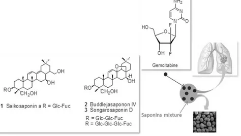
saikosaponin a, buddlejasaponin IV and songarosaponin D. In our previous study, these compounds have been tested in vitro for their potential antiproliferative activity against different human tumor cell lines. A remarkable activity was detected against COR-L23 (large cell lung carcinoma) cells with IC50 values in the range 0.4-0.6 µM (16). The interest in these compounds is related also to their ability to exerted significant inhibition of nitric oxide (NO) production in LPS induced RAW 264.7 macrophages. Recently, special attention is giving to combinations of saponins and other anticancer drugs (e. g. docetaxel, cisplatin, doxorubicin, paclitaxel, etc.) as efficient treatment against cancer cell line (27, 28). The improved therapeutic success of saponin-anticancer drug combination is related to the potentiation of cancer growth inhibition and the possibility to by-pass drug resistance (16, 19). In view of their potential value and of the considerations reported above, a mixture of saikosaponin a (1), buddlejasaponin IV (2) and songarosaponin D (3) was used to prepare and investigate the feasibility of microspheres loaded with gemcitabine to obtain synergic effects (Figure 1).
35
Figure 1: Representative scheme of gemcitabine loaded microparticles
triterpene saponins based.
The formation of microspheres could enhance the saponins selectivity towards cancer cells and reduce their toxicity, increasing their therapeutic index. In fact, it is known that saponins can cause side effects because of their hydrophobic-lytic properties resulting in trapping at the site of administration, causing cell and tissue destruction with local and systemic adverse reactions and a low therapeutic index.
2. Materials and Methods
2.1 ChemicalsAcetone, chloroform, dichloromethane, ethanol, methanol, isopropanol, n-hexane, acetonitrile, tetrahydrofuran (THF) were obtained from Carlo Erba
36
Reagents (Milan, Italy). n-Hexane, chloroform and tetrahydrofuran were purified by standard procedures. Acrylic acid, dicyclohexylcarbodimide (DCC), dimethylaminopyridine (DMAP), N,N-dimethylacrylamide,
sorbitan trioleate (Span 85), polyoxymethylene sorbitan (Tween 85),
N,N,N',N'-tetramethylethylenediamine (TMEDA), ammonium persulfate
(APS) and gemcitabine hydrochloride were purchased from Sigma-Aldrich (Sigma Chemical Co., St. Louis, MO, USA).
2.2 Plant materials
The roots of Physospermum verticillatum (Waldst & Kit) (Apiaceae) were collected in Calabria (Southern Italy) and authenticated by dr. D. Puntillo, Natural History Museum of Calabria and Botanical Garden, University of Calabria (Italy). A voucher specimen (No. CLU 21188) was deposited in the Botany Department Herbarium at the University of Calabria, Italy.
2.3 Extraction procedure
The triterpene saponins mixture was obtained by extraction from the roots of P. verticillatum as previously reported (21).Briefly, the roots (650 g) were subjected to maceration with methanol (5 × 72 h). The total extract (87.8 g) was suspended in distilled water and partitioned successively with ethyl acetate and water-saturated n-butanol. An n-butanol fraction of 52.5 g was obtained. 5 g of this was chromatographed on a silica gel column (0.040-0.063 mm) eluted with CHCl3/MeOH/H2O gradient and fractions of 10 ml were collected. After monitoring by thin layer chromatography (TLC), fractions were combined into six main parts (1-5). The fifth one (3.5 g) was subjected to flash column chromatography over silica gel by using CHCl3/MeOH/H2O mixture as eluent to afford a mixture (2.7 g) of saikosaponin a, buddlejasaponin IV and songarosaponin D (ratio of 2.1:1.8:1).
2.4 Measurements
The FT-IR spectra were carried out using a spectrometer FT-IR Jasco 4200 (Jasco Analytical Instrument, Japan) and KBr diskes. The structures of the synthesized compounds were confirmed by GC-MS Hewlett Packard 5972 (Hewlett-Packard Company, CA) instrument. The UV-Vis spectra were
37
performed using UV-Vis spectrophotometer V-530 JASCO (Jasco Analytical Instrument, Japan). Dimensional analysis of the microparticles was made by light scattering using a Brookhaven 90 Plus Particle Size Analyzer (Brookhaven Instruments Corporation, USA) by measuring the autocorrelation function at 90° scattering angle. The polydispersity index (PI), which indicates the measure of the distribution of nanoparticle populations, was also determined. Six separate measurements were made in order to obtain the average. Data were fitted by the method of inverse ‘‘Laplace transformation’’ and Contin. Samples were lyophilized using a ‘‘Freezing-drying’’ Micro module apparatus, Edwards (Crawley, West Sussex, UK). Photomicrografies of microparticles were obtained using a scanning electron microscope JEOL JSMT 300 A (JSMT, Jeol Ltd., Tokyo, Japan); the surface of the samples was made conductive by deposition of a gold layer in a vacuum chamber.
2.5 Esterification of triterpene saponins with acrylic acid
The reaction was conducted in accordance with the procedure of Steglich esterification (29). In a three-necked flask fitted with reflux condenser and magnetic stirrer, accurately flamed and maintained in an inert atmosphere, 19.75 µl (0.288 mmol) of acrylic acid were dissolved in 25 ml of dry tetrahydrofuran. After dissolution was added 0.040 g of DCC (0.192 mmol), 0.0012 g of DMAP (0.0096 mmol). After 30 minutes 0.15 g of saponins mixture (0.192 mmol) were added. The reaction mixture was kept cold, in a cold water bath, under stirring for 72 hours and monitored by TLC (silica gel, eluent mixture n-hexane/chloroform 9:1). At the end of the reaction the solvent was removed at reduced pressure. The dicyclohexylurea (DCU) was removed using methanol. The product was then dried with a mechanical pump and subsequently purified by column chromatography on silica gel using as eluent n-hexane/chloroform 8:2. The product was characterized by FT-IR.
2.6 Preparation of microspheres based on triterpene saponins
Microspheres based on were produced by radical polymerization technique. Briefly, in a cylindrical reactor of glass (100-150 ml) equipped with mechanical stirrer, dropping funnel and a screw cap with puncture-proof
38
rubber septum, was flamed in a current of nitrogen, and after cooling was placed in a bath thermostatically controlled at 40 ° C. Then, the required amounts of n-hexane (20 ml) and chloroform (18 ml), distillates, constituting the dispersant phase, were introduced in the reactor. The polymerization reaction was conducted according to the procedure reported in the literature (30).The dispersing phase was kept in a mechanical shaker for 30 minutes. A quantity of 0.15 g of ester (0.593 mmol) was suitably dissolved in 4 ml of distilled water, sonicated for a few minutes to facilitate the dissolution process. In the environmental reaction were added, under nitrogen stream, the solution containing the ester, 122.2 µl of N, N-dimethylacrylamide (1.186 mmol) and 0.80 g of ammonium persulfate ((NH4)2S2O8) (3.5 mmol) which acts as a radical initiator. The density of the organic phase was adjusted with the addition of one of the two solvents to obtain an aqueous phase in equilibrium with the organic phase. Then was added under nitrogen stream 150 μl of sorbitan trioleate (Span 85), after 10 minutes 150 µl of polyoxyethylene sorbitan trioleate (Tween 85) and after additional 10 minutes 150 µl of tetramethylenediamine (TMEDA). The system was kept under stirring for 3h and the temperature of 40 ° C. The content of the reactor was then filtered and washed in succession with 100 ml of isopropanol, 100 ml of ethanol and 100 ml of acetone. The filtrate was than analysed and characterizated.
2.7 Microspheres characterization
The obtained samples were characterized by FT-IR spectrophotometry, scanning electron microscopy (SEM) and measurement of the swelling degree in aqueous solutions at different pH values (6.2 and 7.4).
2.7.1 Dimensional analysis
The size of the microparticles was determined by light scattering using a particle size analyzer 90 Plus, at 25 °C. It was also determined the polydispersity index (PI), indicating the extent of the distribution of the microparticle population (31). The microparticles were also observed by means of (SEM).
39 2.7.2 Swelling studies
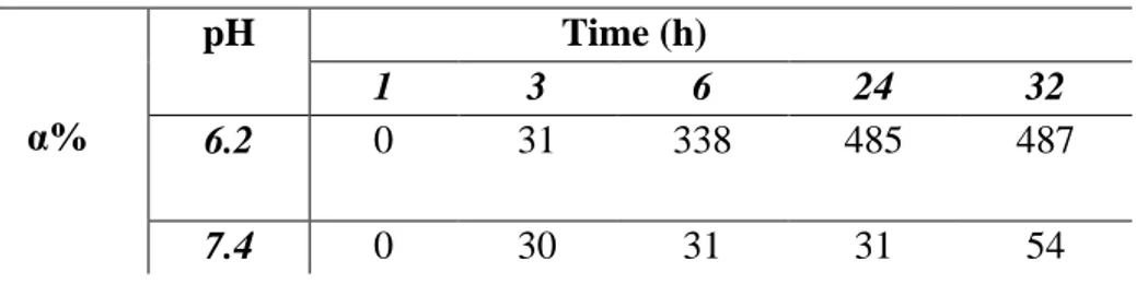
The affinity of the hydrophilic microparticles towards the aqueous environment was determined by their swelling degree (% WR). Aliquots notes of dry material (20 mg) were placed in glass filters (porosity G2/3) previously wetted, centrifuged (5 min at 2000 rpm), and then weighed. Subsequently, the filters were put in contact with a solution of phosphate buffer (Na2HPO4) at pH 6.2 (to mimic the conditions typical of the lungs tumor) and at pH 7.4 (to simulate the physiological environment) at the temperatures of 37 °C until reaching swelling equilibrium. At predetermined intervals time (1h, 2h, 3h, 12h, 18h, 24h) the excess of water was removed from the filters by means of percolation at atmospheric pressure. Three replicates were used for each pH value. Subsequently, the filters were centrifuged at 2000 g/min for 5 min. Finally, after centrifugation, the filters are weighed. The weights recorded to the time listed above were averaged and used to give the equilibrium swelling degree by the following equation:
𝑊𝑅(%) =(𝑊𝑠−𝑊𝑑)
𝑊𝑠 × 100 (Eq. 1)
where Ws and Wd are the weights of the swollen and dried microspheres respectively (30).
2.8 Impregnation of the microspheres with gemcitabine
The preformed matrix (about 50 mg) was placed in contact with a solution of known concentration of drug obtained by dissolving 5 mg of gemcitabine in 20 ml of distilled water. The impregnation was carried out under stirring at room temperature for 3 days, during which the solution of drug was absorbed by the matrix with consequent swelling (15). Finally, the solvent was removed by filtration and the microparticles were dried to constant weight. The analysis of filtrated water through spectrophotometer UV-Vis allowed to calculate the drug loading percentage (LE%) through the next equation:
40 𝐿𝐸% =𝑀𝑖−𝑀0
𝑀𝑖 × 100 (Eq. 2)
where Mi represents the drug concentration in the solution before loading and M0 the drug concentration in the solution after loading respectively. 2.9 In vitro release studies of gemcitabine from microparticles
Dried and loaded microparticles (10 mg) were dispersed in 6ml of swelling media with pH 6.2 or 7.4 (32).The test tubes were put in a water bath at 37 °C under stirring. At predetermined time intervals (1h, 2h, 3h, 6h, 12h, 24h), the samples were centrifuged, 5 ml of supernatant were removed and the medium was replaced with fresh solution to maintain the same total volume throughout the study. The gemcitabine concentration in the different solutions was monitored by the use of UV-Vis spectrophotometry at 275 nm. Each in vitro release study was performed in triplicate and under sink conditions. The release was calculated in terms of percentage of drug released.
2.10 Statistical analysis
Data are represented as the mean value ± standard deviation (S.D.) of three independent experiments. Differences within and between groups were evaluated by nonparametric ANOVA tests (Krushal-Wallis) completed by and with a multicomparison Dunn test using GraphPad softwere. A P value of <0.05 was considered significant.
3. Results and discussion
The microspheres are prepared using a triterpene saponins mixture containing saikosaponin a, songarosaponin D and buddlejasaponin IV, with a known antineoplastic activity (33).From the chemical point of view these saponins have a triterpene aglycone linked to several sugar moieties. Moreover, in the structure are present numerous hydroxyl groups, which have been exploited in the acrylation reaction to obtain the corresponding acrylic ester. The most reactive OH groups are the primary ones; in saikosaponin a are three primary -OH groups, four in songarosaponin D and
41
three in buddlejasaponin IV. Their esterification with acrylic acid furnished a derivatized saponins mixture, with numerous polymerizable moieties, that was used to prepare multifunctional microspheres, through radical polymerization. These particles were characterized and analyzed by means of common analyses methods. In particular, were effected studies to verify the microparticles swelling degree at the typical pHs of healthy and cancer cells.
3.1 Esterification of triterpene saponins with acrylic acid
The first step involves the esterification of the hydroxyl groups of saponins with acrylic acid through esterification of Steglich (29).This reaction allows obtaining an ester with practically unitary yields (Figure 2).
Figure 2: Synthetic route of triterpene saponins derivatization with acrylic acid. Respect to Fischer esterification this procedure provides the advantage of not using a protic acid as catalyst and avoids the formation of water molecules, making easier the purification operations of the final product. The obtained product was dried, purified by column chromatography and characterized by FT-IR. FT-IR (KBr), ν (cm-1): 3422, 2963, 2920, 1764, 1752, 980, 905.
3.2 Preparation of microspheres
The preparation of the microspheres was carried out using the technique of emulsion polymerization in reverse phase. This technique consists in the addition of a monomer solution (dispersed phase) in an excess of organic