Multi-physics interactions drive
VEGFR2 relocation on endothelial
cells
Valentina Damioli
1, Alberto Salvadori
2,4, Gian Paolo Beretta
1, Cosetta Ravelli
3,4&
Stefania Mitola
3,4Vascular Endothelial Growth Factor Receptor-2 (VEGFR2) is a pro-angiogenic receptor, expressed on endothelial cells (ECs). Although biochemical pathways that follow the VEGFR2 activation are well established, knowledge about the dynamics of receptors on the plasma membrane remains limited. Ligand stimulation induces the polarization of ECs and the relocation of VEGFR2, either in cell protrusions or in the basal aspect in cells plated on ligand-enriched extracellular matrix (ECM). We develop a mathematical model in order to simulate the relocation of VEGFR2 on the cell membrane during the mechanical adhesion of cells onto a ligand-enriched substrate. Co-designing the in vitro experiments with the simulations allows identifying three phases of the receptor dynamics, which are controlled respectively by the high chemical reaction rate, by the mechanical deformation rate, and by the diffusion of free receptors on the membrane. The identification of the laws that regulate receptor polarization opens new perspectives toward developing innovative anti-angiogenic strategies through the modulation of EC activation.
Tyrosine Kinase Receptors (RTKs) transmit information from the extracellular to the intracellular microen-vironment and play a central role in physiological and pathological conditions, including tumor progression. RTKs catalyze the phosphorylation of Tyrosine residues in their sequences as well as in second messengers. The non-homogeneous distribution of lipids and proteins on the cell membrane, both in space and time, is highly dynamic at multiple spatial levels and orchestrates the cellular response to different biochemical and mechanical inputs1. Membrane dynamics and composition2,3 also govern the expression and the activation of Epidermal
Growth Factor Receptor (EGFR) and Vascular Endothelial Growth Factor Receptor-2 (VEGFR2). The latter, expressed by cancer and ECs, modulates angiogenesis and tumor progression4–7 by binding different soluble
lig-ands, including VEGF-A, the non-canonical HIV-1-Tat8,9, and gremlin10,11. Thus, an abnormal spatial regulation
of RTKs may play a role in cancer progression12. Most of VEGFR2 ligands contain a heparin binding domain and
accumulate in the ECM, supporting a long-lasting activation of the cells. Moreover, ligand-enriched ECM recruits VEGFR2 at the basal aspect of ECs13,14, leading to a polarization of intracellular molecules.
Receptor-ligand interactions have been extensively studied from the biological and computational point of view. Several mathematical models have been developed to describe the body distribution of different isoforms of canonical and non canonical ligands of VEGFR2 and their interactions with VEGFRs both in vitro and in vivo15,16. These models confirmed that the amount of matrix-bound VEGF in normal human tissues (e.g. skeletal
muscle) is 30 to 100-fold higher than the amount of free ligands17,18. Some models considered also receptor
inter-nalization, since similarly to other RTKs ligand interaction induces VEGFR2 endocytosis in early endosomes19.
It is worth to point out that VEGFR2 undergoes efficiently in the endocytic compartment even in the absence of VEGF20,21. Mathematical models were also used to simulate and describe the competitive and/or synergic effects
of different ligands on VEGFR2 interaction and biological cell responses22.
The goal of this study is threefold: formulating a mathematical model of VEGFR2 recruitment in EC, simulat-ing the dynamics of VEGFR2 in EC seeded on ligand-enriched ECM, and finally co-designsimulat-ing experimental and 1Università degli Studi di Brescia, DIMI Department of Mechanical and Industrial Engineering, Brescia, 25123, Italy. 2Università degli Studi di Brescia, DICATAM, Department of Civil, Environmental, Architectural Engineering and Mathematics, Brescia, 25123, Italy. 3Università degli Studi di Brescia, DMMT, Department of Molecular and Translational Medicine, Brescia, 25123, Italy. 4Laboratory for Preventive and Personalized Medicine (MPP Lab), Università degli Studi di Brescia, Brescia, 25123, Italy. Correspondence and requests for materials should be addressed to C.R. (email: [email protected]) or S.M. (email: [email protected])
Received: 9 March 2017 Accepted: 4 November 2017 Published: xx xx xxxx
numerical investigations to characterize the dynamic lateral distribution (diffusion) of VEGFR2 receptors on the plasma membrane and their interactions (reaction) with immobilized ligands. The key features of our experimen-tal evidence on VEGFR2 relocation are well captured by a diffusion-reaction model, whereby the evolving geom-etry of the membrane is extremely simplified. The model is mathematically rigorous and self-consistent, in that it stems from continuity equations (for mass, energy, and entropy), standard chemical kinetics, thermodynamic restrictions, and constitutive specifications23,24. This sequence provides the governing equations in a strong form,
stated for dimensionless unknown fields and converted in a weak form prior to the numerical approximation via the Finite Element Method. Thermodynamic parameters have been inferred from the experimental analyses and from the literature, whereas a few have been calibrated.
The present model will be further developed, to include the spreading/deformation of the cell, its focal adhe-sion and the evolution of the stress fibers, the internalization of the complex, the ligand competition, ECM com-position, co-receptor partners, and the cell cortex structure12,25. Moreover, the predictive capabilities of the model
will be exploited in future studies to foresee alterations of the receptor behavior induced by mutations or admin-istration of anti-angiogenic drugs.
Results
Free and immobilized ligands induce VEGFR2 rearrangement on EC plasma membrane.
Different canonical and non canonical ligands, including VEGF-A, HIV-1-Tat, and gremlin are able to activate VEGFR2 and induce VEGFR2-mediated EC proliferation and migration9,10. To assess whether VEGFR2
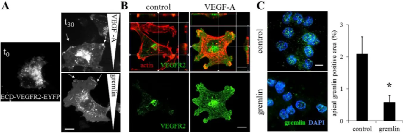
redis-tributes on the EC membrane when challenged by free ligands, adherent ECs over-expressing the Enhanced Yellow Fluorescent Protein (EYFP)-labeled extracellular domain (ECD) of VEGFR2 (ECD-VEGFR2-EYFP) were exposed for 2 hours to a linear concentration gradient of free ligands, including gremlin or VEGF-A, in a 2D chemotaxis assay. According to our previous investigations data, ECD-VEGFR2-EYFP exerts binding and dimerization activities similar to the VEGFR2 full length10,11,14. Figure 1A shows that ECD-VEGFR2-EYFP is
equally distributed on non-treated ECs (t0), while the gradient of ligands induces the ECD-VEGFR2-EYFP to be recruited in the lamellipodia at the leading edge of migrated ECs (t30). Together, these data demonstrate that free ligands are able to induce EC polarization, leading to VEGFR2 relocation on the surface of ECs. Although VEGFR2 ligands are usually considered as soluble molecules, in vivo they are bound and immobilized in the ECM or on the cell membrane by heparan-sulphate proteoglycans26.
Soluble and ECM-bound VEGF impact VEGFR2 trafficking rate. The clustering and the slower internali-zation rate of VEGFR2 complexes activated by ECM-bound VEGF elicits a prolonged activation of VEGFR2 and Extracellular signal Regulated Kinase (ERK) with a different pattern of site-specific phosphorylation14,27,28.
To characterize the influence of the immobilized VEGFR2-ligands on the VEGFR2 rearrangement on the cell membrane, we plated ECs on ligand-coated cell plates. Similarly to immobilized gremlin14, immobilized VEGF-A
induces the recruitment of VEGFR2 to the plasma membrane at the basal aspect of ECs, thus leading to a local-ized and directional receptor activation (Fig. 1B). The concentration of VEGFR2 at the apical side of the cell is diminished by the recruitment of VEGFR2 at the basal portion of adherent cells, as demonstrated by the reduc-tion of soluble ligand binding ability (Fig. 1C). Similar data were obtained with immobilized-VEGF-A.
Figure 1. VEGF-A and gremlin induce VEGFR2 rearrangement on EC surface. (A) ECD-VEGFR2-EYFP ECs were stimulated by a VEGF-A or gremlin gradient for 2 hours, fixed and analysed using a Zeiss Axiovert 200M system (630×; white bar: 10 μm). Arrows indicate ECD-VEGFR2-EYFP-enriched cell lamellipodia. (B) HUVECs adherent on Fibrinogen or VEGF-A-enriched substrates were stained for VEGFR2 (green) and actin (red) and analysed using a LSM510 Meta confocal microscope. Images show the basal portion of adherent cells with the orthogonal z reconstruction of the whole cell (630×; white bar: 10 μm). (C) VEGFR2-EC, seeded on immobilized gremlin or on coverglass for 4 hours, were incubated with 150 ng/mL of gremlin for 90 minutes at 4 °C and washed with 1.5 mol/L NaCl. VEGFR2-bound gremlin, in the apical portion of the cells, was detected by immunofluorescence analysis using a Zeiss Axiovert 200 M microscope system (630x; white bar: 10 μm). Data are expressed as percentage ± s.d. of gremlin positive area with respect to the total cell area (n = 20 cells/ sample; *P < 0.001, Student’s t-test).
Ligand binding decreases VEGFR2 diffusion on plasma membrane.
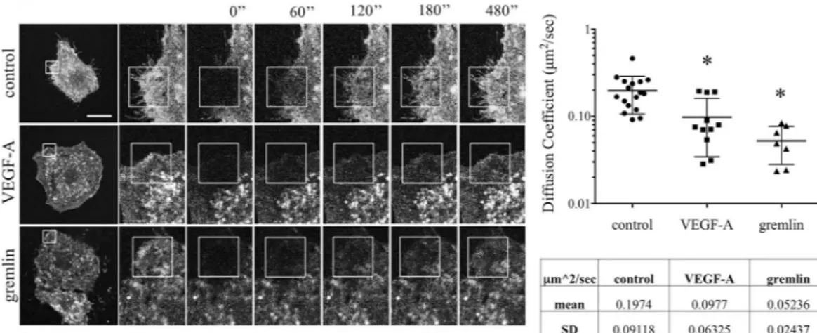
In order to measure the mobility of VEGFR2 on the cell membrane, we performed Fluorescence Recovery After Photobleaching (FRAP) analysis on EC culture expressing ECD-VEGFR2-EYFP. The rate of fluorescence recovery provides quantitative information about the kinetics of diffusion of fluorescent molecule in the photo-bleached area. To measure the dynamics of VEGFR2 on the cell membrane, fluorescence was recorded every minute for 10 minutes in an irreversibly photo-bleached mem-brane region of ECD-VEGFR2-EYFP EC in the absence or in the presence of 50 ng/mL of VEGF-A or gremlin. In our experimental conditions, 77% of ECD-VEGFR2-EYFP in the plasma membrane turns out to be in a mobile form, with a lateral diffusion coefficient of 0.198 μm2/s in untreated ECs. Both VEGF-A or gremlin treatments decrease the receptor mobility respectively to 0.098 μm2/s and 0.052 μm2/s (Fig. 2). All these data highlight that non-activated receptors are mainly free to move on the cell membrane, thus suggesting that VEGFR2 phosphorylation, its dimeriza-tion, and its interaction with membrane co-receptors or intracellular signaler reduce its motility. FRAP data support our former observations14 that VEGFR2 is rapidly recruited and immobilized in the membrane in close contact withligands. These events lead to increase the receptor concentration in the basal side of the cell.
A chemo-transport-mechanical model describes VEGFR2 relocation on EC surface.
We describe the relocation of VEGFR2 on the cell membrane during its adhesion to ligand-enriched ECM by means of a model that accounts for the ligands-receptors chemical interaction, the diffusion of receptors along the mem-brane, and the mechanical deformation of the cell. It will be denoted henceforth as a chemo-transport-mechanical model. It is defined on the cell surface only, and it has been validated against co-designed experimental investi-gations. The cell adhesion to a ligand-enriched substrate results in VEGFR2 polarization to the basal membrane and entails several concurrent phenomena, including cell deformation and cytoskeletal remodeling, which lead to an increased interaction between the basal cell membrane and the ligand enriched-substrate. While the intra-cellular tail of VEGFR2 and in particular the tyrosine residues are required for the correct VEGFR2 internaliza-tion29,30, the kinase domain is not required for the receptor relocation, as demonstrated either by the recruitmentof VEGFR2 in the presence of the Sugen 541614 or by the recruitment of the receptor without its intracellular
tail (ECD-VEGFR2). VEGFR2 polarization is not mediated by integrin interaction even though, once recruited, VEGFR2 will form an active complex with integrin in lipid rafts. We neglect accordingly the intracellular subset of VEGFR2 and the interaction with coreceptors in our chemo-transport-mechanical model. To simulate the interaction between VEGFR2 and its immobilized ligand, we assume a fixed membrane geometry and account for the effects of cell adhesion with a supply of ligands onto the cell surface at a prescribed rate, sL (ligands/μm2 s),
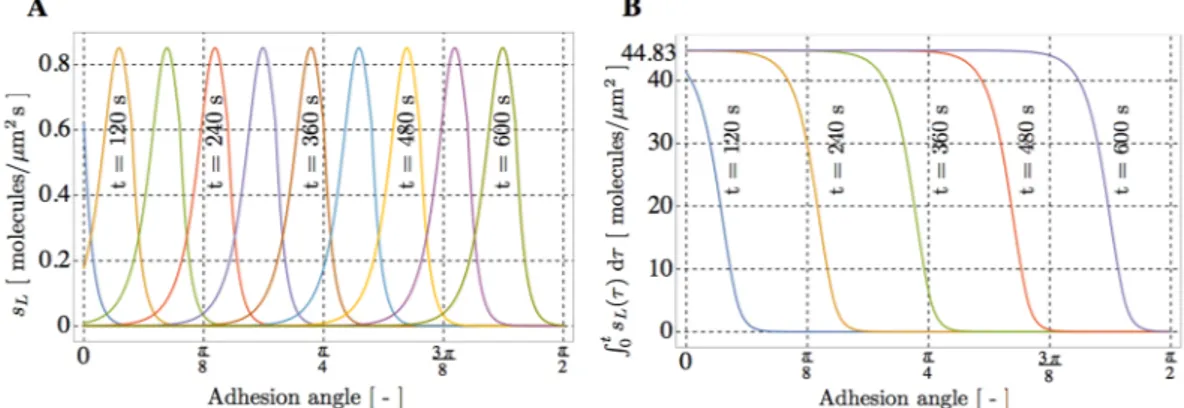
detailed in Eq. (8). Figure 3 shows the spatial evolution of the mass supply sL and of the total amount of
lig-ands parametrized in time: at each location, liglig-ands smoothly reach the saturation limit of 44.83 liglig-ands/μm2 s (Fig. 3B). Owing to this modeling simplification, the actual time-evolving geometry of the membrane becomes relatively unimportant and thus, for maximal simplicity, we analyze it as a circumference of radius l = 20 μm and assume that the time-dependent concentrations depend on the curvilinear coordinate.
The model was implemented in a finite element code as a script in Wolfram Mathematica version 10 and calibrated. The simulations run until the final time tF = 7200 s at the constant temperature 310.15 K with a
substrate-adsorbed ligand concentration of 44.83 ligands/μm2 (Fig. 3B).
Multi-physics drive the complex formation with three different mechanisms.
As depicted in Fig. 4, that represents the overlay of the outcomes of simulation (green line) and in vitro experiments (red dots)14normalized to the value of VEGFR2 at the final time tF, VEGFR2 recruitment induced by immobilized ligands
Figure 2. VEGF-A and gremlin reduce VEGFR2 motility on EC surface. (A) FRAP analysis was performed on cell plasma membrane of serum-starved ECD-VEGFR2-EYFP over-expressing GM7373 cells treated or not with VEGF-A or gremlin (50 ng/mL). Images were acquired at one per minute for 12 minutes, 2 before and 10 after bleaching. The bleached area is indicated by a square and the recovery time is indicated over the images as seconds after photobleaching (630x; white bar: 20 μm). (B) Collected images were analyzed using simFRAP ImageJ plugin to calculate diffusion coefficients. The graph shows diffusion coefficient mean ± s.d. of control, VEGF-A, and gremlin treated cells (n = 7 – 15; *P < 0.01, Student’s t-test).
shows three phases of complex formation marked by circled roman numbers: an initial plateau (I), a steep branch (II), and finally an evolution with a lower formation rate (III). Our numerical simulations allow connecting these three phases to three distinct mechanisms dominated by different limiting factors. The initial plateau is governed by the cell-ligand contact (I), the second steep phase (that ends at 600 s) is due to a chemo-mechanical evolution - induced by the cell attachment and deformation (II), and the final slow phase reflects the diffusive slow motion of the receptors from the apical to the basal membrane that is in contact with the substrate (III).
The quantitative correspondence between experimental and numerical outcomes suggests that the number of well-oriented ligands available for the receptor binding is much smaller than the total amount of immobilized lig-ands. As shown in Fig. 4, the simulated evolution in time of the overall amount of bound VEGFR2-ligands com-plex on the membrane overlaps the experimental outcomes we previously observed14, validating of the model.
The chemo-mechanical transport model describes VEGFR2 dynamics.
Numerical simulations pre-dict the evolution of the concentration of free receptors (cR) during 2 hours of cell stimulation. Figure 5Aquanti-fies cR at each location along the membrane at different times. Exploiting the axial symmetry of the simulations,
each curve on the right side of Fig. 5A depicts the spatial concentration profile every minute. At t = 0 the distri-bution of receptors is uniform at the concentration cR = 4.8 receptors/μm2. After 60 s, the concentration profile
is perturbed and decreases at the bottom of the cell due to receptor-ligand complex formation. As time goes by, starting from 120 s, an enlarging zone with negligible concentration cR ~ 0 of free receptors is visible at the basal
side of the cell (point A), due to the engagement of free receptors by immobilized ligands. At the end of the simu-lation, at tF = 7200 s, the concentration of unbound receptors at the apical side amounts at cR = 0.5 receptors/μm2.
The chemo-mechanical transport model allows concluding that the depletion of free receptors is due to three concurrent factors: i) the infinitely fast kinetics of the ligand-receptor interaction; ii) the high equilibrium con-stant, that favors the formation of ligand-receptor complex; iii) the evidence that diffusion of the receptor on the cell membrane is much slower than interaction kinetics.
Figure 3. Effects of cell deformation mimicked through a supply of ligands sL onto the membrane. (A) Spatial
evolution of the mass supply sL and (B) of its time-cumulate.
Figure 4. Time evolution of the VEGFR2-gremlin complex formation on the EC membrane. Comparison is made between the VEGFR2 total fluorescence intensity (free and bound) in contact the substrate (red dots) and the numerical simulation data (green lines). To allow comparisons, both sets of data have been normalized to the values reached at the final time tF = 7200 s.
The depletion “propagates” with time, so that at tf = 600 s, after the cell is completely adhered, the lower
por-tion of the cell membrane is essentially empty of free receptors. Since no further supply sL is provided afterwards,
the process becomes diffusion-dominated, and it slowly evolves towards a final steady state. The thick blue curve plots the distribution of free receptors at the end of the simulation at time tF. The maximum concentration of free
VEGFR2 at tF is 0.49 receptors/μm2 and a steady state has not yet been reached.
Ligand-receptor complex accumulates at the boundary of the EC basal membrane.
Numerical simulations predict that after 2 hours of adhesion (at tF = 7200 s) a zone with high ligand-receptor complexcon-centration manifests at the boundary of the contact area. Figure 5B depicts the evolution of complex cC in space
(X axis) and time (different colors) at the basal aspect of ECs. Such distribution profile was confirmed experimen-tally in EC VPMs, as shown in Fig. 5C. VPMs were obtained by an osmotic shock of ECs, that preserves only the basal portion of cell membrane in close contact with ECM, allowing the visualization of the recruited receptors VGEFR2.
Discussion
VEGFR2 is a transmembrane protein, a key signal transducer in angiogenesis, and a potential therapeutic target in angiogenesis-related diseases. It has a lateral mobility of about 0.198 μm2/s, as measured in this work by means of FRAP experiments, and can interact with both soluble and ECM-immobilized ligands. Indeed several VEGFR2 ligands contain one or more heparin binding domains in their sequences. After ligand interaction, VEGFR2 dimerizes and transduces an intracellular signaling via its relocation on the cell membrane and the recruitment of intracellular proteins.
We developed a multi-physics model to describe and predict the effects of ligands on VEGFR2 relocation during the EC activation. The interaction between ligands and receptors has been modeled by a chemical reac-tion that produces a complex, as customary in literature19,28. The model accounts for finite reaction kinetics,
although simulations have been carried out assuming that the reaction kinetics is infinitely fast. The time-scale of Figure 5. The chemo-mechanical transport model describes VEGFR2 relocation dynamics. (A) Time evolution of the spatial concentration cR of free VEGFR2 along the cell membrane. Each curve plots the distribution of
free receptors at different times t = 60 n, with n = 0, 1, 2, … 120 s from the beginning of the experiment at t = 0 to the final time tF = 7200 s. (B) Spatial evolution of the concentration cC of the receptor-ligand complex at
various times. The curves report the numerical simulation: points A, B, and C correspond to those in (A). (C) VPM staining for VEGFR2 confirms peaks in the intensity of fluorescence at the boundary of the substrate/ membrane contact surface.
the VEGFR2-gremlin binding reaction is in fact assumed to be much faster than the time-scale of the mechan-ical deformation of the cell and of the diffusion of receptors on cell membrane. Under these assumptions, we recovered the experimental evidence that the motion of receptors and their subsequent trapping into immobile VEGFR2-ligands complexes proceed in a sequence of three phases, and we characterized those phases with dif-ferent rate-controlling factors. The first phase starts when a small portion of membrane gets in contact with the substrate, which provides a sudden supply of ligands that immediately trap the available receptors. This phase is very rapid, because the reaction rate is the controlling factor (in our model assumed infinite), and fully depletes the concentration of free receptors, because the equilibrium constant is very large. The second phase (calibrated in 10 minutes from experiments) is rate-controlled by the mechanical deformation of the cell, which provides additional supply of ligands that afresh immediately react with the available receptors on the newly formed con-tact area. The mechanical deformation of the cell and the VEGFR2 recruitment14 are influenced by the
chemi-cal affinity of the VEGFR2-gremlin binding reaction coupled with intracellular cortichemi-cal actin dynamics. In our co-designed experimental and theoretical study, the cell adhesion is not mediated by integrin engagement, even though integrin involvement cannot be completely ruled out. We observed that in our experimental conditions substrate-immobilized growth factors act as a cell-adhesive stimulus for ECs, which is weaker than the ECM13.
Although a few papers consider the integrin-mediated focal adhesions and their interactions with the cytoskel-eton31–33, they do not apply to our experimental conditions since the involved β
3 integrin is not recruited in the focal adhesion complexes. Bound VEGFR2 forms a complex with β3 integrin in membrane lipid raft domains and not in focal adhesion. Our current simplified model does not capture the complexity of this binding. Instead, it surrogates the details of the mechanical deformation with the experimentally-guided assumption of an empirical ad-hoc time-sequence of gremlin supply on increasing portions of the cell membrane. The cell surface becomes depleted of free receptors very rapidly where the cell adheres to the substrate. When the mechanical deforma-tion terminates and the cell is eventually spread, the diffusion of receptors becomes the rate-controlling mecha-nism. During this final phase, receptors that diffuse through the boundary of the contact surface are immediately trapped and immobilized by the ligands on the substrate. Therefore, the VEGFR2-gremlin complex tends to accu-mulate at the boundary of the basal aspect of the cells in close contact with ECM. Such a localization was observed in the ventral plasma membrane of ECs adherent on gremlin- or HIV-1 Tat13 enriched surfaces. A higher
concen-tration of receptors at the cell boundary could have relevant biological implications for the cell, which may sense ligand concentration variation and migrate in the direction of stimulus production.
The key features of our experimental evidence on VEGFR2 relocation are captured well. The effect of the mechanical deformation of the cell has been accounted for in the model, by surrogating the explicit description of the cell spreading/deformation with a given increase in time of the surface area available for the chemical reac-tion. The model accounts for finite reaction kinetics, although simulations have been carried out assuming that the reaction kinetics is infinitely fast. Moreover, the model shown predictive capability. Numerical simulations highlighted a zone with high complex concentration at the boundary of the contact area at the final time of the simulations. This concentration were experimentally confirmed Fig. 5B and has been understood.
Other authors19 proposed meta-models that involve the integration of extracellular cues with intracellular
processes, such as receptor endocytosis and phosphorylation. Those models are typically point-wise in nature and focus on the evolution in time disregarding the space distribution, so that ordinary differential equations suf-fice. Modeling the motion of membrane receptors concurrently with several other aspects of endothelial biology (as internalization or cooperativity in ligand action) is more intricate and can only be dealt with incrementally, moving from simple yet not simplistic assumptions and adding complexities stepwise. The current model will be enriched toward the ambitious target of modeling the angiogenesis process as a whole. The role of kinetic constants will be investigated34–37, including internalization38,39 as well as the involvement of other co-receptors
(including integrins and heparansulphateproteoglycans) and the cytoskeleton re-organization that leads to cell motility and morphogenesis. The interaction between the ligand complex and non engaged integrins will be con-sidered, because β3 integrin mediates and triggers the long-lasting VEGFR2 activation14.
The surrogated mechanics will be replaced by a detailed analysis of the structural behavior of the cell, fol-lowing recent studies31–33,40–42, thus coupling the evolution for the Laplace-Beltrami-like operator that presides
the formulation with the large deformation of the cell. Mechanical models for cell spreading involve sophis-ticated descriptions of active and passive behavior of cells with equations that are much harder to follow than the ones presented here. Simulations of these models require an impressive computational burden, so large that high performance computing is mandatory. In conclusion, there is a substantial increment of theoretical and computational complexity in adding further phenomena of endothelial biology. Since the simplified model with surrogated mechanics resulted capable to reproduce experimental evidences, we expect that the real mechanical evolution may add further insights and fine tune the results, without altering the general picture presented here. A simple yet effective surrogate model may have merits, though, and it will be used to save computational costs when it comes to perform uncertainty quantification studies, which require a large number of computationally expensive simulations.
Methods
Cell cultures.
Foetal bovine aortic endothelial GM7373 cells43 were grown in Dulbecco’s modified Eaglemedium (DMEM, Gibco, Life Technologies) containing 10% FCS, vitamins, essential and non-essential amino acids. GM7373 cells were transfected with a pcDNA3/Enhanced Yellow Fluorescent Protein (EYFP) vector har-boring the extracellular domain of human VEGFR2 (ECD-VEGFR2) cDNA (provided by K. Ballmer-Hofer, PSI, Villigen, Swiss) to generate ECD-VEGFR2-EYFP EC14, or with a pcDNA3.1 vector harboring the mouse VEGFR2
Chemotaxis assay.
This was performed as described44. Briefly, ECD-VEGFR2-EYFP overexpressing ECswere seeded at 3.0 × 10⁶ cells/mL in μ-slide chemotaxis chambers (IBIDI, Martinsried, Germany) and incubated in complete medium for 24 hours. Cells were then stimulated with gremlin11 and VEGF-A (100 ng/mL)45 and
observed during gradient formation. After 2 hours, cells were photographed under a Zeiss Axiovert 200 M epi-fluorescence microscope equipped with a Plan-Apochromat 63x/1.4 NA oil objective. Z-stack images acquired using ApoTome imaging system were elaborated through AxioVision Extended Focus module (Zeiss Axiovert 200 M system).
ECs adhesion on immobilized protein.
2.0 μg/mL of gremlin, VEGF or Fibrinogen (Fb) were added to polystyrene tissue culture plates or glass coverslips. After 16 hours of incubation at 4 °C, samples were washed and uncovered glass was blocked with 1.0 mg/mL bovine serum albumin (BSA) for 1 hour at room temperature14.Ventral plasma membrane (VPM) preparation.
VPMs were prepared by osmotic shock using a modi-fication of the squirting lysis technique14. Briefly, cells were washed twice with ice-cold water; after 1 minute cellswere squirted over by using a jet of ice-cold water and immediately fixed for immunocytochemistry analysis. In all the experiments, the absence of DAPI staining and the persistence of actin filaments were used to unequivocally identify the VPM remnants bound to the substratum. For total VEGFR2 staining, samples were incubated over-night with a rabbit polyclonal anti-VEGFR2 antibody (1:200, Santa Cruz Biotechnology) followed by a 1-hour incubation with AlexaFluor 488-conjugated anti-rabbit IgG (1:500). For actin staining samples were incubated for 30 minutes with TRITC-phalloidin (0.9 mg/mL in PBS, Sigma). All reagent dilutions were in PBS containing 3% BSA. VPMs were acquired under an Axiovert 200 fluorescence microscope equipped with a Plan-Apochromat 63x/1.4 NA oil objective and ApoTome system (Carl Zeiss). VEGFR2-positive areas and total VPM areas, defined by actin staining, were quantified using Image-Pro Plus software.
Ligands/cell binding assay.
GM7373-VEGFR2 (VEGFR2-ECs) cells were seeded on VEGF-A or gremlin or uncoated coverslip for 4 hours and then incubated with soluble gremlin (150 ng/mL) for 90 minutes at 4 °C in PBS added with Ca2+ and Mg2+ and 0.1% gelatin. Then, cells were washed three times with PBS or with PBS plus 1.5 mol/L NaCl to remove HSPGs-bound ligands. Immunofluorescence analysis was performed using a goat polyclonal anti-gremlin antibody (R&D Systems) or goat polyclonal anti VEGF-A antibody followed by AlexaFluor 488 anti-goat IgG (Molecular Probes, Life Technologies). Cells were analysed using a Zeiss Axiovert 200 M system.Fluoresence Recovery After Photobleaching (FRAP).
ECD-VEGFR2-EYFP ECs were seeded at 5.0 × 10⁵ cells/mL μ-slides (IBIDI, Martinsried, Germany). Cells were starved in minimal medium for 4 hours, stimulated with gremlin or VEGF-A (50 ng/mL) and analysed using LSM510-META confocal microscope equipped with an incubation chamber (Zeiss). Confocal images were recorded with 4–5% of the intensity of the 514-nm line from living transfected cells. EYFP fluorescence was eliminated using a 350-iteration bleach cycle at 100% intensity of the 514-nm line. Control bleach experiments performed over the entire cell surface demonstrated that the EYFP chromophore was completely inactivated by this treatment and that recovery of flu-orescence due to newly synthesized EYFP proteins was not detectable during the period of recovery (10 minutes). Image series were analysed using simFRAP ImageJ plugin (https://imagej.nih.gov/ij/plugins/sim-frap/index. html)46. To calculate diffusion coefficients and using FRAP Caculator ImageJ macro (https://www.med.unc.edu/microscopy/resources/imagej-plugins-and-macros/frap-calculator-macro) to calculate mobile and immobile fractions following author instructions.
Data representation.
Data are expressed as mean ± SD or mean ± SEM. Statistical analyses were performed using the Student’s t-test. The significance level was set at P < 0.01.Chemo-transport-mechanical model formulation.
A general formulation for the chemo-transport- mechanics problem with trapping is here tailored to model the relocation of VEGFR2 on the lipid bilayer membrane. The interaction between receptors (R) and ligands (L) is described as a chemical reaction, which produces a receptor-ligand complex (C), + − . + R L C (1) k k 1 1 The ligand, whose degradation is negligible, and the complex are assumed to be immobile. Complex internaliza-tion and the return back to the surface will be elaborated in future publicainternaliza-tions. Since receptors are free to move along the membrane, reaction (1) portrays a conversion of mobile to trapped receptors and vice-versa. Its rate is denoted with w(1) (molecules/μm2 s). Therefore, the mass balance equations for the three species involved in reaction (1) read: ∂ ∂ + → + = c tR div hR w(1) 0, (2a) ∂ ∂ + = c tL w(1) s ,L (2b)
c
tC w(1) 0 (2c)
∂
∂ − = .
Since Eq. (2) are defined on the cell membrane only, derivatives in the divergence operator are defined on a curved surface, in the Laplace-Beltrami sense. Concentrations cR, cL, cC (molecules/μm2) are constrained fields,
since they must be positive and cannot exceed the saturation amounts cRmax, c
Lmax, cCmax, respectively. Symbol sL
denotes a mass supply (molecules/μm2 s), which will be illustrated later in the paper. Vector →h
R (molecules/sμm) denotes the flux of receptors along the membrane surface. It is constitutively described by a Fickian law, by means of the tangential gradient of the concentration,
→
= − ∇
hR DR [ ],cR (3)
thus entailing the Clausius-Duhem inequality. Symbol DR denotes the receptors’ diffusivity along the membrane (μm2/s). The chemical kinetics of reaction (1) is modeled via the law of mass action47,
w k k 1 1 1 , (4) L L R R C C (1) 1 θ θ θ θ 1 θ θ = − − − − + − where k±
1 denote the forward (+) and reverse (−) rate constants (molecules/μm2 s) for reaction (1), θ = c cJ J J/ max for J = R, L, C, and the chemical potentials are assumed of the form
μ μ θ θ = + − RT ln 1 , J J J J 0
with each μJ0 independent of all the concentrations. At chemical equilibrium the concentrations obey the relation θ θ θ θ θ θ − − − = = −∆ K G RT 1 1 1 exp , (5) C C L L R R eq eq eq eq eq eq eq(1) (1) 0
where Keq(1) is the equilibrium constant and ∆G(1)0 =μC0−μR0−μL0 is the Gibbs free energy of the reaction. Mass balance equation (2) shall be accompanied by the balance of force, to model the mechanical (and thus geometrical) evolution of the cell shape, whose boundary - the membrane - is the geometrical support of Eq. (2). Modeling the evolution of the Laplace-Beltrami operator that presides formulation (2–4) concurrently with the large deformation of the cell is a phenomenally ambitious task, which is in progress motivated by the promising outcomes here shown. In the present work, we surrogate the mechanics with some simplifying assumptions. They are collected in the following section.
Simplifying assumptions.
Two major simplifications, pertaining to reaction kinetics and mechanical mod-eling, will be undertaken henceforth. They allow verifying the capability of the chemo-transport model (2–4) to reproduce experimental evidences with much less theoretical and computational burden.The assumption is taken henceforth that the reaction kinetics is infinitely fast, considering that the time required to reach chemical equilibrium is orders of magnitude smaller than the time-scale of the other processes. This assumption is usually taken in conceptually similar problems, as the diffusion of species in metals48 or the
electro-mechanics of batteries49–53. Then, concentration of the complex is governed by thermodynamic
equilib-rium at all times by Eq. (5), which with trivial algebra becomes: c c c c K ( , ) 1 1 1 1 (6) C L R C L L R R max eq(1) 1 θ θ θ θ = + − − . −
The two final parabolic governing equations in the unknown fields cL and cR can be easily inferred from Eqs
(2a) and (2b), by replacing (2c) with (6) c t c c c t h ( , ) div[ ] 0, (7a) R C L R R ∂ ∂ + ∂ ∂ + → = ∂ ∂ + ∂ ∂ = . c t c c c t s ( , ) (7b) L C L R L
The change of the geometry of the membrane with time is dictated by the mechanical evolution of the cell, which in turn is governed by several complex phenomena (such as focal adhesion and stress-fiber reorganiza-tion32, integrins-ECM interactions54, curvature changes of cell membrane55, cooperation between integrins and
VEGFR245,56) triggered by the adhesion of the cell onto the ligand-coated plate described in the experimental
sec-tion. Roughly, adhesion induces cells to deform from an initially spherical shape to a final spread configuration33,57,
resulting in increased interaction between basal cell membrane and the ligand enriched-substrate. We replace the explicit modeling of cell spreading/deformation with a supply of ligands sL, calibrated from experimental data and
We assume therefore that the membrane geometry is fixed and thus account for the effects of cell adhesion by a supply of gremlin on the membrane at a prescribed rate, sL (molecules/μm2 s), inferred from experimental
evidence and defined as follows:
s x t c t t x v t t x v ( , ) (8) L = L − − +
and plot in Fig. 3. In Eq. (8) is the Heaviside step function, cL=72 ligands/ mμ 2 is the concentration of substrate-immobilized ligand available for reaction (1), tf is the time required for the complete mechanical
defor-mation of the cell, v=π/2tf is the velocity of mechanical deformation (assumed to be constant until tf), is the
cell radius, t is a parameter that identifies a finite time required for binding, x is the curvilinear abscissa of tf our simplified geometry, t the generic time. In view of Eq. (8), the supply of ligands at point x on the membrane remains zero until t < x/v; then, in the time span between t = x/v and =t x v/ +t, it increases rapidly from zero to cL.
Weak form and discretization.
The governing equations for the relocation of VEGFR2 on the membrane under the above modeling assumptions have been made dimensionless and multiplied by test functions. The weak form obtained by their integration over the spatial domain can be transformed to a first order Ordinary Differential Equation (ODE) in time if the discretization is performed via separated variables. Therefore, nodal unknowns depend solely on time, while test and shape functions solely on space. Time advancing has been achieved by finite differences, using a backward Euler scheme. Discretization of the unknown fields by means of standard linear shape functions leads to the numerical approximation via the finite element method in each time step.Material parameters.
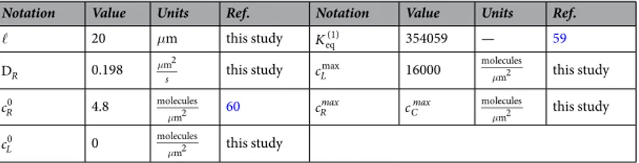
Parameters for the in silico simulation (see Table 1) were defined by in vitro assays. The cell radius was calculated from the measure of radius of 50 ECs using Zeiss Axiovert 200 M microscope; receptor diffusivity was obtained by FRAP analysis (see above). The amount of VEGFR2 on cell membrane per area was calculated by dividing the number of high affinity binding sites, obtained by radiolabeled binding exper-iments58 for cell surface area. The kinetic parameters ligand/receptor were measured by Surface plasmonres-onance (SPR) measurements (BIAcore X, GE Healthcare). The extracellular domain of human VEGFR2 was immobilized onto CM5 sensorchips (BIAcore) and increasing concentrations (from 100 ng/mL to 4 μg/mL) of ligand was injected in HBS-EP buffer (BIAcore) for 10 minutes (sample volume: 50 μL; flow rate: 5 μL/minute; dissociation time: 2 minutes). Binding parameters were calculated by the nonlinear-curve-fitting software pack-age BIAevaluation 3.2 (Biacore)58. The maximal surface density of immobilized ligand on surface was determined
by SPR. The sensorgram recorded during immobilization gives direct check on the amount of ligand attached to the surface. Full details and references about this procedure can be found at the dedicated page of the SPR pages website: http://www.sprpages.nl/Experiments/Howmuch.php.
Model calibration.
Analyzing the evolution in time of VEGFR2 recruitment induced by immobilized lig-ands experimentally measured in14 as fluorescence intensity of the overall VEGFR2 (free and bound), threephases of complex formation were identified, namely: an initial plateau, lasting few seconds; a steep branch, that takes place until about 600 s; and finally an evolution with a lower formation rate after the first 600 s. Accordingly, we calibrated tf = 600 s as the time for completion of the mechanical deformation and =t 1 as the parameter that s
identifies a finite time required for binding.
References
1. Grecco, H., Schmick, M. & Bastiaens, P. Signaling from the living plasma membrane. Cell 144, 897–909 (2011).
2. Ichinose, J., Murata, M., Yanagida, T. & Sako, Y. EGF signalling amplification induced by dynamic clustering of EGFR. Biochem
Bioph Res Co 324, 1143–1149 (2004).
3. Nacev, B., Grassi, P., Dell, A., Haslam, S. & Liu, J. The Antifungal Drug Itraconazole Inhibits Vascular Endothelial Growth Factor Receptor 2 (VEGFR2) Glycosylation, Trafficking, and Signaling in Endothelial Cells. J Biol Chem 286, 44045–44056 (2011). 4. Chung, G. et al. Vascular endothelial growth factor, FLT-1, and FLK-1 analysis in a pancreatic cancer tissue microarray. Cancer 106,
1677–1684 (2006).
5. Lee, T. et al. Vascular endothelial growth factor mediates intracrine survival in human breast carcinoma cells through internally expressed VEGFR1/FLT1. Plos Med 4, e186 (2007).
6. Silva, S. et al. VEGFR-2 expression in carcinoid cancer cells and its role in tumor growth and metastasis. Int J Cancer 128, 1045–1056 (2011).
7. Yang, F. et al. Increased VEGFR-2 gene copy is associated with chemoresistance and shorter survival in patients with non-small-cell lung carcinoma who receive adjuvant chemotherapy. Cancer Res 71, 5512–5521 (2011).
Notation Value Units Ref. Notation Value Units Ref.
20 μm this study Keq(1) 354059 — 59
DR 0.198 μ
s
m2 this study cLmax 16000
μ molecules m2 this study cR0 4.8 μ molecules m2 60 cR
max cCmax molecules
m2
μ this study
cL0 0 moleculesμm2 this study
8. Albini, A. et al. The angiogenesis induced by HIV–1 Tat protein is mediated by the Flk–1/KDR receptor on vascular endothelial cells.
Nature Medicine 2, 1371–1375 (1996).
9. Mitola, S. et al. Identification of Specific Molecular Structures of Human Immunodeficiency Virus Type 1 Tat Relevant for Its Biological Effects on Vascular Endothelial Cells. Journal of Virology 74, 344–353 (2000).
10. Mitola, S. et al. Gremlin is a novel agonist of the major proangiogenic receptor VEGFR2. Blood 116, 3677–3680 (2010).
11. Grillo, E. et al. Monomeric gremlin is a novel vascular endothelial growth factor receptor-2 antagonist. Oncotarget 7, 35353–35368 (2016).
12. Casaletto, J. & McClatchey, A. Spatial regulation of receptor tyrosine kinases in development and cancer. Nature 12, 387–400 (2012). 13. Urbinati, C. et al. Substrate-Immobilized HIV-1 Tat Drives VEGFR2/v 3-Integrin Complex Formation and Polarization in
Endothelial Cells. Arterioscl Throm Vas 32, e25–e34 (2012).
14. Ravelli, C. et al. β3 Integrin Promotes Long-Lasting Activation and Polarization of Vascular Endothelial Growth Factor Receptor 2 by Immobilized Ligand. Arterioscl Throm Vas 35, 2161–2171 (2015).
15. Gabhann, F. M., Yang, M. T. & Popel, A. S. Monte carlo simulations of VEGF binding to cell surface receptorsin vitro. Biochimica et
Biophysica Acta (BBA) - Molecular Cell Research 1746, 95–107 (2005).
16. Gabhann, F. M. & Popel, A. S. Dimerization of VEGF receptors and implications for signal transduction: A computational study.
Biophysical Chemistry 128, 125–139 (2007).
17. Gabhann, F. M. & Popel, A. S. Interactions of VEGF isoforms with VEGFR-1, VEGFR-2, and neuropilinin vivo: a computational model of human skeletal muscle. Am J Physiol-Heart C 292, H459–H474 (2007).
18. Kut, C., Mac Gabhann, F. & Popel, A. Where is VEGF in the body? a meta-analysis of VEGF distribution in cancer. Br J Cancer 97, 978–985 (2007).
19. Weddell, J. & Imoukhuede, P. Integrative meta-modeling identifies endocytic vesicles, late endosome and the nucleus as the cellular compartments primarily directing RTK signaling. Integr. Biol. 9, 464–484 (2017).
20. Gampel, A. et al. VEGF regulates the mobilization of VEGFR2/KDR from an intracellular endothelial storage compartment. Blood
108, 2624–2631 (2006).
21. Basagiannis, D. & Christoforidis, S. Constitutive endocytosis of VEGFR2 protects the receptor against shedding. J Biol Chem 291, 19892–903 (2016).
22. Mac Gabhann, F. & Popel, A. A model of competitive binding of vascular endothelial growth factor and placental growth factor to VEGF receptors on endothelial cells. Am J Physiol 286, H153–64 (2004).
23. Gurtin, M., Fried, E. & Anand, L. The Mechanics and Thermodynamics of Continua (Cambridge University Press, 2010). 24. Paolucci, S. Continuum Mechanics and Thermodynamics of Matter (Cambridge University Press, 2016).
25. Bussolino, F., Serini, G., Mitola, S., Bazzoni, G. & Dejana, E. Dynamic modules and heterogeneity of function: a lesson from tyrosine kinase receptors in endothelial cells. ENBO reports 2, 763–767 (2001).
26. Chiodelli, P. et al. Heparan Sulfate Proteoglycans Mediate the Angiogenic Activity of the Vascular Endothelial Growth Factor Receptor-2 Agonist Gremlin. Arterioscl Throm Vas 31, e116–27 (2011).
27. Chen, T. T. et al. Anchorage of vegf to the extracellular matrix conveys differential signaling responses to endothelial cells. J Cell Biol
188, 595–609 (2010).
28. Clegg, L. & Mac Gabhann, F. A computational analysis of in vivo vegfr activation by multiple co-expressed ligands. PLoS Comput
Biol 13, e1005445 (2017).
29. Ewan, L. et al. Intrinsic tyrosine kinase activity is required for vascular endothelial growth factor receptor 2 ubiquitination, sorting and degradation in endothelial cells. Traffic 7, 1270–82 (2006).
30. Mattila, E., Auvinen, K., Salmi, M. & Ivaska, J. The protein tyrosine phosphatase TCPTP controls VEGFR2 signalling. J Cell Sci 121, 3570–80 (2008).
31. Deshpande, V., Mrksich, M., Mc Meeking, R. & Evans, A. A bio-mechanical model for coupling cell contractility with focal adhesion formation. J Mech Phys Solids 56, 1484–1510 (2008).
32. Deshpande, V., Mc Meeking, R. & Evans, A. A model for the contractility of the cytoskeleton including the effects of stress-fiber formation and dissociation. P R Soc A 463, 787–815 (2007).
33. Ronan, W., Deshpande, V., Mc Meeking, R. & McGarry, J. Cellular contractility and substrate elasticity: a numerical investigation of the actin cytoskeleton and cell adhesion. Biomech Model Mechanobiol 13, 417–435 (2014).
34. Bell, G. Models for the specific adhesion of cells to cells. Science 200, 618–627 (1978).
35. Gilson, M. K., Given, J. A. & Head, M. S. A new class of models for computing receptor-ligand binding affinities. Chemistry &
Biology 4, 87–92 (1997).
36. Klotz, I. M. & Hunston, D. L. Mathematical models for ligand-receptor binding. real sites, ghost sites. Journal of Biological Chemistry
259, 10060–10062 (1984).
37. Myers, A. C., Kovach, J. S. & Vuk-Pavlovi, S. Binding, internalization, and intracellular processing of protein ligands. derivation of rate constants by computer modeling. Journal of Biological Chemistry 262, 6494–6499 (1987).
38. Lauffenburger, J. & Linderman, D. A. Receptors: Models for Binding, Trafficking, and Signaling (Oxford University Press, 1993). 39. Leitner, D. M., Brown, F. L. & Wilson, K. R. Regulation of protein mobility in cell membranes: A dynamic corral model. Biophysical
Journal 78, 125–135 (2000).
40. Rahimi, M. & Arroyo, M. Shape dynamics, lipid hydrodynamics, and the complex viscoelasticity of bilayer membranes. Phys Rev E
86, 011932 (2012).
41. Deseri, L. & Zurlo, G. The stretching elasticity of biomembranes determines their line tension and bending rigidity. Biomech Model
Mechanobiol 12, 1233–1242 (2013).
42. Bentley, K. & Philippides, A. Do Endothelial Cells Dream of Eclectic Shape? Dev Cell 29(4), 146–158 (2014).
43. Grinspan, J., Mueller, S. & Levine, E. Bovine endothelial cells transformed in vitro by benzo(a)pyrene. J Cell Physiol 114, 328–338 (1983).
44. Zantl, R. & Horn, E. Chemotaxis of slow migrating mammalian cells analysed by video microscopy. Method Mol Cell Biol 769, 191–203 (2011).
45. Ravelli, C., Mitola, S., Corsini, M. & Presta, M. Involvement of αvβ3 integrin in gremlin-induced angiogenesis. Angiogenesis 16,
235–243 (2013).
46. Blumenthal, D., Goldstien, L., Edidin, M. & Gheber, L. Universal Approach to FRAP Analysis of Arbitrary Bleaching Patterns.
Scientific Reports 5, 11655 (2015).
47. De Groot, S. & Mazur, P. Non-Equilibrium Thermodynamics (Dover, 1984).
48. Oriani, R. A. The diffusion and trapping of hydrogen in steel. Acta Metall Mater 18, 147–157 (1970).
49. Salvadori, A., Bosco, E. & Grazioli, D. A computational homogenization approach for Li-ion battery cells. Part 1 - Formulation. J
Mech Phys Solids 65, 114–137 (2014).
50. Salvadori, A., Grazioli, D. & Geers, M. Governing equations for a two-scale analysis of Li-ion battery cells. Int J Solids Struct 59, 90–109 (2015).
51. Salvadori, A., Grazioli, D., Geers, M., Danilov, D. & Notten, P. A novel approach in modeling ionic transport in the electrolyte of (Li-ion) batteries. J Power Sources 293, 892–911 (2015).
52. Salvadori, A. et al. On the role of saturation in modeling ionic transport in the electrolyte of (Li-ion) batteries. J Power Sources 294, 696–710 (2015).
53. Grazioli, D., Magri, M. & Salvadori, A. Computational modeling of Li-ion batteries. Comput Mech, https://doi.org/10.1007/s00466-016-1325-8 (2016).
54. Kim, S. H., Turnbull, J. & Guimond, S. Extracellular matrix and cell signalling: The dynamic cooperation of integrin, proteoglycan and growth factor receptor. J Endocrinol 209, 139–151 (2011).
55. Deserno, M. Fluid lipid membranes: From differential geometry to curvature stresses. Chemistry and Physics of Lipids 185, 11–45 (2015).
56. Somanath, P. R., Malinin, N. L. & Byzova, T. V. Cooperation between integrin αvβ3 and VEGFR2 in angiogenesis. Angiogenesis 12,
177–185 (2009).
57. Golestaneh, A. & Nadler, B. Modeling of cell adhesion and deformation mediated by receptor-ligand interactions. Biomech Model
Mechanobiol 15, 371–387 (2016).
58. Maiolo, D. et al. Role of Nanomechanics in Canonical and Noncanonical Pro-angiogenic Ligand/VEGF Receptor-2 Activation. J Am
Chem Soc 120823144733008 (2012).
59. Brozzo, M. et al. Thermodynamic and structural description of allosterically regulated VEGFR-2 dimerization. Blood 119, 1781–1788 (2012).
60. Stabile, H. et al. Bone morphogenic protein antagonist drm/gremlin is a novel proangiogenic factor. Blood 109, 1834–1840 (2007).
Acknowledgements
Work supported by grants from Associazione Italiana per la Ricerca sul Cancro (IG 17276) to S.M., from New Opportunity and Ways toward ERC (NOW-ERC project 2014-2256), Fondazione Cariplo, and Regione Lombardia to C.R., and from Fondazione Berlucchi to V.D.
Author Contributions
V.D., A.S., GP.B., C.R. and S.M. designed research, S.M. and C.R. conceived the experiments, C.R. conducted the experiments, V.D., A.S., G.B. formulated the model, A.S. implemented the FEM algorithm and performed the numerical simulations, V.D., A.S., GP.B., C.R. analyzed the results. All authors reviewed the manuscript.
Additional Information
Competing Interests: The authors declare that they have no competing interests.
Publisher's note: Springer Nature remains neutral with regard to jurisdictional claims in published maps and institutional affiliations.
Open Access This article is licensed under a Creative Commons Attribution 4.0 International License, which permits use, sharing, adaptation, distribution and reproduction in any medium or format, as long as you give appropriate credit to the original author(s) and the source, provide a link to the Cre-ative Commons license, and indicate if changes were made. The images or other third party material in this article are included in the article’s Creative Commons license, unless indicated otherwise in a credit line to the material. If material is not included in the article’s Creative Commons license and your intended use is not per-mitted by statutory regulation or exceeds the perper-mitted use, you will need to obtain permission directly from the copyright holder. To view a copy of this license, visit http://creativecommons.org/licenses/by/4.0/.