Full Terms & Conditions of access and use can be found at
http://www.tandfonline.com/action/journalInformation?journalCode=irnf20
Download by: [Univ Degli Studi di Foggia] Date: 05 November 2015, At: 06:21
Renal Failure
ISSN: 0886-022X (Print) 1525-6049 (Online) Journal homepage: http://www.tandfonline.com/loi/irnf20
Oral manifestations in chronic uremia patients
Mario Dioguardi, Giorgia Apollonia Caloro, Giuseppe Troiano, Giovanni
Giannatempo, Luigi Laino, Massimo Petruzzi & Lorenzo Lo Muzio
To cite this article: Mario Dioguardi, Giorgia Apollonia Caloro, Giuseppe Troiano, Giovanni Giannatempo, Luigi Laino, Massimo Petruzzi & Lorenzo Lo Muzio (2015): Oral manifestations in chronic uremia patients, Renal Failure, DOI: 10.3109/0886022X.2015.1103639
To link to this article: http://dx.doi.org/10.3109/0886022X.2015.1103639
Published online: 29 Oct 2015.
Submit your article to this journal
Article views: 5
View related articles
ISSN: 0886-022X (print), 1525-6049 (electronic) Ren Fail, Early Online: 1–6
!2015 Taylor & Francis. DOI: 10.3109/0886022X.2015.1103639
STATE OF THE ART REVIEWS
Oral manifestations in chronic uremia patients
Mario Dioguardi1, Giorgia Apollonia Caloro2, Giuseppe Troiano1Giovanni Giannatempo1, Luigi Laino1, Massimo Petruzzi3, and Lorenzo Lo Muzio1
1
Department of Clinical and Experimental Medicine, University of Foggia, Foggia, Italy,2Department of Emergency and Organ Transplantation – Nephrology, Dialysis and Transplantation Unit, University of Bari, Bari, Italy, and3Dental Clinic, Faculty of Medicine and Surgery, University of Bari, Bari, Italy
Abstract
The incidence of chronic renal failure (CRF) is approximately 200 cases per million people in different Western countries. Recent data indicate that the incidences of these pathologies are increasing. Ninety percent of patients with CRF report oral signs and symptoms that affect both the bone and soft tissues. A broad range of lesions may be observed in chronic uratemia patients, including the following: gingival hyperplasia, enamel hypoplasia, petechiae, gingival bleeding, and others lesions. These patients require various types of treatment ranging from dietary and lifestyle changes to dialysis and kidney transplantation. CRF often leads to multiple oral manifestations that are difficult for dentists to manage. The present study examined the characteristics of this disease, the existing therapeutic options and the relevant considerations for dental professionals.
Keywords
Chronic renal disease, gingival hyperplasia, kidney transplantation, oral manifestations in hemodialysis
History
Received 8 April 2015 Revised 21 July 2015 Accepted 28 September 2015 Published online 27 October 2015
Introduction
Chronic renal failure (CRF) is a progressive clinical condition that is characterized by gradual and permanent reductions of both the homeostatic and emunctory functions of the kidneys. The incidences of CRF are approximately 200 cases per million people in different Western countries.
The pathological presentation is characterized by the progressive loss of functioning nephrons due to a reduction in the glomerular filtrate rate (GFR), which is approximately 100–130 mL/min in normal conditions.1,2
Depending on the level of renal impairment, CRF can be classified into the following progressive stages according to the Kidney Disease Outcomes Quality Initiative (K/DOQI): ! Stage I, chronic nephropathy with a GFR490 mL/min. ! Stage II, mild CRF with 605GFR589 mL/min. ! Stage III, moderate CRF with 595GFR544 mL/min
(stage III A) or 435GFR530 mL/min (stage III B). ! Stage IV, acute CRF with 295GFR515 mL/min. ! Stage V, terminal CRF (TRI) with GFR515 mL/min
(also called the uratemic phase).
The followings are among the recognized causes of terminal CRF: diabetes; renovascular diseases; glomerulo-nephritis; infectious and obstructive nephropathies; genetic diseases (polycystic kidney disease, Alport’s syndrome, oxalosis syndrome, cystinosis, and Fabry’s disease); and various other diseases (e.g., amyloidosis, myeloma, and
sarcoidosis). Uratemic symptoms include hematological manifestations (anemia, bleeding, and a tendency to bruise easily); cardiovascular manifestations (hypertensive cardio-vascular disease, peripheral edema, and arrhythmia); nervous manifestations (alterations in the sensitivities of the hands and feet and neuropathies); endocrine manifestations (secondary hyperparathyroidism); bone manifestations (renal osteody-strophy); and gastrointestinal manifestations (nausea, hiccup-ing, and vomiting). Therapy may be conservative (e.g., pharmacological or fluid restrictions and changes in the diet) or more aggressive, including dialysis and kidney transplant-ation.3In the last few years, great interest in the literature has been directed to the treatment of patients suffering from CRF and the oral manifestations of renal diseases. Increases in kidney transplantation and chronic long-term therapy in these patients have resulted in more frequent requests for dental therapies. Occasionally, dentists feel unprepared to know-ledgeably address health problems linked with the renal diseases of the patients. In contrast, nephrologists might face oral diseases that are not easy to understand in terms of both complex differential diagnoses and problems that are essen-tially dental.
Materials and methods
A literature search was performed with the aim of identifying the articles related to the oral manifestations of chronicle uratemia patients. An initial literature research of the Italian and European books of nephrology and oral pathology was conducted. Subsequently, an informatics-based bibliography search was performed via the following online search engines:
Address correspondence to Dr. Giuseppe Troiano, Department of Clinical and Experimental Medicine, Foggia University, Via Rovelli, 50 71122 Foggia, Italy. Tel: 0039 0881 588090; Fax: 0039 0881 588081; E-mail: [email protected]
Pub Med Medline, the Ebsco Library, and the Web of Science Core Collection. The citations have been searched according to key words and titles using the following combinations of terms: ‘‘oral manifestations AND renal failure’’, ‘‘oral signs AND hemodialysis’’, and ‘‘oral health AND chronic uratemia patient’’. A preliminary evaluation of the abstracts determined the inclusion in this review. The references lists of other articles were carefully evaluated to identify interesting articles that were not found in the previous database searches. Only articles written in English were included in this study. After a preliminary evaluation of 164 articles, 47 were deemed eligible for inclusion in this study.
Oral manifestations
Manifestations involving the oral cavity often represent clinical signs of an advanced uratemic state that affects both the hard and soft tissues. Occasionally, such manifestations can also be due to therapeutic measures that include the following: fluid restrictions, dietary changes, side effects of systemic therapy, and dialysis and/or kidney transplantation. In these patients, the following changes in the oral microflora can be observed: the rate of colonization by yeasts is higher than normal, and variabilities in species distributions and antifungal susceptibility profiles can be observed.4,5
The manifestations related to soft tissue involvement are summarized in Table 1.
Uratemic stomatitis
Uratemic stomatitis is an uncommon complication of uratemia that follows the accumulation of nitrogenous waste products in the blood.6 Four variants of uratemic stomatitis exist: erythematosus membranous stomatitis, ulcerative stomatitis, hemorrhagic stomatitis, and the hyper-parakeratotic stomatitis. Erythematosus uratemic stomatitis is characterized by the formation of pseudomembranes that consist of a thick sticky exudate on the erythematous mucosa. The ulcerative form represents the most common presentation and involves ulcerated and erythematous mucosa. The presence of bleeding characterizes the hemorrhagic form, which can be observed in any site of the oral cavity but is most frequent on the gingiva. The hyperkeratotic presentation is a rare form that occurs in end-stage renal failure and is likely due to the effects of chemical substances on the oral mucosa.7 Uratemic stomatitis primarily occurs in advanced renal failure particularly when the disease has progressed gradually for many years. An acute form of necrotic pseudomembranous gingivostomatitis can occasion-ally be observed in patients who rapidly develop high blood urea nitrogen levels.8,9 The possible causes include the following: tissue reactions to toxins or to uratemic catabolites; the hydrolyzing action of the oral bacterial flora on the urea that results in the formation of irritant ammonium compounds; and hemorrhagic diathesis, which is common manifestation of uratemic syndromes.10 A small local hemorrhage in the oral cavity can cause tissue dystrophy, which is a condition that is predisposing to the development of bacterial infection. Bacterial infection in combination with immune uratemic deficiency can cause ulcerations and the formation of pseudomembranes. The lesions often appear on the ventral surface of the tongue and are very painful and can even make the ingestion of fluids
impossible. Uratemic stomatitis is basically diagnosed clinic-ally, but it mimics other common pathologies of the oral cavity, and there are few clinical aspects that are useful for its differential diagnosis.11To avoid the development of potentially fatal uratemia, uratemic patients soon become dependent on renal function replacement therapy (i.e., dialysis or transplant-ation).12 Several studies have proven that the injuries that develop after months or years of uratemic conditions are resistant to local treatments (e.g., antiseptic and antifungal mouthwashes) if the plasma levels of the nitrogenous catabolites are continually elevated; symptom regression is only possible with a reduction in the serum level of urea.7
Impairment of salivation and dry mouth (xerostomia) with or without dysgeusia
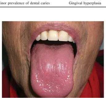
Xerostomia is a symptom of oral dryness that may be distinct from salivary gland hypofunction, which involves an object-ive reduction of the salivary flow. Patients with hyposaliva-tion often show exhibit the following: glossitis, cervical caries, cracked, peeled and atrophic lips, candidiasis, and pale, corrugated, and dry buccal mucosa13(Figure 1). Dry-mouth may be debilitating and affect the patient’s quality of life via the presence of dysphagia, dysgeusia, oral pain, dental caries, oral infections, and periodontal diseases. Xerostomia seems to be present in 28–59% of end-stage renal disease patients due to polyuria caused by the inability of the kidneys to reabsorb sodium.14
Mucosal lesions
Oral lichenoid lesions and oral hairy leukoplakia (OHL) are most commonly observed in transplanted patients. OHL can
Figure 1. Impairment of salivation and dry mouth (xerostomia) with dysgeusia.
Table 1. Common oral manifestations of chronic uratemic patients. Hard tissues disease Soft tissues disease Enamel hypoplasia Uremic stomatitis Increased formation of tartar Xerostomia Dental calculus Gingival bleedings Osteodystrophy Mucosal lesions as ulcers
and neoformations Minor prevalence of dental caries Gingival hyperplasia
2 M. Dioguardi et al. Ren Fail, Early Online: 1–6
be unilateral or bilateral, typically involves the lateral and dorsolateral tongue and is often observed in patients undergo-ing cyclosporine therapy. OHL is probably caused by drug-induced reactivation of the Epstein-Barr virus in the basal layers of the oral epithelium,15and it presents as a painless white patch with an irregular surface and prominent folds and projections. Immunosuppression can also elicit oral candid-iasis, dysplasia, carcinomas of the lip and non-Hodgkin lymphoma in the oral cavity.16Other common oral infectious lesions include candidiasis with the peculiar oral thrush and cytomegalovirus infection with vesicles and blisters.17 Gingival overgrowth
In pre-dialysis and dialysis patients, gingival hyperplasia is related to calcium channel blockers, while in patients with kidney transplants, gingival hyperplasia is due to immuno-suppressive therapy with cyclosporine18 (Figure 2). Some studies have hypothesized that these two drugs may have an additive effect.19 Cyclosporine-related gingival hyperplasia appears in approximately 70% of adult transplant patients. Cyclosporine interferes with the metabolism of human gingival fibroblasts and the extracellular components of the lamina propria, which causes increments in the deposition of intercellular matrix and the degree of vascularization.20 Petechiae, bruises and gingival bleeding
Chronic uratemic patients often suffer from spontaneous gingival bleeding, which is probably caused by bacteriemia (Figure 3). The bacterial toxins lead to platelet dysfunction and subsequent bleeding. This bleeding is aggravated by renal anemia, the use of anticoagulants, and the dysfunction of the endothelial cells, which results in vessel fragility.21 Furthermore, endothelial dysfunction due to hyperlipidemia is often present and associated with calcium and oxalate precipitation into the vessels that leads to the formation of petechiae in the oral tissues22(Figure 4).
The manifestations to hard tissue involvement are detailed in the subsequent section.
Enamel hypoplasia
Enamel renal syndrome (ERS) is a hereditary condition that is characterized by quantitative or qualitative enamel defects
and nephrocalcinosis. In ERS patients, the teeth present with yellow-brown discolorations. The surfaces may be either rough or smooth, and the latter is often associated with microdontia even when normal enamel hardness is present.23 The amount of enamel is substantially reduced and can even be absent in both the primary and permanent teeth.24In the pulp chambers, pulp stones can be observed in both the primary and secondary dentitions. Renal exams reveal nephrocalcinosis that is asymptomatic in children who are affected by ERS. ERS seems to be associated with recessive mutations in FAM20A, which is a gene that is normally expressed by osteoblasts, ameloblasts (during the secretion stage), and odontoblasts and plays an essential role in tooth development.25
Osteodystrophy of the maxillary bone
Renal osteodystrophy is a bone pathology that occurs as a side effect of alterations in the mineral metabolisms of patients with chronic renal disease. Renal osteodystrophy involves bone demineralization with the loss of the trabeculae and thinning of the cortex.26 The results are radiolucent bone lesions that are histologically similar to giant cell bone tumors. Furthermore, some metastatic calcifications that are caused by altered calcium–phosphorus products that facilitate the precipitation of crystals can be observed in the soft tissues of the periodontium.27 The consequences of this condition
Figure 3. Spontaneous gingival bleeding in a chronic uratemia patient.
Figure 4. Petechiae on the mucosa of the hard palate. Figure 2. Gingival hyperplasia involving the labial surface of the
interdental papilla.
include defects of the temporomandibular joint, dental malocclusions, calcifications of the pulp chamber, delayed eruption, and potential bone fractures following tooth extractions.28
Halitosis and increased tartar formation
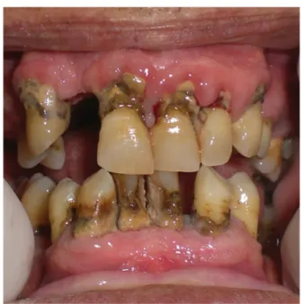
Microbiological degradation in the oral cavity leads to the production of volatile sulfur compounds (VSCs) that are the main cause of halitosis. VSCs seem to be produced by Gram-negative anaerobic oral bacteria.29 Hydrogen sulfide (H2S), methyl mercaptan (CH3SH), and dimethyl sulfide (CH3)2S are the VSCs that are primarily involved in halitosis. In approximately 85% of cases, bad breath is due to intraoral factors that include the following: tongue coating, dry mouth, poor oral hygiene, and the presence of prostheses and periodontitis.30 Approximately 10% of oral halitosis cases derive from the ears, nose and throat region, and within this region, 3% seems of cases seem to originate from the tonsils. Patients with CRF associated with high levels of urea nitrogen in the blood and salivary production31 report more severe halitosis when the blood urea levels are455 mg/dL. Urea is a buffer that prevents increases in the pH of the bacterial biofilm and thus leads to a higher rate of dental calculus and a lower incidence of caries32(Figure 5). The lower incidence of caries seems to be due to acid neutralization and inhibition of the growth of bacteria in the plaque.17 Patients with renal-associated halitosis might benefit from peritoneal dialysis therapy because this therapy elicits a reduction in blood nitrogen urea.33
Erosions on the lingual surfaces of the teeth
These lesions are mainly present on the lingual surface of the teeth, present as enamel abrasions, and may be considered to be consequences of reflux and the frequent vomiting episodes caused by the electrolyte imbalances in uratemic patients34 (Figure 6).
Dentists can aid other clinicians in the diagnosis of carotid artery calcification via panoramic radiography. A recent study demonstrated higher levels of carotid artery calcification in patients with renal injuries that are accompanied by changes in triglyceride and potassium levels that can be diagnosed via panoramic radiography.35Zhao et al.36evaluating a Chinese population of chronic kidney disease patients who were undergoing hemodialysis and demonstrated increased alveolar resorption at the sites of the upper premolars and first molars. Some authors have focused on decayed, missing and filled teeth index scores (DMFT) and periodontal status. These authors have observed worsening gingival indices and probing depth measurements in patients who have undergone hemo-dialysis for more than 3 years.30 However, data from other authors do not accord with these results.28 Craig37 recom-mended careful monitoring of the periodontal statuses of patients with chronic renal disease because periodontitis can increase the inflammatory burden and atherosclerotic com-plications. Differences have also been found regarding the cause of hemodialysis. Indeed, Teratani et al.38reported the presence of fewer teeth and worse periodontal health in diabetic nephropathy patients compared with chronic glom-erulonephritis subjects. However, the data related to peri-odontal health are not homogeneous in the recent literature.39
Dental protocols in uremic patients
Patients with renal insufficiency require special consider-ations related to both the dental treatment itself and the side effects that follow some of the administered therapies. First, it is important to refer the patient to a renal specialist who can provide the dentist information concerning the state of the disease and the adopted therapeutic measures. Any alterations in drugs that are routinely used by the patient or other aspects of the treatment must be previously agreed upon by the renal specialist.40
An intense collaboration between the specialist and the dentist is therefore needed to create a program of dental assistance that involves a multidisciplinary approach to improve the oral and general health of the patient. Some authors have focused on decayed, missing and DMFT and periodontal status. These authors have observed worsening gingival indices and probing depth measurements in patients
Figure 5. High levels of tartar formation in an uratemic patient.
Figure 6. Erosions and sores on the lingual surfaces of the teeth.
4 M. Dioguardi et al. Ren Fail, Early Online: 1–6
who have undergone hemodialysis for more of 3 years.29 However, data from other authors do no accord with these results.27 Craig37 recommended the careful monitoring of periodontal status in patients with chronic renal disease because periodontitis can increase the inflammatory burden and atherosclerotic complications. Regarding the latter aspect, dentists can aid other clinicians in the diagnoses of carotid artery calcification via panoramic radiography. A recent study demonstrated that higher levels of carotid artery calcification in patients with renal injuries and changes in triglyceride and potassium levels can be diagnosed via panoramic radio-graph.34 Prior to any invasive dental treatment, a complete blood count and coagulation tests must be requested and evaluated by the dentist. Moreover, it is fundamental to eliminate any infection from the oral cavity, and prophylactic antibiotic therapy should be considered if bleeding is expected and/or there is the risk of bacteriemia (as a consequence of tooth extractions, periodontal treatments, endodontic treatments, periapical surgery, scaling or implant surgery).41
Metabolism and the elimination of drugs are altered in conditions of renal insufficiency. In such cases, dose modi-fications or changes in the therapeutic range are necessary. Antibiotics of the aminoglycoside and tetracycline families need to be avoided due to their nephrotoxicities. The antibiotics of choice are penicillins, clindamycin, and ceph-alosporins, which can be administered at normal doses even if the therapeutic range will be extended.42
Regarding painkillers, paracetamol is the option of choice for cases of episodic pain. Aspirin is characterized by an anti-platelet activity and thus its use should be avoided in uratemic patients.
In most of the advanced stages of renal insufficiency, other medications, such as indomethacin, ibuprofen and naproxen, should be avoided or at least administered at lower dosages because they inhibit the formation of prostaglandin and generate a hypertensive effect. Benzodiazepines can be prescribed without the need for dosage adjustments. Narcotic painkillers (e.g., codeine, morphine, and fentanyl) are metabolized by the liver and therefore usually do not require dosage adjustments. Regarding dialyzed patients, those on peritoneal dialysis do not require special measures unlike patients who are undergoing hemodialysis and are at an increased risk of bleeding due to heparin. Therefore, it is recommended to provide dental treatments on non-dialysis days to ensure the absence of circulating heparin, which has a half-life of approximately 4 h. In any case, the topical use of hemostatic drugs may useful in cases of need, and it is necessary to use traditional manual acupressure, sutures, microfibrillar collagen, and oxidized regenerated cellulose. Moreover, tranexamic acid, both as a rinse and orally administered (10–15 mg/kg per day, distributed in 2–3 doses), can also be helpful, whereas the use of desmopressin has recently been proposed for the control of severe bleeding.43 Moreover, an antibiotic therapy with 2 g of orally administered amoxicillin 1 h before the dental proced-ure is recommended for a prophylactic purpose; endocarditis is actually a potential complication in this type of patients despite the continuing debate in literature concerning the real need for coverage with the antibiotic. For patients with
penicillin allergies, clindamycin is the drug of choice (600 mg, administered orally, 1 h before the operation).44 Dialyzed patients are subject to several transfusions, which in the past have caused particularly increased risks of paren-terally transmitted infections such as HIV, HBV, and HCV.The adoption of measures to prevent the infection of dental personnel and cross-contamination in the dental clinic is considered very important.
For patients who have to be placed on a waiting list for renal transplant, it is important to conduct dental evaluations to eliminate existing infectious foci, and teeth that present uncertain prognoses must be removed. The potential for infections in the oral cavity following transplant is effectively very high. Such patients receive long-term immunosuppres-sive therapy that consists of corticosteroids, calcineurin inhibitors (i.e., cyclosporin or tacrolimus) and inhibitors of lymphocyte proliferation (i.e., mycophenolate mofetil or azathioprine). As stated previously, prophylactic antibiotic therapy is therefore indicated for transplant patients before invasive dental procedures are performed.45
Moreover, prolonged corticosteroid therapy may necessi-tate the administration of a supplementary dose in stressful situations, for example, during dental procedures, to prevent an adrenal crisis. The most recent guidelines recommend a dose of 25 mg of hydrocortisone via the intravenous route prior to an intervention. Finally, during the first 6 months after the transplant, patients should avoid any elective dental treatment.46
Declaration of interest
All authors declare that they do not have any conflicts of interest.
References
1. Piccoli G. [The Canadian Society of Nephrology 2014 clinical practice guideline for timing the initiation of chronic dialysis: A paradigm shift and the return of the clinical nephrologist]. G Ital Nefrol. 2014;31:pii: gin/31.3.8.
2. Raubenheimer EJ, Noffke CE, Mohamed A. Expansive jaw lesions in chronic kidney disease: Review of the literature and a report of two cases. Oral Surg Oral Med Oral Pathol Oral Radiol. 2015;119: 340–345.
3. Raubenheimer EJ, Noffke CE, Hendrik HD. Chronic kidney disease-mineral bone disorder: An update on the pathology and cranial manifestations. J Oral Pathol Med. 2015;44:239–243. 4. Akin L, Herford AS, Cicciu M. Oral presentation of disseminated
histoplasmosis: A case report and literature review. J Oral Maxillofac Surg. 2011;69:535–541.
5. Godoy JS, de Souza Bonfim-Mendonca P, Nakamura SS, et al. Colonization of the oral cavity by yeasts in patients with chronic renal failure undergoing hemodialysis. J Oral Pathol Med. 2013;42: 229–234.
6. Halazonetis J, Harley A. Uremic stomatitis. Report of a case. Oral Surg Oral Med Oral Pathol. 1967;23:573–577.
7. Antoniades DZ, Markopoulos AK, Andreadis D, et al. Ulcerative uremic stomatitis associated with untreated chronic renal failure: Report of a case and review of the literature. Oral Surg Oral Med Oral Pathol Oral Radiol Endod. 2006;101:608–613.
8. Hovinga J, Roodvoets AP, Gaillard J. Some findings in patients with uraemic stomatitis. J Maxillofac Surg. 1975;3:125–127. 9. Larato DC. Uremic stomatitis: Report of a case. J Periodontol.
1975;46:731–733.
10. Ruospo M, Palmer SC, Craig JC, et al. Prevalence and severity of oral disease in adults with chronic kidney disease: A systematic
review of observational studies. Nephrol Dialys Transplant. 2014; 29:364–375.
11. Strippoli GF, Palmer SC, Ruospo M, et al. Oral disease in adults treated with hemodialysis: Prevalence, predictors, and association with mortality and adverse cardiovascular events: The rationale and design of the ORAL Diseases in hemodialysis (ORAL-D) study, a prospective, multinational, longitudinal, observational, cohort study. BMC Nephrol. 2013;14:90.
12. Leao JC, Gueiros LA, Segundo AV, et al. Uremic stomatitis in chronic renal failure. Clinics (Sao Paulo). 2005;60:259–262. 13. Mortazavi H, Baharvand M, Movahhedian A, et al. Xerostomia due
to systemic disease: A review of 20 conditions and mechanisms. Ann Med Health Sci Res. 2014;4:503–510.
14. Dirschnabel AJ, Martins Ade S, Dantas SA, et al. Clinical oral findings in dialysis and kidney-transplant patients. Quintessence Int. 2011;42:127–133.
15. McCreary CE, Flint SR, McCartan BE, et al. Uremic stomatitis mimicking oral hairy leukoplakia: Report of a case. Oral Surg Oral Med Oral Pathol Oral Radiol Endod. 1997;83:350–353.
16. de la Rosa-Garcia E, Mondragon-Padilla A, Irigoyen-Camacho ME, Bustamante-Ramirez MA. Oral lesions in a group of kidney transplant patients. Med Oral Patol Oral Cir Bucal. 2005;10: 196–204.
17. Sobrado Marinho JS, Tomas Carmona I, Loureiro A, et al. Oral health status in patients with moderate–severe and terminal renal failure. Med Oral Patol Oral Cir Bucal. 2007;12:E305–E310. 18. Ciavarella D, Guiglia R, Campisi G, et al. Update on gingival
overgrowth by cyclosporine A in renal transplants. Med Oral Patol Oral Cir Bucal. 2007;12:E19–E25.
19. Klassen JT, Krasko BM. The dental health status of dialysis patients. J Can Dent Assoc. 2002;68:34–38.
20. Daley TD, Wysocki GP, Day C. Clinical and pharmacologic correlations in cyclosporine-induced gingival hyperplasia. Oral Surg Oral Med Oral Pathol. 1986;62:417–421.
21. de la Rosa Garcia E, Mondragon PA, Aranda RS, Bustamante Ramirez MA. Oral mucosa symptoms, signs and lesions, in end stage renal disease and non-end stage renal disease diabetic patients. Med Oral Patol Oral Cir Bucal. 2006;11:E467–E473. 22. Jaspers MT. Unusual oral lesions in a uremic patient. Review of the
literature and report of a case. Oral Surg Oral Med Oral Pathol. 1975;39:934–944.
23. Subramaniam P, Gupta M, Mehta A. Oral health status in children with renal disorders. J Clin Pediatr Dent. 2012;37:89–93. 24. Wang SK, Reid BM, Dugan SL, et al. FAM20A mutations
associated with enamel renal syndrome. J Dent Res. 2014;93: 42–48.
25. de la Dure-Molla M, Quentric M, Yamaguti PM, et al. Pathognomonic oral profile of enamel renal syndrome (ERS) caused by recessive FAM20A mutations. Orphanet J Rare Dis. 2014;9:84.
26. Davidovich E, Davidovits M, Eidelman E, et al. Pathophysiology, therapy, and oral implications of renal failure in children and adolescents: An update. Pediatr Dent. 2005;27:98–106.
27. Kerr AR. Update on renal disease for the dental practitioner. Oral Surg Oral Med Oral Pathol Oral Radiol Endod. 2001;92:9–16. 28. Bots CP, Poorterman JH, Brand HS, et al. The oral health status of
dentate patients with chronic renal failure undergoing dialysis therapy. Oral Dis. 2006;12:176–180.
29. Bollen CM, Beikler T. Halitosis: The multidisciplinary approach. Int J Oral Sci. 2012;4:55–63.
30. Bayraktar G, Kurtulus I, Duraduryan A, et al. Dental and periodontal findings in hemodialysis patients. Oral Dis. 2007;13: 393–397.
31. Proctor R, Kumar N, Stein A, et al. Oral and dental aspects of chronic renal failure. J Dent Res. 2005;84:199–208.
32. Gulsahi A, Evirgen S, Oztas B, et al. Volatile sulphur compound levels and related factors in patients with chronic renal failure. J Clin Periodontol. 2014;41:814–819.
33. Keles M, Tozoglu U, Uyanik A, et al. Does peritoneal dialysis affect halitosis in patients with end-stage renal disease? Perit Dial Int. 2011;311:68–172.
34. Sampson E, Meister Jr F. Dental complications in the end stage of renal disease. Gen Dent. 1984;32:297–299.
35. Lee JY, Antoniazzi MC, Perozini C, et al. Prevalence of carotid artery calcification in patients with chronic renal disease identified by panoramic radiography. Oral Surg Oral Med Oral Pathol Oral Radiol. 2014;118:612–618.
36. Zhao D, Chen X, Yue L, et al. Assessment of residual alveolar bone volume in hemodialysis patients using CBCT. Clin Oral Invest. 2015;19:1619–1624.
37. Craig RG. Interactions between chronic renal disease and peri-odontal disease. Oral Dis. 2008;14:1–7.
38. Teratani G, Awano S, Soh I, et al. Oral health in patients on haemodialysis for diabetic nephropathy and chronic glomerulo-nephritis. Clin Oral Invest. 2013;17:483–489.
39. Vesterinen M, Ruokonen H, Furuholm J, et al. Oral health in predialysis patients with emphasis on diabetic nephropathy. Clin Oral Invest. 2011;15:99–104.
40. Atassi F. Oral home care and the reasons for seeking dental care by individuals on renal dialysis. J Contemp Dent Pract. 2002;3:31–41. 41. Werner CW, Saad TF. Prophylactic antibiotic therapy prior to dental treatment for patients with end-stage renal disease. Spec Care Dent. 1999;19:106–111.
42. Sgan-Cohen HD, Saadi S, Weissman A. Dental knowledge and attitudes among Arab schoolteachers in northern Israel. Int Dent J. 1999;49:269–274.
43. Mannucci PM. Treatment of von Willebrand’s disease. New Engl J Med. 2004;351:683–694.
44. Tong DC, Walker RJ. Antibiotic prophylaxis in dialysis patients undergoing invasive dental treatment. Nephrology (Carlton). 2004; 9:167–170.
45. Andrade MR, Salazar SL, de Sa LF, et al. Role of saliva in the caries experience and calculus formation of young patients undergoing hemodialysis. Clin Oral Invest. 2015;19:1973–1980. 46. Bayraktar G, Kurtulus I, Kazancioglu R, et al. Effect of educational
level on oral health in peritoneal and hemodialysis patients. Int J Dent. 2009;2009:159767.
6 M. Dioguardi et al. Ren Fail, Early Online: 1–6