Protein-Tyrosine Phosphatase–Dependent Mechanism 1
Stefania Mitola, Barbara Brenchio, Marco Piccinini, Leon Tertoolen, Luca Zammataro, Georg Breier,
Maria Teresa Rinaudo, Jeroen den Hertog, Marco Arese, Federico Bussolino
Abstract—During angiogenesis, a combined action between newly secreted extracellular matrix proteins and the repertoire
of integrins expressed by endothelial cells contributes in the regulation of their biological functions. Extracellular matrix-engaged integrins influence tyrosine kinase receptors, thus promoting a regulatory cross-talk between adhesive and soluble stimuli. For instance, vitronectin has been reported to positively regulate VEGFR-2. Here, we show that collagen I downregulates VEGF-A–mediated VEGFR-2 activation. This activity requires the tyrosine phosphatase SHP2, which is recruited to the activated VEGFR-2 when cells are plated on collagen I, but not on vitronectin. Constitutive expression of SHP2C459S
mutant inhibits the negative role of collagen I on VEGFR-2 phosphorylation. VEGFR-2 undergoes internalisation, which is associated with dynamin II phosphorylation. Expression of SHP2C459S impairs receptor internalisation suggesting that SHP2-dependent dephosphorylation regulates this process. These findings demonstrate that collagen I in provisional extracellular matrix surrounding nascent capillaries triggers a signaling pathway that negatively regulates angiogenesis. (Circ Res. 2006;98:45-54.)
Key Words: endothelial cell 䡲 extracellular matrix 䡲 tyrosine kinase receptor 䡲 tyrosine phosphatase
A
ngiogenesis takes place during development, tissue growth and repair, and aberrantly in several pathological settings. During angiogenesis, endothelial cells (ECs) modify their genetic program. The final consequences are the as-sumption of a migratory phenotype, cell cycle activation, and the secretion of proteases and proteins of the extracellular matrix (ECM).1Changes in ECM physico-chemical featuresare crucial for EC biological functions.2,3 In the resting
vasculature, ECM is mainly composed by collagens, laminin, tenascin, proteoglycans, and perleclan;4 such environment
favors cell– cell adhesion, survival, and inhibits cell prolifer-ation and migrprolifer-ation.2,3 In contrast, provisional ECM
sur-rounding angiogenic ECs include new proteins such as fibronectin, fibrin, vitronectin, collagen I, and throm-bospondin. Furthermore, this ECM contains proteolytic cleavage products of laminin, fibronectin, and collagens generated by metalloproteinases released by activated ECs.2,3
A balance between negative and positive cues triggered by provisional ECM is permissive for neovascularization.2,3For
example, collagen I5and thrombospondin-16prevent
angio-genesis, whereas fibronectin shows opposite activities.7
Fur-thermore, the proteolytic of laminin, collagen, and fibronectin generates pro- and antiangiogenic fragments.2,3
Coordinated with these ECM modifications, ECs adapt their repertoire of integrins to allow adhesion to new ECM
components and to receive instructive signals from surround-ing microenvironment.2,3 In particular, the induction of an
angiogenic phenotype is associated with the expression of ␣v3, ␣v5, and ␣51.8 –10 Actually, vascular endothelial
growth factor (VEGF)-A and fibroblast growth factor-2 induce angiogenesis that is, respectively, inhibited by block-ing␣v5and␣v3function.11
VEGF-A represents a rate-limiting step for physiologic and pathologic angiogenesis.12VEGF-A activates VEGF receptor
(R) R-1 and VEGFR-2, the latter being the principal target of the ligand in adult life. Different mechanisms are involved in the fine-tuning of VEGFR-2 signaling:12,13 (1) VEGFR-2
transcription is regulated by soluble molecules and hypoxia; (2) the catalytic activity of VEGFR-2 may be negatively regulated by VEGFR-1, tissue inhibitor of metalloproteases (TIMP)-2 and dopamine; (3) the association of VEGFR-2 with neuropilin-1 is instrumental for increasing its affinity VEGF-A165; and (5) the localization in specific plasma
membrane domains is another mechanism to control VEGFR-2 function. For example, in confluent ECs, VEGFR-2 is mainly associated with adherens junctions and mediates survival signals.14On the contrary, during
VEGF-A–mediated EC migration, VEGFR-2 forms a complex with ␣v3 integrin.15–17 Finally, caveolae depletion results in an
increase of VEGFR-2 phosphorylation but leads to the inhibition of downstream signals and EC motility.18
Original received February 15, 2005; revision received May 31, 2005; final revision received November 22, 2005; accepted November 29, 2005. From the Institute for Cancer Research and Treatment (S.M., B.B., L.Z., M.A., F.B.), Department of Oncological Sciences (S.M., B.B., L.Z., M.A., F.B.), Department of Medicine and Experimental Oncology (M.P., M.T.R.), University of Torino, Torino, Italy; Netherlands Institute for Developmental Biology (L.T., J.d.H.), Utrecht, The Netherlands; Max Planck Institute for Physiological and Clinical Research (G.B.), D-61231 Bad Nauheim, Germany; University Clinic Carl Gustav Carus (G.B.), Dresden, Germany.
Correspondence to Federico Bussolino, MD, IRCC, Strada provinciale 142, Km 3.95. 10060 Candiolo, Italy. E-mail [email protected] © 2006 American Heart Association, Inc.
Circulation Research is available at http://circres.ahajournals.org DOI: 10.1161/01.RES.0000199355.32422.7b
insights into how ECM surrounding nascent vessels could generate regulatory signals that are critical for EC response to VEGF-A.
Materials and Methods
For details on reagents and plasmids, cells, cell motility, and immunoprecipitation and immunoblotting, please see the online data supplement available at http://circres.ahajournals.org.
Tyrosine Phosphatase Assay
Tyrosine phosphatase activity was measured on EC immunoprecipi-tates anti-SHP1 and anti-SHP2 by HitHunter Fluorescence detection tyrosine phosphatase assay kit (Discover, Fremont, Calif). Alterna-tively, VEGFR-2 was immunoprecipitated as described from 107
human ECs stimulated with VEGF-A165 (0.23 nmol/L) and beads
were washed twice in 50 mmol/L 2-(N-morpholino)ethanesulphonic acid, pH 6.5. VEGFR-2 immunoprecipitate was used as substrate and incubated with the aforementioned immunoprecipitates in 2-(N-morpholino)ethanesulphonic acid buffer for 1 hour at 37°C. At the end of incubation, proteins were denatured, separated by SDS-PAGE, and probed with mAb anti-pY or Ab anti–VEGFR-2.
VEGFR-2 Internalization
VEGFR-2 internalization was determined by evaluating the amount of receptor that was resistant to trypsinization23or by evaluating the
ratio between the amount of [125I-VEGF-A internalized and bound to
the cell surface.24The methods are detailed in the online
supplemen-tary materials.
Fluorescence Resonance Energy Transfer Analysis
For fluorescence resonance energy transfer (FRET), PAE cells carrying VEGFR-2-CFP and YFP-SHP2 were observed by a Leitz orthoplan upright microscope (Leitz GMBH, Wetzlar, Germany) equipped with an epi-illumination fluorescence detection system and a temperature-controlled specimen holder at 33°C. Measurements were made in a DMEM containing 2% fetal calf serum and 10 mmol/L HEPES (pH 7.4) exactly as previously described.25
Further details are described online.
Results
Effect of Type I Collagen and Vitronectin on
VEGF-A165–Induced EC Chemokinesis
Previous works demonstrated that ECM proteins modulate the autophosphorylation activity of VEGFR-2 and its biolog-ical responses.16,17,26To further support this hypothesis, EC
chemokinesis-stimulated by VEGF-A165 was evaluated on
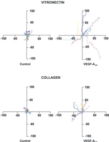
cells plated on collagen I or vitronectin by time-lapse video microscopy of individual cells (Figure 1). ECs exhibited a baseline mean migration speed of 17.31⫾0.09m/hour on vitronectin and 14.51⫾0.12 m/hour on collagen I (P⬍0.001; t test). In presence of VEGF-A165, EC speed
increased to 36.09⫾0.13 m/hour (P⬍0.0001 versus un-stimulated cells) and to 22.25⫾0.08 m/hour (P⬍0.0002 versus unstimulated cells) on vitronectin and collagen I, respectively. VEGF-A165stimulation resulted in higher
direc-tional persistence of cell migration27; in contrast unstimulated
ECs moved in random directions. The VEGF-A165effect was
more evident in cells plated on vitronectin. Unstimulated cells on collagen I and on vitronectin showed a similar directional persistence (vitronectin: 0.12⫾0.06; collagen I: 0.10⫾0.02), whereas VEGF-A165 increased this parameter in a greater
extent in cells on vitronectin (0.43⫾0.05) than on collagen I (0.19⫾0.04) (P⬍0.0001) (Figure 1).
To exclude that the observed effects were caused by different levels of VEGFR-2 expression, the high-affinity binding sites of VEGF-A on the surface of EC plated on collagen I or vitronectin were studied. Scatchard analysis indicated that the type of ECM did not influence the number of receptors and their affinity (supplemental Figure I).
Type I Collagen Inhibits VEGFR-2 Phosphorylation by Activating SHP2 Tyrosine Phosphatase
When VEGF-A165 stimulated ECs on vitronectin, VEGFR-2
phosphorylation was markedly higher as compared with cells on collagen I,17 suggesting a modulating role of tyrosine
phosphatases.13,28 –30In particular, both SHP1 and SHP2 have
been demonstrated to negatively regulate VEGFR-2.13,28
Therefore, we measured phosphatase activity in SHP1 and SHP2 immunocomplexes from VEGF-A165–stimulated ECs
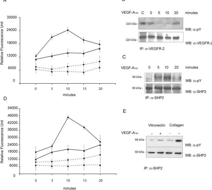
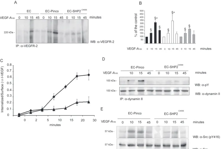
adherent on native ECM. VEGF-A165 promoted a rapid
activation of SHP2 that peaked at 10 minutes and then declined (Figure 2A). SHP-1 activity increased, but in slight manner, without reaching a well-defined peak within 20
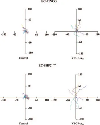
Figure 1. Tracks of ECs plated on vitronectin or collagen I.
Pan-els represent paths of cells (n⫽9) tracked by video-lapse microscopy in presence of VEGF-A165(10 ng/mL) or vehicle over
a period of 5 hours. Cell paths are replotted such that all paths start from the origin. The unit of measure on axes ism/h.
minutes. On the basis of this observation and previous data indicating that SHP2 is involved in GAS6 inhibition of VEGFR-2,28we pointed to this phosphatase. Our hypothesis
was further confirmed by the ability of SHP2 immunocom-plexes isolated from VEGF-A165–stimulated ECs to
dephos-phorylate in vitro phosdephos-phorylated VEGFR-2 (Figure 2B; supplemental Figure II). VEGF-A165 rapidly phosphorylated
SHP2 in tyrosine residues, an event known to be associated with SHP2 catalytic activity19 (Figure 2C; supplemental
Figure II). Both catalytic activity (Figure 2D) and tyrosine
phosphorylation (Figure 2E; supplemental Figure II) stimu-lated by VEGF-A165 were more pronounced in cells on
collagen I than on vitronectin.
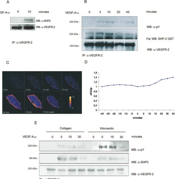
SHP2 Binds VEGFR-2
Figure 3A shows that SHP2 associated with activated VEGFR-2 immunoprecipitated from ECs on native ECM. To further analyze this interaction, we performed a far Western blot analysis using SHP2-GST fusion protein. Figure 3B and supplemental Figure III show that phosphorylated VEGFR-2
Figure 2. VEGF-A165activates SHP2. A and B, ECs (2⫻107) grown on native ECM were stimulated with VEGF-A165(solid line) (0.23 nmol/L) or
vehicle alone (dashed line) for the indicated times. Cell lysates were immunoprecipitated with anti-SHP2 (⽧) or anti-SHP1 (Œ) for phospha-tase assay (mean⫾SD; n⫽5) (A). Alternatively SHP2 immunocomplexes were incubated for 1 hour at 37°C with phosphorylated VEGFR-2 immunopurified from ECs activated by its ligand as detailed in Methods. Denatured proteins were separated by SDS-PAGE and blotted as indicated. Lane C indicates the initial level of VEGFR-2 phosphorylation incubated with a nonimmune anti-rabbit Ig immunocomplex from ECs stimulated for 10 minutes with VEGF-A165(B). C, ECs (2⫻107) grown on native ECM were stimulated as before and SHP2 was
immunopre-cipitated. Solubilized proteins were divided in two aliquots and immunoblotted as indicated. D to E, ECs (2⫻107) plated for 90 minutes on
collagen I or vitronectin were processed as described in (A and B). D, SHP2 phosphatase assay on unstimulated (dashed line) or VEGF-A165
-(0.23 mmol/L) stimulated (solid line) ECs plated on collagen I (⽧) or vitronectin (Œ). Mean⫾SD of five experiments. E, Tyrosine phosphoryla-tion of immunoprecipitated SHP2 from ECs plated on collagen I or vitronectin and stimulated with VEGF-A165(5 minutes; 0.23 mmol/L). B, C,
bound SHP2-GST in a time dependent-manner. To follow this interaction in living cells, we analyzed FRET of VEGFR-2-CFP and YFP- SHP2 in PAE stimulated with VEGF-A165.
Figure 3C and 3D shows that FRET occurred at the plasma membrane level and disappeared when VEGF-A165 was
re-moved from medium.
Type I Collagen Elicits SHP2 Interaction With VEGFR-2
Next, we investigated whether ECM could modulate the association of SHP2 with VEGFR-2. VEGFR-2 was immu-noprecipitated from ECs on collagen I or on vitronectin and the association with SHP2 was analyzed. The amount of
Figure 3. SHP2 binds VEGFR-2. A and B, ECs (3⫻07) were grown on native ECM and stimulated with VEGF-A
165(0.23 nmol/L) or
vehi-cle alone. Cell lysates were immunoprecipitated with Ab anti–VEGFR-2. Immunoprecipitate was divided in 2 (A) or 3 (B) aliquots and probed with the indicated Abs or with GST-SHP2 protein. Results are representative of 4 experiments and the densitometric analysis of panel B is reported in supplemental Figure III. C, FRET analysis of PAE cells coexpressing VEGFR-2-CFP and SHP2- YFP with a ratio 1:1 or 1:2 and stimulated with VEGF-A165for the indicated length of times. Degree of FRET efficiency was indicated by scale color from
blue to yellow. D, From the images in C, the FRET efficiency in a membrane portion was calculated. C and D, Representative of 25 cells, each obtained from 3 independent transfections. Variations within single experiments do not exceed 3% and between different experiments do not exceed 7%. E, ECs (3⫻107) were plated for 90 minutes on collagen I or on vitronectin and stimulated for the
indi-cated times with VEGF-A165(0.23 nmol/L). Cell lysates were immunoprecipitated, divided in 3 aliquots and blotted as indicated. Results
associated SHP2 with VEGFR-2 in cells on vitronectin was much reduced compared with cells on collagen I (Figure 3E; supplemental Figure III). In these experiments EC adhesion to ECM substrates was similar, thus excluding the possibility that the observed effect could be caused by differences in cells adhesion (not shown).
Effect of SHP2C459SOverexpression on
VEGFR-2 Activation
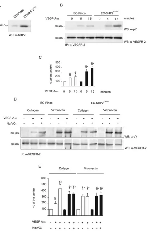
To investigate the involvement of SHP2 in VEGFR-2 signal-ing, ECs were infected with a retroviral vector carrying
SHP2C459Sdominant-negative mutant31(Figure 4A), plated on collagen I, and stimulated with VEGF-A165 for different
lengths of time. The amount of VEGFR-2 was not modified
by the constitutive expression of the transgene (supplemental Figure IV), suggesting that the results obtained were not dependent on a modification of the basal expression of VEGFR-2. The expression of SHP2C459S mutant enhanced
VEGFR-2 phosphorylation after 15 minutes of stimulation with VEGF-A165as compared with ECs infected with vector
alone (Figure 4B and 4C).
When EC infected with vector alone (EC-Pinco) were plated on collagen I (Figure 4D and 4E) VEGFR-2 phosphor-ylation triggered by VEGF-A165was lower than on
vitronec-tin. The pre-incubation with orthovanadate resulted in in-crease VEGFR-2 phosphorylation, which was particularly evident in stimulated cells on collagen I. Accordingly, SHP2C459Soverexpression abrogated the increased VEGFR-2
Figure 4. Effect of overexpression of
SHP2C459Son VEGFR-2 activation. A,
ECs were infected with a retroviral vector encoding SHP2C459Sand the expression
of the proteins was analyzed on cell lysate by immunoblot. B, Infected cells (2⫻107) plated on collagen I were
stimu-lated for the indicated times with VEGF-A165(0.23 nmol/L). Cell lysates were
immunoprecipitated as indicated. C, Densitometric analysis of the experiment shown in (B). Mean⫾SD of 4 experi-ments. ANOVA was F⫽77.55. §P⬍0.05 vs unstimulated cells. *P⬍0.05 vs cells carrying vector alone. White and black bars, respectively, indicate EC-PINCO and EC-SHP2C459S. D, Cells plated on
collagen I or on vitronectin were pre-treated with orthovanadate (1 mmol/L, 15 minutes) and then treated VEGF-A165
(0.23 nmol/L) for 10 minutes. After immu-noprecipitation, samples were divided in 2 aliquots, run in two gels, and immuno-blotted as indicated. E, Densitometric analysis of the experiment shown in (D). Mean⫾SD of 5 experiments. Before ANOVA (F⫽31.04) data were normalized according to z score §P⬍0.05 vs unstimulated cells plated on collagen I or vitronectin. *P⬍0.05 vs cells plated on collagen I and stimulated with VEGF-A165. White and black bars, respectively,
phosphorylation caused by orthovanadate. When EC-SHP2C459Swere plated on vitronectin, VEGFR-2
phosphory-lation was completely unaffected by orthovanadate treatment (Figure 4D and 4E). These data support the concept type I collagen modulates VEGFR-2 through SHP2.
Because the chemokinetic activity of VEGF-A165was low
on EC plated on collagen I (Figure 1), we investigated the rescuing effect of the expression of SHP2C459S. The baseline
migration speed on EC-Pinco and EC-SHP2C459Swas similar
(13.92⫾0.22 m/hour and 14.21⫾0.42 m/hour, respec-tively) and was increased ⬇1.5- to 2-fold by VEGF-A165
(EC-Pinco: 20.33⫾0.26 m/hour; EC-SHP2C 4 5 9 S:
25.82⫾0.28 m/hour). Analysis of directional persistence demonstrated that the expression of the mutant did not modify the resting values (EC-Pinco: 0.09⫾0.04; EC-SHP2C459S: 0.12⫾0.08), whereas it allowed VEGF-A
165to be
more efficient (EC-Pinco: 0.20⫾0.06; EC-SHP2C459S:
0.32⫾0.07) (Figure 5). The expression of SHP2C459Sdid not
affect the chemokinetic parameters of unstimulated or stim-ulated cells plated on vitronectin, which were completely comparable to those shown in Figure 1(data not shown).
SHP2 Regulates VEGFR-2 Internalization
On engagement with its ligands, VEGFR-2 is internal-ized.18,24,32,33 Recent data suggest that dephosphorylation of
tyrosine kinase receptors by protein phosphatases is involved in their downregulation through internalization.34 –36
Therefore, we looked at the internalisation of VEGFR-2 in ECs overexpressing SHP2C459S. To discriminate between the
receptor molecules that remain on the cell surface are cleaved by trypsin. This assay is an indirect indication of endocytosis, even though it does not offer any indication of the intracel-lular fate of the receptor.23,35 VEGFR-2 endocytosis was
detectable 10 minutes after stimulation and reached the peak at 15 minutes (Figure 6A and 6B). Very little amount of internalized VEGFR-2 was detected in ECs overexpressing SHP2C459S. These results were confirmed by internalisation
analysis of [125I]VEGF-A. Expression of SHP2C459S reduced
the internalisation of the ligand (Figure 6C).
The GTPase dynamin II is a regulator of both clathrin- and caveolae-dependent endocytosis and its activity is positively regulated by tyrosine phosphorylation.37,38In ECs dynamin II
is a substrate of Src kinase,39which is activated by
VEGF-A.33,40Furthermore, SHP2 regulates Src by controlling
phos-phorylation of its inhibitory tyrosine 529.41If SHP2
contrib-utes to the VEGFR-2 endocytosis, Src and dynamin II should be inhibited in cells carrying SHP2C459S. Indeed, VEGF-A
165–
evoked tyrosyl phosphorylation of dynamin II and phosphor-ylation of Src Tyr-416 were impaired in these cells (Figure 6D and 6E; supplemental Figure V). These data demonstrate that SHP2 stimulation by VEGFR-2 modulates dynamin II activation, probably through Src kinase activation.
SHP2 Binds Directly to Tyrosine 1173 in the VEGFR-2
C-terminal Tyr-1212 and -1173 are the two major VEGF-A– dependent autophosphorylation sites42 and their removal
impairs VEGFR-2 downregulation,43suggesting that one of
these Tyr residues could binds SHP2. VEGFR-2 and its mutants VEGFR-2Y1173Fand VEGFR-2Y1212Fwere
immunopre-cipitated from transfected PAE cells and the association with SHP2 was analyzed by immunoblot. The VEGF-A165–
depen-dent tyrosine phosphorylation of the receptor was lower in PAE carrying VEGFR-2Y1173F and VEGFR-2Y1212F mutants
than in cells transfected with wild type receptor accordingly to the relevance of these residues in the autophosphorylation event (Figure 7A). Mutation in tyrosine 1173 abrogated the interaction with SHP2, suggesting that this reside is crucial for SHP2 binding to VEGFR-2 (Figure 7B).
Discussion
Our study establishes a role for collagen I, a protein released within the provisional ECM by angiogenic ECs, as a negative regulator of VEGFR-2 through activation of tyrosine phos-phatase SHP2. This concept parallels the observation that ␣11integrin engaged by collagen activates the T-cell protein
tyrosine phosphatase function that inhibits epidermal growth factor receptor signaling.44
We demonstrated that EC adhesion to collagen I reduces VEGF-A165–induced VEGFR-2 autophosphorylation by
re-cruiting SHP2 to the phosphorylated tyrosine 1173. This
Figure 5. Effect of overexpression of SHP2C459Son EC
chemoki-nesis. Panels represent paths of EC-Pinco and EC- SHP2C459S
plated on collagen I (n⫽9) in presence of VEGF-A165as detailed
observation explains our previous data showing that EC proliferation stimulated by VEGF-A165is lower on collagen I
than on vitronectin.17Moreover, we provide evidences that:
(1) EC motility induced by VEGF-A165 is enhanced on
vitronectin as compared with collagen I; (2) SHP2 activity and tyrosine residue phosphorylation are activated by VEGF-A when ECs adhere on native ECM or on vitronectin; however, these effects are greatly increased when EC adhere
Figure 6. Effect of overexpression of SHP2C459Son VEGFR-2 internalization, dynamin II, and Src phosphorylation. ECs (2⫻107)
overex-pressing SHP2C459Swere plated on collagen I and stimulated for the indicated times with VEGF-A
165(0.23 nmol/L). A, Cells were
sub-mitted to trypsin treatment as described in Methods, and cell lysates were immunoprecipitated with an Ab anti the C-terminus of VEGFR-2 and immunoblotted with an Ab anti–VEGFR-2. B, Densitometric analysis of the experiment shown in (A). Mean⫾SD of 4 experiments. ANOVA was F⫽21.50. §P⬍0.05 vs unstimulated cells. *P⬍0.05 vs SHP2C459Scells stimulated for 15 minutes with
VEGF-A165. White, black, and hatched bars, respectively, indicate EC, EC-Pinco, and EC-SHP2C459S. C, EC overexpressing SHP2C459S(Œ) or
vector alone (⽧) were incubated, then incubated at 37°C with [125I]VEGF-A. Internalization was monitored as outlined in Methods.
Mean⫾SD of three samples in one experiment of two. D, Cell lysates were immunoprecipitated with Ab anti-dynamin II, divided in 2 aliquots, separated by SDS-PAGE and blotted as indicated. E, Total amount of Src and pY416 Src was detected by immunoblotting on cell lysates. Results are representative of 7 (C) and 4 (D) experiments, respectively. Densitometric analysis of (C) and (D) is reported in supplemental Figure VI.
Figure 7. SHP2 interacts with Y1173 of
VEGFR2. Wild-type and mutant VEGFR-2 cDNAs were transiently transfected in PAE cells that were stimulated with VEGF-A165. Lysates were
immunoprecipi-tated with anti–VEGFR-2 Ab and immu-noblotted with anti-pY (A) or anti-SHP2 (B). Results are representative of at least 3 experiments.
Downregulation of tyrosine kinase receptors by phospho-tyrosine phosphatases could be considered a simpler and faster way to modify their behavior and functions as com-pared with clathrin-mediated or caveolae-dependent endocy-tosis or to proteasome-mediated degradation.45 SHP2 has
been generally considered a positive downstream signal activated by membrane receptors.46However, emerging
evi-dences suggest that it may positively or negatively regulate signaling pathways depending on the specific type of signal-ing network. For example, SHP2 may act as a negative regulator of platelet-derived growth factor receptor by induc-ing dephosphorylation of the receptor itself or of its cognate substrates.47– 49However, SHP2 is also implicated in positive
regulation of this receptor through regulation of Ras or focal adhesion kinase.50,51 Similarly, SHP2 exerts both negative
and positive influences on T-cell receptor52,53 and JAKs/
STATs pathway.54,55Finally, SHP2 exclusively acts as
neg-ative regulator in angiotensin II receptor A1-mediated signals.56
Here, we do not investigate the precise mechanism by which collagen I favors SHP2-mediated VEGFR-2 dephos-phorylation. Recently, we have demonstrated that SHP2 is involved in the negative control of VEGFR-2 triggered by GAS6-dependent Axl stimulation.28Furthermore, our
obser-vation parallels the results that VEGFR-2 may be negatively regulated by TIMP-2 through SHP1.13In ECs stimulated by
TIMP-2, SHP1 shifts from␣31integrin to VEGFR-2, which
is dephosphorylated.
It has been reported that in vascular smooth muscle cells 3 engagement by vitronectin results in tyrosine phosphory-lation of its cytosolic domain and recruitment of SHP2, which modulate insulin growth factor I receptor.57Thus, we
hypoth-esize a protective role on VEGFR-2 signaling by vitronectin-engaged␣v3, which recruits SHP2 and preserves the receptor
from phosphatase activity. In contrast, EC adhesion on collagen I, which does not depend on␣v3 integrin, could
allow SHP2 interacting with VEGFR-2. Preliminary evi-dences demonstrate that a phosphatase activity is associated to 3 in ECs plated on vitronectin and stimulated by VEGF-A165. However,1 immunocomplexes do not contain
any phosphatase activity both in cells plated on collagen I and vitronectin (S. Mitola, unpublished).
Then we showed that SHP2 plays a relevant role in VEGFR-2 internalisation. VEGFR-2 undergoes endocytosis by a Cbl-dependent ubiquitination process58 or through a
mechanism caveolin-1– dependent.18 Here, we extend these
observations by showing overexpression of SHP2C459S
dra-matically decreases VEGFR-2 internalization, suggesting that dephosphorylation is involved in regulating its trafficking.
VEGF-A165induced tyrosyl phosphorylation of dynamin II,
a GTPase regulator of vesicle fission required for the control of both clathrin- and caveolae-mediated endocytosis.38
Pre-tion. These data suggest that SHP2 activation dephosphory-lates VEGFR-2 and concomitantly activates Src kinase, which triggers a dynamin-dependent receptor internalization. SHP2 binds Tyr-1173 of stimulated VEGFR-2. This residue seems important in internalisation process as demonstrated by studying truncated receptors lacking 1173 and Tyr-1212.61This process occurs in ECs growing on native ECM,
as demonstrated by the data showing that this phosphatase is also activated by VEGF-A165 in ECs adherent to this ECM
(Figures 2A, 2B, and 3) as well as in cells on collagen I. Here, we did not address the behavior of the internalized VEGFR-2 in EC adhering on collagen I. VEGFR-2 could either be accumulated in the endosomal compartment and degraded or recycled to the membrane, thus allowing a different degree of EC activation or a modified spatial regulation of the signal. It is known that tyrosine kinase receptors may be translocated to the nucleus.62 Notably, it has been recently reported that
VEGF-A through VEGFR-2 may be transferred to the nu-cleus of migrating ECs.32We may speculate that collagen I
mediated internalization of VEGFR-2 results in attenuation of VEGFR-2–mediated signals or in activation of undefined EC responses and that a modulation of SHP2-dependent mecha-nism of internalization by ECM characterizes specific steps of angiogenesis.
Acknowledgments
This study was supported by Associazione Italiana per la Ricerca sul Cancro, Istituto Superiore di Sanita`. (AIDS and Stem Cells National Projects), Ministero dell’ Universita` e della Ricerca (MIUR) (60% and PRIN 2004 projects), Fondi incentivazione della Ricerca di Base (RBNE0184P, RBNE01MAWA, RBNE01T8C8), Ministero della Salute (Ricerca Finalizzata 2002 and 2003 to M.A. and F.B.), Regione Piemonte, and European Community (LSHM-CT-2003-503251) (http:://www.engv.org). We thank Letizia Lanzetti, Luca Primo, and Guido Serini for their suggestions, and Vincenzo Bag-nardi for statistical analysis.
References
1. Jain RK. Molecular regulation of vessel maturation. Nat Med. 2003;9: 685– 693.
2. Kalluri R. Basement membranes: structures, assembly and role in tumor angiogenesis. Nature Rev Cancer. 2003;3:422– 433.
3. Stupack DG, Cheresh DA. ECM remodeling regulates angiogenesis: endothelial integrins look for new ligands. Sci STKE. 2002;2002:E7. 4. Bosman TF, Stamenkoovic I. Functional structure and composition of the
extracellular matrix. J Pathol. 2003;200:423– 428.
5. Seandel M, Noack-Kunnaman K, Zhu D, Aimes RT, Quigley JP. Growth factor-induced angiogenesis in vivo requires specific cleavage of fibrillar type I collagen. Blood. 2001;97:2323–2332.
6. Dameron KM, Volpert OV, Tainsky MA, Bouch N. Control of angiognesis in fibroblasts by regulation of thrombospondin-1. Science. 1994;265:1582–1585.
7. George EL, Baldwin HS, Hynes RO. Fibronectins are essential for heart and blood vessel morphogenesis but are dispensable for initial specifi-cation of precursor cells. Blood. 1997;90:3073–3081.
8. Brooks PC, Montgomery AMP, Rosenfeld M, Reisfeld RA, Hu T, Klier G, Cheresh DA. Integrin avb3 antagonists promote tumor regression by
inducing apoptosis of angiogenic blood vessels. Cell. 1994;79: 1157–1164.
9. Kim S, Bell K, Mousa SA, Varner JA. Regulation of angiogenesis in vivo by ligation of␣51with the cell-binding domain of fibronectin. Am J
Pathol. 2000;156:1345–1362.
10. Suzuma K, Takagi H, Otani A, Honda Y. Hypoxia and vascular endo-thelial growth factor stimulate angiogenic integrin expression in bovine retinal microvascular endothelial cells. Invest Ophthalmol Vis Sci. 1998; 39:1028 –1035.
11. Friedlander M, Brooks PC, Shaffer RW, Kincaid CM, Varner JA,
Cheresh DA. Definition of two angiogenic pathways by distinct ␣v
integrins. Science. 1995;270:1500 –1502.
12. Ferrara N, Gerber HP, LeCouter J. The biology of VEGF and its receptors. Nat Med. 2003;9:669 – 676.
13. Seo DW, Li H, Guedez L, Wingfield PT, Diaz T, Salloum R, Wei BY, Stetler-Stevenson WG. TIMP-2 mediated inhibition of angiogenesis: an MMP-independent mechanism. Cell. 2003;114:171–180.
14. Carmeliet P, Lampugnani MG, Moons L, Breviario F, Compernolle V, Bono F, Balconi G, Spagnuolo R, Oostuyse B, Dewerchin M, Zanetti A, Angellilo A, Mattot V, Nuyens D, Lutgens E, Clotman F, de Ruiter MC, Gittenberger-de Groot A, Poelmann R, Lupu F, Herbert JM, Collen D, Dejana E. Targeted deficiency or cytosolic truncation of the VE-cadherin gene in mice impairs VEGF-mediated endothelial survival and angio-genesis. Cell. 1999;98:147–157.
15. Dardik R, Loscalzo J, Eskaraev R, Inbal A. Molecular mechanisms underlying the proangiogenic effect of Factor XIII. Arterioscler Thromb
Vasc Biol. 2005;25:526 –532.
16. Borges E, Jan Y, Ruoslahti E. Platelet-derived growth factor receptor beta and vascular endothelial growth factor receptor 2 bind to the beta 3 integrin through its extracellular domain. J Biol Chem. 2000;275: 39867–39873.
17. Soldi R, Mitola S, Strasly M, Defilippi P, Tarone G, Bussolino F. Role of alphavbeta3 integrin in the activation of vascular endothelial growth factor receptor-2. Embo J. 1999;18:882– 892.
18. Labrecque L, Royal I, Surprenant DS, Patterson C, Gingras D, Beliveau R. Regulation of vascular endothelial growth factor receptor-2 activity by caveolin-1 and plasma membrane cholesterol. Mol Biol Cell. 2003;14: 334 –347.
19. Tonks NK, Neel BG. From form to function: signaling by protein tyrosine phosphatases. Cell. 1996;87:365–368.
20. Feng G. Shp-2 tyrosine phosphatase: signaling one cell or many. Exp Cell
Res. 1999;253:47–54.
21. Whelan MC, Senger DR. Collagen I initiates endothelial cell morpho-genesis by inducing actin polymerization through suppression of cyclic AMP and protein kinase A. J Biol Chem. 2003;278:327–334. 22. Kim S, Bakre M, Yin H, Varner JA. Inhibition of endothelial cell survival
and angiogenesis by protein kinase A. J Clin Invest. 2002;110:933–941. 23. Ceresa BP, Kao AW, Santeler SR, Pessin JE. Inhibition of clathrin-mediated endocytosis selectively attenuates specific insulin receptor signal transduction pathways. Mol Cell Biol. 1998;18:3862–3870. 24. Dougher M, Terman BI. Autophosphorylation of KDR in the kinase
domain is required for maximal VEGF-stimulated kinase activity and receptor internalization. Oncogene. 1999;18:1619 –1627.
25. Tertoolen LG, Blanchetot C, Jiang G, Overvoorde J, Gadella TW Jr, Hunter T, den Hertog J. Dimerization of receptor protein-tyrosine phos-phatase alpha in living cells. BMC Cell Biol. 2001;2:8 –15.
26. Wijelath ES, Murray J, Rahman S, Patel Y, Ishida A, Strand K, Aziz S, Cardona C, Hammond WP, Savidge GF, Rafii S, Sobel M. Novel vascular endothelial growth factor binding domains of fibronectin enhance vascular endothelial growth factor biological activity. Circ Res. 2002;91: 25–31.
27. Gu J, Tamura M, Pankov R, Danen EH, Takino T, Matsumoto K, Yamada KM. Shc and FAK differentially regulate cell motility and directionality modulated by PTEN. J Cell Biol. 1999;146:389 – 403.
28. Gallicchio M, Mitola S, Valdembri D, Fantozzi R, Varnum B, Avanzi GC, Bussolino F. Inhibition of vascular endothelial growth factor receptor 2-mediated endothelial cell activation by Axl tyrosine kinase receptor.
Blood. 2005;105:1970 –1976.
29. Kroll J, Waltenberger J. The vascular endothelial growth factor receptor KDR activates multiple signal transduction pathways in porcine aortic endothelial cells. J Biol Chem. 1997;272:32521–32527.
30. Huang L, Sankar S, Lin C, Kontos CD, Schroff AD, Cha EH, Feng SM, Li SF, Yu Z, Van Etten RL, Blanar MA, Peters KG. HCPTPA, a protein tyrosine phosphatase that regulates vascular endothelial growth factor
receptor-mediated signal transduction and biological activity. J Biol
Chem. 1999;274:38183–38188.
31. Zhao Z, Tan Z, Wright J, Diltz C, Shen S, Krebs E, Fischer E. Altered expression of protein-tyrosine phosphatase 2C in 293 cells affects protein tyrosine phosphorylation and mitogen-activated protein kinase activation.
J Biol Chem. 1995;270:11765–11769.
32. Li W, Keller G. VEGF nuclear accumulation correlates with phenotypical changes in endothelial cells. J Cell Sci. 2000;113(Pt 9):1525–1534. 33. Meyer RD, Dayanir V, Majnoun F, Rahimi N. The presence of a single
tyrosine residue at the carboxyl domain of vascular endothelial growth factor receptor-2/FLK-1 regulates its autophosphorylation and activation of signaling molecules. J Biol Chem. 2002;277:27081–27087. 34. Ostman A, Bohmer FD. Regulation of receptor tyrosine kinase signaling
by protein tyrosine phosphatases. Trends Cell Biol. 2001;11:258 –266. 35. Chiarugi P, Cirri P, Taddei ML, Talini D, Doria L, Fiaschi T, Buricchi F,
Giannoni E, Camici G, Raugei G, Ramponi G. New perspectives in PDGF receptor downregulation: the main role of phosphotyrosine phos-phatases. J Cell Sci. 2002;115:2219 –2232.
36. Haj FG, Verveer PJ, Squire A, Neel BG, Bastiaens PI. Imaging sites of receptor dephosphorylation by PTP1B on the surface of the endoplasmic reticulum. Science. 2002;295:1708 –1711.
37. Hinshaw JE. Dynamin and its role in membrane fission. Annu Rev Cell
Dev Biol. 2000;16:483–519.
38. Orth JD, McNiven MA. Dynamin at the actin-membrane interface. Curr
Opin Cell Biol. 2003;15:31–39.
39. Shajahan AN, Timblin BK, Sandoval R, Tiruppathi C, Malik AB, Minshall RD. Role of Src-induced dynamin-2 phosphorylation in caveolae-mediated endocytosis in endothelial cells. J Biol Chem. 2004; 279:20392–20400.
40. He H, Venema VJ, Gu X, Venema RC, Marrero MB, Caldwell RB. Vascular endothelial growth factor signals endothelial cell production of nitric oxide and prostacyclin through flk-1/KDR activation of c-Src.
J Biol Chem. 1999;274:25130 –25135.
41. Zhang SQ, Yang W, Kontaridis MI, Bivona TG, Wen G, Araki T, Luo J, Thompson JA, Schraven BL, Philips MR, Neel BG. Shp2 regulates SRC family kinase activity and Ras/Erk activation by controlling Csk recruitment. Mol Cell. 2004;13:341–355.
42. Takahashi T, Yamaguchi S, Chida K, Shibuya M. A single autophos-phorylation site on KDR/Flk-1 is essential for VEGF-A-dependent acti-vation of PLC-gamma and DNA synthesis in vascular endothelial cells.
Embo J. 2001;20:2768 –2778.
43. Meyer RD, Singh AJ, Rahimi N. The carboxyl terminus controls ligand-dependent activation of VEGFR-2 and its signaling. J Biol Chem. 2004; 279:735–742.
44. Mattila E, Pellinen T, Nevo J, Vuoriluoto K, Arjonen A, Ivaska J. Negative regulation of EGFR signalling through integrin-alfa1beta1-mediated activation of protein tyrosine phosphatase TCPTP. Nat Cel Biol. 2005;7:78 – 85.
45. Haglund K, Di Fiore PP, Dikic I. Distinct monoubiquitin signals in receptor endocytosis. Trends Biochem Sci. 2003;28:598 – 603. 46. Tonks NK, Neel BG. Combinatorial control of the specificity of protein
tyrosine phosphatases. Curr Opin Cell Biol. 2001;13:182–195. 47. Meng TC, Fukada T, Tonks NK. Reversible oxidation and inactivation of
protein tyrosine phosphatases in vivo. Mol Cell. 2002;9:387–399. 48. Klinghoffer RA, Kazlauskas A. Identification of a putative Syp substrate,
the PDGF beta receptor. J Biol Chem. 1995;270:22208 –22217. 49. Cossette LJ, Hoglinger O, Mou L, Shen SH. Localization and
down-regulating role of the protein tyrosine phosphatase PTP2C in membrane ruffles of PDGF-stimulated cells. Exp Cell Res. 1996;223:459 – 466. 50. Bennett AM, Tang TL, Sugimoto S, Walsh CT, Neel BG.
Protein-tyrosine-phosphatase SHPTP2 couples platelet-derived growth factor receptor beta to Ras. Proc Natl Acad Sci U S A. 1994;91:7335–7339. 51. Qi JH, Ito N, Claesson-Welsh L. Tyrosine phosphatase SHP-2 is involved
in regulation of plateled-derived growth factor-induced migtation. J Biol
Chem. 1999;274:14455–14463.
52. Marmor MD, Yarden Y. Role of protein ubiquitylation in regulating endocytosis of receptor tyrosine kinases. Oncogene. 2004;23:2057–2070. 53. Frearson JA, Alexander DR. The phosphotyrosine phosphatase SHP-2 participates in a multimeric signaling complex and regulates T cell receptor (TCR) coupling to the Ras/Mitogen-activated protein kinase (MAPK) pathway in Jurkat T cells. J Exp Med. 1998;187:1417–1426. 54. Yu CL, Jin YJ, Burakoff SJ. Cytosolic tyrosine dephosphorylation of
STAT5. Potential role of SHP-2 in STAT5 regulation. J Biol Chem. 2000;275:599 – 604.
the insulin-like growth factor I receptor. Mol Endocrinol. 2003;17: 1824 –1833.
58. Duval M, Bedard-Goulet S, Delisle C, Gratton JP. Vascular endothelial growth factor-dependent down-regulation of Flk-1/KDR involves
Cbl-tyrosine kinase Pyk2 as a novel effector of fibroblast growth factor receptor 3 activation. J Biol Chem. 2004;279:28450 –28457.
62. Wells A, Marti U. Signalling shortcuts: cell-surface receptors in the nucleus. NatRev Mol Cell Biol. 2002;3:697–702.