University of Pisa
PhD School in Morphology and Function of Normal and Pathological Cells and Tissues
(XXIV cycle) 2009-2011
Doctor of Philosophy Thesis in
Functional and morphological study of cells in
connected culture in response to interactions
associated with nanoparticles
(SSD BIO/13)
Candidate Supervisor
Nadia Ucciferri Prof. Francesco Fornai
____________ ______________
Prof. Arti Ahluwalia ______________
Advisor
Dr. Claudio Domenici ______________
Prof. Claus-Michael Lehr ______________
Table of Contents
Abstract ... 6
SECTION I, Introduction ... 8
Chapter 1: In vitro models for Toxicology Testing 1. Toxicology ... 9
2. The animal testing debate ... 11
3. In vitro models ... 13
4. Advanced in vitro models and body on a chip model... 18
Chapter 2: Nanotoxicology: an emerging issue 1. Nanoparticles: properties and applications ... 28
2. Nanotoxicology ... 32
3. Nanoparticles exposure and biokinetic ... 35
4. Nanoparticles testing and risk assessment ... 38
Chapter 3: Bioreactor design and improvement for investigating absorption, biotransformation & biodistribution 1. Bioreactor: MCmB ... 42
2. Allometry ... 46
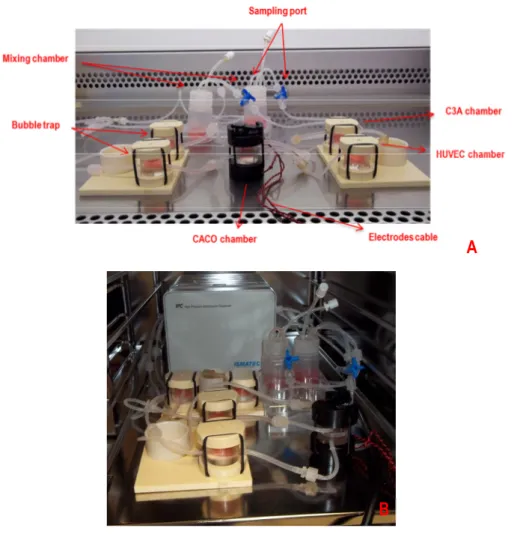
3. InLiveTox bioreactor system ... 48
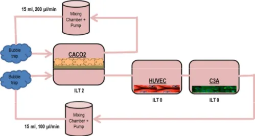
3.1. ILT 2 intestinal bioreactor ... 49
SECTION II, Experimental design, materials and methods ... 52
Chapter 4: Experimental design 1. Establishment of NPs testing on HUVEC static culture ... 53
2. Identification of common dynamic culture conditions ... 54
3. Allometric scaling ... 55
4. NP dynamic testing with single tissue configuration ... 56
5. Evaluation of ILT system baseline ... 56
6. NPs testing in ILT system ... 57
Chapter 5: Materials and methods 1. Cell culture ... 58
1.1. HUVEC extraction and culture ... 58
1.2. C3A culture ... 59
1.3. CaCo-2 culture ... 59
1.4. Dynamic cell culture ... 60
1.4.1. Assembling of ILT 0 ... 60
1.4.2. Assembling of ILT 2 ... 60
2. Nanoparticles ... 61
3. Experimental protocols ... 61
3.1. Establishment of NPs testing on HUVEC static culture ... 62
3.2. Identification of common culture conditions ... 62
3.3. Allometric scaling ... 63
3.4. NP dynamic testing with single tissue configuration ... 65
3.5. Evaluation of ILT complete system baseline ... 66
3.6. NPs testing in ILT system ... 67
4. Assays ... 68
4.1. Assays on cells ... 68
4.2. Assays on medium ... 71
SECTION III, Results and Discussion ... 73
Chapter 6: Results 1. Establishment of NPs testing on HUVEC static culture ... 74
1.1. Cell viability ... 74
1.2. Oxidative stress ... 77
1.3. Inflammation ... 78
1.3.1. Interleukin-8 (IL 8) ... 78
1.3.2. Intercellular Adhesion Molecule-1 or CD54 (ICAM-1) .... 79
1.3.3. Tumor Necrosis Factor-α (TNF-α) ... 80
1.4. Apoptosis ... 80
1.4.1. Fas-Ligand ... 80
1.5. Function of cells: von Willebrand Factor expression ... 81
1.5.1. vWF mRNA quantification ... 81
1.5.2. vWF protein expression ... 82
2. Identification of common culture conditions ... 85
2.1. Media testing ... 85
2.1.1. HUVEC viability ... 86
2.1.2. C3A viability ... 87
2.1.3. CaCo-2 viability ... 88
2.2. Dynamic condition set up ... 88
2.2.1. Viability ... 88
2.2.2. Functional marker of HUVEC ... 89
2.2.3. Functional marker of C3A ... 90
3. Allometric scaling ... 91 3.1. Viability ... 91 3.2. Carbohydrate metabolism ... 91 3.2.1. Glucose ... 92 3.2.2. D-Lactate ... 92 3.3. Fat metabolism... 93
3.3.1. Triglyceride ... 93
3.3.2. Free Fatty Acid ... 94
3.3.3. Glycerol ... 95
3.4. Function ... 95
3.4.1. Cytochrome P450 subfamily 3A4 ... 96
3.4.2. Albumin ... 96
3.4.3. Urea ... 97
4. NP dynamic testing with single tissue configuration ... 98
4.1. HUVEC ... 98
4.1.1. Cell viability ... 98
4.1.2. Inflammation ... 99
4.1.3. Apoptosis ... 100
4.1.4. Cell function: von Willebrand Factor expression ... 100
4.2. C3A ... 101
4.2.1. Cell viability ... 101
4.2.2. Inflammation ... 102
4.2.3. Apoptosis ... 102
4.2.4. Cell function: albumin release and phalloidin expression ... 103
5. Evaluation of ILT system baseline ... 104
5.1. Cell viability ... 104
5.2. Inflammation ... 106
5.3. Apoptosis ... 107
5.4. Function of CaCo-2: TEER ... 107
5.5. Function of HUVEC: von Willebrand Factor expression ... 107
5.6. Function of C3A: albumin release and phalloidin expression . 108 6. NPs testing in ILT system ... 109
6.1. Polystyrene-FITC 55 nm NP ... 110
6.1.1. Viability ... 110
6.1.2. Inflammation ... 110
6.1.4. Function of CaCo-2: TEER ... 111
6.1.5. Function of HUVEC: von Willebrand Factor expression 111 6.1.6. Function of C3A: albumin release and phalloidin expression ... 112
6.1.7. PS-FITC 55 nm NP passage evaluation ... 112
6.2. Ag NP preliminary results ... 114
6.2.1. Viability ... 114
6.2.2. Inflammation ... 114
6.2.3. Apoptosis ... 115
6.2.4. Function of CaCo-2: TEER ... 115
6.2.5. Function of HUVEC: von Willebrand Factor expression 116 6.2.6. Function of C3A: albumin release and phalloidin expression ... 116
Chapter 7: Discussion 1. Establishment of NPs testing on HUVEC static culture ... 117
2. Identification of common dynamic culture conditions ... 122
3. Allometric scaling ... 123
4. NP dynamic testing with single tissue configuration ... 127
5. Evaluation of ILT system baseline ... 128
6. NPs testing in ILT system ... 130
SECTION III, Conclusion ... 134
Chapter 8: Conclusion ... 135
Glossary ... 138
Abstract
The improvement of in vitro and in vivo models for tissue engineering, pharmacology, or metabolic studies, is largely requested. In fact, while in vitro models are usually preferred due to their convenience and compatibility with the 3Rs, unfortunately they lack biochemical interactions, for instance cell-cell cross-talk or important bio-barriers leading to non-physiologically relevant results.The aim of this thesis is to develop a new in vitro system able to recreate the main barrier through ingestion, the intestinal epithelium as well as a connected target organs, the vascular endothelium and the liver. The thesis is focused on the study of nanoparticle (NP) fate after ingestion, the ability of NPs to cross the intestinal barrier and the effects on relevant target tissues. In order to achieve this aim simplified models were firstly used so that the complexity of the system could be increased stepwise to include additional cell types, more complex 3D models of tissues, and more sophisticated and specific tests of cellular responses to the presence of nanoparticles.
The system developed is a new body-on-a-plate device able to study the physiologically relevant doses that actually reach the systemic circulation after intestinal absorption. It has a microfluidic flow which transports messaging molecules from cell to cell and stimulates them with a constant low shear stress.
Hence, the results obtained with this new model were compared with data generated in conventional static cell cultures in order to validate the system and gain a better insight on the systemic effects of NP toxicity.
Besides the study of the toxicity of nanoparticles of industrial and environmental interest, this thesis demonstrates the importance of advanced in vitro testing, pointing out the differences in results from standard simple cultures with respect to those obtained from more relevant physiological model.
Section I
Introduction
Chapter 1
In vitro
models for Toxicology Testing
1. TOXICOLOGY
Toxicology is the study of the adverse effects of chemical, physical or biological agents on people, animals, and the environment. Many studies are required to ensure the safety of medicines, household and gardening chemicals, and industrial and natural chemicals to which everybody is continuously exposed.
Toxicologists know that no substance is risk-free. Paracelsus, in the 16th century, summarized this concept in the sentence “All things are poison, and nothing is without poison; only the dose permits something not to be poisonous", or more commonly, "the dose makes the poison". That is to say, substances considered toxic are harmless in small doses, and conversely an ordinarily harmless substance can be deadly if over-consumed.
For this reason, toxicology research is important for ensuring the health of humans, animals, and their environments. This research is intended to identify harmful effects of potential new products and to provide understanding of the mechanisms by which chemical substances cause injury, in order to use this information in the treatment of poisonings.
Toxicology represents an area of science of growing importance, largely as a consequence of the rapid increase in environmental and safety legislation. A further driver of toxicology is the continuing growth of the industrial sector which develops a greater number of new materials and compounds.
All organisms are composed of chemicals, and chemical reactions power all life processes. When a substance is introduced into a body, it can interact in many places and effects upon one process can cause unexpected consequences in others.
The use of animals in this kind of experiments is fundamental because such complexity cannot be duplicated in normal cell culture or in non-living systems. For example, toxicity can be influenced by the speed with which the substance enters the system, how the metabolic organs change it and how it is taken up by and interacts with various body tissues.
Fig. 1: Schematic diagram explaining how a chemical can change into another thanks to internal metabolism. Adapted from [1].
Moreover, because "the dose makes the poison" at the level of the individual organ, we need to be able to analyze not only how a chemical acts, but the relationship between the dose given to the animal and the dose delivered to the different organs and tissues in the body. Studies in whole animals are required to ensure the proper use of beneficial chemicals such as medicines, because the tissue or organ receiving the beneficial effect might be harmed if exposures are greater than needed. In many cases, laboratory tools simply cannot duplicate these complicated phenomena [2]. Ultimately, animal testing is the best method to detect effects such as cancer and birth defects, even if it sometimes is difficult to identify the right model to test as demonstrated by the Thalidomide case whose
teratogenicity was found only occasionally in the huge set of different animals and species tested [3].
2. THE ANIMAL TESTING DEBATE
The ethical questions raised by performing experiments on animals are subject to much debate, and viewpoints have shifted significantly over the 20th century [4].The case for animal experiments is that they will produce such great benefits for humanity that it is morally acceptable to harm a few animals. The equivalent case against is that the level of suffering and the number of animals involved are both so high that the benefits to humanity don't provide moral justification.
Animal welfare, and animal rights organizations (such as PETA -People for the Ethical Treatment of Animals- and BUAV -British Union for the Abolition of Vivisection-) question the legitimacy of animal testing, arguing that it is cruel, poor scientific practice, poorly regulated, that medical progress is being held back by misleading animal models, that some of the tests are outdated, that it cannot reliably predict effects in humans, that the costs outweigh the benefits, or that animals have an intrinsic right not to be used for experimentation.[3]
Heavy pressure from these associations leads to a high interest of scientist and governments to address the animal testing issue. Moreover, as technology has advanced, new ways to stop animal testing have come into the picture. In 1959, Russel and Burch published The Principles of Humane Experimental Technique in which “the 3 R’s principles” were proposed to encourage scientist reducing the impact of research on animals.
“The 3 R’s” stand for reduce, refine and replace and means:
Reduction:
• Reducing the number of animals used in experiments by: o Improving experimental techniques
o Improving techniques of data analysis o Sharing information with other researchers Refinement:
• Refining the experiment or the way the animals are cared for so as to reduce their suffering by:
o Using less invasive techniques o Better medical care
o Better living conditions Replacement:
• Replacing experiments on animals with alternative techniques such as:
o Experimenting on cell cultures instead of whole animals
o Using computer models
o Studying human volunteers o Using epidemiological studies
In November 2008 the European Union put forward proposals to revise the directive of 1986 for the protection of animals used in scientific experiments in line with the three Rs principle of replacing, reducing and refining the use of animals in experiments. The proposals have three aims:
• to considerably improve the welfare of animals used in scientific procedures
• to ensure fair competition for industry
• to boost research activities in the European Union [5].
Now EU rules for animal experimentation are more restrictive, requiring that experiments where animals are used be subject to authorization and state that alternatives to testing on animals must be used when available and that the number of animals used in projects be reduced to a minimum [6].
The Commission and industry have set up the European Partnership for Alternative Approaches to Animal testing (EPAA) whose aim is to promote
the development of new ‘3R’ methods as alternative approaches to the use of animals in safety assessment.
Another thing to take in account about the reducing of animal testing issue is that concerning the Cosmetics industry. The 7th amendment to the Cosmetics Directive (76/768/EEC) foresees a testing and marketing ban on cosmetics tested on animals. The directive on animal testing (86/609/EEC) does not amend the rules provided for in other pieces of EU legislation. The sales ban means not only that cosmetics’ testing is mostly ended in the European Union but also that even products tested elsewhere cannot be sold there.
The testing ban on cosmetics has been in application since 2009 when testing was prohibited irrespective of alternatives to animal testing being available. The marketing ban applies unconditionally to all human health effects with the exception of three toxicological effects such as repeated-dose toxicity, reproductive toxicity and toxicokinetics. The year 2013 is foreseen as the deadline for a ban on these specific health effects.
The interest for non-animal methods is clearly really high.
3. IN VITRO MODELS
In vitro models are the obvious solution to the replacement of animal testing.
In vitro refers to studies in experimental biology that are conducted using
components of an organism that have been isolated from their usual biological context in order to permit a more detailed or more convenient analysis than can be done with whole organisms. Common examples of in vitro experiments include:
• cells derived from multicellular organisms (cell culture or tissue culture)
• subcellular components (e.g. mitochondria or ribosomes)
• cellular or subcellular extracts (e.g. wheat germ or reticulocyte extracts)
• purified molecules in the test tube (proteins, DNA, or RNA, either individually or in combination) [4].
In vitro studies allow scientists to isolate specific cells, bacteria, and viruses and study them without the complexity of a whole organism. This permits an enormous level of simplification of the system under study, so that the investigator can focus on a small number of components [7]. For example, the knowledge of the mechanism by which a protein target recognizes and binds to a receptor is the result of the extensive use of in vitro work that means isolate the proteins, identify the cells and genes that produce them, study the physical properties of their interaction with molecules, and identify how those interactions lead to cellular signals that activate other components of the response pathway.
In toxicology, in vitro testing methods are employed primarily to identify potentially hazardous chemicals and/or to confirm the lack of certain toxic properties in the early stages of the development of potentially useful new substances such as therapeutic drugs, agricultural chemicals and direct food additives
In vitro toxicity testing:
• provides rapid and effective means of screening and ranking chemicals in food or environment for a number of toxicology endpoint
• allows the understanding of the mechanism by which a toxic lead effects at both the cellular and molecular level, and of both causal and adaptive responses
• is essential for bridging between experimental animal and human, and addressing the correct species choice
• provides well defined and simplified system for studying structure-activity relationship
• provides a means for identification of key molecular events that are involved in toxicity, enabling the development of effective biomarkers of effect
• enables detailed analysis of the toxicological consequences of genetic variation within the population
• enables assessment of cell-specific (e.g. liver, cardiac, kidney, neural, immune system) and, where possible, tissue-specific (e.g. embryo) effects;
• allows establishing the nature of concentration-effect relationships and the existence of effect-specific thresholds in cells from different species and different tissues [8].
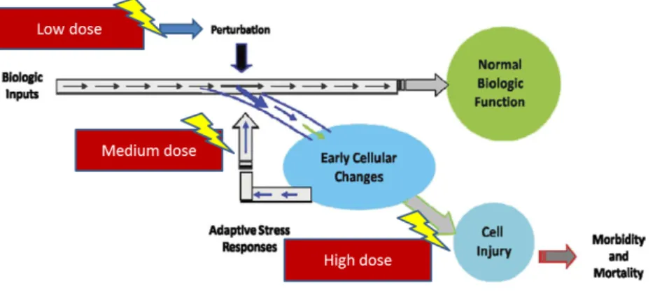
Fig. 2: Progressive activation of toxicity pathways from perturbation of initial target low dose, through activation of stress controlling pathways (medium dose), to overtly toxic responses (high dose). Biologic responses from exposure results in perturbation of biologic pathways,
accumulation and high dose lead to toxicity and disease. Adapted from [9].
As already mentioned, in vitro assays for xenobiotic toxicity are recently carefully considered by key government agencies (e.g. EPA; NIEHS/NTP; FDA) due to the social movement to reduce the use of animals in research, and a desire to better assess human risks.
Most toxicologists believe that in vitro toxicity testing methods can be a useful, time and cost-effective supplement to toxicology studies in living animals. However, it is generally accepted that the available in vitro tests are not presently adequate to entirely replace animal toxicology tests.
For example, the hazardous properties of chemicals cannot be sufficiently determined using currently available in vitro testing methods, as noted in a report published by the European Centre for the Validation of Alternative methods (ECVAM). Until prediction of metabolism can be more reliably accomplished through computational toxicology and in vitro testing, some degree of highly targeted testing using whole animals may be needed to identify metabolites and to assist in developing pharmacokinetic models for the distribution of test compounds and their metabolites in humans [9]. Relying solely on vitro methods can underestimate the potentially hazardous properties of chemicals that could be harmful to humans and the environment.
In vitro models Advantages Disadvantages
2-D cell culture model (conventional monolayer model)
Easy and convenient to maintain, increased viability
Loss of organ-specific cell-to cell interactions and differentiated functions
2-D co-culture model Interactions between multiple cell types
Cell-cell competition, complicated and conflicting culture requirements 3-D cell culture model
(hydrogels, multicellular spheroids, and multilayer cell culture)
Well-defined geometry, co-cultivation of multiple cell types, mimics the in vivo situation very closely, and 3-D scaffold for mechanical stability
Poor viability, diffusion and action of chemicals altered, multicellular resistance, and expensive
Tissue slice/Grafting model 3-D representation of cellular environment, preserve cell-to-cell & cell-to-matrix environment, realistic preclinical model, and
Viability is limited, morphological evaluation alters with the slice, difficult to reproduce, and limited availability for human models
preserve the functional and morphological heterogeneity Micro-scale cell culture
model
(microfluidic cell culture system)
Mimics the in vivo situation very closely, controlled microenvironment, allow reproduction of
biotransformation in vitro, short reaction times, portability, low cost, and small consumption of reagents and cells
Complex fluidic connection and flow control required, expensive detection and analysis system, and formation of air bubble
Tab.1: Commonly used in vitro models in toxicology: advantages and disadvantages. Adapted from [10].
The most commonly used in vitro model is represented by cells in culture. Normally, these cells are cells lines, then derived from tumors or immortalized and are therefore transformed. Another in vitro approach is to use primary cells that can be kept in culture only for a certain time. Both cell cultures can result in loss of differentiated properties, different metabolic components and modified functionality. The weakness of those in vitro tests result from the fact that the cells are isolated from their natural environment and are no longer integrated into an ordered tissue and organ topology. Metabolic conversions of xenobiotics would be studied using subcellular fractions in vitro approach. These systems usually favor only the specific biotransformation step, depending on the type of isolation procedure, on the cofactors added, on the source of tissue, and on the expression level of the involved enzymes. However, the balance of metabolic activation and inactivation requires a highly ordered interplay of many enzymes and cofactors in most cases.
Also, it is difficult to study effects on integrated and at the same time diffusely organized systems like the neuro, immuno and endocrino systems.
Hence, to study effects on these organ systems in vitro is not an easy task [8]
.
4. ADVANCED IN VITRO MODELS AND BODY ON A CHIP MODEL
In this scenario, new tools and technology for in vitro toxicology testing are highly required by industries and governments as an alternative to animal testing but also as an improvement of available cell culture systems.
In 2004, the U.S. Environmental Protection Agency (EPA) and the U.S. National Institute of Environmental Health Sciences (NIEHS) asked the U.S. National Research Council (NRC) to conduct a review and a long-term strategic plan to update and advance toxicity testing. The NRC Committee on Toxicity Testing and Assessment of Environmental Agents produced two reports. The final report outlined four design criteria, which should guide the development of a new toxicity-testing paradigm:
1. achieving broad coverage of chemicals, chemical mixtures, outcomes, and life stages;
2. diminishing the cost and time required for toxicity testing;
3. developing a better scientific basis for assessing human health effects of environmental chemicals, including knowledge of modes of action;
4. minimizing the use of animals in toxicity testing [11] .
The new paradigm has to integrate with the widely used four-stage risk assessment framework originally proposed by the NRC in 1983 in the so-called Red Book: hazard identification, dose-response assessment, exposure assessment, and risk characterization.
The newest techniques, such as in silico models, -omics technologies and high-throughput screens, have been considered in this advanced in vitro toxicology.
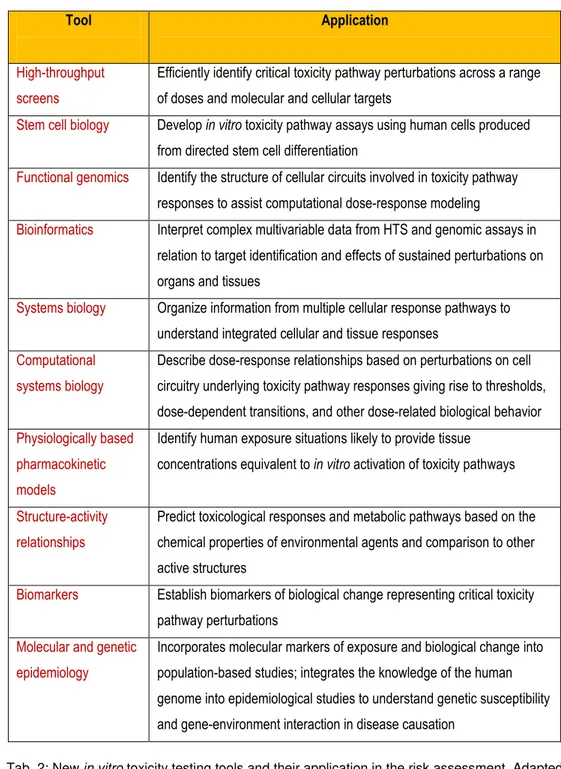
Tab. 2: New in vitro toxicity testing tools and their application in the risk assessment. Adapted from [11].
An approach to overcome in vitro test limits is to perform a more realistic model that mimics animal and human response accurately, increasing
Tool Application
High-throughput screens
Efficiently identify critical toxicity pathway perturbations across a range of doses and molecular and cellular targets
Stem cell biology Develop in vitro toxicity pathway assays using human cells produced from directed stem cell differentiation
Functional genomics Identify the structure of cellular circuits involved in toxicity pathway responses to assist computational dose-response modeling
Bioinformatics Interpret complex multivariable data from HTS and genomic assays in relation to target identification and effects of sustained perturbations on organs and tissues
Systems biology Organize information from multiple cellular response pathways to understand integrated cellular and tissue responses
Computational systems biology
Describe dose-response relationships based on perturbations on cell circuitry underlying toxicity pathway responses giving rise to thresholds, dose-dependent transitions, and other dose-related biological behavior Physiologically based
pharmacokinetic models
Identify human exposure situations likely to provide tissue concentrations equivalent to in vitro activation of toxicity pathways
Structure-activity relationships
Predict toxicological responses and metabolic pathways based on the chemical properties of environmental agents and comparison to other active structures
Biomarkers Establish biomarkers of biological change representing critical toxicity pathway perturbations
Molecular and genetic epidemiology
Incorporates molecular markers of exposure and biological change into population-based studies; integrates the knowledge of the human genome into epidemiological studies to understand genetic susceptibility and gene-environment interaction in disease causation
control over cell–cell and soluble cues typical of in vivo cell environments. Cells sense most extracellular signals (including proteins, peptides and carbohydrates) via transmembrane receptors that activate complex biochemical cascades which regulate cell physiology. Moreover, cells are sensitive to the presence of neighboring cells of similar or different type and often make long-lasting mechanical and biochemical connections to them [12], [13]
.
The first step in this direction is to combine microfabrication of 3D extracellular matrix (ECM) structures and microfluidic networks that transport soluble factors such as nutrients and oxygen. In this way mechanical strain, through shear, in the physiological range is also created [14]
.
Recent advances in using microfabrication with cell culture technology resulted in the rise of a new research field often referred to as cells-on-chip technology, which has allowed researchers to have precise control over in vitro biological systems with microscale resolution [15].
Body on a chip models are physiologically more relevant than single or co-culture in vitro models, stimulating the multi-tissue interactions and crossing-talk given by fluid flow condition.
Those systems also achieve one of the aspects of new desired tools that is the high-throughput screening of several compounds in a cheaper and less time consuming manner.
Microfabrication techniques allow the creation of devices with structures of sizes relevant to biological systems such as cell sizes (on the order of 10 µm) and the sizes of human blood vessels with spacing between capillaries on the order of 100 to 200 µm. The result is a more physiological growth environment with relevant shear stress, liquid-to-cell ratios, and physiological fluid residence times in tissue compartments. Devices that are design with regard to the structure of the human body in scale are known as micro Cell Culture Analog device (µCCA).
Sung and Shuler, in 2009, demonstrated the importance of in vitro microfluidic mimics device in drug testing [16]; their system, contained liver cells (HepG2/C3A), colon cancer cells (HCT-116), and myeloblasts (Kasumi-1) was used to test Tegafur, an oral prodrug of the cancer drug 5-fluorouracil (5-FU), with better bioavailability than that of 5-FU.
The µCCA was able to reproduce the metabolism of Tegafur, in the liver cell compartment, in order to let 5-FU travelled through the microfluidic connections to the cancer cell compartment and caused its action as a decrease in cell viability.
Viravaidya and collaborators developed a µCCA with liver and lung cells to assess toxicity of chemical as naphthalene [17]. Naphthalene is known to cause oxidative stress with subsequent glutathione (GSH, anti-oxidative molecule) depletion assuming due to its metabolite naphthalene epoxide; in this study it was demonstrated that the observed toxicity on lung cells is caused by naphthoquinone rather than naphthalene epoxide being the half-life of naphthalene epoxide in aqueous medium 3.6 min, and the residence time in the other tissue/debubbler compartment is approximately 50 min, so the epoxide cannot reach the lung cells to cause the oxidative stress.
Other technologies can be useful in development of more physiological devices.
Mathematical models can be used to foresee complex processes, for example the absorption, distribution, metabolism, and elimination (ADME) of a new compound. ADME processes are important because determine the concentration-time profiles in the bloodstream and in the tissues of organs. Pharmacokinetic (PK) and pharmacodynamic (PD) models and more complex physiologically based pharmacokinetic (PBPK) models can predict the time-dependent pharmacological effects of a drug and increase the predictability of human response leading to higher success rates in clinical trials.
PBPK models and µCCAs can be used in combination to inform each other. In the development of multicompartment devices, representations of the human body such as PBPK models can be used to guide the device design with regard to the arrangement of chambers and fluidic channel connections. In this way, furthermore, the resulting systems are physical representations of PBPK models and the reactions are that described by the equations of the model. Because PBPK models rely on the input of already known mechanisms, conversely, data obtained with these systems may be used to test and refine mechanistic hypotheses and to amplify the model with new pathways and reactions to reach a more complete system. The µCCA and PBPK model can be used in an iterative manner to test modifications in the proposed mechanism and to validate each others.
Next step in advanced in vitro models is the implementation of body-on-a-chip devices with analytical and detection technology. The ‘micro-total-analysis-system’ (µTAS) framework seeks to create microsystems incorporating several steps of an assay into a single system [14]. Integrated microfluidic devices perform rapid and reproducible measurements on small sample volumes while high sensitivity of analytical methodologies is required due to the small physical dimensions of culture systems inherently contain small numbers of cells, leading to mass-limited quantities of analyte molecules. Optical and electrical techniques have found extensive application in these devices owing to their ready compatibility with microfluidic formats and relative ease of operation. Such systems can provide near-real-time analysis. Indicator testing compound such as autofluorescent substrate can be added to the device to evaluate change in the metabolite fluorescence or absorption in order to evaluate the ability of the cells to perform that metabolic reaction.
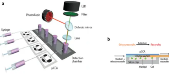
Fig. 3: Micro cell culture analog (µCCA) connected to an in situ fluorescence optical detection system (ISFODS). (a) The system consists of a syringe µCCA chips and the ISFODS. (b) Medium-containing substrate is perfused into a µCCA, above the liver cells, and the fluorescent product from P450 enzyme metabolism is detected at the detection chamber,
where the detection of fluorescent signal is made. Adapted from [18].
The integration of ultraviolet-visible absorption spectroscopy with microfluidic cell culture chips has been used, for example, to study acetaminophen metabolism and cytotoxicity [19].
Many electrical measurement methods such as amperometry with enzymatic bioelectrodes and cellular impedance can be integrated into µCCA devices [20]. Electrical impedance measurements are a particularly attractive option for determining cell viability in microfluidic cell culture devices because they are real time, label free, and non-invasive [21-23].
Another advanced technique in cells culture is the use of scaffolds. More authentic cellular behaviour is facilitated by making a three-dimensional structure that can be a simple hydrogel encapsulation of cells or mixtures of cell types [24] or preformed scaffolds that may represent a high level of authenticity, particularly for autocrine-like compounds. These systems also increase the concentration of cell in the chamber helping the analysis of metabolites and secreted molecules. Although most work with µCCAs has
utilized two-dimensional cell cultures, it has been demonstrated that a 3D culture enhance the cells vitality and growth [25]. The mechano-physical features of the scaffolds can favor cells adhesion and survival and lead to biological signals released [26].
Finally as noticed by Esch et al. in their review about body on a chip devices more useful devices should developed and included barrier tissue analogs. Barrier tissues such as the epithelium of the gastrointestinal (GI) tract, the lung epithelium, the skin, and the blood-brain barrier can significantly reduce the bioavailability of drugs that are taken up orally, through inhalation, or through application to the skin, leading to reducted doses to the target organs.
A study of Mahler et al. improve the µCCA device with a co-culture of intestinal epithelial cells (CaCo-2 cell) and HT-29 goblet-like cells so that the epithelium was covered with mucous [27]. Proof of concept experiments showed that acetaminophen (one of the most widely used analgesic and antipyretic drugs) passes through the epithelial layer where is firstly metabolized and then is further metabolized by liver cells, resulting in liver cell toxicity in a dose-dependent manner. The presence of the GI-tract barrier reduced liver cell death because acetaminophen diffused slowly across the mucous-covered CaCo-2/HT-29 cell layer, and, in addition, intestinal cells also converted some of the drug into nontoxic metabolites.
A future direction in advanced in vitro testing is individualized health care [28]
.
Since the level of enzymes and other metabolites can differ from person to person, everyone responds differently to drugs. Small tissue sample could be taken from a patient (biopsy) and tested for the efficacy of a mixture of drugs or for a range of different biological tests.
New advanced in vitro technologies are less expensive and ethically less questionable than experimenting with animals, moreover they allow a larger experimental space to be explored. The microfluidic format and the presence of possibility to study multiple-cell type in combination afford the devices several advantages compared to others in vitro drug screening models. A big challenge is recreating authentic cell behavior on the microfluidic platform and developing sensors that can monitor the physiology of cells within three-dimensional tissue constructs. These devices can be used as a preliminary screening to determine whether to invest effort and resources in a particular drug testing or biochemical pathway.
Chapter 2
Nanotoxicology: an emerging issue
Nanoparticles (NPs) are generally defined as engineered structures possessing at least one dimension sized from 1 to 100 nm. The science which studies and manipulates those nano-objects is nanotechnology, which consists essentially in a set of techniques that allows creating and using materials and devices at a very small scale.
Although nanoparticles are generally considered an invention of modern science, they actually have a very long history. Probably the earliest use being in glazes for early dynasty Chinese porcelain. A Roman cup, called the Lycurgus cup, used nanosized gold cluster to create different colors depending on whether it was illuminated from front or the back, obviously the cause of this effect was not known to those who exploited it [29].
Even these days, pottery from the Middle Ages and Renaissance often retain a distinct gold or copper colored metallic glitter. The luster was caused by a metallic film applied to the transparent surface of a glazing. The luster originated within the film itself, which contained silver and copper NPs dispersed homogeneously in the glassy matrix of the ceramic glaze. These nanoparticles were created by the artisans by adding copper and silver salts and oxides together with vinegar, ochre and clay, on the surface of previously-glazed pottery [3].
The first description, in scientific terms, of the optical properties of nanometer-scale metals was provided by Faraday in his classic 1857 paper [30]
. In 1908, Turner points out that: "It is well known that when thin leaves of gold or silver are mounted upon glass and heated to a temperature which is well below a red heat, a remarkable change of properties takes place,
whereby the continuity of the metallic film is destroyed. The result is that white light is now freely transmitted, reflection is correspondingly diminished, while the electrical resistivity is enormously increased" [31]. Nowadays, the ability to see nano-sized materials has opened up a world of possibilities in a variety of industries and scientific endeavors.
Fig. 4: Picture represents “The Scale of Things”, it provides a comparison of various objects to help understanding exactly how small a nanometer is. From objects that can be seen
by human eye to objects about a nanometer or less in size, such as the ATP molecule used in humans to store energy from food. (Designed by the Office of Basic Energy
Sciences (BES) for the U.S. Department of Energy using U.S.). Reproduced with permission from [32].
1. NANOPARTICLES: PROPERTIES AND APPLICATIONS
The large interest for NPs is due to the fact that the transition from microparticles to nanoparticles can lead to a number of changes in physical, chemical, electrical, optical, mechanical and magnetic properties. NPs are effectively a bridge between bulk materials and atomic or molecular structures. A bulk material should have constant physical properties regardless of its size, but at the nano-scale size-dependent properties are often observed. Thus, two of the major factors in this are the increase in the ratio of surface-area-to-volume, and size of the particle moving into the realm where quantum effects predominate. The increase in the surface-area-to-volume ratio, which is a gradual progression as the particle gets smaller, leads to an increasing dominance of the behavior of atoms on the surface of particle over that of those in the interior of the particle. For bulk materials larger than one micrometer, the percentage of atoms at the surface is insignificant in relation to the number of atoms in the bulk of the material. The interesting and sometimes unexpected properties of nanoparticles are therefore largely due to the large surface area of the material, which dominates the contributions made by the small bulk of the material.
High surface area is a critical factor in the performance of such technologies as fuel cells and batteries (see below). The large surface area of NPs also results in a lot of interactions between the intermixed materials in nanocomposites, leading to special properties such as increased strength and/or increased chemical/heat resistance [33]. The high surface area provides, also, a tremendous driving force for diffusion, especially at elevated temperatures. Sintering can take place at lower temperatures, over shorter time scales than for larger particles. Moreover, NPs have been found to impart some extra properties to various day to day products. For example the presence of titanium dioxide nanoparticles imparts what we call the self-cleaning effect, and the size being nano-range, the particles cannot
be observed [34]. In fact, the dimension below the critical wavelength of light renders them transparent, a property which makes them very useful for applications in packaging, cosmetics and coating (see below). Zinc oxide particles have been found to have superior UV blocking properties compared to its bulk substitute. This is one of the reasons why it is often used in the preparation of sunscreen lotions [35], and is completely photostable [36].
Clay nanoparticles when incorporated into polymer matrices increase reinforcement, leading to stronger plastics, verifiable by a higher glass transition temperature and other mechanical property tests. These nanoparticles are hard, and impart their properties to the polymer (plastic) [37]
. Moreover, perfectly-formed silicon nanospheres, with diameters of between 40 and 100 nanometers, are not just harder than silicon but among the hardest materials known, failing between sapphire and diamond [38]. NPs are currently made out of a very wide variety of materials such as titanium, zinc, aluminum, iron oxide and silicate NPs. Those new materials are used, or being evaluated for use, with different applications in several fields. The list below introduces some of those fields of use:
• Electronic: to increase the capability of electronics devices, for example display screens, reducing their weight and power consumption; or increasing the density of memory chips and reducing the size of transistors used in integrated circuits [39].
• Energy: to improve the efficiency of energy generation or develop new methods to generate energy. Stronger and lower weight blades are made possible by the use of nanotube-filled epoxy, increasing the amount of electricity generated by each windmill [40]. Companies (Bloo Solar, Solamer Energy, Nanosolar, etc.) have developed nanotech solar cells that can be manufactured at significantly lower cost than conventional solar cells.
• Space: Mavroidis and co-workers from Northeastern University in collaboration with NASA Institute of Advanced Concepts Design
developed two macro-scale devices with important space application that will be using bio-nanocomponent assemblies to build bio-nano robots in spacesuits [41], [42].
• Environment: improve the environment, cleaning up existing pollution and improving manufacturing methods to reduce the generation of new pollution. Researchers at NASA Kennedy Space Center have shown that iron NPs can be effective in cleaning up organic solvents that are polluting groundwater [43], [44].
• Consumer Products: nano has already found its way into lots of products of everyday use, from clothing to tennis racquets. Skin care products use NPs to deliver vitamins deeper into the skin; sunscreens also use NPs to block UV rays without leaving white residue on the skin. Lithium ion batteries use NP based electrodes powering plug in electric cars. Fishing rods use silica NPs to fill spaces between carbon fibers, strengthening the rod without increasing the weight. A nanoporous material called ThermaBlok® Aerogel Insulation is an excellent insulator, needing about one third the thickness compare to conventional insulation [45]. Nanoparticles have also been attached to textile fibers in order to create smart and functional clothing [46].
• Chemical Sensors: nanotechnology can enable sensors to detect very small amounts of chemical vapors. Various types of detecting elements, such as carbon nanotubes, zinc oxide nanowires or palladium nanoparticles can be used in nanotechnology-based sensors. These detecting elements change their electrical characteristics, such as resistance or capacitance, when they absorb a gas molecule [47].
• Medicine: the main application of nanotechnology in medicine currently being developed involves employing NPs for drugs delivery [48], [49], [50]
. Particles are engineered so that they are attracted to diseased cells, allowing direct treatment of those cells (e.g. cancer
cells [51], [52]). This technique reduces damage to healthy cells in the body and allows for earlier detection of disease. In therapy fullerene NPs (also known as buckyballs) may be used to trap free radicals generated during an allergic reaction and block the inflammation that results from an allergic reaction [53]. Z-Medica is producing a medical gauze that uses aluminosilicate NPs that can quickly reduce bleeding in trauma patients by absorbing water, causing blood in a wound to clot quickly [54], [55]. Nanotechnology is also used in diagnostic and imaging techniques [56], [57]: iron oxide NPs can be coated with a peptide that binds to a cancer tumor in order to enhance, once the NPs are attached to the tumor, the images from the Magnetic Resonance Imagining scan thanks to the magnetic property of the iron oxide [58], [59]. NPs can attach to proteins or other molecules, allowing detection of disease indicators in a lab sample at a very early stage. Finally, NPs are involved in anti-microbial techniques as nanocrystalline silver [60] or nanocapsules containing antibiotics [59].
• Food: Companies are developing nanomaterials that will make a difference not only in the taste of food, but also in food safety, and the health benefits that food delivers. The Sharper Image developed FresherLongerTM Miracle Food Storage Containers which keep foods fresher longer than conventional containers thanks to the antimicrobial silver nanoparticles in the polypropylene material that reduce the growth of microorganisms. Researchers are using silicate nanoparticles to provide a barrier to gasses like oxygen, or moisture in a plastic film used for packaging reducing the possibly of food spoiling or drying out [61]. Zinc oxide nanoparticles can be incorporated into plastic packaging to block UV rays and provide anti-bacterial protection, while improving the strength and stability of the plastic film [62]. Nanosensors are being developed detect bacteria and other contaminates, such as salmonella, at a packaging plant.
This point-of-packaging testing has the potential to dramatically reduce the chance of contaminated food reaching grocery store shelves.
All the applications and examples given are only a small part of the products and projects on emerging nanotechnologies; a longer list is available on the web site www.nanotechproject.org.
2. NANOTOXICOLOGY
Nanotechnology offers society the promise of major benefits, and the use of nanotech in consumer and industrial sectors is expected to increase significantly in the future. In parallel however, because NPs have unique properties different from conventional materials, concerns about safety have been raised. The same properties of nanostructured materials that make them so attractive for applications could potentially lead to unforeseen health or environmental hazards [63].
In fact, the high surface to volume ratio can make the particles very reactive or catalytic [64]. Moreover the ability of NPs to pass through cell membranes in organisms, and their interactions with biological systems make them of high potential hazard [65].
In Europe and in the USA, governments, non-governmental organizations, and others have expressed concern that, while the field of nanotechnology and the number of consumer products incorporating nanomaterials increase dramatically, in many cases, the safety of these materials has not been demonstrated and there are still a large number of unanswered questions. For example, little is known about the relationship between nanoparticles (NPs) physicochemical characteristics and their ability to cross biological barriers and to enter the general circulation, their fate within the body (toxico-kinetics), their subsequent toxic impact, or the ability of our bodies to defend against such toxic impact.
Fears over the possible dangers of some nanotechnologies may be exaggerated, but they are not necessarily unfounded. Recent studies examining the toxicity of engineered nanomaterials in cell cultures and animals have shown that size, surface area, surface chemistry, solubility and possibly shape all play a role in determining the potential for engineered nanomaterials to cause harm [66].
First increased surface area enhances contact with their surrounding exposing catalytic or other active sites on the particle surface [67], in some cases inducing the formation of reactive oxygen species (ROS) [68].
Second, due to their small size, NPs are retained in many cells and organs to a larger extent at the intracellular more than at the extracellular level due to solubilization or degradation that takes place inside cells. Toxic effects have also been demonstrated to depend on the uptake mechanism (see below) [69], presumably due to differences in fate, for example, being stored in intracellular vesicles or secreted.
Another propriety to take in account is the shape of NPs that can play a crucial role in determining responses. Geometric effects have been highlighted by the example of high toxic needle-shaped carbon nanotubes, which impale entire cells [70], [71].
Physico-chemical NP properties of relevance for toxicology
Size (airborne, hydrodynamic) Size distribution Shape Agglomeration/aggregation Surface properties Area (porosity) Charge Reactivity
Chemistry (coating, contaminants)
Properties can change:
• With method of production, preparation process, storage
• When introduced into
Defects
Solubility (lipid, aqueous, in vivo) Cristallinity
Tab. 3: List of some physico-chemical properties of NPs that impact on their biological ⁄ toxicological activity. Adapted from [72].
Rivera Gil and collaborators provided the main points that are important for nanotoxicological screening approaches [73], pre-testing criteria that should be kept in mind include:
(1) the need for defined and well-characterized NPs as model systems (2) knowledge of NP properties and potential for exposure during all stages of their life cycle
(3) the need for knowledge about biokinetics
(4) the need for validated in vitro models that are predictive of outcomes following in vivo exposures
(5) the need for evidence that in vitro outcomes are NP-specific via appropriate benchmarking (i.e., does a solute produce the same response? Do larger particles of the same composition produce the same response?) (6) the need for ranking new NPs against well-validated benchmark NPs. Moreover the concepts of dosimetry, dose metrics, exposure assessment, hazard identification, and risk characterization need still to be assessed. The challenge for both health and environmental protection is to ensure that as nanomaterials are developed and use, any unintended consequences of exposures to humans are prevented or minimised. In addition, knowledge concerning how best to apply nanotechnology to detect, monitor, prevent and control is needed.
3. NANOPARTICLES EXPOSURE AND BIOKINETIC
Numerous applications of nanomaterials lead to many possible routes through which synthetic and free nanoparticles can get into the human body.
With the expected increased intentional (e.g., in the field of medical diagnostics) and unintentional (e.g., in occupational settings and chemical waste streams) exposures to NPs nanotoxicology has now become a critical element in safety assessments of nanomaterials. In the beginning interest in the potential toxicity of very small particles starts from studies of workers exposed to metal fumes [74] and inhalation studies with ultrafine particles [75]. As matter of fact the portal of entry for nano-objects firstly and mostly studied is the pulmonary tract.
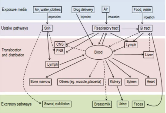
Fig. 5: A schematic overview of biokinetics of NPs. The arrows connect the routes, in dashed arrows the potential routes. Adapted from [66].
Inhaled particles smaller than 2.5 µm have access to the alveolar structures of the deep lung and may induce inflammation. Most of the particles that deposit into the lung are removed by mucociliary transport or, at alveoli level, through macrophage disposal. It was shown that high doses of nanoscale particles are, also capable of crossing the thin air–blood barrier to transmigrate into the blood [76].
Another inhaled route going directly to the brain is the uptake via olfactory nerve, here the particles can reach the brain, even if in a really small amount, and by pass the blood-brain barrier [77].
The healthy skin instead represents an effective barrier against many NPs. The uptake of nanoparticles, especially of the non-lipophilic type involved in cosmetics and in sunscreen applications, is hampered by the anatomic structure of the human skin. The EU FP6 project NANODERM (NanoDerm, 2008) results showed that TiO2 deposited only in the corneocyte layers of the stratum corneum or in the hair follicles but is not detected in the deeper regions of the skin; although it was found by another group that very small particles (<10 nm) are capable of penetrating through to the epidermis or dermis [78]. Particle surface coatings or functionalization which are often used to prevent agglomeration, may strongly influence the penetration [79]. The corneal layer of stressed or diseased skin is more permeable to all kinds of particles and must be regarded independently [80].
Since the use of nano-materials is continuously highlighted in food and packaging, a route of increasing importance is the gastrointestinal tract. The nutrients, as well as everything orally taken, are absorbed by the small and large intestines by the intestinal epithelium and can be distributed in the body via the bloodstream. 98% of the nanoparticles administered orally to the test animals were excreted in the feces, whereas the rest was eliminated via urine, indicating some uptake into the blood circulation. In the same study, but using NPs intravenously administered approximately 80% of the material was found to have accumulated in the liver after one week
[66]
. As matter of fact, injected administration route has to be considered since nanomaterials are really promising in diagnostic and therapeutic medical applications [81].
Going to the cellular level, several uptake mechanisms for NPs have been proposed. In contrast with large particles (> 500 nm) which will be exclusively taken up by phagocytosis, NPs may use other different translocation routes into the cells. NPs transport can use receptor-binding mechanisms [82], [83], diffusion through the plasma membranes, as adhesive interactions [84], [85] and any vesicle transport pathway for particles with diameters below 100 nm [86], [87].
Fig. 6: Proposed uptake mechanisms for nanoparticles with different size. Adapted from [88].
The subsequent effects observed are influenced by the different uptake mechanisms: in the case of uptake by vesicular processes, particles are sheathed by membranes (for example, caveolae). Free transport through the membrane, however, would be assumed to be more critical, as it allows particles to achieve direct contact with the plasma proteins and with other molecules of the cell. The uptake of nano-particles may well have fatal consequences for the cell if the material consists of, for example, an incompatible metal and/or is removed owing to physiological conditions.
Moreover, NPs that do not dissolve but remain stable for a long time (biopersistent) or accumulate in cells may become “active” in another way while obeying the surface principle: as there are considerably more atoms available on the particle surfaces, they can interact with the environment much more efficiently. Finally, for biological systems coming into contact with such objects, the materials constituting the nanofractions are also relevant even with uniform shapes and sizes [89], [90].
Size, shape, surface, and composition can significantly cause biological effects and must be considered separately for each material to evaluate its potential toxicity.
4. NANOPARTICLES TESTING AND RISK ASSESSMENT
New properties and behavior of NPs with respect to bulk materials leads to the need for a systematic risk research as information about safety and potential hazards is required. Toxicology tests and the resulting database would provide information for material safety data sheets for NPs as well as a basis for potential NP risk assessments and risk management.
Toxicity assays will identify the potential hazard by establishing dose-response relationships or stress and inflammatory dose-responses. However, because a risk of adverse effects associated with NPs is a function of hazard and exposure, the generally accepted approach is to incorporate both components into a risk assessment paradigm, consisting of Hazard Identification, Hazard Characterization, Exposure Assessment and Risk Characterization [91] so that appropriate risk management decisions can be made.
Whereas first investigations concerning the toxicity of NPs were based on in vivo experiments (i.e., inhalation studies, etc.), due to the huge amount of a large variety of different NPs it is desirable to develop and validate simple non in vivo assays in order to reduce and avoid extensive testing using laboratory animals. In vitro studies enable the identification of conceptual
models for interactions of NPs with cells [73]. An important issue to take in account within in vitro models is the interference of the NPs with well-established toxicity assays in order to avoid confounding or even conflicting data. Each test should be characterized in advance considering the presence of NPs, moreover it is recommended to perform two different tests for each biological end point to exclude cross-reactions [88].
Cytotoxicity assay Detection principle NP interference Altered readout Particle studied Cell viability MTT Colorimetric detection of mitochondrial activity Adsorption of substrate Reduced indication of cell viability Carbon nanoparticles LDH Colorimetric detection of LDH release Inhibition of LDH enzyme Reduced indication of necrosis Trace metal-containing nanoparticles Annexin V Fluorimetric detection of Phosphatidylserine exposure (apoptosis marker) Ca 2+ depletion Reduced indication of apoptosis Chitosan nanoparticles Propidium Iodide Staining of DNA (necrosis marker)
Dye adsorption Reduced indication of necrosis
Carbon nanoparticles
Neutral red Colorimetric detection of intact lysosomes
Dye adsorption Reduced indication of cell viability Carbon nanoparticles Caspase Fluorimetric detection of Caspase-3 activity (apoptosis marker) Inhibition of Caspase-3 enzyme Reduced indication of apoptosis Trace metal-containing nanoparticles, especially Zn2+
Stress response DFC Fluorimetric detection of ROS production Fluorescence quenching Reduced indication of oxidative stress Carbon nanoparticles Inflammatory response ELISA Colorimetric detection of cytokine secretion
Dye adsorption Reduced indication of cytokine concentration
Metal oxide nanoparticles
Tab. 4: Nanoparticles interference with cytotoxicity assay. Adapted from [92].
In vitro high-dose toxicology and mechanistic studies should be viewed as proof-of-principle studies, though, that ultimately require validation in vivo. A major issue that needs to be carefully considered is the relevancy of the doses applied in vitro for predicting in vivo outcomes. At very high doses assays can certainly identify a NP as hazardous but how realistic is the study for in vivo exposure conditions?
Therefore, it is desirable to develop and validate simple non in vivo assays for the purpose of predicting in vivo responses.
In parallel, efforts should be made to obtain data on exposure levels occurring for workers at NP manufacturing sites despite best occupational hygiene conditions, as well as for anticipated consumer exposures to nano enabled products [72].
Fig. 7: The evaluation process of toxicity of NPs. In the diagram are shown the in vitro-in vivo relationship and its extrapolation to humans. Adapted from [88].
Chapter 3
Bioreactor design and improvement for
investigating absorption,
biotransformation & biodistribution
1. BIOREACTOR: MCmB
The request of more physiological tests that do not involve animals was underlined in chapter 1. Given the concerns regarding animal testing many groups have developed systems that are more representative of human physiology (as described in paragraph 4, chapter 1).
Our group at the Interdepartmental Research Center “E.Piaggio”, developed a “system on a plate” modular MultiCompartmental Bioreactor (MCmB) array [93] commercialized as Quasi-Vivo® (by Kirkstall Ltd). Its main advantage is its size: with no need to change multiwell plate/petri dish culture protocols. The microscale dimensions that make body-on-a-chip devices (described in paragraph 4, chapter 1) so attractive lead also to the need of translating experimental methods to be feasible in the micro-range. The MCmB offers the possibility to use the same protocols as multiwell plates improving them with mechanical, flow and biochemical stimuli, or cross-talk by adding other cell types so as to recreate the physiological environment. It was designed as an easy-to-assemble modular lego-type device according to finite element methods and allometric scaling in order to
have low shear stress, high nutrient turnover, and physiologically relevant cell numbers and fluid residence times. Many cues should be controlled in vitro using engineering and design as shown in figure 8.
Fig. 8: A representation of the more important biochemical, physio-chemical and mechano-structural cues presented in the cellular microenvironment and the parameters that can be
controlled. Adapted from [94].
The level of cross-talk is likely to depend strongly on dynamic stimuli such as flow and concentration gradients. The flow rate and volume will determine the residence and passage times of molecules and oxygen, as well as the shear stress on the cells. The goal of dynamic culture device design is the balance between high oxygen mass transfer and low wall shear stress to cells. Shear stress is given by a fluid moving along a solid boundary, this tangential flow constantly stimulates cells [95] by a low velocity convective motion but in contrast most type of cells cannot support high levels of shear which compromises their function.
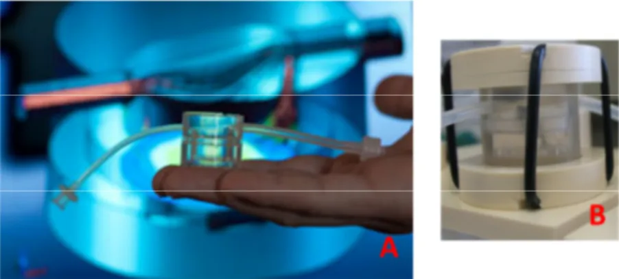
The MCmB is fabricated in PDMS (polydimethylsiloxane), a biocompatible elastomer. The modular chamber is similar in size to that of a 24 well plate with a volume of 2 mL. The choice to design a modular device is justified by the possibility to allow any tissue or organ model to be simulated and performed simply by connecting the chambers in a desired configuration. The bioreactor unit is composed of two self-sealing (male-female joint) parts, of which top piece has smart design that allows bubbles (one of the biggest problem in microfluidic device) to be conveyed to the outlet tube. Varying the flow rate it is possible to set oxygen and nutrient residence time and low shear stress of the order of 10-5 Pa at the bottom of the chamber where cells are housed.
Fig. 9: Pictures of MCmB showing the size and the shape (A). In the right, bioreactor is shown within his closure system (B)
MCmB is well characterized in Mazzei et al. [93] and has been already tested using different high throughput multi compartmental bioreactor experiments [94-101]
.
Hepatocytes were firstly used to test the MCmB because they are the difficult cells to culture for their rapid loss of phenotypic expression in-vitro and because of their sensitivity to oxygen concentration, with high metabolic demands.
The high importance of flow rate has been shown, with a decrease in viability below 180 µL/min, due to lower oxygen concentration, and above 500 µL/min as result of too high shear stress. Albumin production, analyzed as marker of function, confirmed the vitality of cells as the albumin concentration under flow condition was higher with respect to controls till 500 µL/min [93].
In Guzzardi et al. a study on metabolic regulation in dynamic culture conditions in the MCmB was performed and compared with static models. In this work cross talk between hepatocytes and adipocytes was explored. Significant differences in metabolic and functional profiles between static and dynamic settings, and in the mono- versus the connected cultures were found [96].
In Vinci et al. it was shown that the combination of 3-D scaffolds and dynamic flow conditions enhances hepatocyte culture with an increase in cell density compared with monolayer controls and a three-fold increase of metabolic function in dynamic culture compared with static monolayer cultures [97].
In a second work, it was demonstrated that the presence of flow significantly modifies cellular metabolism of hepatocytes, adipocytes and endothelial cells [98]. Hepatocytes, endothelial cells and adipocytes were connected together each in a MCmB chamber to investigate metabolic cross-talk between them in regulating glucose and lipid metabolism [99].
Another work from the same author used the MCmB for primary human hepatocyte culture, showing that medium flow stimulates the expression and activity of detoxification genes [95]. Those results were confirmed by Vozzi et al. [100] who detected diclofenac (arylacetic non‐steroidal anti‐inflammatory drug -NSAID-) toxicity in the MCmB system at concentrations significantly lower than conventional hepatocyte cultures, similar to what has been observed in-vivo.
![Fig. 6: Proposed uptake mechanisms for nanoparticles with different size. Adapted from [88]](https://thumb-eu.123doks.com/thumbv2/123dokorg/7613263.115495/38.748.118.649.376.644/fig-proposed-uptake-mechanisms-nanoparticles-different-size-adapted.webp)