UNCORRECTED
PROOF
Contents lists available at ScienceDirectAmerican Journal of Otolaryngology--Head and Neck
Medicine and Surgery
journal homepage: http://ees.elsevier.com
Menière's disease patients improve specific posturographic parameters following
diagnostic intratympanic injection
GiampieroNeri
a,⁎, DaniloBondi
a,b, AndreaScordella
b, ArmandoTartaro
c, LetiziaNeri
c, FiorellaCazzato
c,
NiccolòPini
a,b, Maria Addolorata Mariggiò
aaDepartment of Neuroscience, Imaging e Clinical Sciences, University “G. d'Annunzio” of Chieti-Pescara, Chieti, Italy bLaboratory of Functional Evaluation, University “G. d'Annunzio” of Chieti-Pescara, Chieti, Italy
cDepartment of Medical and Oral Sciences and Biotechnologies, University “G. d'Annunzio” of Chieti-Pescara, Chieti, Italy
A R T I C L E I N F O
Keywords Menière's disease Stabilometry Computerized posturography Dizziness Gadolinium Columella effectA B S T R A C T
Purpose: Evaluation of specific computerized posturographic parameters in patients with Menière's disease (MD)
following the intratympanic injection of gadolinium, a contrast agent, used in radiological diagnosing.
Materials and methods: We have observed 12 adult patients with unilateral Menière's Disease subjected to inner
ear magnetic resonance imaging (MRI) examination after intratympanic gadolinium injection (ITG). The diag-noses have been performed according to the guidelines of the American Academy of otolaryngology. Before and after 24 h the ITG, all patients were subjected to the clinical evaluation and computerized posturography (CP), in 4 conditions depending on open/closed eyes and with/without foam cushion under feet.
Results: After ITG, in the affected ear the MRI confirmed the endolymphatic hydrops revealing a thin or even
disappeared perilymphatic space. The statokinesigram showed improvement of stability only with closed eyes on a foam cushion. The CP performed 24 h after the contrast intratympanic injection showed a significant reduction of Path Length and Confidence Ellipse Area, due to an improvement of vestibular function on static balance. This improvement could be directly dependent to intratympanic pressure modification mediated by volume of con-trast liquid, by “columella effect”.
Conclusions: This study demonstrates the absence of vestibular damage in patients undergoing intratympanic
gadolinium infiltration and confirms the relationship between intratympanic pressure and vestibular stability modifications providing positive evidences for an applicative use of CP as a functional assessment to better ad-dress diagnosis and follow-up in MD patients treated with intratympanic injections..
1. Introduction
Menière's disease (MD) is an idiopathic disorder of the inner ear, characterized by fluctuating hearing loss, tinnitus, fullness and recurrent episodes of rotatory vertigo [[1]].
Today, this pathology remains a complicate pathology to deal with. Since the studies by Hallpike and Cairns [2] and Rauch and others [3], the typical anatomical sign of MD is the endolymphatic hydrops (EH), present also in some asymptomatic patients. Many crucial fac-tors have been proposed in its development such as ionic alterations, gene mutations, autonomic imbalance, autoimmune reactions, viral in-fections, vascular and allergic alterations, phlogosis and trauma [4–6]. All causes seem to trigger endolymphatic hypertension through differ-ent mechanisms that can range from endolymph over-production to re
duced absorption by the endolymphatic sac, or mechanical obstruction to outflow. For this considerable number of factors, the MD diagnosis is frequently used to describe the symptomatologic triad and from time to time attributed to hypothetical etiological factors. Diagnosis is de-termined according to the 2015 guidelines proposed by the American Academy of Otolaryngology-Head and Neck Surgery (AAO-HNS) with the support of the Barany Society [7].
Due to the impossibility to directly see the hydrops, and the absence of standard instrumental tests or specific clinical signs, clinicians have experienced for a long time a lack of tools for an established diagnosis.
In 2007 a group directed by Nakashima [8] revealed for the first time the presence of hydrops using the MRI 3 T in patients suffering from MD. They demonstrated that the gadolinium (Gd), injected into the middle ear, was carried in the internal ear spreading only in peri Abbreviations: MD, Menière’s disease; Gd, gadolinium; EH, endolymphatic hydrops; MRI, magnetic resonance imaging; CP, computerized posturography; ITG, intratympanic gadolinium injection; PL, Path Length; CEA, Confidence Ellipse Area; RI, Romberg Index.
⁎Corresponding author at: ENT Clinic, University “G. D'Annunzio” Chieti-Pescara, “S.S. Annunziata” Hospital, via dei Vestini, 66100 Chieti Scalo, CH, Italy. E-mail address: [email protected] (G. Neri)
UNCORRECTED
PROOF
lymphatic spaces, first in the scala tympani of the basal turn of thecochlea and in the vestibule, and the day after it appeared in almost all part of the labyrinth [9]. The spreading of the contrast agent was also reported in other studies. Once more, Nakashima and colleagues re-ported that the EH appeared as the contrast-absent area surrounded by contrast agents that disappeared about six days after the injection [10]. Moreover, they described the results from MRI scans performed 4 h after Gd injection, reporting a lowering of Gd concentration during the time [11]. In addition, the same authors showed that the intratympanic injec-tion was more effective than intravenous one [12]. Liu and colleagues [13] reported a less invasive intratympanic injection of Gd through the Eustachian tube, also this method made possible to view the EH in pa-tients with MD. All these pieces of evidence support that these methods are free of side effects in the short and long term [9,11–14]. However, comprehensive knowledge about the side effects needs to be enforced. Indeed, until now adverse reactions have not been reported in humans [14], although in animals effects in the inner ear were shown after Gd administration. Tanaka and Colleagues [15] reported alterations in vestibular hair cells from animals subjected to (gadodiamide) adminis-tration depending on the dilution of contrast agents.
The hydropic alterations on physiological functions can be assessed in a more analytical way using the computerized static posturography (CP, i.e. stabilometry throughout the present work) to test the upright stance posture [16,17]. To be specific, MD patients show poor upright posture stability, greatly dependent on visual and somatosensory infor-mation [18].
CP represents a non-invasive and effective tool in evaluating the sta-tic balance of the patient's body with changes in the vestibular function. This method detects the reflex of the spinal vestibule by recording and analyzing the oscillations and can identify deficiencies in the integration of visual, vestibular and proprioceptive afferents by exposing the patient to various sensory situations [19]. Particularly in MD patients, who have spontaneous vertigo attacks, CP allows evaluating with altered patterns of sensory interaction upon the balance control in the erect position.
Thus, since in the literature no functional alterations related to the intratympanic administration of Gd contrast agent (gadobutrol solution) are described, we aimed to assess whether among MD patients subjected to the injection there were changes in specific posturographic parame-ters.
2. Materials and methods
2.1. Study design and participants' characteristics
This retrospective clinical study was conducted in our ENT Depart-ment of Chieti-Pescara University Hospital on the patients suspected to MD and that were subjected to MRI 3 T with a trans-tympanic injection of gadodiamide. The study protocol was approved by the local ethics committee. All patients provided their written informed consent.
Among all the patients subjected to MRI, 12 adult patients, seven women and five men, aged 38-to-73 years, with unilateral MD were studied. Diagnosis of definitive MD was based on the history of the disease and findings of neuro-otological examinations, which assessed the coexistence of recurrent episodic vertigo and fluctuating cochlear symptoms including hearing loss, tinnitus and aural pressure, accord-ing to the 2015 guidelines proposed by the American Academy of Otolaryngology-Head and Neck Surgery (AAO-HNS) with the support of the Barany Society [7]. All patients underwent audiological and otoneurologic examinations, to exclude other inner-ear disorders and retro labyrinthine disorders. Exclusion criteria were previous otologic surgery, ablative therapy with gentamicin, patients with superior canal dehiscence syndrome (SCDS) or enlarged vestibular aqueduct syndrome (EVAS) or any other temporal bone abnormalities. Subjects with a his
tory of inflammatory middle ear disease and those patients that cannot undergo MRI were also excluded; none of the patients was in an acute ear phase, no one had ever utilized the Meniett device nor had tympanic perforations nor underwent transtympanic drainage with a drain tube insertion.
2.2. Intratympanic gadolinium injection (ITG) and magnetic resonance imaging (MRI)
The injection was performed in the affected ear after administration of local anesthetic applied in the external ear conduct. The patient was lying in a supine position with the head turned 30° to the healthy side, so 0.4–0.5 mL of the solution was injected through the tympanic mem-brane by a 23-gauge needle, using a puncture in the postero-inferior quadrant. The contrast agent was used in the Gadobutrol form (Gadovist - GD) diluted 7-fold in saline solution. After the ITG the patient re-mained 30 min with the head turned 45° to the healthy side to permit the diffusion through the round window. 3 Tesla MRI was executed be-fore and 24 h-after the injection, with the acquisition of 3D FLAIR and 3D T2 SE sequences. Multi Planar Reconstructed (MPR) images were an-alyzed. Vestibular endolymphatic hydrops was graded considering the ratio of the area of the endolymphatic space to the vestibular fluid space (sum of the endolymphatic space and perilymphatic space) [9]. Patients with no hydrops had a ratio of one third or less, those with mild hy-drops had between one-third and a half and those with severe hyhy-drops had a ratio of >50%. Cochlear and semicircular canals endolymphatic hydrops were defined positive when an MRI signal void was detected. 2.3. Computerized static posturography (CP)
Subjects were instructed to stand with their feet 7 cm apart and the arms along the sides on a static balance platform for each test sequence. The CP was performed before and 24 h after the Gd injection. The cen-ter of pressure between both feet was measured with a compucen-terized vertical force platform (Phisionorm NBP Computerized Posturographic System). Measurements were performed while the subject maintained a 51.2-s upright stance position with open or closed eyes, with or with-out the presence of a foam cushion under feet. To be specific, during the open eyes conditions, subjects were looking at a small eye-level tar-get 1 m away; the foam cushion was a 100 mm-thick foam rubber mat, exactly with the same size of the balance platform, posed on top of it. Closed eyes condition limited the role of vision control of upright stance, while foam cushion conditions limited the somatosensory and proprio-ceptive information [20]. Each test was conducted under visually and acoustically standardized conditions, in a room with white walls and uniform brightness. Each test was followed by a 30-s interval. The pa-rameters considered before and after the intratympanic injection were: Path Length (PL), defined as the linear length (mm) of the sway path; Confidence Ellipse Area (CEA), defined as the area of the 95% confi-dence ellipse around the Center of Pressure (CoP) trajectory [21]. We fo-cused on these parameters because of their large use and validity in dou-ble-stance static balance assessment [21]. We then calculated Romberg Index (RI), as the percentage ratio between the value of PL, or CEA, with closed eyes and the same parameter obtained with open eyes.
2.4. Statistics
The statistical analyses were carried out using GraphPad Prism Soft-ware, version 7 (GraphPad SoftSoft-ware, La Jolla, USA), and R-based open-source software Jamovi (The jamovi project 2019 – jamovi, (Ver-sion 0.9) [Computer software]) and Jasp (JASP Team (2019). JASP (Version 0.11.1) [Computer software]). The identification of outliers
UNCORRECTED
PROOF
formed with the ROUT method (Q = 0.5%). The normality of thedistri-butions was assessed with the Shapiro-Wilk test and checking Q-Q plots of residuals. After acceptance of normality assumption checks, we used paired t-test to analyze the pre-post differences. For each comparison, we considered p values and Effect Size (Cohen's d) and calculated 95% Confidence Intervals (CIs) of mean differences. In addition, we calcu-lated the probability of superiority (PS), as the percentage of a randomly sampled member of the higher mean distribution having a higher score than a randomly sampled member of the other distribution, and unbi-ased Cohen's d, to adjust for a likely overestimation of effect size due to the small sample size [22]. We complemented the frequentist approach with the Bayesian approach to provide additional information for hy-pothesis testing [23]; in particular, we used Cauchy's prior distribution, reported Bayes Factor (BF10) and performed robustness check. Our
ap-proach permitted us to rate the various comparisons, and consequently to identify the posturographic parameters and test conditions most re-sponsive to the injection.
3. Results
No adverse reactions and no audiological or vestibular complications and no damage to the eardrum membrane, as persistence of perforation, were observed after Gd injection in all the 12 patients recruited for this study. The MRI indicated the presence of hydrops in 10 patients while in the remaining two cases the exam was negative. In particular, the MRI performed with the contrast medium, indicated the diffusion of Gd in the structures of the inner ear only in those 10 patients bearing the hydrops. The possibility of not observing the diffusion of the contrast medium has already been reported [24]. In all cases the clinical signs of hydrops were present, indeed, the remaining two patients that did not present the diffusion of the contrast agent reported to have suffered from otitis in childhood. The otitis may indeed have altered the anatomy of the district, hindering in MRI examination the diffusion of Gd in peri-lymphatic space.
Data of PL and CEA are shown in Figs. 1 and 2, respectively, and Table 1 collected the results of statistical analyses of these parame-ters. The most relevant difference was found for PL in eyes closed plus foam cushion under feet condition, with a significant reduction 24 h-af-ter injection (p = 0.011, Cohen's d = 0.876, BF10= 5.328). Robustness
check of Bayesian statistics showed that the evidence was moderate and robust, close to the max BF10(5.330) and relatively stable across a
Fig. 1. Path Length (PL) of MD patients before (pre) and 24 h-after (post) the intratym-panic injection; OE: Open Eyes; CE: Closed Eyes; FC: foam cushion under feet. Data are presented as boxes and whiskers (with Tukey method).
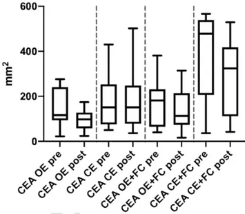
Fig. 2. Confidence Ellipse Area (CEA) of MD patients before (pre) and 24 h-after (post) the intratympanic injection; OE: Open Eyes; CE: Closed Eyes; FC: foam cushion under feet. Data are presented as boxes and whiskers (with Tukey method).
Table 1
Statistical comparison between stabilometric parameters: Path Length or Confidence El-lipse Area, measured pre and 24 h-post intratympanic injection on MD patients. In the table are reported the Eye condition (Eye), the presence of foam cushion under feet (FC), the statistical significance (p, *p < 0.05), the Effect size (Cohen's d) and the Bayes Factor (BF10).
Eyes FC p Cohen'sd BF10
Path Length (PL) Open No 0.199 0.394 0.610
Open Yes 0.668 0.127 0.313
Closed No 0.443 0.230 0.376
Closed Yes 0.011* 0.876 5.328 Confidence Ellipse Area (CEA) Open No 0.145 0.453 0.759
Open Yes 0.877 0.046 0.291
Closed No 0.884 0.043 0.290
Closed Yes 0.046* 0.688 1.832
wide range of prior distributions. Unbiased Cohen's d was calculated as 0.815; PS was calculated as 71%. CI of mean difference was in the range of 31-to-200 mm.
A similar result was obtained analyzing CEA data resulting in a sig-nificant decrease in eyes closed plus foam cushion under feet condi-tion (p = 0.046, Cohen's d = 0.688, BF10= 1.832). Robustness check
showed that the evidence was smaller in respect to the above-mentioned PL comparison, but robust (max BF10= 1.938, relatively stable across a
wide range of prior distributions). Unbiased Cohen's d was calculated as 0.640; PS was calculated as 67%. CI of mean difference was in the range of 2-to-176 mm2.
Concerning RI of PL, there was a significant reduction 24 h-after in-jection (2.15 pre vs 1.97 post) in foam cushion condition, while there was no difference without foam cushion (1.52 pre vs 1.50 post) (see sta-tistical analysis in Table 2). RI of CEA appeared to be decreased 24 h-af-ter injection in foam cushion condition (3.73 pre vs 2.78 post) and ap-peared to be increased without foam cushion (1.09 pre vs 1.73 post), although the statistics did not provide robust evidence (see statistical analysis in Table 2). As well as the above-mentioned statistics of CEA, for RI comparisons robustness checks highlighted robust but weak dif-ferences.
UNCORRECTED
PROOF
Table 2
Statistical comparison between stabilometric parameters: Romberg Index of Path Length or Romberg Index of Confidence Ellipse Area, measured pre and 24 h-post intratympanic injection on MD patients. In the table are reported the presence of foam cushion under feet (FC), the statistical significance (p, *p < 0.05), the Effect size (Cohen's d) and the Bayes Factor (BF10).
FC p Cohen'sd BF10
Romberg Index of PL No 0.675 0.125 0.312
Yes 0.046* 0.648 1.762
Romberg Index of CEA No 0.055 0.654 1.585
Yes 0.077 0.562 1.193
4. Discussion
The MRI with intratympanic injection of the contrast agent is the sole method currently applicable to observe directly the EH in living patients with MD. Once released into the tympanic cavity the Gd goes through the round window into perilymphatic space of the vestibule, and then within 24 h to the cochlea and the semicircular canals. The 3 T MRI with ITG allowed us to correlate MD signs and symptoms [25] and monitor the effects of pharmacological treatments for hydrops.
Firstly, CP is used to evaluate patients with balance alterations: this technique allows not only quantification of the subject's capacity to maintain an upright stance posture, but also allows the analysis of the impact of various sensory information.
Furthermore, it is well known that MD patients are sensitive to low-frequency sounds, probably due to the activation of vestibulo-spinal responses. These responses seem to be the result of the Tullio phenom-enon in which sound energy activates the vestibular end-organ [26]. In this regard, CP protocols indicated a significant relationship between the audiometric hearing threshold and the center of pressure displacement, especially in patients with audiometric advanced disease [27]. In addi-tion, posturography tests are sensitive to the time elapsed since the last typical vertigo attacks [28].
Considering the frequent poor stability in the upright posture of MD patients [18], we aimed to verify if the injection of contrast agent so-lution in the tympanic cavity could affect patients' stability, measured by static CP. Therefore, the CP tests were performed in diverse condi-tions: open and closed eyes and with or without foam cushion. The lack of visual support (closed eyes) and the reduced proprioceptive feedback (presence of foam cushion) produced a stabilometric result mainly led by the vestibular system.
Our results demonstrated a reduction of PL and CEA in closed eyes plus foam cushion condition, 24 h-after the injection, indicating a pos-tural stability improvement in respect to the identical condition before the injection. This suggests that this improvement could be led by the vestibular system.
Some authors found a visual dependency pattern in patients with vestibular dysfunction [29–31]. In our study, the Romberg Index de-creased after the injection in foam cushion condition, both in PL and CEA, suggesting that the vestibular more notable control after the injec-tion may reduce the impairment due to the lack of visual control and proprioceptive feedback.
The intratympanic injection was followed by a period of at least 30 min. During this time, patients must remain in a position that en-sures the maintenance of the contrast medium between the tympanum and the round window. This procedure may lead to the following conse-quences; the first one is tympanum perforation, which inevitably mod-ifies the pressure ratio between the external auditory canal and the tympanic cavity. The pressure ratio is high enough to imply an acces-sory perforation in the epitympanum, to prevent an excessive increase of pressure during the infiltration. If an epitympanic perforation is un
performed, as on our subjects, the insertion of the Gd solution can rapidly increase the pressure in the tympanic cavity. This pressure, as reported by Feijen [32], is transmitted directly into the structures of the inner ear; because in MD patients the symptomatology can be mediated by pressure alteration [33], this effect could modify vestibular hair cells function. A further consideration concerning the contrast liquid inserted into the tympanic cavity is that the injection creates a direct connec-tion between the eardrum and the round window; this effect is known as the “columella effect”, i.e. any sound produced in the environment is transmitted directly to the round window and then in the inner ear dis-tributing sound pressure into perilymphatic spaces [34]. This modifica-tion of perilymphatic pressure, as well as endolymphatic one, can mod-ulate cochlear and vestibular hair cells function; this function, through vestibulo-spinal responses, entails to postural changes [26].
In addition, the conditions generated by ITG are similar to the ones generated by the Meniett device in the treatment of MD [35] with-out tympanostomy. This device delivers intermittent low-pressure pulses through a tympanostomy tube (grommet) to the middle ear space, then acting on the round window membrane. Since the fluids of the inner ear are uncompressible, the energy of the pressure pulses causes a displace-ment of the perilymphatic fluid. This stimulates endolymphatic fluid flow, resulting in a reduction of static endolymphatic fluid. The same functional complications (i.e. tinnitus increase in 5.4% of patients, tran-sient dizziness in 16.9% and vertigo in 1.8% of subjects) were obtained after transtympanic injections, as a consequence of the modification of intratympanic and intralabyrintic pressure [36]. We therefore planned to carry out CP tests before and 24 h-after ITG to study vestibular stabil-ity.
A similar approach, measuring serial posture oscillation and center of pressure displacement and area, was used by Narita et al. [37] who followed up 31 patients with vestibular dysfunction for a period of ap-proximately 260 days. They found a correlation of clinical improvement and reduction of posture oscillations with time, confirming the useful-ness of the test to follow up patients with vestibular dysfunction. From our results, we conclude that the increased stability after the intratym-panic injection was predominantly due to a vestibular function improve-ment.
The CP parameters measured in this study, PL and CEA, are scien-tifically appreciated [21] and we support their use as functional mea-sures in MD patients. These parameters allow discriminating the impact of the vestibular function on upright stance postural control. Romberg Indexes need to be better evaluated in larger samples, to better clarify the eventual role of such indexes in the evaluation of MD patients. PL, rather than CEA, seemed to better adhere to the rationale of postural control in MD patients. Statistics revealed PL in closed eyes plus foam condition as the most adequate functional measurement in evaluating static postural affection in the current study, with moderate and robust evidence [22,23]. Further studies should address the role of Load Distri-bution Difference parameter in the evaluation of MD patients, relating it to the side of the disease.
Recently, Wesseler and colleagues reinforced the diagnostic value of MRI performed 24 h after intratympanic Gd injection in MD: they demonstrated this procedure to be the most reliable to diagnose MD, in respect to other functional tests; indeed, none of those diagnostic vestibular functional procedures served as an indicator of endolym-phatic hydrops [38]. With the current work, we extended this evidence in a functional basis, revealing how injection affected static balance where a vestibular impairment emerged.
Further studies may also discriminate between the specific Gd injec-tion and a control injecinjec-tion, to test the effect of fluid volume and specific substances commonly used in MD analyses. Plus, further studies may in-vestigate the effect of intratympanic injection in control vs MD patients.
UNCORRECTED
PROOF
5. ConclusionsThe intratympanic injection of contrast solution in the middle ear of MD patients improved some specific posturographic parameters 24 h af-ter the injection. In particular, we demonstrated a significant improve-ment of the vestibular function with eyes closed on a foam cushion un-der feet. We think this improvement is causally related to the intratym-panic pressure modification mediated by the volume of contrast liquid, by means of the “columella effect”. This study confirms the relations be-tween intratympanic pressure and vestibular stability modifications that can be also subclinical. CP parameters could be measured, in longitudi-nal studies or pharmacological intervention, to monitor MD patients and to discriminate the vestibular component on the postural control.
CRediT author statement
Conceptualization: GN, AS, MAM. Methodology: GN, AS, MAM. Formal analysis: GN, DB. Investigation: GN, AS, AT, LN, FC. Resources: GN, AS, AT, FC.
Writing - Original Draft: GN, DB, AS, AT, LN, FC, NP, MAM. Writing - Review & Editing: GN, DB, AS, AT, LN, FC, NP, MAM. Visualization: DB, NP.
Supervision: GN, MAM.
Project administration: GN, MAM. Funding acquisition: GN, AT, MAM.
Declaration of competing interest
The authors have no competing interest to declare.
Acknowledgments
The authors thank all the participants.
Funding sources
This work was supported by G. d'Annunzio University grants to GN, AT and MAM.
References
[1] H Sajjadi, M M Paparella Meniere’s disease. Lancet 2008;372:406–414. doi:10.1016/S0140-6736(08)61161-7.
[2] C S Hallpike, H Cairns Observations on the pathology of Ménière’s syndrome. Proc R Soc Med 1938;31:1317–1336.
[3] S D Rauch, S N Merchant, B A Thedinger Meniere’s syndrome and endolymphatic hydrops. Double-blind temporal bone study. Ann Otol Rhinol Laryngol 1989;98:873–883. doi:10.1177/000348948909801108.
[4] H F Schuknecht Pathology of the ear. 2nd ed. Lea & Febiger; 1993.
[5] S N Merchant, S D Rauch, J B Nadol Ménière’s disease. Eur Arch Otorhinolaryngol 1995;252:63–75. doi:10.1007/bf00168023.
[6] J B Nadol Pathogenesis of Ménière’s syndrome. Ménière’s disease. Kugler Publications; 1999.
[7] J A Lopez-Escamez, J Carey, W-H Chung, J A Goebel, M Magnusson, M Mandalà, et al. Diagnostic criteria for Menière’s disease. J Vestib Res 2015;25:1–7. doi:10.3233/VES-150549.
[8] T Nakashima, S Naganawa, M Sugiura, M Teranishi, M Sone, H Hayashi, et al. Visualization of endolymphatic hydrops in patients with Meniere’s disease. Laryngoscope 2007;117:415–420. doi:10.1097/MLG.0b013e31802c300c. [9] S Kasai, M Teranishi, N Katayama, M Sugiura, S Nakata, M Sone, et al.
Endolymphatic space imaging in patients with delayed endolymphatic hydrops. Acta Otolaryngol 2009;129:1169–1174. doi:10.3109/00016480802691143. [10] S Naganawa, H Satake, S Iwano, H Fukatsu, M Sone, T Nakashima Imaging
endolymphatic hydrops at 3 tesla using 3D-FLAIR with intratympanic Gd-DTPA administration. Magn Reson Med Sci 2008;7:85–91. doi:10.2463/mrms.7.85. [11] T Nakashima, S Naganawa, M Teranishi, M Tagaya, S Nakata, M Sone, et al.
Endolymphatic hydrops revealed by intravenous gadolinium injection in patients with Ménière’s disease. Acta Otolaryngol 2010;130:338–343. doi:10.1080/ 00016480903143986.
[12] M Yamazaki, S Naganawa, M Tagaya, H Kawai, M Ikeda, M Sone, et al. Comparison of contrast effect on the cochlear perilymph after intratympanic and intravenous gadolinium injection. AJNR Am J Neuroradiol 2012;33:773–778. doi:10.3174/ajnr.A2821.
[13] F Liu, L-S Yu, W-N Huang, M Chen, C Zhang, Y Fu, et al. Non-invasive visualization of endolymphatic hydrops in patients with Meniere’s disease. Zhonghua Er Bi Yan Hou Tou Jing Wai Ke Za Zhi 2010;45:324–327. [14] J P R Louza, W Flatz, E Krause, R Gürkov Short-term audiologic effect of
intratympanic gadolinium contrast agent application in patients with Ménière’s disease. Am J Otolaryngol 2012;33:533–537. doi:10.1016/j.amjoto.2011.12.004. [15] H Tanaka, T Tanigawa, M Suzuki, K Otsuka, S Inafuku Effects of MRI contrast
agents (Omniscan) on vestibular end organs. Acta Otolaryngol 2010;130:17–24. doi:10.3109/00016480902896147.
[16] S Caruso, D Mauro, L Maiolino, C Grillo, A M C Rapisarda, S Cianci Effects of combined oral contraception containing drospirenone on premenstrual exacerbation of Meniere’s disease: preliminary study. Eur J Obstet Gynecol Reprod Biol 2018;224:102–107. doi:10.1016/j.ejogrb.2018.03.015.
[17] O Sasaki, P-M Gagey, S-I Usami, S Sakura Vertiginous attacks in Meniere’s disease can be anticipated by nonlinear analysis of posturography|Article
Information|J-GLOBAL. Equilib Res 2006;65:35–47.
[18] C Fujimoto, N Egami, M Kinoshita, K Sugasawa, T Yamasoba, S Iwasaki Factors affecting postural instability in Ménière’s disease. Otolaryngol Head Neck Surg 2013;149:759–765. doi:10.1177/0194599813501625.
[19] M E Norré Sensory interaction platform posturography in patients with Ménière’s syndrome. Am J Otolaryngol 1993;14:404–409. doi:10.1016/
0196-0709(93)90114-m.
[20] G Pagnacco, F R Carrick, P B Pascolo, R Rossi, E Oggero Learning effect of standing on foam during posturographic testing – preliminary findings. Biomed Sci Instrum 2012;48:332–339.
[21] G Nagymáté, Z Orlovits, R M Kiss Reliability analysis of a sensitive and independent stabilometry parameter set. PLoS One 2018;13:e0195995. doi:10.1371/journal.pone.0195995.
[22] C O Fritz, P E Morris, J J Richler Effect size estimates: current use, calculations, and interpretation. J Exp Psychol Gen 2012;141:2–18. doi:10.1037/a0024338. [23] D S Quintana, D R Williams Bayesian alternatives for common null-hypothesis
significance tests in psychiatry: a non-technical guide using JASP. BMC Psychiatry 2018;18:178. doi:10.1186/s12888-018-1761-4.
[24] Y J Seo, J Kim, J Y Choi, W S Lee Visualization of endolymphatic hydrops and correlation with audio-vestibular functional testing in patients with definite Meniere’s disease. Auris Nasus Larynx 2013;40:167–172. doi:10.1016/ j.anl.2012.07.009.
[25] F Fiorino, F B Pizzini, A Beltramello, F Barbieri MRI performed after intratympanic gadolinium administration in patients with Ménière’s disease: correlation with symptoms and signs. Eur Arch Otorhinolaryngol 2011;268:181–187. doi:10.1007/s00405-010-1353-5.
[26] H Ishizaki, I Pyykkö, H Aalto, J Starck The Tullio phenomenon in patients with Menière’s disease as revealed with posturography. Acta Otolaryngol Suppl 1991;481:593–595. doi:10.3109/00016489109131479.
[27] M A Sevilla-Garcia, M S Boleas-Aguirre, N Perez-Fernandez The limits of stability in patients with Ménière’s disease. Acta Otolaryngol 2009;129:281–288. doi:10.1080/00016480802226171.
[28] A Soto, T Labella, S Santos, M D Río, A Lirola, E Cabanas, et al. The usefulness of computerized dynamic posturography for the study of equilibrium in patients with Meniere’s disease: correlation with clinical and audiologic data. Hear Res 2004;196:26–32. doi:10.1016/j.heares.2004.06.010.
[29] M E Norré Sensory interaction posturography in patients with benign paroxysmal positional vertigo. Clin Otolaryngol Allied Sci 1993;18:226–230. doi:10.1111/ j.1365-2273.1993.tb00836.x.
[30] H Suarez, P Muse, A Suarez, M Arocena Postural behaviour responses to visual stimulation in patients with vestibular disorders. Acta Otolaryngol
2000;120:168–172. doi:10.1080/000164800750000847.
[31] E Martín Sanz, R Barona de Guzmán, C Comeche Cerverón, J M Baydal Analysis of the interaction between visual and vestibular influence in postural control. Acta Otorrinolaringol Esp 2004;55:9–16. doi:10.1016/s0001-6519(04)78476-9. [32] R A Feijen, J M Segenhout, F W J Albers, H P Wit Change of guinea pig inner ear
pressure by square wave middle ear cavity pressure variation. Acta Otolaryngol 2002;122:138–145. doi:10.1080/00016480252814135.
[33] P Düwel, E Jüngling, M Westhofen, A Lückhoff Potassium currents in vestibular type II hair cells activated by hydrostatic pressure. Neuroscience
2003;116:963–972. doi:10.1016/s0306-4522(02)00776-5.
[34] W L Valk, H P Wit, F W J Albers Evaluation of cochlear function in an acute endolymphatic hydrops model in the guinea pig by measuring low-level DPOAEs. Hear Res 2004;192:47–56. doi:10.1016/j.heares.2003.12.021.
[35] R Stokroos, M K Olvink, N Hendrice, H Kingma Functional outcome of treatment of Ménière’s disease with the Meniett pressure generator. Acta Otolaryngol 2006;126:254–258. doi:10.1080/00016480500388869.
[36] Y-C Liu, F-H Chi, T-H Yang, T-C Liu Assessment of complications due to intratympanic injections. World J Otorhinolaryngol Head Neck Surg 2016;2:13–16. doi:10.1016/j.wjorl.2015.11.001.
[37] S Narita, J Itoh, M Kurose, K Kobayashi, T Himi Factors influencing long-term progress in patients with vestibular neuritis. Nippon Jibiinkoka Gakkai Kaiho 2004;107:793–799. doi:10.3950/jibiinkoka.107.793.
[38] A Wesseler, A Óvári, A Javorkova, A Kwiatkowski, J E Meyer, D E Kivelitz Diagnostic value of the magnetic resonance imaging with intratympanic gadolinium administration (IT-Gd MRI) versus audio-vestibular tests in Menière’s