Case Report
Late-Developing Supernumerary Premolars: Analysis of
Different Therapeutic Approaches
Sergio Paduano,
1Roberto Rongo,
2Alessandra Lucchese,
3,4Domenico Aiello,
5Ambrosina Michelotti,
2and Cristina Grippaudo
51Department of Health, “Magna Graecia” University of Catanzaro, Viale Europa, Loc. Germaneto, 88100 Catanzaro, Italy 2School of Orthodontics, Department of Neurosciences, Reproductive Sciences and Oral Sciences University of Naples “Federico II”,
Via Pansini 5, 80131 Naples, Italy
3Department of Orthodontics, University Vita Salute San Raffaele, Milan, Italy
4Research in Dentofacial Orthopedics, Orthodontics and Pediatric Dentistry, Research Area in Oral Pathology and Implantology,
IRCCS San Raffaele Hospital, Via Olgettina 58, 20132 Milan, Italy
5Postgraduate School of Orthodontics, Universit`a Cattolica del Sacro Cuore, Largo A. Gemelli 8, 00168 Rome, Italy
Correspondence should be addressed to Sergio Paduano; [email protected] Received 14 May 2016; Accepted 3 August 2016
Academic Editor: Maria Beatriz Duarte Gavi˜ao
Copyright © 2016 Sergio Paduano et al. This is an open access article distributed under the Creative Commons Attribution License, which permits unrestricted use, distribution, and reproduction in any medium, provided the original work is properly cited. This case series describes the different potential approaches to late-developing supernumerary premolars (LDSP). LDSP are supernumerary teeth (ST) formed after the eruption of the permanent dentition; usually they develop in the premolar region of the upper and lower jaw. The choice to extract or to monitor the LDSP depends on many factors and has to be carefully planned due to the several risks that either the monitoring or the extraction could provoke. These four cases of LDSP showed different treatment plan alternatives derived from a scrupulous assessment of the clinical and radiographic information.
1. Introduction
Supernumerary teeth (ST) or hyperodontia is one of the less frequent developmental anomalies characterized by an excess number of teeth with respect to the usual configuration of 20 deciduous and 32 permanent. The prevalence of ST varies between 0.1% and 3.8% [1]; they are more reported in the permanent dentition (1–3% of general population) than in primary dentition (0.8% of population) [2]. The etiology of ST is still not completely understood. Several theories were proposed to explain this condition such as the phylogenetic theory [3], the dichotomy theory (splitting of the tooth germ) [4], and the hyperactivity theory (hyperactive dental lamina) [5]; however, it is most likely to be due to a combination of genetic and environmental effects [6]. In fact, due to the higher prevalence of ST in male than female (2 : 1 ratio) an X linked hereditary pattern was presumed [7]. Moreover, certain genetic related syndrome, cleidocranial
dysplasia, Gardner’s syndrome [8, 9], and cleft lip and palate [10] predispose to ST.
Chronology, topography, and morphology are used to classify hyperodontia. Based on chronology, ST can develop before the primary dentition (predeciduous), contemporary to the permanent teeth, or after the permanent dentition (post-permanent dentition). Frequently patients are affected by a single supernumerary (76–86%), less by double supernu-meraries (12–23%), and rarely by multiple supernusupernu-meraries (<1%) [7]. ST are more frequent in premaxilla region but can occur also in the mandible, where the most common ST recurred in the premolars region. Supernumerary premolars represent between 8 and 9.1% of all supernumerary teeth [11] and often look like normal premolars and develop after the formation of the permanent teeth [12].
ST may cause some complications including delayed eruption and/or displacement of permanent teeth, root resorption, and cyst formation [13, 14]. Moreover, the stability Volume 2016, Article ID 2020489, 8 pages
(c) (d)
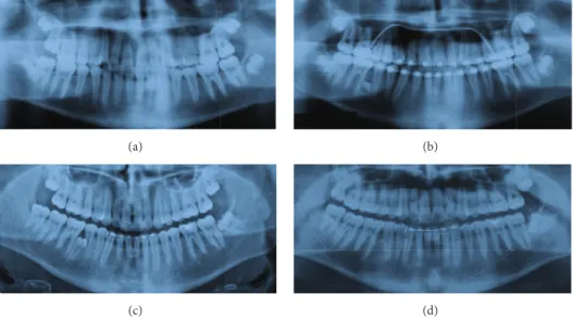
Figure 1: Case 1 radiography: before treatment 2004 (a), before rebonding, ST in 4.5–4.6 region, 2006 (b), 5 years after debonding 2011 (c), and after the extraction of the ST 2013 (d).
of the results is an important objective of the orthodontic treatment [15]; specifically, post-permanent ST can also com-promise this stability and interfere with orthodontic closure mechanics or with implant and mini-implant placement [1]. Generally, supernumerary teeth are detected incidentally during radiographic examination, and late-developing super-numerary premolars (LDSP) are often detected at the end of an orthodontic treatment, because the age of formation is around 12-13 years. There are two options in case of LDSP, follow-up or extraction; and there are several factors to analyze and choose the best therapeutic option. Modern radiographic technologies as three-dimensional computed tomography (3D CT) and cone beam computed tomography (CBCT) allow a better assessment of the risk to extract or not to extract a supernumerary post-permanent tooth and a more detailed analysis of the stomatognathic system’s bone structure [16, 17].
There are some reports on LDSP in the literature [12, 18– 23], and four further cases of LDSP with different approaches for their management are described in this paper, to improve knowledge regarding how to choose the best therapeutic option.
2. Case 1
2.1. Diagnosis and Etiology. A 12-year-old male in the perma-nent dentition was brought to our clinic for orthodontic treat-ment. The patient presented Class II, Division 2 malocclusion on Class I skeletal base. The overjet was reduced (1 mm) and the overbite increased (5 mm). Medical and familiar histories were unremarkable. The pretreatment radiography showed the presence of all the permanent teeth with also the presence of the wisdom teeth (Figure 1(a)). Treatment started in 2004 with thermoelastic wires to have a less painful resolution of the crowding [24]. During the treatment, after two years from the start, a new panoramic radiograph was requested to evaluate the necessity of some brackets rebonding, and ST
between 4.5 and 4.6 was detected. The radiography showed a bicuspid crown with a dental follicle (Figure 1(b)).
2.2. Treatment Objectives. The treatment objective was to avoid the damage of the surrounded tissues and of the con-tiguous teeth and the disruption of the occlusion, balancing the risk/benefit ratio between the follow-up of the tooth and the extraction.
2.3. Treatment Alternatives. There are two options when a LDSP is present; the first one is in the radiographic follow-up and the second one is the extraction. In this case the OPG (Figure 1(b)) revealed that the tooth was still developing, only the crown being formed, and that the tooth did not have any contact with the contiguous teeth. Performing an extraction of this tooth implied a germectomy with a deep mandibular access and a great bone loss. Moreover, no further active orthodontic treatment has to be performed in the lower arch. Hence, due to these considerations the best choice was to monitor the tooth during the time.
2.4. Treatment Progress. The patient’s parents were informed about the LDSP. Clinical and radiographic follow-up were performed to verify the onset of any complication. After 5 years the tooth was still not completely formed but an initial repositioning of the root of 4.5 was present (Figure 1(c)). In 2013, when the tooth achieved a more accessible position it was extracted upon the request of the patient (Figure 1(d)).
3. Case 2
3.1. Diagnosis and Etiology. An 11-year-old male in the mixed dentition was referred to the clinic for an orthodontic treatment. He presented Class II, Division 1 malocclusion on Class II skeletal base. The overjet and overbite were increased, and a molar crossbite was present [25]. Medical and familiar histories were unremarkable; no one of the parents had ST.
(a) (b)
(c) (d)
(e)
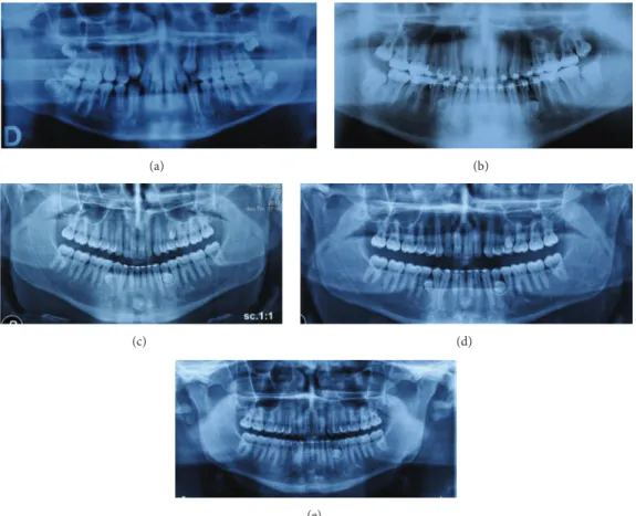
Figure 2: Case 2 radiography: before treatment 2006 (a), before rebonding, ST in regions 3.4–3.5, 4.4–4.5, and 2.5–2.6, 2009 (b), 3 years after debonding 2012 (c), 4 years after debonding 2013 (d), and after the extractions of ST in regions 4.4–4.5 and 2.5–2.6, 2015 (e).
The pretreatment radiography showed the presence of all the permanent teeth with also the presence of the wisdom teeth and the upper canine in eruption phase (Figure 2(a)). The patient was treated by means of aesthetic brackets and wires [26]. In the final stage of the orthodontic treatment in the panoramic radiograph, requested to assess the root parallelism, three supernumerary premolars, two in the lower jaw and one in the upper jaw, were identified (Figure 2(b)). 3.2. Treatment Objectives. The treatment objectives are to avoid that the ST could damage the surrounded tissues and the contiguous teeth and disrupt the occlusion, balancing the risk/benefit ratio between the follow-up of the tooth and the extraction.
3.3. Treatment Alternatives. In this case, the first treatment approach was the follow-up. Even if the ST in regions 3.4–3.5 and 4.4–4.5 created a root displacement with possible resorp-tion, in the upper jaw ST in 2.5–2.6 region did not create any issue. However, due to the very close relationship between the lower jaw supernumerary and the mental foramen it was preferred also in this case to monitor the teeth over time. 3.4. Treatment Progress. The patient’s parents were informed about the LDSP. The patient was subjected to follow-up, but his participation was not very regular, and in 2012 the OPG
showed the formation of the three teeth (Figure 2(c)); in 2013 two out of the three teeth erupted and due to the more accessible position it was decided to extract these two teeth (Figure 2(d)). In 2015 the patient has still the supernumerary in 3.4–3.5 region (Figure 2(e)).
4. Case 3
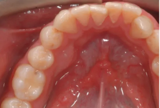
4.1. Diagnosis and Etiology. A 25-year-old male patient in permanent dentition presented to the orthodontic clinic with Class II subdivision on Class I skeletal base, with crooked teeth. At the clinical examination, the patient presented a little bulge in the 4.4–4.5 region (Figure 3) and at the panoramic radiography the presence of a supplemental tooth in this region was discovered (Figure 4(a)). The tooth was completely formed; the root apex was closed and seemed not to create any damage to the circumstance region.
4.2. Treatment Objectives. The treatment objective is to start an orthodontic treatment in the upper and lower jaw. 4.3. Treatment Alternatives. In this case, the only possible choice was the extraction of the ST. The patient came to the clinic to correct his malocclusion by means of an orthodontic treatment. Hence, due to the will of the patient to start an orthodontic treatment, the extraction was the best choice.
Figure 3: Case 3 intraoral image: lingual bulge in 4.4–4.5 region.
4.4. Treatment Progress. Before extracting the tooth, it was decided to perform a CBCT of the lower arch to assess the relationship between the ST and the mandibular nerve. Examining the CBCT ascertained that there was no contigu-ity between the tooth and the nerve (Figures 5(a) and 5(b)) and that the roots of 4.4 and 4.5 were damaged (Figures 5(c)– 5(e)). Hence, due to the favorable tooth position, the root damage, and the patient’s will, the extraction was performed. After the extraction a periapical radiograph of the extraction site was done (Figure 4(b)); then the patient started the orthodontic treatment.
5. Case 4
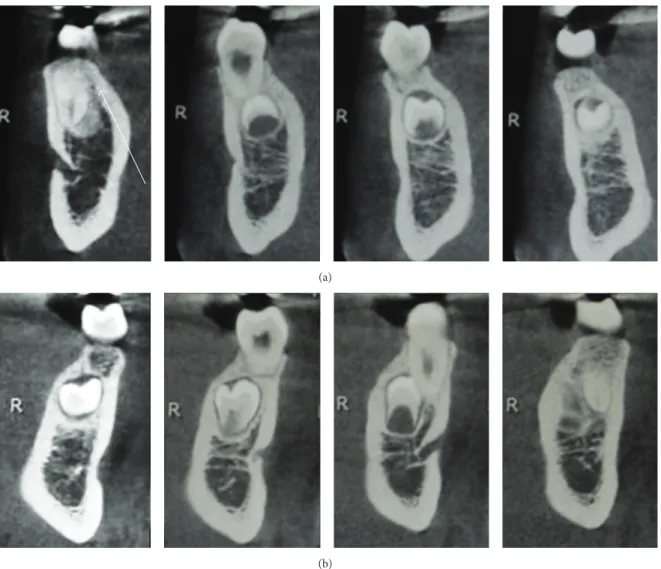
5.1. Diagnosis and Etiology. A 14-year-old patient, in good health, visited the clinic in March 2011. The diagnosis was dentoskeletal Class II malocclusion, with greatly increased overjet, atypical swallowing with tongue thrust, and anterior open bite. Initial OPG revealed all the permanent elements including gems of wisdom teeth with the absence of any number or shape of dental anomalies (Figure 6(a)). The patient was treated with self-ligating brackets [27] for 36 months. In the final OPG in March 2014, two supernumerary teeth in 3.4–3.5 and 4.4–4.5 area were found (Figure 6(b)). A CBCT of the lower arch was performed to better understand the ST position, the relationship with the surrounded tissues, and the presence of any root resorption that were not visible in the OPG. From the CBCT, the root displacement of 3.4 and 4.4 and the root resorption of 4.4 were clear (Figures 7(a) and 7(b)).
5.2. Treatment Objectives. The treatment objectives are to avoid further damage of the contiguous teeth and of the sur-rounded tissues and to maintain a good occlusion, balancing the risk/benefit ratio between the follow-up of the tooth and the extraction.
5.3. Treatment Alternatives. Once root displacement and root resorption were found on the CBCT no other choice than extraction was possible. In this case, even if the ST are in a deep position and the access will remove healthy bone, the damage present on the roots suggests extracting the ST to prevent worsening of the root resorption.
only with the CBCT due to the slight damage and the close superimposition between the ST and the teeth.
6. Results
In all the four cases presented the treatment objectives were achieved. In the first two cases where a follow-up approach was chosen, the occlusion remains stable over time. However, thanks to the radiographic images it was possible to check the evolution of the ST and to decide, in case of any complication, to extract the tooth as in Cases 1 and 2. In Cases 3 and 4 the aims were different; in Case 3 the extraction was mandatory both for the patient’s will to start an orthodontic treatment and for the little bulge present in the lower jaw. Also in Case 4 the extraction was mandatory because contiguous teeth showed root resorption, even if more dangerous than in the other cases; however the extractions stopped this phenomenon and the treatment aim was reached.
7. Discussion
Several cases of LDSP are discussed in the literature [12, 18– 23]; hence, even if uncommon, this condition has to be taken into account in the different phases of an orthodontic treatment. It was reported that the physiological calcification of the premolars starts between the age of 1.5 and 2.5 [28], although usually they are not detectable in radiograph until 3 or 4 years [29]. Otherwise, as reported in the literature, supernumerary premolar teeth start their developing around 10–15 years, a critical age since many children have an orthodontic treatment at that time [12, 18–23]. In fact, in all 4 cases reported, the supernumerary teeth were not detected in patients younger than 14 years old.
There are two solutions after discovering a LDSP, extract-ing or monitorextract-ing. The advantages and disadvantages of both options are different and the final decision has to be taken considering the risk/benefit relationship of the surgical removal. As a matter of fact, a ST could delay the eruption of the adjacent teeth, alter the eruption direction, displace the adjacent tooth, or cause cystic lesion and root resorption [30, 31]. Nevertheless, also the surgical removal is not totally free of risks; extraction of impacted teeth may lead to damage to adjacent structures and/or adjacent teeth with possible ankyloses [30, 31]. Hence, only in the case that the benefits of removal overbalance the risks of surgery, the teeth have to be extracted.
Bodin et al. [32] reported that only 2 percent of supernu-meraries in the premolar region exhibited any pathological change and suggested that the teeth may be left rather than the risk of surgical damage.
(a) (b)
Figure 4: Case 3 radiography: ST in 4.4–4.5 region 2015 (a) and after extraction 2015 (b).
(a) (b) (c)
(d) (e)
Figure 5: Case 3 CBCT: no continuity between the ST’s root and the alveolar nerve (a, b) and root resorption of contiguous teeth, indicated by the white arrows (c–e).
The choice to extract or not to extract the supernumerary teeth was due mainly to the damage that the tooth left in locus might cause. In Case 1, the decision was to monitor the lower jaw supernumerary tooth because it was in a position that unlikely evolves in a problematic situation and did not disrupt the occlusion. The tooth was extracted only due to the patient’s request and for the slight repositioning of the root of the 4.4. Instead, in Case 2 the solution to monitor the development of the teeth was due to the dangerous position of the teeth close to the mental foramen, thus being difficult to extract. In this case, once the teeth were totally formed and
were more accessible the teeth were removed. In the third case, the tooth was extracted before starting the orthodontic treatment and the CBCT helped the clinician to plan the surgical approach and to diagnose the root resorption due to the LDSP. The last case was an example of how important could the 3D imaging be to better evaluate the presence of damage of the adjacent structures due to ST. Probably also in Case 1 a CBCT could have shown root resorption in 4.5 or 4.6.
Therefore, the correct management of a supernumerary tooth depends on risk/benefit relationship between surgical
(c)
Figure 6: Case 4 radiography: before treatment 2011 (a), after treatment, ST in regions 3.4–3.5 and 4.4–4.5, 2014 (b), and after extraction of ST 2014 (c).
(a)
(b)
extraction and monitoring, the type and the position of the tooth and the possible damage in the surrounded tissues, and the orthodontic treatment stage in which the tooth was discovered. In many cases only 3D imaging could help the clinician to clearly evaluate the problem.
8. Conclusions
The cases highlight the late development of supernumerary teeth in the premolar region at different times with respect to the orthodontic treatment. If the post-permanent tooth appears before starting the orthodontic treatment, the sug-gestion is to remove it to avoid interferences during the teeth movement. The same protocol should be used even when the tooth was discovered during active orthodontic treatment. On the other hand, if the tooth was detected at the end of the orthodontic treatment or with no need of orthodontic treatment and did not disrupt the patient occlusion, it should be monitored by means of a regular radiographic and clinical follow-up, and the extraction has to be an option only if the risk/benefit relationship is favorable.
Competing Interests
The authors declare that they have no competing interests.
References
[1] A. Parolia, M. Kundabala, M. Dahal, M. Mohan, and M. S. Thomas, “Management of supernumerary teeth,” Journal of
Conservative Dentistry, vol. 14, no. 3, pp. 221–224, 2011.
[2] D. R. McKibben and L. J. Brearley, “Radiographic determination of the prevalence of selected dental anomalies in children,”
ASDC Journal of Dentistry for Children, vol. 28, no. 6, pp. 390–
398, 1971.
[3] J. D. Smith, “Hyperdontia: report of case,” The Journal of the
American Dental Association, vol. 79, no. 5, pp. 1191–1192, 1969.
[4] J. F. Liu, “Characteristics of premaxillary supernumerary teeth: a survey of 112 cases,” ASDC Journal of Dentistry for Children, vol. 62, no. 4, pp. 262–265, 1995.
[5] R. E. Primosch, “Anterior supernumerary teeth—assessment and surgical intervention in children,” Pediatric dentistry, vol. 3, no. 2, pp. 204–215, 1981.
[6] A. H. Brook, “A unifying aetiological explanation for anomalies of human tooth number and size,” Archives of Oral Biology, vol. 29, no. 5, pp. 373–378, 1984.
[7] L. D. Rajab and M. A. M. Hamdan, “Supernumerary teeth: review of the literature and a survey of 152 cases,” International
Journal of Paediatric Dentistry, vol. 12, no. 4, pp. 244–254, 2002.
[8] M. Fader, S. N. Kline, S. S. Spatz, and H. J. Zubrow, “Gardner’s syndrome (intestinal polyposis, osteomas, sebaceous cysts) and a new dental discovery,” Oral Surgery, Oral Medicine, Oral
Pathology, vol. 15, no. 2, pp. 153–172, 1962.
[9] B. L. Jensen and S. Kreiborg, “Development of the dentition in cleidocranial dysplasia,” Journal of Oral Pathology and Medicine, vol. 19, no. 2, pp. 89–93, 1990.
[10] P. N. Tannure, C. A. G. R. Oliveira, L. C. Maia, A. R. Vieira, J. M. Granjeiro, and M. De Castro Costa, “Prevalence of dental anomalies in nonsyndromic individuals with cleft lip
and palate: a systematic review and meta-analysis,” Cleft
Palate-Craniofacial Journal, vol. 49, no. 2, pp. 194–200, 2012.
[11] M. M. Nazif, R. C. Ruffalo, and T. Zullo, “Impacted supernu-merary teeth: a survey of 50 cases,” The Journal of the American
Dental Association, vol. 106, no. 2, pp. 201–204, 1983.
[12] S. M. Cochrane, J. R. Clark, and N. P. Hunt, “Late develop-ing supernumerary teeth in the mandible,” British Journal of
Orthodontics, vol. 24, no. 4, pp. 293–296, 1997.
[13] E. C. Stafne, “Supernumerary teeth,” Dental Cosmos, vol. 74, pp. 653–659, 1932.
[14] S. Paduano, R. Uomo, M. Amato, F. Riccitiello, M. Simeone, and R. Valletta, “Cyst-like periapical lesion healing in an orthodontic patient: a case report with five-year follow-up,”
Giornale Italiano di Endodonzia, vol. 27, no. 2, pp. 95–104, 2013.
[15] A. Lucchese, M. Manuelli, L. Bassani et al., “Fiber reinforced composites orthodontic retainers,” Minerva Stomatologica, vol. 64, no. 6, pp. 323–333, 2015.
[16] J. Mossaz, D. Kloukos, N. Pandis, V. G. A. Suter, C. Katsaros, and M. M. Bornstein, “Morphologic characteristics, location, and associated complications of maxillary and mandibular supernumerary teeth as evaluated using cone beam computed tomography,” European Journal of Orthodontics, vol. 36, no. 6, pp. 708–718, 2014.
[17] M. Portelli, G. Matarese, A. Militi, G. Lo Giudice, R. Nucera, and A. Lucchese, “Temporomandibular joint involvement in a cohort of patients with juvenile idiopatic arthritis and evalua-tion of the effect induced by funcevalua-tional orthodontic appliance: clinical and radiographic investigation,” European Journal of
Paediatric Dentistry, vol. 15, no. 1, pp. 63–66, 2014.
[18] D. E. Bowden, “Post-permanent dentition in the premolar region,” British Dental Journal, vol. 131, no. 3, pp. 113–116, 1971. [19] M. L. Kantor, C. S. Bailey, and E. J. Burkes Jr., “Duplication
of the premolar dentition,” Oral Surgery, Oral Medicine, Oral
Pathology, vol. 66, no. 1, pp. 62–64, 1988.
[20] J. J. Breckon and S. P. Jones, “Late forming supernumeraries in the mandibular premolar region,” British Journal of
Orthodon-tics, vol. 18, no. 4, pp. 329–331, 1991.
[21] N. Gibson, “A late developing mandibular premolar supernu-merary tooth,” Australian Dental Journal, vol. 45, no. 4, pp. 277– 278, 2000.
[22] R. S. Jamwal, P. Sharma, and R. Sharma, “Late-developing supernumerary mandibular premolar: a case report,” World
Journal of Orthodontics, vol. 11, no. 4, pp. 94–98, 2010.
[23] S. Yassaei, M. Goldani Moghadam, and S. M. Tabatabaei, “Late developing supernumerary premolars: reports of two cases,”
Case Reports in Dentistry, vol. 2013, Article ID 969238, 4 pages,
2013.
[24] I. Cioffi, A. Piccolo, R. Tagliaferri, S. Paduano, A. Galeotti, and R. Martina, “Pain perception following first orthodontic archwire placement—thermoelastic vs superelastic alloys: a randomized controlled trial,” Quintessence International, vol. 43, no. 1, pp. 61–69, 2012.
[25] G. Iodice, G. Danzi, R. Cimino, S. Paduano, and A. Michelotti, “Association between posterior crossbite, masticatory muscle pain, and disc displacement: a systematic review,” European
Journal of Orthodontics, vol. 35, no. 6, pp. 737–744, 2013.
[26] R. Rongo, G. Ametrano, A. Gloria et al., “Effects of intraoral aging on surface properties of coated nickel-titanium arch-wires,” Angle Orthodontist, vol. 84, no. 4, pp. 665–672, 2014. [27] S. Paduano, I. Cioffi, G. Iodice, A. Rapuano, and R. Silva,
the American Dental Association, vol. 131, no. 5, pp. 623–632,
2000.
[31] A. Shah, D. S. Gill, C. Tredwin, and F. B. Naini, “Diagnosis and management of supernumerary teeth,” Dental Update, vol. 35, no. 8, pp. 510–520, 2008.
[32] I. Bodin, P. Julin, and M. Thomsson, “Hyperodontia. I. Fre-quency and distribution of supernumerary teeth among 21,609 patients,” Dentomaxillofacial Radiology, vol. 7, no. 1, pp. 15–17, 1978.
Submit your manuscripts at
http://www.hindawi.com
Hindawi Publishing Corporation
http://www.hindawi.com Volume 2014
Oral Oncology
Journal ofDentistry
International Journal ofHindawi Publishing Corporation
http://www.hindawi.com Volume 2014
Hindawi Publishing Corporation
http://www.hindawi.com Volume 2014
International Journal of
Biomaterials
Hindawi Publishing Corporation
http://www.hindawi.com Volume 2014
BioMed
Research International
Hindawi Publishing Corporation
http://www.hindawi.com Volume 2014
Case Reports in
Dentistry
Hindawi Publishing Corporation
http://www.hindawi.com Volume 2014
Oral Implants
Journal ofHindawi Publishing Corporation
http://www.hindawi.com Volume 2014
Anesthesiology Research and Practice
Hindawi Publishing Corporation
http://www.hindawi.com Volume 2014 Radiology
Research and Practice
Environmental and Public Health
Journal of
Hindawi Publishing Corporation
http://www.hindawi.com Volume 2014
The Scientific
World Journal
Hindawi Publishing Corporationhttp://www.hindawi.com Volume 2014
Hindawi Publishing Corporation
http://www.hindawi.com Volume 2014
Dental Surgery
Journal ofDrug Delivery
Journal of Hindawi Publishing Corporationhttp://www.hindawi.com Volume 2014
Hindawi Publishing Corporation
http://www.hindawi.com Volume 2014
Oral Diseases
Journal ofHindawi Publishing Corporation
http://www.hindawi.com Volume 2014 Computational and Mathematical Methods in Medicine
Scientifica
Hindawi Publishing Corporationhttp://www.hindawi.com Volume 2014
Pain
Research and TreatmentHindawi Publishing Corporation
http://www.hindawi.com Volume 2014 Hindawi Publishing Corporation
http://www.hindawi.com Volume 2014
Endocrinology
International Journal ofHindawi Publishing Corporation
http://www.hindawi.com Volume 2014
Hindawi Publishing Corporation
http://www.hindawi.com Volume 2014