Dottorato di Ricerca in
Qualità degli Alimenti e Nutrizione umana
Coordinatore: Prof.ssa Annunziata Giangaspero
TESI DI DOTTORATO
XXVIII Ciclo
Fennel (Foeniculum vulgare):
a novel food allergen of the Mediterranean Diet
Dottoranda:
Dott.ssa Mariangela Di Giacomo
Relatore:
Prof. Luigi Macchia
Correlatore:
Prof.ssa Maria Filomena Caiaffa
Index
Abstract 4
1. Introduction 6
1.1. The Mediterranean Diet benefit on human health 6 1.2. Food allergy pathogenic mechanisms 12
1.3. Food allergy diagnosis 14
1.4. Foeniculum vulgare 16
1.4.1. Foeniculum vulgare and food allergy 16
1.4.2. Foeniculum vulgare botanical and agronomical characteristics 16
1.4.3. Foeniculum vulgare nutritional aspects and health properties 18
1.4.4. Foeniculum vulgare production in Italy 20
1.5. Foeniculum vulgare allergy. Background 21
1.6. The Foeniculum vulgare allergy project 25
2. Materials and methods 26
2.1. Fennel allergy diagnosis work up 26
2.1.1. Convincing clinical history 26
2.1.2. In vivo tests 27
2.1.3. In vitro tests 28
2.2. Generation of semi-purified food extracts 28
2.2.1. Preparation of a semi-purified fennel extract 28
2.2.2. Preparation of semi-purified peach and celery 100,000 x g supernatants 29
2.3. Biotinylation of the semi-purified fennel extract 29
2.4. The ELISA capture in vitro assay 31
2.5. RAST inhibition experiments 32
2.6. Immunodetection of fennel allergens 33
2.6.1. Sodium dodecylsulfate-polyacrylamide gel electrophoresis (SDS-PAGE) of the semi-purified Foeniculum vulgare extract 33
2.6.2. IgE Immunoblotting analysis 34
2.7. Statistical analysis 35
3. Results 36
3.1. Selection of study patients 36
3.2. Generation of a semi-purified fennel extract 43
3.3. Clinical features of the selected fennel allergy population 44
3.4. Studies on mast cell-bound specific IgE by precision SPT and prick by prick 45
3.5. In vitro studies: ImmunoCAP Thermo Fisher analysis and development of an in-house RAST-capture assay 53
3.5.1. ImmunoCAP Thermo Fisher analysis of sera from 41 fennel allergy patients 53 3.5.2. Development of an in-house RAST-capture assay and analysis
3.5.3. Comparison between the two in vitro experimental approaches:
ImmunoCAP Thermo Fisher assay and in-house RAST-capture assay 59
3.5.4. Finding the biotinylated fennel extract amount suitable for the in-house RAST-capture assay 61
3.6. RAST inhibition experiments 62
3.6.1. Production of the semi-purified extracts of peach and celery 63
3.6.2. RAST inhibition experiments with the semi-purified peach extract 64
3.6.3. RAST inhibition experiments with the in-house Foeniculum vulgare extract 65
3.6.4. RAST inhibition experiments performed with celery 100,000 x g supernatant 66
3.7. Immunoblotting analysis of the semi-purified Foeniculum vulgare extract 67
3.7.1. Experimental settings and technical experiments 68
3.7.2. Artefactual nature of an apparently immunoreactive doublet 69
3.7.3. The sensitivity problem 72
3.7.4. Detection of putative immunoreactive bands in 40 patients with fennel allergy 73
4. Discussion and Conclusion 79
5. Acknowledgements 85
6. References 87
incubation of the sera with either the fennel extract or the celery extract caused RAST to become negative, thus, providing evidence of cross-reactivity between fennel and celery, as expected.
Moreover, 18 fennel extract protein bands were detected when the in-house fennel extract was analyzed by SDS-PAGE, under reducing conditions. Immunoblotting analysis was performed in 40 out of the 44 fennel allergy subjects, in order to detect putative fennel allergen bands. These experiments showed that some bands were recognized more frequently, but also that there were rather diverse immunoreactive band profiles, among these patients. Thus, 3 major immunoreactive bands, putatively involved in fennel allergy, were detected. These bands were: a 33 kDa band (detected by sera from 8 patients), a 45 kDa band (detected by 5 patients) and, finally, a 50 kDa band (also detected by 5 patients).
In conclusion, the results showed that F. vulgare can be considered a major food allergen, at least in those Countries where the Mediterranean Diet prevails, accounting for a substantial proportion of all food allergy cases (possibly, up to 30%). Moreover, it is also possible to regard fennel allergy as a well-characterized and self-consistent food allergy, as the results of the in vivo investigations and the RAST and RAST inhibition experiments pointed out.
Keywords
1. Introduction
1.1. The Mediterranean Diet benefit on human health
“The Mediterranean Diet is a set of traditional practices, knowledge and skills passed on
from generation to generation and providing a sense of belonging and continuity to the
concerned communities”.
This is the first reason cited by UNESCO for recognizing the Mediterranean Diet as an Intangible Cultural Heritage, in 2010 (http://www.unesco.org/culture/ich/en/RL/00394). This most authoritative international organization officially ratified what science has been consistently demonstrating during the prevoius decades.
It appears that UNESCO did not consider the Mediterranean Diet just as a mere collection of some selected foods but, rather, ascribed to the eating habits typical of the Mediterranean basin a cultural-promotional role, as stated in the description of this type of diet: “The Mediterranean Diet encompasses more than just food. It promotes social interaction, since communal meals are the cornerstone of social customs and festive events. It has given rise to a considerable body of knowledge, songs, maxims, tales and legends. The system is rooted in respect for the territory and biodiversity, and ensures the conservation and development of traditional activities and crafts linked to fishing and farming in the Mediterranean communities of which Soria in Spain, Koroni in Greece, Cilento in Italy and Chefchaouen in Morocco are examples”.
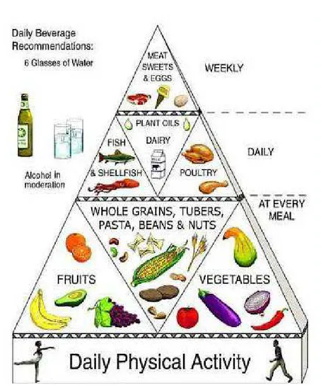
The Mediterranean Diet is a complete and balanced nutritional model, combining various ancient culinary patterns and traditions. Usually, it is represented as a pyramid (Fig. 1.), where plant foods constitute the core of the daily intake, whereas foods from animals are more peripheral.
Examples of traditional Mediterranean dietary patterns, with foods from plants at the center of the plate, include the use of couscous, vegetables, and legumes in North Africa; pasta, polenta, rice, or potatoes, along with vegetables and legumes in Southern Europe; rice, along with vegetables and beans in the Eastern Mediterranean regions.
Fresh vegetables, salads, fruits, nuts, olive oil, low fat cheese and yogurt are consumed frequently and garlic, onions, and herbs are used as condiments. Poultry and fish are also
advisable; instead, sweets, eggs and meat should be eaten weekly and in moderation. A moderate intake of wine (one glass for women and two drinks for men per day, preferably during meals) is recommended, since it is an important source of resveratrol, a polyphenolic compound that plays an anti-inflammatory and anti-atherogenic role (http://www.mediterraneandiet.com/). According to the Mediterranean food pyramid, physical activity is very much encouraged, since it promotes well-being and maintenance of a healthy weight (Bonaccio et al., 2012).
Fig. 1. The Mediterranean Diet food pyramid.
In 2009 an interesting study, published in the "British Medical Journal", underlined that the principal component of the Mediterranean Diet, as a predictor of lower mortality, was the moderate intake of ethanol, followed by low consumption of meat and meat products and high
Thus, the moderate consumption of alcohol has been considered a key element of the food pyramid, since it has been proven to be involved in the prevention of cardiovascular risk (Costanzo et al., 2010; Di Castelnuovo et al., 2002).
In fact, life-style and eating habits are the major modifiable factors on which it is possible to intervene in order to prevent the onset of the so-called "diseases of wellness" (obesity, diabetes, cardiovascular diseases, tumors), characteristics of our times. Thus, increasing the level of physical activity and introducing healthy food in everyday meals is regarded as highly beneficial for health, therefore affecting positively the quality of human life.
There is a substantial body of evidence linking the Mediterranean Diet to cardiovascular risk reduction and prevention of other major chronic diseases (Willett et al., 2009). A meta-analysis study accounting for a particularly large number of subjects and studies, showed a significant and consistent protection provided by adherence to the Mediterranean Diet, in relation to the onset of major chronic degenerative diseases (Sofi et al., 2010).
The beneficial effects of the Mediterranean Diet have been reported to be due to the synergistic interaction of various constituents. However, numerous studies have demonstrated that single components, such as ω-3 polyunsaturated fatty acids (PUFAs) and polyphenols, play important roles in the prevention of neuro-degenerative diseases and inflammation (Widmer et al., 2015). Inflammation is recognized as the major risk factor in the etiopathogenesis of many chronic diseases, such as cardiovascular diseases (CVD), cancer, type 2 diabetes, Alzheimer's disease and obesity (Ostan et al., 2015).
A recent review, by Barbaresko and co-workers, demonstrated that a diet consistent with the Mediterranean Diet contributes to a reduction of C-reactive protein levels (a marker of inflammation) and leads to an increase of adiponectin (Barbaresko et al., 2013). Furthermore, this diet reduces postprandial inflammation, and the postprandial oxidative stress that can result from postprandial hyperglycemia and hyperlipemia (Camargo et al., 2012).
A link between oxidative stress and inflammation is represented by the transcription factor NF-kB, which is induced by oxidative stress. This factor, in turn, increases the expression of pro-inflammatory genes for cytokines and chemokines (Reuter et al., 2010).
A substantial antioxidant activity is attributed to compounds of plant origin, since these molecules are able to reduce inflammatory responses, as in the case of polyphenols present in extra-virgin olive oil. These polyphenols include hydroxytyrosol, potent antioxidant and free radical scavenger, and oleocanthal, inhibitor of cyclooxygenase activity and best known for its anti-inflammatory properties. Oleocanthal also possesses neuro-protective effects and a
positive effect on markers of inflammation implicated in Alzheimer’s disease (Parkinson et al., 2014).
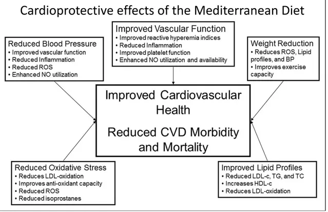
Foods belonging to the Mediterranean Diet are able to prevent the onset of cardiovascular diseases, improving blood lipid profile and reducing blood pressure, presumably through reduced expression of inflammatory biomarkers, reduced oxidation of low-density lipoprotein cholesterol (LDL cholesterol) and neutralization of free radicals (Fig. 2.).
Fig. 2. Mechanisms proposed to underlie the protective effect of foods belonging to the Mediterranean Diet, against cardiovascular diseases.
Moreover, adherence to the Mediterranean Diet showed a protective effect on allergic rhinitis, asthma-like symptoms and atopy (Chatzi et al., 2007). This study was conducted in Crete, Greece, among school children aged between 7 and 18 years. Children in Crete consume high quantities of locally produced fruits and vegetables, whose regular and frequent consumption may have a beneficial role against wheezing and rhinitis symptoms. The results obtained were consistent with results from previous epidemiological studies on the protective effects of citrus fruits, apples and tomatoes on asthma and rhinitis symptoms (Farchi et al., 2003; Garcia
observed. Grapes consumption was shown to be inversely associated with wheezing and allergic rhinitis. Red grape skin contains a mix of non-bioflavonoid and bioflavonoid polyphenols. Oligomeric proanthocyanidins are antioxidants with an anti-inflammatory action that is relevant to oxygen-free radical scavenging, anti-lipid peroxidation and inhibition of the formation of inflammatory cytokines (Li et al., 2001). Moreover, resveratrol, a polyphenolic stilbene found in grapes skin, is able to inhibit cytokine-stimulated inducible nitric oxide synthase expression and nitrite production in human primary airway epithelial cells. It is possible that the beneficial effect of grape intake on wheezing and allergic rhinitis symptoms is mediated by the antioxidant effect of grape phenolic content.
This study also showed a protective effect by nut consumption on the occurrence of wheezing symptoms. Nuts are rich in vitamin E, the body’s principal defence against oxidant-induced membrane injury in human tissue, via its role in breaking the lipid peroxidation chain reaction. It was also observed that weekly intake of margarine had a harmful effect on asthma and rhinitis symptoms in this population. Margarine is a source of omega-6 polyunsaturated fatty acids such as linoleic acid, by which arachidonic acid is generated. In turn, arachidonic acid represents the substrate of the cyclooxygenase pathway that leads to prostaglandins bio-synthesis, important pro-inflammatory as well as anti-inflammatory lipid mediators.
Therefore, a general protective effect of a high level of adherence to the Mediterranean Diet on allergic rhinitis, asthma and atopy was assessed. Thus, this study indicated that a high dietary intake of commonly consumed fruits and vegetables and nuts might have a protective effect on the occurrence of asthma-like symptoms and allergic rhinitis.
Because of its benefit on human health, in recent years, the Mediterranean Diet has attracted substantial interest by the biomedical community. In general, foods belonging to the Mediterranean Diet cause allergy less frequently, but the Mediterranean Diet also includes potentially allergenic foods, most of them are poorly characterized or, simply, unrecognized. Moreover, food allergy, in recent years, has represented the focus of considerable research interest, as epitomized by the inclusion of this topic in the list of the thematic areas of the
Sixth and Seventh Framework Programmes of the European Union
(http://ec.europa.eu/research/fp6/index_en.cfm; https://ec.europa.eu/research/fp7/pdf/fp7-brochure_it.pdf).
Thus, the Work Programme of Thematic Priority 5: “Food Quality and Safety” of the VI Framework Programme, at point 5.4.2. “Epidemiology of food-related diseases and allergies”, read: “ The objective is to examine the complex interactions between food intake and
risk factors and develop common European databases. Many diseases and disorders prevalent in Europe today can be linked to diet, genetic make-up and life-style. Research in this area will use pan-European epidemiological studies concentrating on most important nutrition-related diseases and disorders in order to identify vulnerable population groups, links to diet, genetic factors, and assess how improved diet might reduce the prevalence of these disorders in the future.”
Moreover, at point T2.2 – “Epidemiology of food allergy”, the same document read: “The focus should be on integrated epidemiological studies on well-defined food allergens influencing the occurrence, prevalence, prevention and distribution of allergic diseases and hypersensitivity disorders in the European population (children, adults, influences during foetal life). Immunological and genetic studies with defined criteria for diagnosis, well-defined and/or developed allergy markers and other factors, such as: diet, environment and infections, should be taken into account. The approach should include also an assessment of the socio-economic impact of food allergy, including costs to society and the effect on quality of life of sufferers and their relatives. The generation of new diagnostic tools and methodologies to predict the outcome of allergen interaction with susceptible individuals should be considered. Dissemination plans for patients and the scientific community should be clearly defined”.
On the other hand, the Seventh Framework Programme – Knowledge-Based Bio-Economy (KBBE), with the Theme 2 - “Food, Agriculture and Fisheries, and Biotechnologies” Cooperation Work Programme, at point KBBE-2007-2-2-06 – “Impact of exogenous factors in the development of allergy”, read: “The aim will be to investigate the mechanisms of early protective effect of exposure to exogenous factors such as diet, life-style and/or living environment on allergy development, using cohort studies and well defined model systems to study the immunological pathways involved, particularly in relation to events in early life. The primary focus will be on the preventable causes, rather than triggers, of allergy onset and on the elucidation of how exogenous factors affect the immune system, mucosal barrier, gut colonization and development of allergy. The expected impact will be represented by the creation of comprehensive and innovative knowledge on the immunological and physiological mechanisms underlying the cause of allergy and its rise in prevalence; promoting health and quality of life of allergy sufferers, by addressing health determinants, such as diet and life-style conditions; finally, providing new knowledge on when exposure to these putative protective agents or mechanisms can improve health by preventing further increase of
production of substantial amounts of specific IgE antibodies. A protein eliciting an anomalous IgE immune response is called an allergen.
1.3. Food allergy diagnosis
Food allergy diagnosis begins with the collection of patient’s clinical history. Based on the information derived from this initial step, diagnosis can be completed and achieved by different laboratory studies.
The importance of clinical history depends on the patient’s ability of remembering the symptoms and on the examiner’s capacity of distinguishing reactions due to food from those with other etiologies. A tool used in addition to clinical history is represented by diet diaries, where patients report all foods ingested during a specific period of time, in a chronological way and the symptoms. Finally, these diaries are reviewed to determine the relationship between food ingested and symptoms experienced by the patient.
Symptoms associated with allergic reactions can affect the gastrointestinal tract, the skin and the respiratory tract. Gastrointestinal symptoms can manifest with mouth itching, lips and palate edema, nausea, vomiting and diarrhea. The skin symptoms are characterized by generalized itching, erythematous rash, urticaria and angioedema. Respiratory symptoms, instead, can affect the larynx and the upper and/or lower respiratory tract, with symptoms similar to those of allergic asthma and allergic rhinoconjunctivitis, such as periocular itching, conjunctival erythema and lachrymation, nasal itching, rhinorrhoea, laryngeal edema, coughing, wheezing and dyspnea.
Another method also used to collect additional clinical information is represented by the elimination diets, which are employed not only in diagnosis but also in food allergy management, when certain foods are suspected to cause allergic reactions. Thus, these foods can be completely eliminated from the diet. The success of these diets depends on the food allergens identification and on patient’s ability to follow a diet free from all forms of the allergen suspected of causing the allergic reaction.
As regards the diagnostic evaluation methods, skin prick test (SPT) are the major tool in detecting patients with IgE-mediated food reaction. SPT are performed by pricking the patient’s skin (usually, at the volar surface of the foreorms) through a droplet of allergen
solution, which allows the allergen molecules to interact with potential IgE specific for that given allergen, on the surface of skin-resident mast cells.
The skin usually is punctured using a sterile lancet and the results are read 20’ after pricking. Histamine hydrochloride 10 mg/ml in 50% glycerol solution is used as the positive control in skin prick testing, while saline with 1% albumin serves as the negative control. Positive responses consist in a wheal-and-flare reaction, whose magnitude essentially reflects the amount of endogenous histamine released by activated cutaneous mast cells (Fig. 4). To measure the elicited cutaneous reactions, the wheal can be outlined with a felt-tip pen and the outline can be blotted on removable tape and recorded on paper, for convenience. The area can be calculated by superimposing a transparent cellulose acetate sheet with a 1 mm square lattice grid and counting the squares comprised within the outline (each square 1 mm2; Corallino et al., 2007).
Criteria for food allergy SPT interpretation were determined by Bock and May (Bock et al., 1978). According to these Authors a wheal (elicited by an allergen) with a diameter at least 3 mm larger than that of the negative control, is considered positive. However, positive SPT indicate an immunological sensitization that not always associates with clinical manifestations. Therefore this diagnostic approach is highly sensitive but not high specific (Sicherer et al., 2010). Thus, the positive predictive value of this method is slightly less than 50%. Conversely, negative SPT support the hypotesis of absence of IgE-mediated reactions. Hence, the negative predictive value of this test is greater than 95%, provided that the quality of food extracts is optimal (Bock et al., 1978).
When diagnosing IgE-mediated sensitizations to fruits or vegetables, SPT can be replaced by prick by prick tests, in order to avoid false negatives, since commercial food extracts could be altered by commercial preparation, because of proteins lability. These tests are performed pricking the skin through a slice of the suspected food.
Finally, in routine diagnosis of allergic diseases, there is a second level exam, known as RAST (Radio Allergo Sorbent Test), which allows determination of specific IgE in serum and other biological fluids. This method is characterizaed by a lower negative predictive value compared to SPT, but it has a higher positive predictive value (Asero et al., 2007).
1.4. Foeniculum vulgare
1.4.1. Foeniculum vulgare and food allergy
In order to assess the potentially hypoallergenic properties of the Mediterranean Diet, definition of occurrence and socioeconomic impact of food allergy in our area, recognition and characterization of novel food allergens, elucidation of the immune mechanisms associated with their intake and tolerance need to be clarified. These aspects of the problem, of course, in turn, need to be supported by larger epidemiological studies, providing the rationale for the investigative work to develop. Moreover, since increasingly strict regulations on marketed pre-packed foods within the European Union also include allergen specification and labeling, we think that the thorough characterization of novel food allergens of the Mediterranean Diet cannot but add to the general appreciation by consumers of the Mediterranean Diet itself, with the exact identification of the related (possibly marginal) health risks.
In this research project, the attention has been focused on the characterization of a novel food allergen belonging to the Mediterranean Diet: Foeniculum vulgare.
Foeniculum vulgare allergy has been studied only marginally, so far. Therefore, fennel is not included in the allergen list of the International Union of Immunological Societies (IUIS) database (http://www.allergen.org/).
1.4.2. Foeniculum vulgare botanical and agronomical characteristics
Foeniculum vulgare, commonly called fennel, is a member of the Apiaceae family (also called Umbrelliferae), a large group of plants encompassing approximately 300 genera and more than 3000 species.
Fennel is an ancient seasonal herb that grows wild in most of temperate Europe, but is generally considered indigenous to the Mediterranean Countries.
Fennel was well-known to the ancient Egyptians, Greeks and Romans, who grew it for its aromatic seeds and the edible fleshy shoots. In various Italian regions, especially in Southern Italy, in stony and sub-mountainous areals up to a height of 700 metres, and along the coasts, wild Foeniculum species with perennial plants, characterized by strong roots that do not form the false bulb (the so-called “grumolo”, in Italian) and resistance to drought are still present.
From the alimentary point of view, the young leaves of these wild species are used as flavoring in several food preparations.
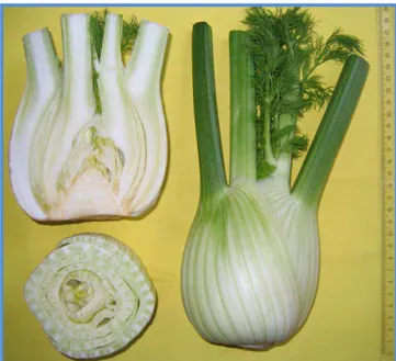
The genus Foeniculum was described by Miller in 1768, who separated it from Anethum, as previously indicated by Linnaeus. Moreover, Miller distinguished two species in the genus Foeniculum: F. vulgare, which includes F. vulgare (var. dulce), cultivated as a vegetable for consumption of the “grumoli”, and F. sativum, cultivated for its seeds. A further subdivision (within F. vulgare) was proposed by Tutin in 1968: F. vulgare subspecies piperitum, whose inflorescences and the stem upper part, are used for pickled storage, and F. vulgare subspecies capillaceum, whose seeds are employed in alcohol preparations as flavoring. F. vulgare (var. dulce) is a herbaceous plant with a biennial cycle. In the first year, it develops the vegetative part represented by the so-called “grumolo” or false bulb which is the edible part, made of the basal sheaths of the leaves, overlapping with each other with the function of accumulating nutrients (Fig. 4.), and by the root system. Instead, during the second year, fennel produces flowers, fruits and seeds (Siviero et al., 2005).
Fig. 4. Fennel “grumolo” or false bulb growth during the first year of vegetative cycle. Fennel is a crop characterized by an autumn-winter production cycle and, therefore, it has reduced water needs. Its ideal minimal temperature is around 7 °C, while the optimum
ranging is between 15 °C and 20 °C. Fennel is also frost-resistant, providing that the temperature does not fall below -2 °C.
Germination requires at least 10 °C, but it is hindered over 30 °C, with an optimum temperature between 20 °C and 25 °C.
The irrigation is an important practice expecially during the early cycle stages, since the plant has to grow in a warm period. Thus, it is particularly sensitive to water stress that can be responsible for physiological problems, besides bacterial/fungal infections.
The most important features for variety choice are the cultivation period, the production area and the cycle duration. Thus, fennel plant prefers deep grounds with an optimal pH between 6 and 7.5.
( http://agronotizie.imagelinenetwork.com/vivaismo-e-sementi/2015/05/04/il-finocchio-una-coltura-per-tutte-le-stagioni/9292).
1.4.3. Foeniculum vulgare nutritional aspects and health properties
Fennel is one of the most pleasing and sweetest vegatebles. It can be consumed either as raw fresh food, in salads, or cooked.
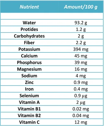
From a nutritional point of view, F. vulgare is characterized by a low-energy content, with only 9 kcal/100 g (38 kJ/ 100 g). It is also worth noticing that it is characterized by a high amount of fiber and mineral salts, in particular potassium (Food Data Bank INRAN, Rome). This vegetable also has a high content of vitamin C, equal to 12 mg/100 g, corresponding to 20% of the recommended daily intake (Recommended Daily Allowance, RDA). See Tab. 1. (http://nut.entecra.it/646/tabelle_di_composizione_degli_alimenti.html?idalimento=005320& quant=100).
Nutrient Amount/100 g Water 93.2 g Protides 1.2 g Carbohydrates 2 g Fiber 2.2 g Potassium 394 mg Calcium 45 mg Phosphorus 39 mg Magnesium 16 mg Sodium 4 mg Zinc 0.9 mg Iron 0.4 mg Selenium 0.9 µg Vitamin A 2 µg Vitamin B1 0.02 mg Vitamin B2 0.04 mg Vitamin C 12 mg
Tab. 1. Nutritional features and content of vitamins and minerals per 100 g of fennel (edible parts).
Furthermore, phytochemical research carried out on F. vulgare has led to the isolation of fatty acids, phenolic components, hydrocarbons, volatile components, and few other classes of secondary metabolites from its different parts. Mostly these phytochemicals are found in essential oil. For example, the anise odor of F. vulgare is due to its essential oil content. It makes an excellent flavoring agent in various types of food and food related products. The essential oil of fennel has been reported to contain more than 87 volatile compounds. The accumulation of these volatile compounds inside the plant is variable, appearing practically in any of its parts: roots, stem, shoots, flowers, and fruits. The main components are the trans anethol phenolic ether and fenchone. Other important components, present in various amounts are estragole, p-anisaldehyde and terpenes. All these molecules give the plant not only an intense characteristic flavor, but also interesting pharmacological properties, such as antimicrobial, antiviral, anti-inflammatory, antimutagenic, antipyretic, antispasmodic, antithrombotic, apoptotic, cardiovascular, chemomodulatory, antitumor, hepatoprotective, hypoglycemic, hypolipidemic and memory enhancing property.
In fact, F. vulgare is well-known for the cure of numerous infectious disorders of bacterial, fungal, viral and mycobacterial origin.
Finally, not only fennel exhibits pharmacological activities but also reveals some environmental active properties. These activities play a key role in the management of nematode, insect, mosquitoes and some harmful larvae of malaria producing vector. Thus, F. vulgare extracts and isolated biologically active compounds are evaluated for their insecticidal, repellent, acaricidal, larvicidal and nematicidal activity (Badgujar et al., 2014).
1.4.4. Foeniculum vulgare production in Italy
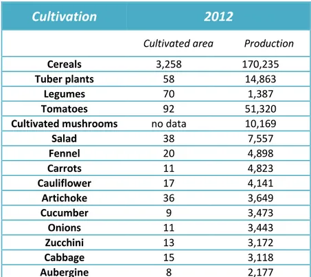
Fennel is cultivated exclusively in open fields. According to 2008-2012 ISTAT data, in 2012 F. vulgare cultivation occupied 20,000 hectares and 4,898 thousands of quintals of fennel were produced (Tab. 2.).
Cultivation
2012
Cultivated area Production
Cereals 3,258 170,235
Tuber plants 58 14,863
Legumes 70 1,387
Tomatoes 92 51,320
Cultivated mushrooms no data 10,169
Salad 38 7,557 Fennel 20 4,898 Carrots 11 4,823 Cauliflower 17 4,141 Artichoke 36 3,649 Cucumber 9 3,473 Onions 11 3,443 Zucchini 13 3,172 Cabbage 15 3,118 Aubergine 8 2,177
Tab. 2. Last available (2012) ISTAT data regarding some important cultivations in Italy. Cultivated area and production are expressed in thousands of hectares and in thousands of quintals, respectively.
The Italian regions that are most involved in F. vulgare production are: Apulia (30%), Lazio (11%), Sicily and Marche (9%), Abruzzo (5%), Calabria (4.5%) and Emilia-Romagna (4%). Italy contributes to about 85% of the so-called “grumoli” production, worldwide. In
Other foreign Countries where fennel grows are Turkey, Syria, Egypt, Morocco and Iran. It is worth noticing that, although fennel has an autumn-winter production cycle, thanks to the introduction of new hybrids during the last few decades, fennel now is cultivated throughout the year. In particular, F. vulgare is characterized by a spring-autumn cycle in the Fucino area (Abruzzo), while it has an autumn-spring cycle in Southern Italy (Campania, Apulia, Molise, Basilicata and Calabria), with different cycles according to the variety. Thus, early varieties have a cycle of 70-80 days; medium varieties a cycle of 90-120 days; medium-late varieties a cycle of 130-160 days and, finally, late varieties are characterized by a cycle of 180-200 days.
1.5. Foeniculum vulgare allergy. Background
Fennel allergy has been seldom reported in literature only occasionally and the few publications that have addressed this type of allergy focused on the relationship between fennel allergy and birch and mugwort pollen allergy, in the so-called birch-weed or fruit-spice syndrome (Asero, 1997; 2000). One of these few previous papers regarding fennel allergy, by Jensen-Jarolim E. and co-workers, studied 15 patients with allergic reactions to spiced foods, including foods containing fennel (F. sativum) seeds as an ingredient (Jensen-Jarolim et al., 1997). The Authors concluded that Bet v1 and profiling-related allergens could be responsible for allergy to a variety of Apiaceae spices, including fennel, thus demonstrating the immunological basis of the clinical association between fennel seeds allergy and birch or mugwort pollen allergy.
More recently, Pastorello and co-workers studied the association between fennel and peach allergy (Pastorello et al., 2013). Thus, since a high number of individuals with fennel allergy symptoms had been observed in a group of severe peach-allergic patients, this study aimed at investigating the clinical and immunological relationship between peach and fennel allergy and, therefore, at detecting and characterizing the putative allergens responsible for this relationship.
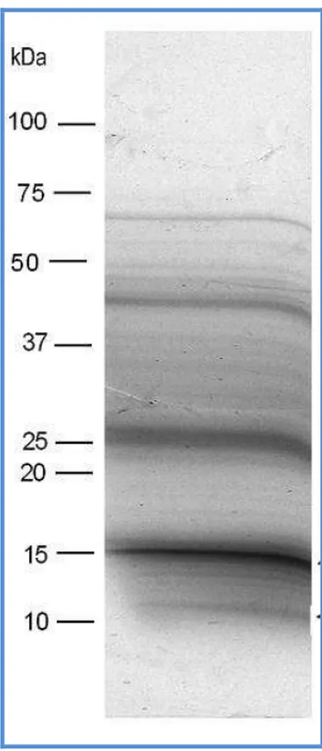
In this paper, at first, an in-house fennel extract was analyzed by Sodium dodecylsulfate-polyacrylamide gel electrophoresis (SDS-PAGE), which revealed numerous components with a molecular weight ranging approximately 9 to 100 kDa (Fig. 5.).
Fig. 5. SDS-PAGE of fennel extract and protein separation according to their molecular weight. The proteins were separated in a discontinuous gel with a 6% stacking gel and a 7.5-20% separation gel, at 6 mA for 16 h (Pastorello et al., 2013).
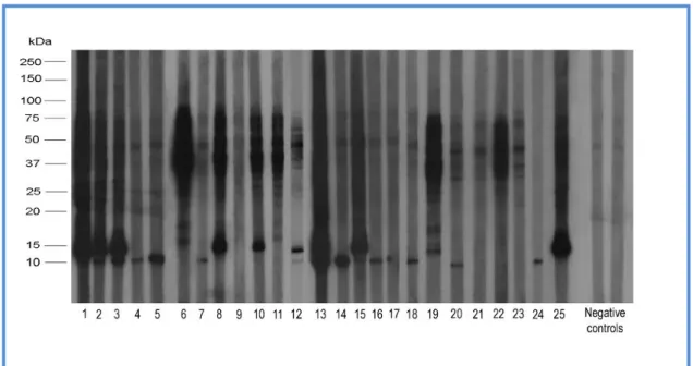
As it is shown in Fig. 6., sera from 25 fennel allergy patients were used for IgE Immunoblotting analysis and IgE-binding proteins were detected by incubation with an 125 I-labeled anti-human IgE antibody. Sixty per cent (60%) of patients’ sera reacted toward an approximately 9 kDa band, whereas 44% of the sera recognized a protein of approximately 15 kDa and 96% reacted with bands in the range of 65-75 kDa.
Fig. 6. IgE Immunoblotting of fennel extract using sera from 25 fennel allergy patients and 2 negative controls. After electrophoresis, fennel proteins were electrotransferred to a nitrocellulose membrane, at 0.45 A and 100 V for 4 h at 4 °C. Moreover, sera from the 25 fennel allergy patients and 2 negative controls, diluted 1:5, were used as primary antibody; while IgE-binding proteins were detected by incubation with an 125I-labeled anti-human IgE antibody, diluted 1:2. Exposure to autoradiographic films, at – 70 °C for 4-7 days, was also performed (Pastorello et al., 2013).
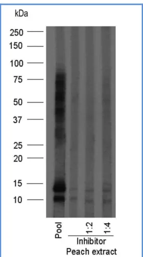
An immunoblotting inhibition experiment was also performed to evaluate the cross-reactivity between fennel and peach extracts, using a pool of sera from patients selected on the basis of their Immunoblotting response pattern to fennel proteins. This experiment revealed that pre-incubation of the pool with a peach extract at different dilutions completely inhibited IgE binding to fennel proteins, suggesting a high cross-reactivity between these two plant foods (Fig. 7.).
Fig. 7. IgE Immunoblotting inhibition of the ability of binding fennel proteins by pooled sera from some fennel allergy patients, pre-incubated with different concentrations of peach extract (Pastorello et al., 2013).
Moreover, the paper demonstrated that the 9 kDa band could be identified, by Liquid Chromatography - tandem Mass Spectrometry (LC-MS/MS), as a lipid-transfer protein (LTP), characterized by a high homology with Pru p3 protein in peach, suggesting its importance in the cross-reactivity evaluation. For this reason, fennel allergy is considered a LTP-related food hypersensitivity and therefore fennel has been included in the list of foods that cause “LTP syndrome” (Pastorello and Robino, 2004).
1.6. The Foeniculum vulgare allergy project
This research project aimed at estimating the occurrence of fennel allergy, in a population with a typically Mediterranean Diet, from Apulia Southern Italy, and characterizing, from an immunological point of view, F. vulgare proteins involved in food allergy to this vegetable. To this purpose, a number of patients with well-diagnosed fennel allergy was detected and non-allergic subjects were recruited as negative controls. Fennel allergy diagnosis was based essentially on clinical history and demonstration of IgE toward fennel, achieved in vivo by skin prick testing. In particular, quantitative SPT were carried out with a commercial extract and with a semi-purified fennel extract produced in our laboratory, respectively. Also quantitative prick by prick tests with raw and microwaved fennel, respectively, were performed. Both these types of skin tests allow detection of mast cell-bound specific IgE for fennel.
The detection of the lesser fraction of circulating fennel-specific IgE was performed in vitro, using the commercial available ImmunoCAP RAST, marketed by Thermo Fisher, and a RAST-capture assay, developed in our laboratory. To this purpose, the in-house fennel extract was first biotinylated (by conjugation with biotin disulphide N-hydroxysuccinimide ester). The biotinylated reagent was used for this ELISA technique. This approach was also adopted in RAST inhibition experiments, in order to evaluate the possible cross-reactivity between fennel and peach and between fennel and celery, respectively.
Finally, the protein profile of the fennel extract was examined by Immunoblotting analysis in order to identify fennel proteins bands that react with IgE from F. vulgare allergy patients. Thus, in these experiments, patients’ sera and anti-human IgE peroxidase-conjugated antibody were used as primary and secondary antibodies, respectively.
2. Materials and methods
2.1. Fennel allergy diagnosis work up
The diagnosis of fennel allergy was based essentially on: a convincing clinical history; in vivo tests (SPT and prick by prick) and in vitro tests (RAST).
2.1.1. Convincing clinical history
A convincing clinical history is definitely important in the diagnosis of food allergy.
There are many specific factors to be considered that make a clinical history convincing, in the field of allergology. First, the timing. The allergic reaction onset, after consumption of food regarded as responsible for the allergic reaction reported by the patient, has to be immediate. Thus, since food allergy is based on an IgE-mediated reaction, in most cases, the allergic reaction takes place within few minutes, after the suspected food consumption. Secondly, the nature of symptoms. The symptoms associated with food allergy reactions can affect primarily the skin (itching, cutaneous rash, urticaria, angioedema). The gastrointestinal tract may be also involved (lips, mouth, tongue and palate itching and edema, nausea, vomiting, diarrhea). Finally, symptoms related to the larynx and the upper and/or lower respiratory tract, may also occur.
Reproducibility. It is also important noticing if the allergic reaction is repetitive, i.e., if it occurs whenever the suspected food is consumed.
Nature of the offending allergen. Moreover, we need to take into consideration the type of food involved in the allergic reaction reported by the patient. Chocolate, for example, very seldom causes allergy (inspite of the general belief at the lay public level). Also important causing is the amount of the suspected food assumed.
Food allergy reactions often occur when having special meals, associated with ceremonies, or when dining out. In both cases, we can eat much more than usual and, sometimes, we do not know if potentially allergic foods are used as ingredients. In addition, alcohol intake as well as physical activity (e.g., dancing, etc.) may facilitate the onset of symptoms associated with food allergy reactions, such as urticarial and cutaneous rash, because of the alcohol ability to determine vasodilation and other similar mechanisms.
These principles were adopted in collecting the anamnesis of the study patients. See also the Results section.
2.1.2. In vivo tests
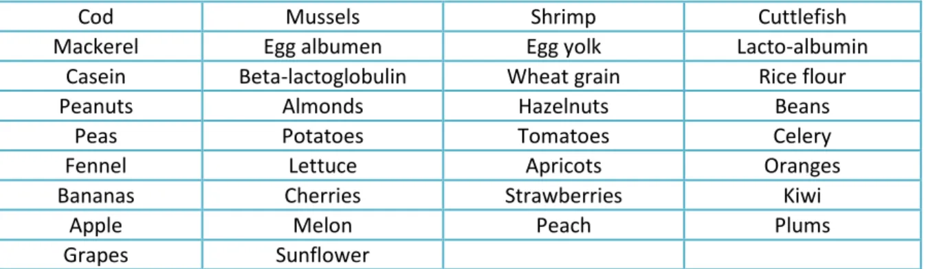
The in vivo tests included SPT and prick by prick. SPT were performed by pricking the patient’s skin through a droplet of allergen solution, which allowed the allergen to interact with potential IgE specific for that given allergen, on the surface of skin-resident mast cells. In food allergy diagnosis, usually, a collection of 34 commercial food allergen extracts was used (Tab. 3.). In particular, SPT were performed with commercial extracts marketed by Lofarma (Milan, Italy). An in-house semi-purified 100,000 x g supernatant fennel extract was also used.
In the highly standardized technique of skin prick testing, the skin is usually punctured using an Østerballe and Weeke-type 1 mm point length standardized needle (Østerballe & Weeke, 1979), supplied by Stallergenes. Histamine hydrochloride 10 mg/ml in 50% glycerol solution (from Stallergenes, Antony, France) was used as the positive control in skin prick testing. Saline with 1% albumin served as the negative control. Results were read 20’ after pricking. Positive responses consist in a wheal-and-flare reaction, whose magnitude essentially reflects the amount of endogenous histamine released by activated cutaneous mast cells. In turn, the magnitude of mast cell activation essentially reflects the amount of the existing specific IgE bound to their surface by high affinity receptors (IgE cross-linking, by allergen molecules penetrated into the skin, triggers mast cell activation and histamine release). In quantitative skin testing, to measure the elicited cutaneous reactions, the wheal can be outlined with a felt-tip pen and the outline can be blotted on removable tape and recorded on paper, for convenience. The area is calculated by superimposing a transparent cellulose acetate sheet with a 1 mm square lattice grid and counting the squares comprised within the outline (each square 1 mm2).
This methodology was rigorously used in this study.
Quantitative prick by prick procedure, instead, was carried out both with the fresh fennel and microwaved fennel (50 g, 2’, 2450 MHz). Microwaving was meant to test the thermostability of fennel proteins. Thus, 50 g of the edible part of fennel were subjected to treatment in a Philips M305 microwave hoven at a 2450 MHz for 2’. The patient’s skin was pricked through a thin piece, usually a thin slice, of the fresh food using a sterile pricker needle and, if the
response was positive, the area of the wheals was measured in the same way described for skin prick testing.
Cod Mussels Shrimp Cuttlefish
Mackerel Egg albumen Egg yolk Lacto-albumin
Casein Beta-lactoglobulin Wheat grain Rice flour
Peanuts Almonds Hazelnuts Beans
Peas Potatoes Tomatoes Celery
Fennel Lettuce Apricots Oranges
Bananas Cherries Strawberries Kiwi
Apple Melon Peach Plums
Grapes Sunflower
Tab. 3. Collection of 34 commercial food allergen extracts used in skin prick testing.
2.1.3. In vitro tests
Sera from patients with fennel allergy were tested for fennel-specific IgE by ImmunoCAP (Thermo Fisher Scientific, Milan, Italy), according to the instructions of the manufacturer (http://www.phadia.com/it/5/Prodotti/Dosaggi/1/#ImmunoCAP).
2.2. Generation of semi-purified food extracts
2.2.1. Preparation of a semi-purified fennel extract
A suitable amount (100 g) of the edible part of fresh F. vulgare was washed properly, minced and homogenated for 15’ by a Heidolph DIAX 900 homogenizer with a Heidolph 10 F probe, at 25,000 revolutions per minute, on ice, in the presence of 10 ml PBS 10X w/o Ca++ e Mg++ (final concentration ~ 1X), 400 μl Ethylenediaminetetraacetic acid (EDTA) 0,5 M (final concentration 2 mM) and 700 μl of plant cell-specific protease inhibitor cocktail (Sigma, Milan, Italy). The inhibitor cocktail contained: 4-(2-Aminoethyl) benzenesulfonyl fluoride hydrochloride (AEBSF), 1,10-phenantroline, pepstatin A, bestatin and trans-epoxysuccinyl-L-leucylamido-(4-guanidino)butane (E64), in unknown concentration. The homogenate was then centrifuged at 12,000 x g, for 20’ at 4 °C and the supernatant was recovered. This step was repeated two more times. Successively, the sample was ultracentrifuged at 100,000 x g,
for 2 hs, at 4 °C. Forty ml of 100,000 x g supernatant were obtained from 100 g of fresh F. vulgare. The extract was kept at -80 °C, until used.
The protein content, determined according to the colorimetric Bradford method (Bradford, 1976), was 3.5 mg/ml.
2.2.2. Preparation of semi-purified peach and celery 100,000 x g
supernatants
A suitable amount (50 g) of the edible portion of fresh Prunus persica and Apium graveolens, respectively, was washed, minced and homogenated for 15’, as above, on ice, in the presence of 5 ml PBS 10X w/o Ca++ e Mg++, 200 μl EDTA 0,5 M and 350 μl of plant cell-specific protease inhibitor cocktail, as above. The homogenates underwent three centrifugation cycles at 12,000 x g, for 20’ at 4 °C. Successively, the homogenates were subjected to ultracentrifugation at 100,000 x g, for 2 h at 4 °C, thus generating semi-purified 100,000 x g supernatants. The protein content, determined by the colorimetric Bradford method (Bradford, 1976), was 0.228 mg/ml for the peach extract and 0.8 mg/ml for the celery extract. The two extracts were maintained at -80 °C, until needed.
2.3. Biotinylation of the semi-purified fennel extract
Upon thawing, 2 ml of the semi-purified fennel extract, containing 7 mg of total proteins [3.5 mg/ml], were applied to a gel filtration Sephadex G 25 column (PD-10 columns, Amersham Biosciences, Sweden), previously conditioned with 30 ml PBS w/o Ca++ e Mg++ 1 mM EDTA. The column was eluted with 13 ml PBS w/o Ca++ e Mg++ 1 mM EDTA. As it is known, gel filtration is a chromatography technique that allows not only separation of biomolecules, such as peptides, proteins, nucleic acids, etc., on the basis of their size, but also to change the composition and pH of the sample buffer. Molecules move through a bed of porous beads, diffusing into the beads to a greater or lesser degree. Smaller molecules enter the pores of the beads and therefore move through the bed slowly, while larger molecules enter less or do not enter at all, moving through the bed quickly. Based on this principle, the elution of the sample through the gel filtration column may also determine the pH shift of the sample of interest.
2.4. The ELISA capture in vitro assay
To perform the in-house RAST-capture assay, an ELISA 96-wells microplate, with anti-IgE antibodies coated on the surface of wells, was used (RADIM, Pomezia, Rome). The wells were first blocked with a blocking solution made of 10% non-fat dried milk in PBS w/o Ca++ e Mg++ 1 mM EDTA (350 μl/well were dispensed) and incubated at 37 °C overnight. The next day, the blocking solution was removed and the IgE microplate was processed using a commercial kit, also marketed by RADIM, including all the necessary reagents.
In the microplate, two blanks were set up and six wells were used for human IgE calibrators, with the following concentrations: 0.36, 0.72, 3.6, 18, 50 and 100 kU/l. The calibrators were needed to draw the calibration curve.
The protocol used was as follows:
step 1: 50 μl of incubation buffer were first dispensed into all the wells;
step 2: 50 μl of each of the calibrators or 50 μl of each of the samples were loaded in the respective wells; instead, other 50 μl of incubation buffer were loaded into the blanks;
step 3: the microplate was incubated for 60 ± 5’, at 37 °C;
step 4: the microplate was washed 3 times (the working solution was preparared from a 10X solution, provided with the kit);
step 5: either 100 μl of anti-IgE biotin conjugated antibody or 100 μl of the biotin conjugated fennel allergen, respectively, were dispensed into the calibrator wells and the sample wells, respectively. The biotinylated allergen amount selected as suitable for the assay was 1 μg per well. Since the protein concentration of the biotinylated fennel extract was 1.9 mg/ml and the volume of the biotinylated allergen solution to be used in the test was 100 μl per well, a 1:200 dilution of the biotinylated fennel extract was performed. Hundred μl of this diluted solution were added to the relevant wells (containing ~ 1 μg of the biotinylated fennel extract);
step 6: the microplate was then incubated for 30 ± 5’, at 37 °C; step 7: then it was washed again 3 times;
step 8: 100 μl of HRP (horseradish peroxidase)-streptavidin conjugated were added to all the wells;
step 9: the microplate was incubated for 30 ± 5’, at 37 °C; step 10: the wells were washed for 3 times;
step 13: finally, 100 μl of stop solution were dispensed in all the wells;
step 14: the resulting colorimetric reaction intensity was read at 450 and 405 nm with the reference filter set at 595 nm in a Microplate Reader (model 450, Bio-Rad). The reaction was read at 405 nm, when the absorbance at 450 nm was above 2.0 O.D. IgE title of the samples, expressed as kU/l, was determined by interpolation on the calibration curve.
2.5. RAST inhibition experiments
RAST inhibition experiments were performed to evaluate the possible cross-reactivity between fennel and peach extracts and between fennel and celery extracts. RAST inhibition experiments were also performed between the semi-purified fennel extract and biotinylated fennel allergen in order to validate the results obtained with the other two settings above. Regarding the peach RAST inhibition experiments, 50 μl of serum from 4 fennel allergy patients (B.L., F.M., P.A. and P.G.; see below the Results section) were pre-incubated for 24 h at 4 °C, while gently shaking, with peach extract in PBS w/o Ca++ e Mg++ 2.5 mM EDTA with 4 different amounts of peach proteins, i.e., 0 μg, 0.228 μg, 1.14 μg and 4.5 μg (4 samples for each of the 4 patients). In particular, the following volumes of the original peach 100,000 x g supernatant [0.228 mg/ml] included in the buffer mentioned above, up to a final total volume of ~ 50 μl were: 0 μl, 1 μl, 5 μl and 20 μl.
The celery RAST inhibition experiments were performed following the same protocol as above. However, the celery protein content of the 4 samples generated for each of the same 4 patients as above, were 0 μg, 0.8 μg, 1.6 μg and 8 μg. In particular, the following volumes of the original celery 100,000 x g supernatant [0.8 mg/ml] were included in the buffer mentioned above, up to a final total volume of ~ 50 μl: 0 μl, 1 μl, 2 μl and 10 μl.
Finally, for the fennel RAST inhibition experiments, sera from the 4 patients with fennel allergy were pre-incubated in the presence of 0 μg, 0.35 μg, 1.05 μg and 10.5 μg of fennel proteins. In this case, the volumes of the original fennel 100,000 x g supernatant [3.5 mg/ml] were included in the buffer mentioned above, up to a final total volume of ~ 50 μl: 0 μl, 1 μl and 3 μl from a dilution 1:10 of the semi-purified fennel extract, respectively and 3 μl from the undiluted fennel extract.
As in the ELISA-capture assay, a 96-wells microplate, with anti-IgE antibodies coated on the surface of wells, was used (from RADIM). The wells were first blocked with a blocking solution made of 10% non-fat dried milk in PBS w/o Ca++ e Mg++ 1 mM EDTA (350 μl/well) and incubated at 37 °C overnight. The next day, the pre-incubated samples were centrifuged at 20,000 x g for 30’ at 4 °C. Upon removal of the blocking solution from the wells, 50 μl of the supernatant of the pre-incubated samples (after centrifugation) were added to each well. Successively, the IgE microplate was processed following the instructions of the commercial kit marked by RADIM, as described above.
2.6. Immunodetection of fennel allergens
2.6.1. Sodium dodecylsulfate-polyacrylamide gel electrophoresis
(SDS-PAGE) of the semi-purified Foeniculum vulgare extract
The in-house fennel extract was analyzed by SDS-PAGE. Upon thawing, SDS-PAGE samples were prepared, with a 12.5 µl volume of fennel extract, corresponding to a protein content of about 45 μg (the protein concentration of the semi-purified F. vulgare extract was 3.5 mg/ml), included in a 12.5 µl volume of 2X Laemli loading buffer (125 mM Tris HCl pH 6.8, 4% SDS, 20% glycerol, 10% 2-β-mercaptoethanol, 0.02% bromophenol blue). The samples were boiled at 100 ºC for 5’, chilled on ice for 5’ and then briefly centrifuged, at 20,000 x g. Moreover, prestained SDS-PAGE Standards – low range samples, with known molecular weight (Bio-Rad, Richmond, CA) were prepared by adding 10 µl of these markers to an equal volume of 1X Laemli loading buffer (62.5 mM Tris HCl pH 6.8, 2% SDS, 10% glycerol, 5% 2-β-mercaptoethanol, 0.01% bromophenol blue). Unlike fennel extract samples, standards were unboiled. Other prestained SDS-PAGE standards were also used in selected experiments: Kaleidoscope Prestained Standards (Bio-Rad, Richmond, CA) and Precision Plus Protein Dual Color Standards (Bio-Rad, Richmond, CA).
Protein bands were separated in a continuous gel. SDS-PAGE was performed on 1.5 mm slab gels (10% resolving gel and 5% stacking gel). Acrylamide/bis-acrylamide mixture with a ratio 29:1 was used (Sigma, Milan, Italy).
Millipore water, 3 ml Tris HCl 1.5 M pH 8.8, 150 µl 10% Sodium Dodecyl Sulphate (SDS), 15 µl Tetramethylethylenediamine (TEMED; Bio-Rad, Richmond, CA) and 100 µl Ammonium Persulfate (APS; Sigma, Milan, Italy). The 5% stacking gel solution was prepared
with 1.62 ml acrylamide/bis-acrylamide mixture, 5.8 ml Millipore water, 2.5 ml Tris HCl 0.5 M pH 6.8, 100 µl 10% SDS, 15 µl TEMED and 100 µl APS.
The run was carried out at 80 V for 3 h on a refrigerated Mini-Protean II vertical electrophoresis apparatus (Bio-Rad, Richmond, CA).
2.6.2. IgE Immunoblotting analysis
After electrophoresis, the separated proteins were electro-transferred to a nitrocellulose membrane (GE Healthcare, Buckinghamshire UK), using a Mini Transphor TE22 (Hoefer Scientific Instrument, San Francisco U.S.A), at 200 mA for 2 h at 4 ºC.
After electroblotting, the gel was stained with 0.1% Coomassie Brilliant Blue G 250 (Merck, Darmstadt, Germany) in water:methanol:acetic acid (50:40:10), overnight at room temperature and, successively, rinsed in water:methanol:acetic acid (50:40:10) destaining solution, three times for 20’, at room temperature. Finally, it was dried with a gel-dryer (Drygel Sr., Slab Gel Dryer, Hoefer Scientific Instrument, San Francisco U.S.A), in order to assess transfer efficiency.
The blotted nitrocellulose membrane was subjected to blocking of the non-specific binding sites (5% non-fat dried milk in 50 mM TBS, pH 7.5), for 1 h at room temperature. Then the membrane was cut into strips, each corresponding to a gel lane. The strips were incubated overnight at 4 ºC, while rotating (11 rpm) with 1 ml of serum from a fennel allergic patient (the primary antibody), diluted 1:5 in 50 mM TBS pH 7.5, with 5% inactivated fetal calf serum (FCS). Alternatively, the strips were incubated with serum from control subjects. Successively, strips were rinsed in 50 mM TBS pH 7.5 with the addition of 0.1% Nonidet NP40, 2 times for 10’, at room temperature. Typically, the strips were re-incubated with 2 ml of an anti-human IgE peroxidase-conjugated polyclonal antibody, raised in goat (Sigma, Milan, Italy), diluted 1:330 in 50 mM TBS pH 7.5, in the presence of 5% non-fat dried milk, for 3 h at room temperature, on rotator (11 rpm). Alternatively, an anti-human IgE peroxidase-conjugated monoclonal antibody (Abcam, Cambridge, UK) was also adopted in selected experiments. After incubation with the secondary antibody, each strip was rinsed in 50 mM TBS pH 7.5 added to 0.1% Nonidet NP40, 3 times for 10’ at room temperature. The immunoreactive proteins were detected by enhanced chemiluminescence (ECL, GE
Patient Wheal area (mm2) Commercial extract Raw fennel
B.A. 10 B.G. 38 B.L. 25 C.A. 36 C.C. 12 C.G. 4 C.R. 6 C.G. 7 C.M. 8 C.A.R. 10 C.A. 28 C.G. 10 C.M. 22 39 D.A.G. 17 D.B.A. 25 D.V.M. 11 D.A. 31 D.B.A. no data 41 D.C.A. 27 D.V.L. 85 D.F. 19 F.M. 12 F.F. 23 L.A. 0 L.V. 41 L.V. 11 L.C. no data L.A. 15 M.D. 19 M.V. 9 M.F. 9 M.C. 15 M.G. no data 13 M.A. 29 M.D.P. 46 M.F. 14 M.B. 11 M.D. 45 M.R. 11 P.A. 7 P.T. 28 P.A. 23 P.R. 17 P.G. 6
Food allergens
Occurrence
(%)
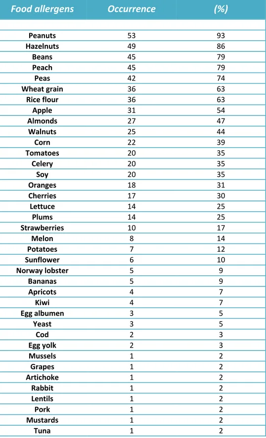
Peanuts 53 93 Hazelnuts 49 86 Beans 45 79 Peach 45 79 Peas 42 74 Wheat grain 36 63 Rice flour 36 63 Apple 31 54 Almonds 27 47 Walnuts 25 44 Corn 22 39 Tomatoes 20 35 Celery 20 35 Soy 20 35 Oranges 18 31 Cherries 17 30 Lettuce 14 25 Plums 14 25 Strawberries 10 17 Melon 8 14 Potatoes 7 12 Sunflower 6 10 Norway lobster 5 9 Bananas 5 9 Apricots 4 7 Kiwi 4 7 Egg albumen 3 5 Yeast 3 5 Cod 2 3 Egg yolk 2 3 Mussels 1 2 Grapes 1 2 Artichoke 1 2 Rabbit 1 2 Lentils 1 2 Pork 1 2 Mustards 1 2 Tuna 1 2Tab. 7. Accociated cutaneous sensitizations to other, then fennel, food allergens (n=43 patients).
Fig. 8. SDS-PAGE analysis of fennel extract. 45 µg of the semi-purified fennel extract were loaded in lanes 2, 3, 4, 6, 7 and 8. In lane 5 prestained SDS-PAGE standards - low range (Bio-Rad, Richmond, CA) were loaded. The relevant calibrated molecular weights are indicated.
3.3. Clinical features of the selected fennel allergy
population
In Tab. 9. the main symptoms and associated diagnoses reported by the patients upon fennel consumption are listed. Thus, 40 out of the 44 fennel allergy patients (91%) reported lip angioedema and oral itching after fennel’s ingestion (oral allergy syndrome). Thirteen (29%) lamented cutaneous itching, of which 9 localized to neck, ears or face and 4 generalized, scattered itching in other parts of the body. Moreover, 9 patients (20%) had urticaria; 16 patients (36%) Quincke's edema; 22 patients (50%) respiratory symptoms (in particular, 16 fennel allergy patients reported dyspnea; 3 subjects lamented chest tightness; 2 patients reported wheezings and 1 coughing); 14 patients (32%) gastrointestinal symptoms (such as nausea, vomiting, epigastric and abdominal pains, diarrhea) and, finally, two patients (4%) experienced severe anaphylaxis, after eating raw fennel (Tab. 9). Moreover, 3 patients (7%) stated that they had never eaten fennel, because they disliked it or even felt repulsion if one just mentioned this vegetable.
1 2 3 4 5 6 7 8 9 10 103 kDa 81 kDa 47 kDa 34 kDa 27 kDa 18 kDa
Patient Wheal area (mm2)
Commercial extract 1 Fennel extract 1 Raw fennel 2 Microwaved fennel 2
B.G. 52 134 50 83 B.L. 48 85 27 57 C.A. 81 48 27 50 C.C. 24 67 45 27 C.R. 7 8 10 8 C.M. 10 28 24 0 C.A.R. 18 27 24 15 C.A. 14 40 39 97 C.M. 18 60 39 29 D.A.G. 5 27 16 7 D.V.M. 9 48 40 22 D.A. 25 38 18 22 D.B.A. 48 96 254 158 D.C.A. 11 60 64 76 D.V.L. 30 241 35 64 D.F. 17 20 19 8
F.M.* no data no data no data no data
L.A. 8 12 29 7 L.V. 34 74 32 60 L.V. 24 50 49 46 M.D. 28 68 56 24 M.C. 12 20 27 26 M.G. 33 42 38 29 M.A. 53 77 57 16 M.D.P. 88 54 60 103 M.B. 17 18 16 6 M.D. 33 66 12 11 M.R. 19 22 27 27 P.A. 11 14 17 0 P.T. 33 197 117 42
P.A.* no data no data no data no data
P.R. 11 99 15 44 P.G. 15 65 140 27 R.A. 41 30 23 49 R.G. 28 42 42 47 R.M.L. 26 45 17 13 S.V. 26 46 22 20 S.G. 8 14 15 17 S.N. 10 36 43 24 S.R. 11 39 10 18 T.G. 31 80 22 36 V.V. 16 17 16 5 V.D.C. 23 121 88 46 V.R. 21 110 39 38
Cutaneous sensitizations
fennel celery carrot parsley
Patie nt s ( n) 0 10 20 30 40 50
Fig. 9. Allergic sensitizations to other food allergens belonging to the Apiaceae family (n=42 patients). SPT with fennel, celery and carrot commercial extracts, respectively were performed. Only in the case of parsley, prick by prick tests were performed, because no parsley commercial extract was available.
Moreover, wheal areas obtained in SPT, carried out with celery and carrot commercial extracts, marked by Lofarma (Milan) and in prick by prick tests, performed using fresh parsley, were calculated (Tab. 11.).
Patient Wheal area (mm2)
Celery extract Carrot extract Parsley*
B.G. 15 0 37 B.L. 22 10 0 C.A. 11 0 52 C.C. 17 0 22 C.R. 6 0 0 C.M. 13 0 0 C.A.R. 15 15 12 C.A. 9 0 60 C.M. 11 8 19 D.A.G. 0 0 6 D.V.M. 10 0 0 D.A. 0 0 10 D.B.A. 11 0 36 D.C.A. 13 0 25 D.V.L. 18 0 22 D.F. 23 0 8
F.M.* no data no data no data
L.A. 0 0 21 L.V. 0 0 14 L.V. 0 0 19 M.D. 12 0 33 M.C. 0 0 4 M.G. 11 0 11 M.A. 13 7 16 M.D.P. 9 8 120 M.B. 21 3 5 M.D. 12 0 17 M.R. 0 13 19 P.A. 9 0 6 P.T. 10 0 33
P.A.* no data no data no data
P.R. 0 0 10 P.G. 10 0 14 R.A. 9 0 8 R.G. 0 0 13 R.M.L. 0 0 16 S.V. 15 0 6 S.G. 9 8 21 S.N. 10 0 7 S.R. 9 11 19 T.G. 12 0 14 V.V. 0 0 0 V.D.C. 17 0 42
Average area of the wheals 9 2 20 Standard deviation 7 4 21
Tab. 11. SPT carried out with celery and carrot commercial extracts marketed by Lofarma (Milan), respectively. *In this case, prick by prick tests were performed using fresh parsley, because no parsley commercial extract was available. In both cases, results are expressed as areas of the wheals (mm2). N=42 patients. *These 2 fennel allergy patients refused to repeat the in vivo tests.
Associated multiple sensitizations to other allergens (not belonging to the Apiaceae family) were also analyzed in these 44 fennel allergy patients. Thus, SPT with the 9 food allergens most frequently associated with fennel sensitization were performed. The 9 food allergens were: peanuts, beans, hazelnuts, peas, apple, peach, almonds, rice flour and wheat grain. These allergens had been selected on the basis of their rate of co-occurrence with fennel sensitization ( > 50% for all of them, except almonds: 47%) as assessed by the analysis of the data obtained by routine skin testing in the 57 fennel allergy patients described above.
The results of these investigations are reported in Tab. 12. As it can be seen, all patients (42 out of 42, because among the 44 fennel allergy patients recruited 2 had declined undergoing the in vivo tests) had a positive shin prick test for peanuts, 40 patients (95%) for beans, 39 patients (93%) for hazelnuts, 37 patients (88%) for peas, 35 patients (83%) for apple and peach, 33 patients (79%) for almonds, 32 patients (76%) for rice flour and, finally, 30 patients (71%) for wheat grain. Also in Tab. 11, the number of patients sensitized to the 9 food allergens, respectively, and lamenting symptoms clearly associated with consumption of the relevant food are reported.
Food allergen Positive patients (n) % 1 Patients with relevant symptoms (n) % 2 Peanuts 42 100 32 76 Beans 40 95 9 22 Hazelnuts 39 93 30 77 Peas 37 88 14 38 Apple 35 83 13 37 Peach 35 83 33 94 Almonds 33 79 18 54 Rice flour 32 76 5 16 Wheat grain 30 71 1 3
Tab. 12. Multiple cutaneous sensitizations to other important food allergens in n=42 patients with fennel allergy. The number of allergic patients with clinically significant sensitization is also reported. %1: positive patients out of the 42 patients with fennel allergy. %2: patients with clearly associated symptoms out of those who were sensitized.
Moreover, the areas of the wheals elicited (mm2) in quantitative skin prick testing, performed
using the commercial extracts (marketed by Lofarma, Milan) of the 9 food allergens more
frequently associated with fennel sensitization were measured. The results of these tests are shown in Tab. 13.