M.R. Giuca***, S. D’Ercole**
Dental School, University “G. D’Annunzio” of Chieti-Pescara, Chieti, Italy
*Department of Medical, Oral, and Biotechnological Sciences **Department of Experimental and Clinical Sciences
***Department of Surgical Pathology, Medicine, Molecular and Critical Area, University of Pisa, Pisa, Italy
e-mail: [email protected]
abstract
Aim Assess prevalence, familial predisposition and
susceptibility to caries of Black Stains (BS). Evaluate the microbiological composition of BS, saliva and subgingival plaque.
Materials and methods Sixty nine subjects with BS
(test group) and 120 subjects without BS (control group) were analysed for oral status. For each BS-patient, a BS-deposit, 1 ml of saliva and subgingival plaque were collected and microbiologically analysed. Five deciduous teeth with BS were observed under SEM.
Results This study showed a BS prevalence similar to
that of the Mediterranean area and a familiality. The microbiological origin of BS was confirmed by SEM and culture method and the BS flora differ from that of supragingival plaque. .
Conclusions Predominance in BS and saliva of
Actinomycetes and the low salivary prevalence of S. mutans and L. acidophilus may be related with low caries incidence in BS patients. The high presence of Actinomyces spp can be a causative factor for BS.
Black Stains: a
microbiological analysis
and a view on familiarity
and susceptibility to
tooth decay of patients
in childhood
The teeth become darker due to age, probably due to deposition of secondary dentine, incorporation of pigments and extrinsic substances, and thinning of the enamel. Consequently, this phenomenon is indicated by the term of discolouration [Watts, 2001]. Currently, the most commonly used classification of discoloration is that proposed by Nathoo in 1997, which divides the discolouration by mechanism of action and time of onset. However, there is another classification based on the colour of the pigment which is of greater clinical interest and identifies four distinct classes: Orange stains, Green stains, Brown and Black Stains [Nathoo, 1997]. The black stains (BS) were described by Wilkins in 2005 as black spots with a linear aspect that also present incomplete coalescing points that are characteristic of this stain and that rarely extend beyond the cervical third of the crown, but the base of the grooves and pits may also be affected [Wilkins, 2005]. BS have a prevalence between 1% and 20% as reported by various authors representing a very common event [Renz, 1976]. It is not possible to find an age and sex that present a greater risk, and BS can be found on both deciduous and permanent teeth. Frequently BS disappear spontaneously at the end of the second decade of life. The differential diagnosis is with intrinsic discolouration and tooth decay from which they are easily distinguishable due to the different clinical presentation. Unlike tooth decay, in fact, BS are not associated with loss of substance or with any evidence of demineralised areas. The location close to the gingival margin is of great help in their identification [Watts, 2001]. Hillman and Socransky [1987] hypothesised that the microbiota of BS can be a substitution model of oral pathogenic flora, and understanding this mechanism may result in a possible prophylactic approach to caries. By contrast, Gasparetto et al. [2003] reported that a lower prevalence of caries is not statistically significant in children with BS, but were able to demonstrate a strong correlation between severity of the disease and presence of BS (DMFT >3 only in 10.3% of children with BS vs. 31.3% of unaffected). Chromogenic bacteria, especially Prevotella melaninogenica were suspected to be the main cause of these characteristic pigmentations. Slots in 1974, in a bacteriological examination of BS revealed a characteristic and relatively stable microflora composed for 90% by Actinomyces spp., while P. melaninogenica accounted for less than 1% were [Slots, 1974]. Theilade and Pang [1987] described this lesion as a special type of dental plaque flora characterised by \a tendency to calcification. The electron microscopy examination revealed that the deposits consisted of microorganisms embedded in an intermicrobical granular or filamentous matrix [Theilade, 1977]. Slots also showed that they contained a high percentage of calcium and phosphate and insoluble iron salts, responsible for the typical black colour of these spots [Slots, 1974].
The purpose of the present study was to assess the
Keywords Black stain; Cariogenic microorganisms;
incidence and the prevalence of BS in the Chieti area, Italy, to assess the possible familiality of this pathology and the possible positive or negative correlation between black stains and susceptibility to caries. Moreover, we conducted a microbiological analysis of the dyschromies, saliva and subgingival plaque to correlate the aetiopathogenetic factors involved in patients with extrinsic tooth stain.
Materials and methods
Patients’ selection
A sample of 69 subjects (test group) with at least one tooth with BS and 120 subjects without BS (control group), all in good health, were randomly selected among all patients of the Paediatric Dentistry Unit, Department of Medical, Oral and Biotechnological Sciences, University of Chieti, in the period 2011–12. The selected subjects participated voluntarily in the study. Patients and their parents were first made aware of the study purpose with oral and written information. Consent was given by signing a protocol (Privacy Law DL 196/2003). All procedures were conducted according to the principles expressed in the Declaration of Helsinki.
Clinical monitoring
A self-administered questionnaire was used for children and parents to obtain data on medical and dental history, family history, oral hygiene practices, snacking habits and fluoride intake. Two clinicians visited each patient and the following parameters were recorded: presence/absence and location of the BS on the tooth surface; decayed, missing and filled index (DMFT) to assess caries prevalence according to the WHO criteria (Oral health survey basic methods. World Health Organization, 1997); Plaque Index (PlI) according to Löe and Silness to evaluate oral hygiene; bleeding index (BOP) to assess periodontal status; bad habits; malocclusion and parafunctional habits.
SEM analysis
Five deciduous teeth, with black stains, were selected from the whole patients and retrieved at the time of exfoliation. After collection, each sample was washed in a saline solution at room temperature and fixed in 14% formalin for at least 12 hours. Then each sample was washed in PBS, dehydrated in ascending series of alcohols, included in resin (LR White EM, TAAB Laboratories Equipment Ltd, England), sectioned along the median axis (Micromet, Remet sas Casalecchio, Bologna, Italy), polished with sandpaper of decreasing grits (320, 600, 800, 1200) using a lapping machine “LS2” (Micromet, Remet sas Casalecchio, Bologna, Italy), etched with 37% phosphoric acid, metallised with gold (550 K Emitech, Emitech Ltd, Ashford, Kent, UK), and then observed under scanning electron microscope (EVO 50, Carl Zeiss SMT AG, Germany) using backscattered and secondary electrons.
Black Stains collection
For each BS-positive patient, after isolation of the
operative field with cotton rolls and wash of the surface with a jet of air/water, the BS deposit was removed with a curette and immersed in a sterile tube containing 2 ml of transport medium (RTF). The procedure was done by two skilled clinicians.
Saliva collection
For all patient (BS and control), 1 ml of stimulated saliva – using a paraffin chewing gum – was collected and immediately placed in a sterile Eppendorf tube.
Subgingival plaque collection
Following isolation of the operative field with cotton rolls, subgingival plaque was collected by insertion of three #30 standardised sterile endodontic paper points into the base of the sulcus until slight resistance was perceived. The paper points were left in situ for 15 s, transferred in an Eppendorf tube containing 1 ml of RTF and immediately sent to the microbiology laboratory.
Microbiological assessment
Samples of saliva, subgingival plaque and BS deposit were dispersed by vortexing for 60 s, and each was then subjected to series of 10-fold dilutions in 0.1 M phosphate buffer. Aliquots of 100 μl of each dilution were spread in duplicate onto Columbia Blood Agar (CBA) (Oxoid Italia SpA, Garbagnate Milanese, Milan, Italy) and Trypticase Soy Agar (ETSA) (Oxoid) plates enriched with 5% defibrinated sheep blood to quantify the number of all cultivable oral bacteria (TBC), which was recorded as the colony count that formed per ml (CFUs/mL) on the growth plate. In particular, the CBA plates were used to cultivate anaerobic bacteria under strict anaerobic conditions at 37°C for 7-12 days in an anaerobic chamber (80/10/10, N2/H2/ CO2, Don Whitley Scientific Ltd, Shipley, UK, International PBI SpA), and the ETSA plates were incubated for 2-4 days at 37°C aerobically. Furthermore, to determine specific strains of bacteria, special microbiological procedures were applied. In brief, the following plates were inoculated and incubated at 37°C for 48–72 h in an anaerobic chamber: Rogosa SL Agar (Becton, Dickinson and Company, Sparks, USA) to assess Lactobacillus spp.; DifcoTM Mitis Salivarius Agar (BD) containing 1% Chapman Tellurite solution to assess oral streptococci and enterococci; Brain Heart Infusion Agar (BHNM) (Oxoid) supplemented with nalidixic acid (30mg/l) (Sigma Aldrich, Milan, Italy) and metronidazole (10 mg/l) (Sigma Aldrich) to determine Actinomyces spp.; Trypticase Soy crystal violet erythromycin (4 mg/l) (CVE) for the determination of Fusobacterium spp.; Trypticase Soy Agar (TSBY) (Oxoid) with the addition of serum, bacitracin (Sigma) (75 ug/ml), vancomycin (Sigma) (5 ug/ ml) for A. actinomycetemcomitans. The growth of fungi was determined on Sabouraud maltose agar (DifcoTM, BD) after aerobic incubation for 4 days at 37°C. A definitive identification of all representative isolates was obtained by subculturing onto Brucella Blood Agar (Oxoid) followed by inoculation of purified cultures onto a commercially packaged automated system (bioMérieux, Inc. 100 Rodolphe Street, Durham, NC 27712.). Each microbial
data was recorded as the colony count that formed per ml (CFUs/ml) on the growth plate.
Statistical analysis
Data are summarised as mean and standard deviation (SD) for quantitative variables and as frequency and percentage for categorical variables. Statistical significance of differences between groups for qualitative variables were assessed using the Chi-squared test or Fischer’s Exact Test, when appropriate; t-test for unpaired data was applied for assessing the comparison of the quantitative variables between groups. A two-tailed p-value of 0.05 or less was considered significant. All statistical analyses were conducted using SPSS® software 11.0 (SPSS Inc., Chicago, IL, USA).
Results
A total of 42 males and 27 females (mean age 9.82 ± 4.43 years) were selected among the total of 930 paediatric patients referred to Department of Medical, Oral and Biotechnological Sciences of University of Chieti, Italy, showing a prevalence of BS of 7.41% on the study population (Table 1). The control group, i.e. the 120 patients without BS, had a mean age of 11 ± 3.2 years. In addition, 33 first-degree relatives as test subjects (47.8%) presented equally BS and were included in the study. The inclusion criteria in the test group were a good general health and the presence of BS. The intraoral evaluation showed a variety of presentation of the BS that in 70% of patients were present in localised form (Fig. 1). All pigmentations were black, tenaciously adherent to the tooth surface. Buccal and palatal surfaces were the most affected without significant differences between the various teeth. As demonstrated in Table 1, DMFT of the test group was 0.07 ± 2.22, while the dmft had a value of 0.12 ± 3.15. The control group showed a DMF-T value of 0.05 ± 2.45 and a dmf-t of 0.1 ± 3.07. Relatives of subjects belonging to the test group had a DMFT of 0.12 ± 3.16.
Active caries were present in the 44.12% of control group and in the 26.08% of test group, so the difference was statistically significant (Table 2). Mean values of PlI and BOP are reported in Table 2, with no statistically significant differences between the different groups.
All subjects reported a balanced and varied diet with regular meals during the day (Table 3). Only 25% of the sample reported regular and abundant ingestion of substances rich in sugar or with high acid loads such as sweets and drinks, although a large part of the sample (91%) consumed sweets and soft drinks occasionally. Moreover, 74% of test group reported the a daily use of fluoride toothpaste. Similar results can be found among the relatives, 57% of which used daily fluride toothpaste.
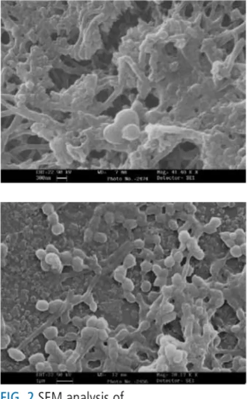
The SEM observation (Fig. 2) showed that the lesions were composed primarily of rod-shaped bacteria, arranged perpendicularly to the enamel surface and closely adhering to each other to form a dense “mesh” similar to the dental plaque, with lack of intercellular matrix. Areas of where rods adeshion was lower showed the presence of calcareous deposits and a greater amount of cocci. Culture method demonstrated that BS deposits (Fig. 3) were composed mainly by Actinomyces spp (63% of total cultivable flora-TBC), confirming the results obtained by SEM observation. Streptococcus spp accounted for 31% of the sample and the remaining part was represented by a mixed microbial flora distributed as follows: Capnocytophaga spp. 2%, A. actinomycetemcomitans 1%, Neisseria spp 1%, Fusobacterium spp 2%, P. melaninogenica 0.45% and L. acidophilus 0.55%. The growth of Candida spp has been detected only in 1 sample. Similarly Actinomyces spp accounted for 81% of the microflora isolated in the subgingival plaque at the black spot sites (Fig. 4). Streptococcus spp accounted for 16% of the total cultivable flora and the rest showed a mixed microbial flora : Capnocytophaga spp 0.1%, A. actinomycetemcomitans 0.3%, Fusobacterium spp 0.5%, P melaninogenica 0.1% and L. acidophilus 2%.
As shown in Fig. 5, saliva microbiological analysis of the patients with black stains showed that the microbial flora was made up to 51% by Streptococcus spp and to 35% by Actinomyces spp. It should be underscored that L. acidophilus accounted for 4% of the microbial flora and the remaining part was constituted by anaerobic strains: Capnocytophaga spp 0.2%, A. actinomycetemcomitans 0.8%, Fusobacterium spp 10%, P. melaninogenica 1%.
The total bacterial count was statistically significantly
Brazil (State of Paranà) Gasparetto et al., 2003 Brazil (Pelotas) França Pinto et al., 2012 Philippines Heinrich-Weltzien et al., 2009 India Tirth et a l., 2009 Italy Potenza) Koch eta/., 2001 Spain Paredes Gallardo et al., 2005 Abruzzo (this study) Subjects 263
(6-12 aa) 1129(5aa) 780 1748(11.7 aa) 1086(6-12 aa) 1100(4-11 aa) 189(2-21)
Prevalence 14.8% 3,5% 16% 20% 6.3% 7.54%
DMF-T BS 1.46± 1.39 N/A 1.5 ± 2.1 N/A 0.49 ± 1.05 N/A 0,07 ± 2,22
DMF-TCTRL 2.42 ± 2.09 N/A 2.5 ± 2.1 N/A 0.97 ± 1.40 N/A 0,05 ± 2,45
TABLE 1 Study results of prevalence of BS in different countries.
FIG 1 Spots spread across the anterior region and involving the first deciduous molars.
with the data recorded in South America [Gasparetto, 2003] and Asia [Heinrich-Weltzien, 2009; Tirth, 2009] which report higher prevalences, even if França-Pinto et al. [2012] reported a prevalence of 3.5% in a homogenous population of Brazilian children aged 5 years. The DMF-T and dmf-t indices recorded in this study show no significant difference between the two groups. Koch reported a DMF-T of 0.49 ± 5.1 in BS patients and 0.97 ± 1.40 in subjects without BS [Koch, 2001]. In the Brazilian population, Gasparetto [2003] reported a DMF-T of 1.46 ± 1.39 and 2.42 ± 2.09 respectively for two groups, with no difference between the groups but with values higher than those recorded for the Italian and Spanish populations. This study demonstrate a lower prevalence of caries in children with black spots. This association is nowadays supported by several authors [Koch, 2001; Sutcliffe, 1967; Shourie, 1947] but not confirmed by others [Paredes Gallardo, 2005; Hillman, 1987] and this topic needs to be further studied. Contrary to our expectations based on the equal indices of plaque and bleeding, we observed that the control group brushed their teeth more frequently. The BS relatives of the test group showed excellent periodontal indices equal to or tending to zero. For all subjects, eating habits consisted in a balanced and varied diet with regular meals spread throughout the day. Trying to understand the onset of BS and the low prevalence of caries in BS patients, particular attention should be directed towards the type of diet, especially regarding sweets and soft drinks, which consumption is more frequent in the test group compared to the control (91% vs. 27.5% for sweets and 96% vs. 40% for soft drinks respectively). There is no correlation between consumption of sweets and beverages and presence of active caries in BS patients. Several authors also noted a higher salivary pH in these children, while the amount of saliva was not different higher in the saliva of patients with black stains compared
to controls (Table 4). As shown in Table 5, the microbial load of S. mitis was statistically significantly higher in the saliva of subjects with black stains compared to controls. Significant differences were observed for S. mutans between saliva of the test group compared to controls.
Discussion
This study showed a BS prevalence of 7.41%. This value is similar to that reported by Koch in 2001 related to the Basilicata region of Italy (6.3%) and Paredes Gallardo [2005] in Spain (7.54%), showing similarity between the data recorded in the Mediterranean area and in contrast
TABLE 2 Prevalence (No.) of active caries, mean values of PII and BOP.
BS Group Control Group Relatives Caries 26,08% (18)* 44,12% (54)* N/A
PI 0,66 0,90 0
BOP 0,24 0,45 0
FIG. 2 SEM analysis of black stains lesions.
FIG 3 Microbiological composition of the black stains.
FIG 4 Microbial composition of the subgingival plaque associated with the black stain lesions.
TABLE 3 Alimentary habits and fluoride intake of the studied population.
BS Group Control Group Relatives
Soft dr. 66 (96%)* 48 (40%)* 24 (72%) Sweets 63 (91%)* 33 (27,5%)* 18 (54,5%) Veget. 45 (65%) 72 (60%) 30 (90.9%) Fluoride 51 (74%) 60 (50%) 24 (72%) S. salivaris S. mutans S. mitis S. milleri Streptococcus spp A. viscosus A. israeli A. naeslundi A. odontolyticus S. sanguis 8% 1% 10% 5% 1% 6% 15% 9% 7% 22% Actinomyces spp 10% Others 6% S. salivaris S. mutans S. mitis S. milleri Streptococcus spp A. viscosus A. israeli A. naeslundi A. odontolyticus S. sanguis 4% 3% 0% 3% 4% 5% 14% 24% 13% 17% Actinomyces spp 13%
from controls [Ronay, 2011; Tripodi, 2011]. The results of this study also allows hypothesising an involvement of fluoride in the low susceptibility to caries of the test group. In fact, 74% of BS patients had taken or assumed regularly fluoride, and the same goes for the examined relatives who used fluoride in 72% of cases, compared to 50% of controls. Ronay et al. [2011] supported the possibility of an individual predisposition for BS, because not all family members with identical or similar eating habits show the presence of BS lesions, thus diminishing the importance of the environmental component. The results of this study gave reason to a positive connection of familiarity, in fact, the family component of the study had a specific high severity of 47.8%. In support of a familial trait, there are the 33 first-degree relatives, of whom 24 were siblings, with a positive match to BS. The microbiological origin of BS was already established by Slots in 1974, reporting that 67% of bacteria were represented by catalase-negative Actinomyces, 15% by catalase-positive Actinomyces, 8% by Corynebacterium, 5% by Streptococci, 4% by Neisseria. Unfortunately, the resources available at that time did not allow further identification of the bacterial constituents. Similarly, Theilade et al. [1973] with ultrastructural examination of dyschromies, showed that the deposit consisted of microorganisms closely entangled in a intermicrobial substance, which led to the classification of BS as a special type of plaque characterised by a simple flora consisted predominantly of filamentous microorganisms, and having the tendency to calcify. In this study, SEM observation revealed that the stains were composed primarily by rod-shaped organisms, closely adhering to each other, often associated in groups to form a dense “mesh” similar to plaque, without a real intercellular
matrix. Furthermore, these filamentous microorganisms were disposed perpendicularly to the enamel surface, organised as in the dental biofilm. In the areas where rods were less adherent, on the tooth surface were detected calcareous deposits as an amorphous material, most likely of salivary origin, which in this case it is covered but not encompassed by a greater amount of coccus forms, as happens in the structure of the supragingival plaque. The microbiological analysis of the black stains showed that these rods, which accounted for 68% of the total flora, could be identified in 63% of cases as Actinomycetes and in particular A. odontolyticus (22%), A. viscosus (15%), A. israelli (9%), A. naeslundii (7%), Actinomyces spp (10%). Streptococci, and in particular Streptococcus spp (6%), S. milleri (1%), S. sanguis (8%), S. mutans (10%), S. mitis (5%), S. salivarius (10%), accounted for 31% of the total flora and the remaining portion was represented by mixed microbial flora. The predominant flora of BS detected in this study, therefore differs from that of normal supragingival plaque, with is mainly formed of Gram-positive cocci. Gram-Gram-positive rods are at most 35-42% of the total cultivable flora [Lamont, 2006]. The results of this study are in agreement with previous microbiological semi-quantitative studies [Slots, 1974; Theilade, 1973] and confirm the hypothesis of Slots [1974] that BS differ from the dental plaque and exhibit a very low degree of individual variation, as opposed to plaque which is even site-specific. The predominance of Actinomycetes may be related to the low frequency of caries detected in subjects with BS. Few studies report on the potential cariogenicity of Actinomycetes, which conversely are implicated in the progression of active root caries and in the aetiology of gingivitis (Gram-positive cocci). In addiction, the low prevalence of the main cariogenic agents, such as S. mutans (10%) and L. acidophilus (0.55%) in in agreement with the low incidence of caries associated with BS. It is believed that the typical color of the black deposit depends on the microbial origin of the lesion and bacterial metabolism. For a long time P. melaninogenica (formerly B. melaninogenicus), microorganism belonging to the group of “black-pigmented Bacteroids”, was held responsible for the colouration of the deposit, but the results of this study confirmed that it actually represents less than 1% of the total flora and therefore cannot have this role. Instead, several studies indicated that actinomycetes might produce a black pigment in dentin [Slots, 1974]. This study
FIG 5 Microbial composition of saliva of patients with black lesions. S. salivaris S. mutans S. mitis S. oralis S. sanguis 6% 6% 11% 14% 7% S. milleri Streptococcus spp A. viscosus A. israeli A. naeslundi A. odontolyticus 1% 6% 16% 8% 7% 3% Actinomyces spp 1% Fusobacterium 10% L. acidophilus 4%
TABLE 5 Putative cariogenic microbial species detected in saliva.
Control Group Black Stains Group S. milis 2,35 x 10⁴ ± 3,38 x 104* 9,8 x 105 ± 1,8 x 10⁴*
L. acidoph. 2,30 x 10⁵ ± 2,55 x 10⁴ 2,5 x 10⁵ ± 2,88 x 10⁴
S. mutans 4,09 x 10⁶ ± 4,19 x 105* 7,63 x 105 ± 1,31 x 10⁵*
TABLE 4 Total bacterial counts in the saliva.
Control Group Black Stains Group salivary CBT 3,70 x 10⁵ ± 2,91 x 105* 7,65 x 106 ± 7,48 x 10⁵*
reported that the microbial composition of the subgingival plaque adjacent to the black lesions, was very similar to that found in BS. The composition of flora, matched the typical microbial composition of newly-formed sub-gingival flora, not associated with periodontal and gingival problems, as demonstrated by the clinical results. It is interesting to note that these stains did not cause problems to the tissues adjacent to the deposits, but it may have an indirect effect in promoting the development of a typical subgingival plaque that – if associated with a poor oral hygiene – could result in gingival and/or periodontal inflammation. It is therefore necessary that such patients undergo regular follow-ups to prevent periodontal damage due to the confirmed presence of A. actinomycetemcomitans in BS and its associated subgingival plaque. It was found in much lower proportions (respectively 1% BS and 0.3% subgingival plaque) compared to Saba et al. [2006], but since it is considered the main etiological agent of early onset periodontitis, it is clear that the mere presence is a warning sign. As shown in this study, the salivary flora of patients BS was very different from the composition described for BS, contrary to what reported for the subgingival flora. The total aerobic and anaerobic bacterial counts were statistically significantly higher in the saliva of patients with black stains compared to controls. The microbial load of S. mitis was statistically significantly higher in the saliva of patients with BS compared to controls. Significant differences between saliva of test patients compared to controls, were observed for S. mutans, a microorganism universally recognised as a pathogen for dental caries. This result is in perfect agreement with the low incidence of caries associated with BSand the highest incidence in the controls, as shown by clinical results. From the results of this study, it is possible to argue that the presence of BS is related to a decreased caries activity of rather than a localised caries-protective effect. This low caries experience is also reported by Heinrich-Weltzien [2014] even if other authors [Shmuly, 2014] suggested for the first time that BS were predictive of a low caries rate.
The role of Actinomyces spp. has not yet been fully clarified, although it was seen that high levels of A. naeslundii is present on surfaces free of caries and with low adherence by S. mutans [Stenudd, 2001], and the same high levels of A. naeslundii were reported by other authors [Heinrich-Weltzen, 2014]. The determination of the low percentages of S. mutans observed both in association with BS and in the saliva of patients with BS and, on the contrary, the high load of S. mutans found in controls who had a significantly higher incidence of active caries, seems to be in agreement with the low level of caries linked to this condition. Moreover, the widespread use of fluoride of this study, could decrease the susceptibility to tooth decay as demonstrated by BS-positive subjects, despite the widespread consumption of sweets and drinks, to identify more specifically, might underlie the onset of stains. The reason why some people develop BS is not yet entirely clear, but certainly a high presence of Actinomyces spp in saliva and subgingival plaque and – as reported in this study – a familial predisposition, can be considered causative factors.
References
› D'Ercole S, Catamo G, Tripodi D, Piccolomini R. Comparison of culture methods and multiplex PCR for the detection of periodontopathogenic bacteria in biofilm associated with severe forms of periodontitis. New Microbiol 2008;31:383-91.
› Ferro R, Besostri A, Giuca MR, Docimo R, Gatto R, Marzo G. The Italian perspective on fluoride intake in children and adolescents. Eur J Paediatric Dent 2014; 15(1): 558.
› França-Pinto CC, Cenci MS, Correa MB, Romano AR, Peres MA, Peres KG, A Matijasevich, Santos IS, Barros AJD, Demarco FF. Association between Black Stains and dental caries in primary teeth: finding from a Brazilian population-based birth cohort. Caries Res 2012; 46:170-176.
› Gasparetto A, Conrado CA, Maciel SM, Miyamoto EY, Chicarelli M, Zanata RL. Prevalence of black tooth stains and dental caries in Brazilian school children. Braz Dent J 2003; 14: 157–161.
› Heinrich-Weltzien R, Monse B, van PalensteinHelderman W. Black stain and dental caries in Filipino schoolchildren. Community Dent Oral Epidemiol 2009; 37: 182-187.
› Heinrich-Weltzien R, Bartsch B, Eick S. Dental caries and microbiota in children with black stain and non-discoloured dental plaque. Caries Res 2014;48(2):118-25. Epub 2013 Dec 5.
› Hillman JD, Socransky SS. Replacement therapy of the prevention of dental disease. Adv Dent Res 1987; 1:119–125.
› Koch MJ, Bove M, Schroff J, Perlea P, Garcia-Godoy F, Staehle HJ. Black stain and dental caries in schoolchildren in Potenza, Italy. ASDC J Dent Child 2001; 68: 353-5.
› Lamont RJ, Burne RA, Lantz MS, LeBlanc DJ. Oral Microbiology and Immunology. ASM Press American Society For Microbiology: Washington DC; 2006.
› Llena C, Leyda A, Forner L, Garcet S. Association between the number of early carious lesions and diet in children with a high prevalence of caries. Eur J Paediatric Dent 2014; 16(1):712.
› Nathoo SA. The chemistry and mechanisms of extrinsic and intrinsic discoloration. J Am Dent Assoc 1997; 128: 6-10.
› Paolantonio M, D'Ercole S, Perinetti G, Tripodi D, Catamo G, Serra E, Bruè C, Piccolomini R. Clinical and microbiological effects of different restorative materials on the periodontal tissues adjacent to subgingival class V restorations. J Clin Periodontol 2004; 31:200-7.
› Paredes Gallardo V, Paredes Cencillo C. Tinciòn cromògena: un problema habitual en la clinica pediatrica. An Pediatr (Barc) 2005; 62: 258-60. › Renz C. Statistical study of dental caries in children aged 5 ½ to 6 ½ years
of Geneva in 1973. Study of 614 children following administration of fluoride tablets, In collaboration with the Geneva Youth Dental Clinic. SSO Schweiz Monatsschr Zahnhellkd 1976; 86: 429-447.
› Ronay V, Attin T. Black Stain – A Review. Oral Health Prev Dent 2011; 9:37-45.
› Saba C, Solidani M, Berlutti F, Vestri A, Ottolenghi L, Polimeni A. Black stains in the mixed dentition: a PCR microbiological study of the etiopathogenic bacteria. J Clin Pediatr Dent 2006; 30:219–224.
› Shourie KL. Mesenteric line or pigmented plaque: a sign of comparative freedom from caries. J Am Dent Assoc 1947; 35:805–807.
› Shmuly T, Zini A, Yitschaky M, Yitschaky O. Can black extrinsic tooth discoloration predict a lower caries score rate in young adults?. Quintessence Int 2014 May;45(5):439-44.
› Slots J. The microflora of black stain on human primary teeth. Scand J Dent Res 1974; 82: 484–490.
› Stenudd C, Nordlund A, Ryberg M, Johansson I, Kallestal C, Stromberg N. The association of bacterial adhesion with dental caries. J Dent Res 2001; 80:2005-2010.
› Sutcliffe P. Extrinsic tooth stains in children. Dent Pract Dent Rec 1967; 17:175-179.
› Theilade J. Development of bacterial plaque in the oral cavity. J Clin Periodontol 1977; 4:11-2.
› Theilade J, Pang KM. Scanning electron microscopy of black stain on human permanet teeth. Scanning Microsc 1987; 1:1983-1989.
› Theilade J, Slots J, Fejerskov O. The ultrastructure of black stain on human primary teeth. Scand J Dent Res 1973; 81:528-532.
› Tirth A, Srivastava BK, Nagarajappa R, Tangade P, Ravishankar TL. An investigation into Black Tooth Stain among schoolchildren in ChakkarKaMilak of Moradabad City, India. J Oral Health Comm Dent 2009; 3: 41-44.
› Tripodi D, D’Ercole S, Pasini M, Nastasio S, Bonini S, Giuca MR. Inflammatory and immunitary modifications in saliva of subjects with labial and tongue piercing. Eur J Inflamm 2011; 9: 175-183.
› Watts A, Addy M. Tooth discolouration and staining:a review of the literature. Br Dent J 2001; 190: 309-16.
› Wilkins EM. Clinical practice of the dental hygienist, ed 9. Philadelphia: Lippincott Williams & Wilkins; 2005. 316-317.