1
UNIVERSITÀ DEGLI STUDI DI FOGGIA
FACOLTÀ DI MEDICINA E CHIRURGIA
DOTTORATO DI RICERCA IN
MEDICINA SPERIMENTALE E RIGENERATIVA
XXXI CICLO
Tesi clinico-sperimentale
RISK STRATIFICATION FOR IN- HOSPITAL
COMPLICATIONS IN PATIENTS WITH TAKOTSUBO
SYNDROME: THE GEIST SCORE
Relatore Dottorando di ricerca
Chiar.mo Prof. Nazzareno Capitaneo Dott. Francesco Santoro
Correlatore
Prof. Natale Daniele Brunetti Prof. Matteo Di Biase
3
Index of contents
Introduction: 4 Methods 5 Statistical analysis 8 Results 9 Discussion 11 Conclusion 17 Limitations 18Figures and tables 19
4 Introduction
Takotsubo syndrome (TTS) is an acute heart failure syndrome, featured by transient and reversible left ventricular (LV) dysfunction 1. The mechanism leading to transient left ventricular systolic dysfunction characteristic of TTS is still unclear, however increased serum levels of catecholamines may be one of the main drivers 2.
Although considered for a long time a relatively “benign” condition, recent data showed high rates of in-hospital and long-term follow-up adverse events in subjects with TTS. During hospitalization, about 25% of patients suffer of in-hospital complications (IHCs), while the rate of major adverse cardiac and cerebrovascular events is 9.9% per-year 3. When compared to STEMI patients, the mortality rate at 3-year follow-up is even higher in TTS (24.7% vs. 15.1%) 4.
IHCs during TTS hospitalization are mainly represented by cardiovascular adverse events. The most common IHC is acute heart failure including pulmonary edema and cardiogenic shock 56; however, in-hospital death and stroke are not rare, with a prevalence of about 5%. Moreover, life-threatening arrhythmias 7 and rare cases of ventricular septal rupture have also been described 8.
Reported predictors of IHCs in previous TTS cohorts are right ventricular (RV) involvement, male gender, physical stressor and diabetes 3 9 10. However, data on in-hospital risk stratification are poor and mainly related to small single center study cohorts.
The aim of this study therefore was to assess potential predictors of IHCs in a large, international, multicenter registry and to develop a simple
5
clinical prognostic score able to stratify the risk of IHCs in TTS patients at admission.
Methods
Study population: The study population included 1007 consecutive
patients with TTS who were enrolled in a multicenter-international registry (GErman and Italian STress Cardiomyopathy [GEIST] registry) involving twelve institutions from Italy and Germany (University Heart Center Lübeck, Germany; Heart Center Leipzig – University Hospital, Germany; University Medical Center Mannheim, Germany; Asklepios Klinik – St Georg, Department of Cardiology, Hamburg, Germany; Ospedali Riuniti, University Hospital of Foggia, Italy; Casa Sollievo della Sofferenza Hospital, San Giovanni Rotondo, Italy; San Paolo Hospital, Bari, Italy; Lorenzo Bonomo Hospital, Andria, Italy; University Hospital of Palermo, Italy; University Hospital of Rome “Tor Vergata”, Italy, University Hospital "Umberto I-Lancisi-Salesi,", Ancona, Italy and San Giovanni di Dio Hospital, University of Cagliari, Cagliari, Sardinia, Italy).
Inclusion criteria: All patients with suspected TTS underwent coronary
and LV angiography. The diagnosis of TTS was based on the revised Mayo Clinic criteria: (a) transient hypokinesis, akinesis, or dyskinesis of the LV mid segments, with or without apical involvement; the regional wall-motion abnormalities extend beyond a single epicardial vascular distribution; a stressful trigger is often, but not always, present; (b) absence of obstructive coronary disease or angiographic evidence of
6
acute plaque rupture; (c) new electrocardiographic abnormalities, either ST-segment elevation and/or T-wave inversion, or modest elevation in cardiac troponin; and (d) absence of pheochromocytoma and myocarditis 11.
Clinical and echocardiographic examination: All patients underwent
clinical examination and baseline characteristics like age, gender, medical history and kind of triggering events/stressors were recorded. Previous diseases as history of previous neurological disorders (cerebrovascular accidents, neurodegenerative and epilepsy) were also recorded. A two-dimensional Doppler echocardiographic examination was performed on admission and serially according to clinical condition. The left ventricular ejection fraction (LVEF) was calculated biplane using the Simpson method from the apical four-chamber and two-chamber view 12. The pattern of LV dysfunction was classified as follows: apical ballooning type (akinesia/dyskinesia of the LV apex), mid-ventricular ballooning type (akinesia/dyskinesia mid ventricular LV segments) and basal type (akinesia/dyskinesia of the LV basal segments) 13. In two institutions cardiac magnetic resonance imaging was also performed in patients without contraindications during hospitalization in order to confirm the diagnosis of TTS 14.
Definition of outcome: The primary clinical end point was in-hospital
major cardiac adverse events (MACEs) including overall-survival, pulmonary edema, need of invasive ventilation, cardiogenic shock. Pulmonary edema was considered present in case of respiratory distress and pulmonary rales due to pulmonary congestion, as confirmed by chest
7
radiography, a respiratory rate of more than 20 breaths per minute, and an arterial hydrogen ion concentration of greater than 45 nmol/l (pH <7.35) 15.
Cardiogenic shock was considered present if a patient had a systolic blood pressure of <90 mmHg for >30 minutes. Moreover, the patient had to exhibit clinical signs of pulmonary congestion and impaired organ perfusion, defined as at least one of the following: (a) altered mental status; (b) cold, clammy skin and extremities; (c) oliguria (≤30 ml per hour); or (d) arterial lactate level >2 mmol/l 16.
All patients gave a written informed consent for participation in the GEIST-registry. The study received approval by an institutional review board in each center.
8
Statistical analysis
Continuous variables were expressed as mean±standard deviation and compared with Student’s t-test or Mann-Whitney U-test as required. Categorical variables were presented as percentages and compared with
χ2 or Fisher test as required. The Kolmogorov-Smirnov test was used to identify variables with normal distribution.
Linear regression was assessed with Pearson’s test. Logistic regression analysis was used to estimate the risk of IHCs associated with clinical variables; odds ratio and 95% confidence intervals (CI) were also calculated.
First, variables significantly related to IHCs in univariable testing (p < 0.10) were further examined in multivariable
analysis. Herein, 4 variables remained statistically significant associated with IHCs. These variables constitute the score parameters. Only patients with complete datasets for these 4 score candidate variables were considered for further testing. Regression coefficients were used to estimate the impact of each predictor on IHCs and to derive a clinical score for the prediction of IHCs. Three categories of risk (low, intermediate, high) were therefore identified and compared with logistic regression and odds ratios. The accuracy of the score was assessed with receiving operator curves (ROC) analysis and validated in an adjunctive population of 946 patients from the RE-TAKO registry (Spanish REgistry for TAKOtsubo cardiomyopathy) 17. The final model was tested for goodness of fit by use of the Hosmer-Lemeshow statistic.
9 Results
Of the 1007 patients enrolled in the GEIST registry, 235 patients were excluded because of incomplete/insufficient data. For the same reasons, 117 of the 946 patients enrolled in the RE-TAKO registry were not included in the validation cohort. Consequently, the final derivation cohort consisted of 772 patients, and the validation cohort included 829 patients with complete clinical data (eFigure 1). Baseline population features are summarized in Table 1. IHCs rate in the GEIST cohort was 23% (death 4%, pulmonary edema 6%, invasive ventilation 6%, cardiogenic shock 9%). Similarly, IHCs rate was 20% in the RE-TAKO population (death 2%, pulmonary edema 9%, invasive ventilation 6%, cardiogenic shock 10%) (Table 1).
Derivation of the GEIST prognosis Score
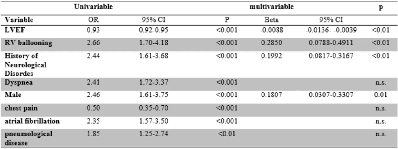
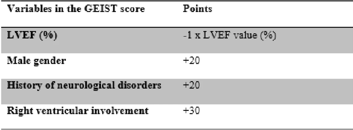
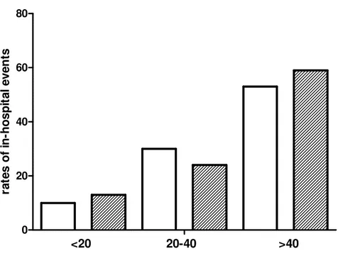
Beside several predictors of IHCs at univariate logistic regression analysis (Table 2) the following values were identified as independent predictors of IHCs: LVEF, RV involvement, history of neurological disorders and male gender. According to the coefficients in multivariable regression analysis, the GEIST prognosis score was derived by attributing 20 points for male gender and history of neurological disorders, 30 points for RV involvement, and subtracting the value in percent of LVEF (Table 3). Stratification into 3 risk groups (<20 vs 20-40 and >40 points) classified 41% of patients as at low risk, 44% as intermediate, and 15% as high risk. The observed IHC rates were 13%, 24% and 59%, respectively (p for trend <0.001) (Figure 1, eTable1). Patients in the intermediate group were
10
characterized by an increased risk of IHCs (odds ratio 2.11, 95% C.I. 1.39-3.19, p<0.001) when compared with the lower risk group; odds ratio for IHCs for the high-risk group vs lower risk was 9.84 (95% C.I. 5.97-16.20, p<0.001).
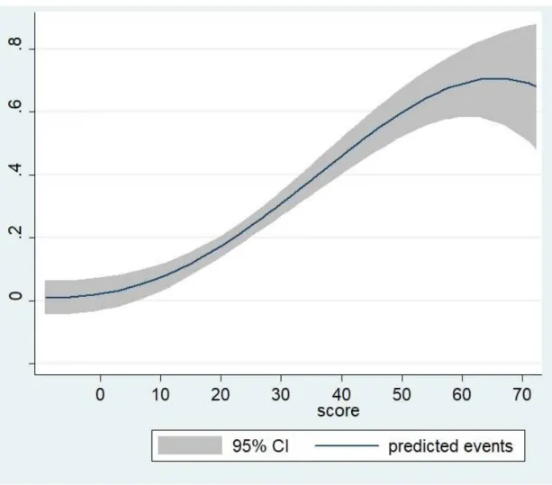
The GEIST prognosis score showed a good prediction of IHCs with an area under the curve of 0.71 (95% CI, 0.68–0.75, p<0.001) (Figure2,
eFigure2-3-4). At ROC curve analysis negative predictive power was 87% in case of
score values <20. P values for GEIST score goodness of fit tested by use of the Hosmer-Lemeshow statistic was non-significant (eFigure5).
Validation of the GEIST Score
Of the 829 patients with complete data in the validation cohort, 52% were in the low-risk, 39% in the intermediate, and 9% in the high-risk group. IHCs rates were: 10% (low risk), 30% (intermediate), and 53% (high risk) (p<0.001) (Figure1). C-statistic revealed a good discrimination of the GEIST score even in the validation cohort with an area under the curve of 0.74 (95% CI, 0.70–0.77, p<0.001, p=n.s. vs GEIST population) (eFigure2).
Long term Follow-up
Sixty-two patients (6%) were lost at long term follow-up. After 2.6±2.0 years follow-up patient with IHCs had statistically higher rates of mortality 40% (93 out of 232 pts) vs 10% (65 out of 647 patients, p=0.01).
11 Discussion
This is the first study proposing a simple clinical score for the prediction of IHCs based on four clinical variables from a large multicenter international registry of TTS patients. Main findings of the study are: (1) male gender, history of neurological disorder, RV involvement and LVEF
are independent predictors of IHCs and could be combined into a novel risk score;
(2) the newly developed GEIST prognosis score can accurately stratify the risk of IHCs in TTS patients and its performance is validated also in an external cohort with good predictive value;
(3) Patients with TTS and IHCs are characterized by high mortality rates at long-term follow-up.
TTS is featured by a transient left ventricular dysfunction and during hospitalization it is associated with several complications. However, there is a paucity of data regarding IHCs and how to promptly recognize and manage them. Several studies showed that TTS may have severe prognostic implications at long-term follow up; indeed 88% of TTS patients have persisting heart failure symptoms and cardiac limitation on exercise testing18. Despite recovery of LVEF, patients with prior TTS may present with an impaired cardiac energetic status 18. A recent study showed that a chronic inflammation could be also present in TTS patients; during the acute and sub-acute phase of TTS, higher serum levels of anti-inflammatory interleukins are present in TTS than in acute myocardial infarction 19.
12
Interestingly secondary forms of TTS, with a stressor represented by exacerbation of a comorbidity like chronic obstructive pulmonary disease (COPD) or kidney insufficiency, are associated with a worse outcome at short and long-term follow-up in other TTS cohorts 20. However, in the present study secondary forms of TTS were not associated with a higher risk of in-hospital complications.
In-hospital complications in TTS
IHCs in TTS patients mainly consist of acute heart failure including cardiogenic shock and pulmonary edema, with an incidence ranging from 12 to 45%. Cardiogenic shock is featured by an increased 28-day mortality (28.6% vs. 4.1%) 21 and a need for prompt treatment (pharmacological with levosimendan 22, esmolol 23, catecholamines 24) or early mechanical circulatory support. LVEF in a recent study was the only significant predictor of cardiogenic shock 21. Age >70 years, presence of physical stressor and LVEF <40% were found as predictors of acute heart failure in TTS in the Mayo clinic score. The presence of one, two and three variables was associated with a 28%, 58%, and 85% risk of acute HF, respectively 25. Our proposed risk score is the first that targets not only heart failure but also other important IHC reflecting severe morbidity and mortality in TTS.
In-hospital mortality rate for TTS is about 4.2%, with a higher rate among male patients (8.4% vs 3.6%, P <0.01) and in patients with secondary TTS 26. Indeed, 81.4% of patients with in-hospital mortality had underlying critical illnesses 26. In the present study male patients showed higher in-hospital mortality rates (9.3% vs 3.3% p= <0.01). However, differences comparing secondary TTS to primary/emotional TTS were not statistically
13
significant (3.6% vs 2.7% p=0.51). TTS patients without a clear stressor were featured by higher in-hospital mortality rates (6%).
Risk stratification during TTS hospitalization
Recently a European consensus paper defined high risk TTS patients as those with >75 years, systolic blood pressure <110 mmHg, presence of pulmonary edema, LVEF < 35%, LV outflow tract gradient >40 mmHg, mitral regurgitation and LV thrombi, while minor risk factors were biventricular involvement, persistent ST-elevation or Q-wave at admission and physical stressors 1.
Several predictors of IHCs have been also previously reported. Templin et al. found that physical triggers, acute neurologic or psychiatric diseases, high troponin levels, and low ejection fraction on admission were independent predictors of IHCs 3. Murakami et al. found that in a Japanese multicenter TTS registry blood cell count and brain natriuretic peptide were independent predictors of IHCs 27. In another registry, lower systolic blood pressure on admission, diabetes mellitus and β-blocker use before admission were independent predictors of IHCs 28. In a German registry (n=209) TTS patients with complications were older (70 vs 67 years), had higher heart rates (91 vs 83 bpm), Q-waves on admission ECG (36% vs 21%) and a lower LV ejection fraction (47% vs 54%). At multivariate regression analysis, Q-waves on admission and LVEF ≤30% were independent predictors of IHCs 29.
Echocardiography performed at admission could also well stratify the risk of TTS patients. Citro et al. found in a cohort of 227 patients that three echocardiographic parameters (LV ejection fraction, E/e’ ratio and
14
reversible moderate to severe mitral regurgitation) were associated with MACE 30.
Actually, our study is in line with previous reports and shows the importance of echocardiographic parameters for risk stratification. Two out of four parameters included in the prognosis score are echocardiographic variables: LVEF and RV involvement. Our study further supports the concept that echocardiographic evaluation at admission of both left and right ventricle is crucial in patients with TTS. LVEF has also important prognostic implications, especially in case of moderate/severe reduction of systolic function. Therefore, especially apical ballooning pattern which is featured by reduced LVEF may be associated with higher complication rates31. RV involvement in TTS has been proved in several studies to be a predictor of poor outcome at short and long-term follow-up 3233 and, in the present study, plays a major role with the highest score (30 points vs 20 points of the other variables). Moreover, RV involvement seems to be associated with a more severe impairment in LV systolic function 34.
Male gender was also associated in the GEIST registry with a poorer outcome. Male TTS patients are younger than females and physical stressors are more common in men than emotional triggers. During hospitalization, male patients have a higher incidence of severe pump failure (defined as Killip Class > III) and need for cardiopulmonary supportive therapies 35. Hormones, especially estrogens, may have a protective role, especially in female TTS patients 36.
Interestingly history of neurological disease is also associated with IHCs. These data further support the heart-brain connection as one potential pathophysiological mechanism of TTS 37. This group of diseases includes
15
stroke (both ischemic and hemorrhagic), neurodegenerative disease (Alzheimer/Parkinsonism) and epilepsy. The potential links between TTS and such conditions are heterogenous and are mainly associated with sympathetic upregulation/central autonomic dysfunction and increased circulating catecholamine levels 29.
A recent brain imaging study showed substantial anatomical differences between TTS patients and healthy control subjects in the limbic network (reduced connectivity) comprising the left amygdala, both hippocampi, left parahippocampal gyrus, left superior temporal pole and right putamen all of which are strongly involved in the control of emotional processing, cognition and the autonomic nervous system 38.
In-hospital complication and long-term follow-up
IHCs may be associated also with a worse outcome at long-term follow-up. TTS patients with IHCs showed in our study higher mortality rates at 2.6±2 year follow-up (40% vs 10%, p=0.01). In line with this high mortality rate Stiermaier et al reported a 24.7% mortality rate at 3-year follow-up 4.
Patients with IHCs may have more comorbidities as neurological disease and higher prevalence of biventricular involvement during hospitalization. The finding implies that TTS patients with IHCs may require a strict follow-up and remarks the concept that TTS is not a benign disease as it may have been considered.
16 Clinical implications of the GEIST score
Categorization of TTS patients according to the proposed GEIST sore might serve as a tool for early clinical risk stratification of TTS patients, which can be performed in the first days after hospital admission. All variables can easily be assessed and are readily available. We believe that it might be also be helpful for clinical decision making with respect to the selection of management strategies (e.g., length of stay in the ICU and hospital or supporting early discharge in low-risk patients).
17 Conclusions
The GEIST prognostic score, a simple score based on 4 clinical and echocardiographic variables, may be useful in early risk stratification of TTS. IHCs in TTS patients may be associated with increased risk of mortality at long-term follow-up.
18 Limitations
The GEIST score was derived from a multi-center observational registry in which data were collected from several different institutions. The accuracy of the score is appreciable and comparable with other clinical scores but does not exceed 70% and some high-risk patients may be missed. However, the specificity of the score is appreciable and might help to facilitate clinical decisions in TTS.
Another limitation was that a certain number of patients needed to be excluded from the analyses due to missing data regarding the score variables. However, the GEIST and Re-TAKO registry are one of the largest existing TTS registries to date. Cardiac magnetic resonance (CMR) imaging may represent the benchmark method to evaluate the presence of RV involvement in TTS; however, it was performed routinely only in 2 out of 12 centers (288 out of 1007 (28.5%) patients received CMR)
Acknowledgments
No funding was needed to perform the study; FS and NDB had full access to all the data in the study and take responsibility for the integrity of the data and the accuracy of the data analysis.
Conflicts of interest
19
20 GEIST registry Derivation Cohort (N=772) RE-TAKO registry Validation Cohort (N=829) Age 70 ± 11 70 ± 15 Gender, male % 11% 13% CV risk factors Hypertension 70% 64% Dyslipidemia 40% 35% Diabetes 21% 19% Smoke 22% 26% Comorbidities Past or chronic neurological disorder 15% 14% - Cerebrovascular accidents 48% 56% - Neurodegenerative disease 35% 32% - Epilepsy 17% 12% History of cancer 14% 13% Clinical presentation Angina 54% 64%
Atypical chest pain 12% 11%
No chest pain 34% 24% Dyspnoea 32% 42% Kind of stressors Emotional stressors 44% 41% Physical stressors 31% 29% No stressor 24% 34% Admission echocardiographic features LVEF (%) 40 ± 10% 43 ± 12% Apical ballooning 81% 86% Mid-ventricular ballooning 17% 11% Basal ballooning 2% 3%
Admission ECG features
ST elevation 59% 58% Negative T waves 52% 37% Atrial fibrillation 15% 8% In-hospital Complications: 23% 20% Pulmonary edema 6% 9% Invasive ventilation 6% 6.4% Cardiogenic shock 9% 10% Death 4% 2%
21
Table 1: Baseline features and in-hospital complications of patients admitted for Takotsubo syndrome
22
Table 2. Predictors of in-hospital complications at univariate logistic regression analysis and beta
23
Table 3: Clinical predictors for in-hospital complications included in the GEIST score and their
24 0 20 40 60 80 RETAKO GEIST <20 20-40 >40 ra te s o f in -h o s p it a l e v e n ts
Figure 1 Observed In-hospital complications according to risk stratification into low (<20 points),
intermediate (20-40 points) and high (>40 points) risk in the GEIST population and validation RE-TAKO population.
25
Figure 2. Predicted probability of In-hospital complication during admission for takotsubo syndrome
26
ADDITIONAL FIGURES AND TABLES
GEIST risk class GEIST score incidence of in-hospital complications Low <20 13% Intermediate 20 - 40 24% High >40 59%
27
28
eFigure 2: Receiver Operating Characteristic Curves Comparing the Areas Under the Curve of Risk
Scores from the derivation (GEIST registry, continuous line) and the validation cohort (RE-TAKO registry, dotted line) (p=n.s.).
29
eFigure 3. Predicted probability of in-hospital complications during admission for Takotsubo syndrome
30
31
eFigure 5. Expected vs observed incidence of in-hospital complications according to GEIST score
32
References
1 Lyon AR, Bossone E, Schneider B, Sechtem U, Citro R et al. Current state of knowledge on Takotsubo syndrome: a Position Statement from the Taskforce on Takotsubo Syndrome of the Heart Failure Association of the European Society of Cardiology. Eur J Heart Fail. 2016;18:8-27.
2 Wittstein IS, Thiemann DR, Lima JA, Baughman KL, Schulman SP et al. Neurohumoral features of myocardial stunning due to sudden emotional stress. N Engl J Med. 2005;352:539-48.
3 Templin C, Ghadri JR, Diekmann J, Napp LC, Bataiosu DR, Jaguszewski M, Cammann VL, Sarcon A, Geyer V, Neumann CA, Seifert B, Hellermann J, Schwyzer M, Eisenhardt K, Jenewein J, Franke J, Katus HA, Burgdorf C, Schunkert H, Moeller C, Thiele H, Bauersachs J, Tschöpe C, Schultheiss HP, Laney CA, Rajan L, Michels G, Pfister R, Ukena C, Böhm M, Erbel R, Cuneo A, Kuck KH, Jacobshagen C, Hasenfuss G, Karakas M, Koenig W, Rottbauer W, Said SM, Braun-Dullaeus RC, Cuculi F, Banning A, Fischer TA, Vasankari T, Airaksinen KE, Fijalkowski M, Rynkiewicz A, Pawlak M, Opolski G, Dworakowski R, MacCarthy P, Kaiser C, Osswald S, Galiuto L, Crea F, Dichtl W, Franz WM, Empen K, Felix SB, Delmas C, Lairez O, Erne P, Bax JJ, Ford I, Ruschitzka F, Prasad A, Lüscher TF. Clinical Features and Outcomes of Takotsubo (Stress) Cardiomyopathy. N Engl J Med. 2015;373:929-38.
4 Stiermaier T, Moeller C, Oehler K, Desch S, Graf T, Eitel C, Vonthein R, Schuler G, Thiele H, Eitel I. Long-term excess mortality in takotsubo
33
cardiomyopathy: predictors, causes and clinical consequences. Eur J Heart Fail. 2016;18:650-6.
5 Madhavan M, Rihal CS, Lerman A, Prasad A. Acute heart failure in apical ballooning syndrome (TakoTsubo/stress cardiomyopathy): clinical correlates and Mayo Clinic risk score. J Am Coll Cardiol. 2011;57:1400-1
6 Citro R, Rigo F, D'Andrea A, Ciampi Q, Parodi G, Provenza G, Piccolo R, Mirra M, Zito C, Giudice R, Patella MM, Antonini-Canterin F, Bossone E, Piscione F, Salerno-Uriarte J; Tako-Tsubo Italian Network Investigators. Echocardiographic correlates of acute heart failure, cardiogenic shock, and in-hospital mortality in tako-tsubo cardiomyopathy. JACC Cardiovasc Imaging. 2014;7:119-29.
7 Stiermaier T, Eitel C, Denef S, Desch S, Schuler G, Thiele H, Eitel I. Prevalence and Clinical Significance of Life-Threatening Arrhythmias in Takotsubo Cardiomyopathy. J Am Coll Cardiol. 2015;65:2148-50.
8 Kumar S, Kaushik S, Nautiyal A, Choudhary SK, Kayastha BL, Mostow N, Lazar JM. Cardiac rupture in takotsubo cardiomyopathy: a systematic review. Clin Cardiol. 2011;34:672-6.
9 Citro R, Bossone E, Parodi G, Carerj S, Ciampi Q, Provenza G, Zito C, Prota C, Silverio A, Vriz O, D'Andrea A, Galasso G, Baldi C, Rigo F, Piepoli M, Salerno-Uriarte J, Piscione F. Clinical profile and in-hospital outcome of Caucasian patients with takotsubo syndrome and right ventricular involvement. Int J Cardiol. 2016; 219:455-61.
34
10 Kato K, Sakai Y, Ishibashi I, Himi T, Fujimoto Y, Kobayashi Y. Predictors of in-hospital cardiac complications in patients with Takotsubo syndrome. Heart Vessels. 2018 Apr 25. doi: 10.1007/s00380-018-1172-y.
11 Prasad A, Lerman A, Rihal CS. Apical ballooning syndrome (Tako- Tsubo or stress cardiomyopathy): a mimic of acute myocardial infarction. Am Heart J. 2008;155:408–417.
12 Lang RM, Bierig M, Devereux RB, Flachskampf FA, Foster E, Pellikka PA, Picard MH, Roman MJ, Seward J, Shanewise JS, Solomon SD, Spencer KT, Sutton MS, Stewart WJ; Chamber Quantification Writing Group; American Society of Echocardiography's Guidelines and Standards Committee; European Association of Echocardiography. J Am Soc Echocardiogr 2005;18:1440-1463.
13 Eitel I, von Knobelsdorff-Brenkenhoff F, Bernhardt P, Carbone I, Muellerleile K, Aldrovandi A, Francone M, Desch S, Gutberlet M, Strohm O, Schuler G, Schulz-Menger J, Thiele H, Friedrich MG. Clinical characteristics and cardiovascular magnetic resonance findings in stress (takotsubo) cardiomyopathy. JAMA. 2011;306:277-86.
14 Eitel I, Behrendt F, Schindler K, Kivelitz D, Gutberlet M, Schuler G, Thiele H. Differential diagnosis of suspected apical ballooning syndrome using contrast-enhanced magnetic resonance imaging. Eur Heart J. 2008;29:2651-9.
35
36
Noninvasive ventilation in acute cardiogenic pulmonary edema. N Engl J Med. 2008;359:142-51
16 Thiele H, Zeymer U, Neumann FJ, Ferenc M, Olbrich HG, Hausleiter J, Richardt G, Hennersdorf M, Empen K, Fuernau G, Desch S, Eitel I, Hambrecht R, Fuhrmann J, Böhm M, Ebelt H, Schneider S, Schuler G, Werdan K. Intraaortic balloon support for myocardial infarction with cardiogenic shock. N Engl J Med. 2012;367:1287-96.
17 Núñez Gil IJ, Andrés M, Almendro Delia M, Sionis A, Martín A, Bastante T, Córdoba Soriano JG, Linares Vicente JA, González Sucarrats S, Sánchez-Grande Flecha A; RETAKO investigators. Characterization of Tako-tsubo Cardiomyopathy in Spain: Results from the RETAKO National Registry. Rev Esp Cardiol (Engl Ed). 2015;68:505-12.
18 Scally C, Rudd A, Mezincescu A, Wilson H, Srivanasan J, Horgan G, Broadhurst P, Newby DE, Henning A, Dawson DK. Persistent Long-Term Structural, Functional, and Metabolic Changes After Stress-Induced (Takotsubo) Cardiomyopathy. Circulation. 2018;137:1039-1048.
19 Santoro F, Costantino MD, Guastafierro F, Triggiani G, Ferraretti A, Tarantino N, Saguner A, Di Biase M, Brunetti ND. Inflammatory patterns in Takotsubo cardiomyopathy and acute coronary syndrome: A propensity score matched analysis. Atherosclerosis. 2018; 274:157-161.
20 Núñez-Gil IJ, Almendro-Delia M, Andrés M, Sionis A, Martin A, Bastante T, Córdoba-Soriano JG, Linares JA, González Sucarrats S,
Sánchez-Grande-37
38
Pérez-Castellanos A, Rueda Sobella F, Cambeiro C, Piqueras-Flores J, Vidal-Perez R, Bodí V, García de la Villa B, Corbí-Pascua M, Biagioni C, Mejía-Rentería HD, Feltes G, Barrabés J; RETAKO investigators. Secondary forms of Takotsubo cardiomyopathy: A whole different prognosis. Eur Heart J Acute Cardiovasc Care. 2016;5:308-16.
21 Stiermaier T, Eitel C, Desch S, Fuernau G, Schuler G, Thiele H, Eitel I. Incidence, determinants and prognostic relevance of cardiogenic shock in patients with Takotsubo cardiomyopathy. Eur Heart J Acute Cardiovasc Care. 2016;5:489-496.
22 Santoro F, Ieva R, Ferraretti A, Ienco V, Carpagnano G, Lodispoto M, Di Biase L, Di Biase M, Brunetti ND. Safety and feasibility of levosimendan administration in takotsubo cardiomyopathy: a case series. Cardiovasc Ther. 2013;31:e133-7.
23 Santoro F, Ieva R, Ferraretti A, Fanelli M, Musaico F, Tarantino N, Martino LD, Gennaro LD, Caldarola P, Biase MD, Brunetti ND. Hemodynamic Effects, Safety, and Feasibility of Intravenous Esmolol Infusion During Takotsubo Cardiomyopathy With Left Ventricular Outflow Tract Obstruction: Results From A Multicenter Registry. Cardiovasc Ther. 2016;34:161-6.
24 Ansari U, El-Battrawy I, Fastner C, Behnes M, Sattler K, Huseynov A, Baumann S, Tülümen E, Borggrefe M, Akin I. Clinical outcomes associated with catecholamine use in patients diagnosed with Takotsubo cardiomyopathy. BMC Cardiovasc Disord. 2018;18:54.
39
25 Madhavan M, Rihal CS, Lerman A, Prasad A. Acute heart failure in apical ballooning syndrome (TakoTsubo/stress cardiomyopathy): clinical correlates and Mayo Clinic risk score. J Am Coll Cardiol 2011;57:1400– 1401.
26 Brinjikji W, El-Sayed AM, Salka S. In-hospital mortality among patients with takotsubo cardiomyopathy: a study of the National Inpatient Sample 2008 to 2009. Am Heart J. 2012;164:215-21.
27 Murakami T, Yoshikawa T, Maekawa Y, Ueda T, Isogai T, Konishi Y, Sakata K, Nagao K, Yamamoto T, Takayama M. Characterization of predictors of in-hospital cardiac complications of takotsubo cardiomyopathy: multi-center registry from Tokyo CCU Network. J Cardiol. 2014;63:269-73.
28 Kato K, Sakai Y, Ishibashi I, Himi T, Fujimoto Y, Kobayashi Y. Predictors of in-hospital cardiac complications in patients with Takotsubo syndrome. Heart Vessels. 2018 Apr 25. doi: 10.1007/s00380-018-1172-y
29 Schneider B, Athanasiadis A, Schwab J, Pistner W, Gottwald U, Schoeller R, Toepel W, Winter KD, Stellbrink C, Müller-Honold T, Wegner C, Sechtem U. Complications in the clinical course of tako-tsubo cardiomyopathy. Int J Cardiol. 2014;176:199-205.
30 Citro R, Rigo F, D'Andrea A, Ciampi Q, Parodi G, Provenza G, Piccolo R, Mirra M, Zito C, Giudice R, Patella MM, Antonini-Canterin F, Bossone E, Piscione F, Salerno-Uriarte J. Echocardiographic correlates of acute heart
40
failure, cardiogenic shock, and in-hospital mortality in tako-tsubo cardiomyopathy. JACC Cardiovasc Imaging. 2014;7:119-29.
31 Stiermaier T, Möller C, Graf T, Eitel C, Desch S, Thiele H, Eitel I. Prognostic Usefulness of the Ballooning Pattern in Patients With Takotsubo Cardiomyopathy. Am J Cardiol. 2016;118:1737-1741.
32 Kagiyama N, Okura H, Tamada T, Imai K, Yamada R, Kume T, Hayashida A, Neishi Y, Kawamoto T, Yoshida K. Impact of right ventricular involvement on the prognosis of takotsubo cardiomyopathy. Eur Heart J Cardiovasc Imaging. 2016;17:210-6.
33 Citro R, Bossone E, Parodi G, Rigo F, Nardi F, Provenza G, Zito C, Novo G, Vitale G, Prota C, Silverio A, Vriz O, D'Andrea A, Antonini-Canterin F, Salerno-Uriarte J, Piscione F. Independent Impact of RV Involvement on In-Hospital Outcome of Patients With Takotsubo Syndrome. JACC Cardiovasc Imaging. 2016;9:894-895.
34 Haghi D, Athanasiadis A, Papavassiliu T, Suselbeck T, Fluechter S, Mahrholdt H, Borggrefe M, Sechtem U. Right ventricular involvement in Takotsubo cardiomyopathy. Eur Heart J. 2006;27:2433-9.
35 Murakami T, Yoshikawa T, Maekawa Y, Ueda T, Isogai T, Sakata K, Nagao K, Yamamoto T, Takayama M. Gender Differences in Patients with Takotsubo Cardiomyopathy: Multi-Center Registry from Tokyo CCU Network. PLoS One. 2015;10(8):e0136655.
41
36 El-Battrawy I, Zhao Z, Lan H, Schünemann JD, Sattler K, Buljubasic F, Patocskai B, Li X, Yücel G, Lang S, Nowak D, Cyganek L, Bieback K, Utikal J, Zimmermann WH, Ravens U, Wieland T, Borggrefe M, Zhou XB, Akin I. Estradiol protection against toxic effects of catecholamine on electrical
properties in human-induced pluripotent stem cell derived
cardiomyocytes. Int J Cardiol. 2018 ;254:195-202.
37 Samuels MA. The brain-heart connection. Circulation. 2007;116:77-84.
38 Hiestand T, Hänggi J, Klein C, Topka MS, Jaguszewski M, Ghadri JR, Lüscher TF, Jäncke L, Templin C. Takotsubo Syndrome Associated With Structural Brain Alterations of the Limbic System. J Am Coll Cardiol. 2018;71:809-811.