University of Molise and
Department of Angiocardioneurology IRCCS
Neuromed
Ph.D. in Translational and Clinical Medicine
XXX COURSE
SDS MED/50
Neuropilin1 signaling in
macrophages mediates the
hypertensive functions of Placental
Growth Factor upon Angiotensin II
Ph.D. Program Coordinator: Prof. Ciro Costagliola
PhD. Student: Maria Piacenti Ph.D. Tutor:
Chapter I:
Hypertension and inflammation
2
1.1 HYPERTENSION: AN OVERVIEW
Cardiovascular changes furnish the fundamental process in the largest number of deaths after the 40th year. Each year cardiovascular disease (CVD) causes 3.9 million deaths in Europe and over 1.8 million deaths in the European Union (EU). CVD accounts for 45% of all deaths in Europe and 37% of all deaths in the EU. CVD is the main cause of death in men in all but 12 countries of Europe and is the
main cause of death in women in all but two countries
(http://www.ehnheart.org/cvd-statistics.html), in fact hypertension generates over 7 million early deaths per year and is responsible of 4.5% of the total diseases (Buford, 2016). Current data from the National Health and Nutrition Examination Surve show that 70% of older adults have hypertension, compared to only 32% for adults aged 40-59 years.
Figure 1: Prevalence of hypertension among adults by age and sex according to the National Health and Nutrition Examination Survey:2007-2012. Adapted from Buford TW(2016): Hypertension and aging.
“Although hypertension is relatively easy to prevent, simple to diagnose, and relatively inexpensive to treat, it remains the second leading cause of death among
3
Americans, and as such should rightly be called a neglected disease” said David W. Fleming, MD, chair of the committee that prepared the report (Buford, 2016). More than ninety percent of cases that do not have a recognizable cause are classified as essential or primary hypertension (Singh, Chapleau, Harwani, & Abboud, 2014)
Over 60 years ago, Dr. Irvine Page suggested the Mosaic Theory of hypertension, which declares that different factors, including genetics, environment, adaptive, neural, mechanical and hormonal perturbations cooperate to raise blood pressure. Now it is known that the production of reactive oxygen species (ROS) and inflammation are two among the main common molecular and cellular events in various organs that inspired the Mosaic Theory. Neuronal firing in specific brain centers and sympathetic outflow are increased by these factors. Furthermore, they alter vascular tone and morphology and support sodium retention in the kidney.
Figure 2: The contribution of inflammation to components of the Mosaic Theory Adapted from Harrison (2013): The Mosaic Theory revisited: common molecular mechanisms coordinating diverse organ and cellular events in hypertension.
4
1.2 HYPERTENSION AND INFLAMMATION
Low-grade inflammation performs a key role in hypertension and innate and adaptive immune system and seems to concur to hypertension progress and maintenance (Bomfim, Rodrigues, & Carneiro, 2017)
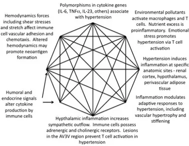
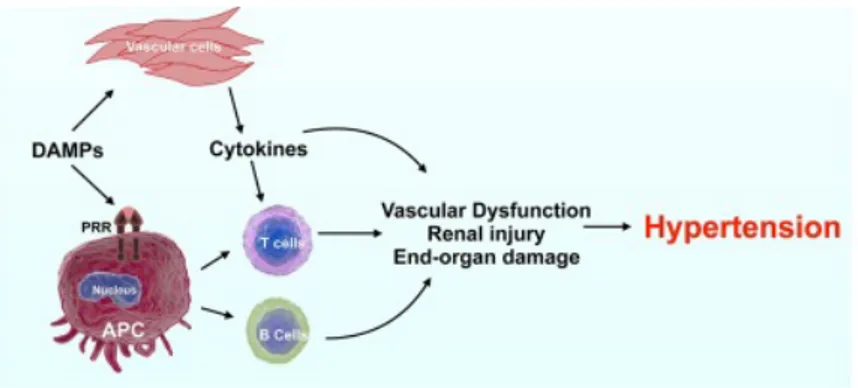
There is a strong association between hypertension and immune system but the mechanism that stimulate immune system and inflammation are totally unknown. Cells from the innate immune system express pattern recognition receptors (PRR), which detect conserved pathogen-associated molecular patterns (PAMPs) and damage-associated molecular patterns (DAMPs) that induce innate effector signals to give endogenous signals such as inflammatory cytokines and chemokines, to warn the host about peril. Furthermore Antigen-presenting cells (APC) act as sentinels that are activated by PAMP and DAMPs to feel the presence of the antigen/neoantigen, following the adaptive immune system activation.
Figure 3: Immune system and Hypertension.
5
The major effectors of the innate immune system are monocytes, macrophages, granulocytes, neutrophils and dendritic cells (DC) and tissue infiltrated by these cells are part of inflammatory response in distinct hypertension model. In particular, lysozyme M-positive monocytes have been involved in Angiotensin II (AngII)-induced hypertension and vascular dysfunction (Wenzel et al., 2011). Similarly, mice with functionally-deficient macrophages develop blunted hypertension and vascular remodeling in response to AngII or deoxycorticosterone acetate-salt (DOCA-salt) (De Ciuceis et al., 2005).
Monocyte/macrophage function can be modulated by mineralocorticoid receptors (MR), whose (Rickard et al., 2009) deletion from macrophages protects against cardiac fibrosis and hypertension in response to DOCA-salt. Macrophages are also involved in salt-dependent volume and blood pressure, via a VEGF-C-dependent mechanism (Machnik et al., 2009). All these results have been obtained in animal models, but the innate system is as well important in human hypertension. In hypertensive patients, blood neutrophils are increased, and this increase has been suggested as a useful biomarker (Liu et al., 2015).
Another immune cell population important for the development of hypertension are dendritic cells. Actually, some DAMPs, such as the γ-ketoaldehydes isoketals, are able to activate dendritic cells in hypertensive conditions, after being oxidized and phagocytized by these leukocytes (Kirabo et al., 2014; Wu et al., 2015). Moreover, dendritic cells can be stimulated by AngII, determining superoxide production and accumulation of isoketals (Harrison, 2016). When isoketals are scavenged, there is not activation of dendritic cells in the kidney. Consequently, dendritic cells fail to induce proliferation of T-cells, and blood pressure does not increase in response to AngII.
Activation of innate immune responses is also associated by AngII induced hypertension. Infusion of AngII for 14 days increases tumor necrosis factor-alpha (TNF-α) (Ahn et al., 2007) and increases TLR2 gene expression and DC number into the kidney. In fact, TLR4 upregulation seems to contribute to AngII induced hypertension (De Batista et al., 2014; Ding et al., 2010).
6
The innate and adaptive immune systems work in a very coordinated manner. The role of the adaptive immune system in hypertension has been progressively demonstrated, in the last decade. Therefore, the question whether both systems communicate during hypertension development has come to light. Various studies demonstrate that they play a key role in the development of adaptive immune responses in TLR signaling pathways, upregulating co-stimulatory molecules of APCs (Brentano, Kyburz, Schorr, Gay, & Gay, 2005), important for the interaction with T-cells. These co-stimulatory molecules have been involved in the development of hypertension (Vinh et al., 2010).
There are five different main subtypes of lymphocytes: CD4 T-cell, CD8 T-cell, B-cell, γδ T-cell and Natural Killer T (NK-T) cell. Evidence from experimental and clinical studies indicates that alterations in immune cell populations, such as activation of different lymphocyte subtypes, play an important role in the pathogenesis of hypertension (Idris-Khodja, Mian, Paradis, & Schiffrin, 2014; Trott & Harrison, 2014) .
7 Figure 4: Proposed role of T effector and regulatory lymphocytes in hypertension. Slight elevation in blood pressure (BP) in response to hypertensive stimuli (angiotensin II, aldosterone, endothelin-1, salt and genetic susceptibility) occurs due to increased central signaling, perhaps causing mild tissue injury and formation of damage-associated molecular patterns (DAMPs) and neoantigens. This may lead to activation of innate antigen-presenting cells (APCs) and, subsequently, activation and polarization of naïve CD4+ T effector lymphocytes (Th0) towards pro-inflammatory T helper (Th)1/Th17 phenotypes. Th1/Th17 may contribute to vascular and kidney damage via production of reactive oxygen species (ROS), interferon (IFN)-γ and interleukin (IL)-17 and lead to maintenance of hypertension and progression of end-organ damage. T regulatory lymphocytes counteract hypertension and associated injury by producing IL-10 or by other mechanisms, and suppression of innate and adaptive immune responses. CD, cluster of differentiation; CNS, central nervous system; MHC-II, major histocompatibility complex-II; PAMPs, pathogen-associated molecular patterns; TCR, T-cell receptor. Adapted from Idris-Khodja (2014): Dual opposing roles of adaptive immunity in hypertension.
The important role of lymphocytes in hypertension has started to be revealed fifty years ago. Researcher of University of Texas showed that transfer of lymphocytes from rats with renal damage induces hypertension in normotensive rats (Okuda & Grollman, 1967).
Then in 1986 a research of University of Lyon demonstrated that neonatal thymectomy prevents the natural rise in systolic blood pressure in spontaneously hypertensive rats (Bataillard, Freiche, Vincent, Sassard, & Touraine, 1986). Hypertensive patients with rheumatoid arthritis or psoriasis, treated with a T-cell suppressing agent, have an important reduction of blood pressure, and these results was confirmed in animal models (Bravo, Quiroz, Ferrebuz, Vaziri, & Rodríguez-Iturbe, 2007; Herrera, Ferrebuz, MacGregor, & Rodriguez-Rodríguez-Iturbe, 2006; Muller et al., 2002). This was confirmed by Guzik et al. in 2007. In this study the researchers showed that T lymphocytes concur to the progression of hypertension by the study of RAG1 KO mouse, which lack T and B lymphocytes. This mouse model have
8
tick hypertension, aortic endothelial dysfunction, vascular remodeling and oxidative stress in response to AngII. (Guzik et al., 2007). This phenotype is re-established by adoptive transfer of T-cells, but not B-cells, in response to AngII. Similar T-cells are shown their importance in other experimental models of hypertension (Mattson et al., 2013).
Recently, researches of Duke University showed that, Scid mice without lymphocyte responses have a dampen hypertensive response to chronic AngII infusion, develop less cardiac hypertrophy and kidney injury. At the same time, Scid mice display upregulation of TNF-α, endothelial nitric oxide synthase (eNOS) and COX-2 in the kidney. The authors hypothesize that increased TNF-α expression in these animals might lead to dysregulation of macrophage function or upregulation of innate immune responses in the absence of adaptive immunity (Crowley et al., 2010). Figure 5: Hypertension and organ damage Schematic representation of sites between autonomic and immune system. A Inflammation in the CNS induces autonomic activity, B autonomic-induced mobilization of hematopoietic progenitor and immune cells from bone marrow and spleen, C migration of immune cells into brain, vasculature, heart and kidney causing end-organ damage and D autonomic receptor and TLRs on immune cells interact to regulate cytokine release. Adapted from Singh (2014): The immune system and hypertension.
9
Angiogenic growth factors, besides inducing vessel growth and promoting inflammation, contribute to the adaptation of cardiac muscle to high blood pressure. For example, defects in cardiac vascular endothelial growth factor (VEGF) impair the adaptive remodeling in overloaded hearts, accelerating the transition to heart failure (Izumiya et al., 2006). The research group I am working with has explored a similar role for placental growth factor (PlGF), a protein belonging to a family of vascular endothelial growth factor (VEGF)-related angiogenic factors. They started from the consideration that PlGF increases in hypoxic/ischemic myocardium (Torry et al., 2009), and contributes to pathological angiogenesis, such as vascular regeneration under tissue ischemia (Autiero, Luttun, Tjwa, & Carmeliet, 2003). On the other hand, it has been demonstrated that the cytokine face of PlGF acts as a chemotactic agent for monocytes (Perelman et al., 2003; Pipp et al., 2003), which participate in the cardiac inflammatory response to pressure overload and support adaptive cardiac remodeling (Endo et al., 2007). Moreover, TNFα activation, one of the earliest inflammatory events in overloaded hearts (Wang et al., 2009), has been linked to PlGF to recruit myelomonocytic cells in inflamed tissues (Ding et al., 2010). In fact, TNFα needs to be shed from a membrane-bound form by TNFα–converting enzyme (TACE), and the main natural inhibitor of TACE is tissue inhibitor of metalloproteinases-3 (TIMP-3), the most abundant TIMP expressed in cardiac tissue (Vanhoutte & Heymans, 2010). Interestingly, the lack of TIMP-3, producing excessive TNFα activation (Kassiri et al., 2005), promotes maladaptive cardiac remodeling, favoring the transition to heart failure. On the other hand, anti–TNFα therapy fails to be successful for heart failure treatment (Chung, Packer, Lo, Fasanmade, & Willerson, 2003; Mann et al., 2004), thus suggesting that, even if excessive inflammatory responses are detrimental, impaired inflammation also could be harmful, hampering an adequate tissue response to injury.
Thus, PlGF was a good candidate to play a role in the complex cytoarchitectural response of the heart to hemodynamic overload. Thus, my research group demonstrated the pathophysiological role of PlGF in cardiac remodeling to pressure overload as both an angiogenic factor and a cytokine (Carnevale et al.,
10
2011). They clarified that PlGF is increased in overloaded hearts and that, in this context, it influences mainly vessel dimensions. More important, we identified that PlGF finely tunes a balanced regulation of the TIMP-3/TACE axis and the consequent TNFα activation in response to TAC, thus ensuring an adequate inflammatory background for adaptive cardiac hypertrophic remodeling.
11
Chapter II:
Placental Growth Factor
12
2.1 PLACENTAL GROWTH FACTOR
Placental Growth Factor (PlGF) is a growth factor and a cytokine, involved in angiogenesis and inflammation. It is a member of the vascular endothelial growth factor family, and its 3D structure is very similar to that of VEGF-A, even if the two proteins only share 42% amino acid sequence identity (Nguyen et al., 2016).
Figure 6: Structural model of human placental growth factor (PGF)-1 Adapted from Nguyen 2016: Placental growth factor and its potential role in diabetic retinopathy and other ocular neovascular diseases.
Human PlGF gene has been mapped to chromosome 14q24, and is encoded by seven exons spanning an 800-kb-long DNA interval. As a protein, PlGF exists in four isoforms, generated by alternative splicing (PlGF-1, -2, -3 and -4), and acts as a dimeric protein held together by disulphide bonds (Nguyen et al., 2016). It was called Placental Growth Factor since it was first identified in placenta, in particular in trophoblastic giant cells associated with the parietal yolk sac at early stages of embryogenesis, suggesting a role to coordinate vascularization in the deciduum and placenta. PlGF has also been identified in lung, heart, thyroid, skeletal muscle, and adipose tissue under normal physiological conditions (Achen,
13
Gad, Stacker, & Wilks, 1997; De Falco, 2012; Persico, Vincenti, & DiPalma, 1999; Viglietto et al., 1995; Voros et al., 2005).
In cardiovascular pathophysiology, PlGF has a role in the adaptive response for cardiac remodeling during transverse aortic constriction (Carnevale et al., 2011) characterized by harmonized cardiomyocyte growth, angiogenesis and inflammation (De Falco, 2012; Frey & Olson, 2003; Hunter, Chien, & Chien, 1999).
This previous work, by the group I have worked with for my PhD, showed that genetic deletion of PlGF caused a dysregulation of cardiac remodeling negatively affecting muscle growth, mainly ascribable to a defective inflammatory response. At molecular level, this piece of research identified an impaired activity of TNF-α converting enzyme (TACE) due to a strong increase of its main natural inhibitor, tissue inhibitor of metalloproteinases (TIMP)-3 (De Falco, 2012; Vanhoutte & Heymans, 2010). TACE is essential to activate TNF-α, among the first cytokines released by inflammation in overloaded hearts (De Falco, 2012; Ding et al., 2010; Wang et al., 2009). PlGF, by regulating the TIMP-3/TACE axis, allows the establishment of an inflammatory response needed for adaptive cardiac remodeling (De Falco, 2012).
The angiogenic role of PlGF is controversial. In some in vitro studies PlGF binding to VEGFR-1 failed to produce endothelial cells (EC) growth and angiogenesis, while other studies show that PlGF/VEGFR-1 signaling promotes EC viability and angiogenesis (Roy et al., 2005; Roy, Bhardwaj, & Ylä-Herttuala, 2006). In placenta and in PlGF-1 expressing tumors higher PlGF levels have been shown to inhibit EC growth (Ahmed, Dunk, Ahmad, & Khaliq, 2000; Roy et al., 2006). PlGF has direct eff ects on ECs, both by inducing its own signaling and by amplifying VEGF-driven angiogenesis (Autiero et al., 2003; Roy et al., 2006). PlGF binds the VEGFR-1 receptor, but not VEGFR-2 (Autiero et al., 2003; Carmeliet et al., 2001; De Falco, Gigante, & Persico, 2002; Otrock, Makarem, & Shamseddine, 2007; Roy et al., 2006). Moreover, the isoforms carrying heparin-binding domain bind to neuropilin (De Falco et al., 2002; Nguyen et al., 2016). PlGF binding to VEGFR-1 has been observed to stimulate EC migration and
14
recruit cells involved in angiogenesis, such as monocyte macrophages, smooth muscle cells and pericytes ((De Falco, 2012; Nguyen et al., 2016). It has been hypothesized that PlGF binding to VEGFR-1 may stimulate angiogenesis also indirectly, by freeing up more VEGF-A to bind to VEGFR-2 (Autiero et al., 2003; Carmeliet et al., 2001; De Falco, 2012; Feeney et al., 2003; Fischer, Mazzone, Jonckx, & Carmeliet, 2008).
Figure 7: Angiogenic effects of VEGFB and PlGF on endothelial cells. Adapted from Fisher 2008: FLT1 and its ligands VEGFB and PlGF: drug targets for anti-angiogenic therapy?
In addition, leucocyte infiltration is connected with angiogenesis. VEGFR-1 ligands are monocyte attractants and contribute to the recruitment of leucocytes. These recruited leucocytes provide an indirect (VEGF-independent) pathway of angiogenesis through the secretion of proangiogenic factors (Mantovani, Allavena, Sica, & Balkwill, 2008).
15
2.2 PLGF RECEPTORS
2.2.1 Vascular Endothelial Growth Factor- Receptor 1
As stated above, the canonical receptor of PlGF is VEGR-1 (Vascular Endothelial Growth Factor Receptor-1), also known as FLT-1, a tyrosine kinase receptor (TK receptor) (Olofsson et al., 1998).
Figure 8: VEGF and its receptor. Adapted from Otrock 2007: Vascular endothelial growth factor family of ligands and receptors: Review.
Flt-1 consists of seven extracellular Ig-like domains, a transmembrane domain and an intracellular TK domain. Dimerization and phosphorylation of Flt-1 is induced by the binding of ligands. During development, VEGFR-1 is first expressed in angioblasts and in the endothelium and its expression decreases during later embryonic stages (Fong, Rossant, Gertsenstein, & Breitman, 1995; Peters, De Vries, & Williams, 1993). VEGFR-1 is expressed in osteoblasts,
16
monocytes/macrophages, placental trophoblasts, renal mesangial cells and also in some hematopoietic stem cells (Zachary, 2001). Then VEGFR-1 expression is up-regulated by hypoxia via a pathway activated by hypoxia-inducible factor-1 (HIF-1) (Gerber, Condorelli, Park, & Ferrara, 1997). In 1996 Barleon and his collaborators showed that VEGFR-1 participates in monocyte migration (Barleon et al., 1996).
Selvaraj group has shown that PlGF binding to VEGFR-1 in monocytes results in activation of PI3 kinase/AKT and ERK-1/2 pathways, leading to chemotaxis and the induction of a series of inflammatory cytokines in sickle cell disease (Selvaraj et al., 2003). Hattori et al. have shown that PlGF promotes recruitment of VEGFR-1(+) hematopoietic stem cells from a dormant to a proliferative bone marrow environment, favoring differentiation and reconstitution of hematopoiesis (Hattori et al., 2002). Furthermore, VEGFR-1-specific PlGF binding can promote collateral vessel growth and arteriogenic activity in myocardial and limb ischemia through the recruitment of bone marrow cells such as monocytes (Luttun et al., 2002; Pipp et al., 2003).
In the end Flt-1 has a low tyrosine kinase activity, and deletion of its tyrosine kinase domain does not affect vascular development (Autiero et al., 2003; Hiratsuka, Minowa, Kuno, Noda, & Shibuya, 1998). In fact mouse embryos lacking Flt-1 generates angioblasts, although blood vessel assembly and tube formation is faulty (Ahmed et al., 2000; Fong et al., 1995; Otrock et al., 2007).
17 2.2.2 Neuropilin- 1
Neuropilin (Nrp), a 130-kDa to 140-kDa cell-surface glycoprotein, was first identified as a receptor for semaphorin/collapsins, a large family of secreted and transmembrane proteins, which are repulsive guidance signals in axonal and neuronal development (Fuh, Garcia, & de Vos, 2000; Miao & Klagsbrun, 2000). Later, it was found that Nrp-1 is able to bind VEGF-A, VEGF-B and PlGF. A similar receptor, Nrp-2, binds VEGF-A, VEGF-C and PlGF (Bagri & Tessier-Lavigne, 2002).
During embryonic development, Nrp-1 is expressed in the nervous, cardiovascular, and skeletal systems (Kitsukawa, Shimono, Kawakami, Kondoh, & Fujisawa, 1995). In adults, its expression is extended to ECs, tumor cells, lung, heart, liver, kidney, pancreas, osteoblasts, and bone marrow stromal cells (Soker, Takashima, Miao, Neufeld, & Klagsbrun, 1998; Tordjman et al., 1999). In humans, the gene coding for Nrp-1 is located on chromosome 10, while the gene coding for Nrp-2 is located on chromosome 2; both genes are composed of 17 exons (BIELENBERG, PETTAWAY, TAKASHIMA, & KLAGSBRUN, 2006; Michael Klagsbrun, Takashima, & Mamluk, 2002).
Nrp-1 acts as a co-receptor enhancing VEGF-A–VEGFR-2 interactions, forming complexes with VEGFR-1 and augmenting tumor angiogenesis in vivo (M Klagsbrun & D’Amore, 1991). Overexpression of Nrp-1 in chimeric mice leads to excessive capillary and blood vessel formation and hemorrhages associated to cardiac malformations (Kitsukawa et al., 1995). In chick embryos, endothelial Nrp-1 expression is mainly restricted to arteries, whereas Nrp-2 is primarily expressed in veins (Herzog, Kalcheim, Kahane, Reshef, & Neufeld, 2001). It is thought that Nrp-1 is required for cardiovascular development because it regulates VEGF165 levels (Kawasaki et al., 1999).
The role of Nrp-1 in the development of the vascular system has been demonstrated by gene-targeting studies; in fact, genetic deletion of Nrp-1 leads to embryonic lethality (Kawasaki et al., 1999). Similar to VEGFR-1, a soluble naturally occurring isoform of Nrp-1 acts as a natural antagonist. Soluble Nrp-1
18
inhibits VEGF165 binding to tumor cells and endothelium and inhibits VEGF165-induced tyrosine phosphorylation of VEGFR-2 in ECs (Otrock et al., 2007). Nrp-1 is expressed in angiogenic vessels in animal models (Gelfand et al., 2014; Lanahan et al., 2013; Nguyen et al., 2016) and in humans (Cui et al., 2003; Nguyen et al., 2016). The blockade of neuropilin-1 in neonatal mouse retinas inhibits vascular remodelling, andrenders vessels more susceptible totreatment with anti-VEGF agents (Pan et al., 2007).
2.3 PLGF-HYPERTENSION-IMMUNITY
The role of PlGF in hypertension is coherent with what I have already discussed about an interaction between hypertension and inflammation. My researcher group gave a contribution to clarify the importance of PlGF in the connection between hypertension and inflammation. In particular, we showed a splenic mechanism activated by PlGF, responsible for T cells co-stimulation via CD86 and T-cells egression and homing in target organs (Carnevale et al., 2014).
In particular, we found that, in PlGF WT mice, chronic infusion of AngII induce inflammation and fibrosis, that were absent in PlGF null mice. Furthermore, the echocardiographic analysis after chronic AngII, showed a hypertrophic response in WT mice but not in PlGF deficient mice, which are protected from cardiac remodeling and renal damage.
Subsequently, to extricate the molecular mechanism
underlying the protection from hypertension showed that PlGF doesn’t have a direct effect on vascular tissue.
Also PlGF deficient mice vessels and kidneys, after AngII infusion, were devoid of T-cells infiltration. This protection is clear yet in pre-hypertensive phase (pre-HTN -3 days).
19
Furthermore, by the splenic transplantation, my laboratory group, demonstrate that splenectomized mice are protected from AngII–induced BP increase.
Surprisingly, the transplantation of PlGF deficient spleen in WT background protected to raise BP after chronic infusion of AngII and protect to infiltration of T-cells in organ target.
In order to track unambiguously the fate of T-cells from the spleen to the target organ during hypertension, my group studied CD45.2 mice that given CD45.1 spleen by transplantation and then infused with AngII to induce hypertension. They observed increased numbers of donor (CD45.1) T cells in aorta and kidneys of Ang-II pre-HTN animals, demonstrating that the immune cells infiltrating target organs in hypertension are of splenic origin.
PlGF is expressed in the marginal zone (MZ) of spleen; this zone is enriched in macrophages and dendritic cells acting as antigen presenting cells (APC) and activating T-cells. They found that PlGF is crucial to allow T-cells co-stimulation via CD86 and their egress for the deployment toward vasculature of target organs, a hallmark of hypertension.
20
Chapter III:
Results
21 3.1 BACKGROUND
An area of emerging interest in mechanistic studies on arterial hypertension revealed a crucial role of adaptive immunity. On this regard, our group is dedicated to the exploration of new mechanisms underlying the interaction between immune system and hypertension. In particular, after identifying Placental Growth Factor (PlGF) as a crucial factor in the dynamics of T-cell activation during hypertension, we aimed here at exploring the molecular mechanisms at the basis of this signaling pathway. The main PlGF "canonical" receptor is VEGR1, known as Flt1, belonging to the same family as the better known VEGFR2, main receptor of VEGF. However, more recent data suggest the existence of an alternative PlGF signaling mechanism mediated by Neuropilin1 (NRP1), a transmembrane protein mainly known for regulating different aspects of vascular and neuronal development.
Moreover, high expression of PlGF receptor neuropilin 1 (Nrp1) correlates with poor overall survival in patients. It was demonstrated that PlGF and Nrp1 are required for the growth and spread of medulloblastoma: PlGF/Nrp1 blockade results in direct antitumor effects in vivo, resulting in medulloblastoma regression, decreased metastasis and increased mouse survival (Snuderl et al., 2013).
By the way, we have established the co-expression and activation in response to AngII of both receptors in the spleen, where PlGF is produced, we explored these two different pathways of intracellular signaling using two transgenic models: 1) VEGFR1/Flt1-TK-/-, mice with a deletion in the intracellular tyrosine kinase domain of VEGFR1, unable to activate the intracellular signal upon interaction with its ligand PlGF.
2) Nrp1flox; LysM-Cre transgenic mice for conditional deletion of NRP1 exon 2 through the strategy of the Cre/LoxP system exclusively in the monocyte-macrophage line, since we had previously identified this cell line as a target of PlGF in the spleen.
22 3.2 VEGFR1 SIGNALING IS NOT ESSENTIAL FOR
ANGII-INDUCED HYPERTENSION
VEGFR1 is the first receptor that we investigated to explore the role of PlGF. First of all, we found that VEGFR1 is expressed in the spleen, in basal condition. Then we evaluated the expression of VEGFR1 in the spleen after chronic infusion of AngII. We found that VEGFR1 is upregulated after chronic infusion of AngII.
We evaluatedits expression in the marginal zone (MZ) of the spleen, in a network
of follicular dendritic cells, identified by the ERTR7 and CD169 antigens. This zone of the spleen is particularly enriched in macrophages and dendritic cells with a highly specialized phenotype (Mueller & Germain, 2009) acting as antigen-presenting cells (APCs) and directly or indirectly activating T-cells. So far, it is unknown how hypertensive stimuli are able to condition and organize adaptive immune responses in the spleen. Classically this happens when, after antigen uptake, APCs undergo a maturation process characterized by an increased surface expression of major histocompatibility complex II (MHCII) and the B7 ligands, CD80 and CD86 (Song, Lei, Xiong, & Haque, 2008).
Interestingly, it has been demonstrated that the AngII hypertensive challenge is able to increase the percentage of cells expressing CD86 in the splenic reservoir, while the number of cells expressing CD80 and MHCII remained unchanged (Vinh et al., 2010). However, how hypertensive stimuli could check the costimulation axis of immune system is unknown.
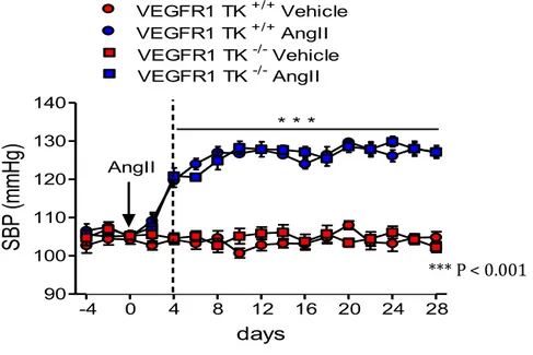
To clarify the real role of VEGR1 as a receptor of PlGF in hypertension-immune
system scenario, we infused chronic AngII in VEGFR1 TK-/- mice.
The VEGFR1 TK-/- mice lacked the TK domain but had the ligand-binding domain
and TM domain.
23 Figure 9: VEGFR-1 −/− mice die in the embryonic stage. The reason is an excessive proliferation of endothelial cells and a disorganized angiogenesis. Thus, mice in which the VEGFR1 receptor was modified to lose only its tyrosine kinase domain were used in this study. Adapted from Shibuya (2011): Involvement of Flt-1 (VEGF receptor-1) in cancer and preeclampsia.
They were basically healthy, and their angiogenesis was almost normal (Hiratsuka et al., 1998). In contrast Fong et al. showed that VEGFR1-/- mice die in the embryonic stage due to the overgrowth and disorganization of blood vessels, thus excluding the possibility of using knockout mice for this receptor (Fong et al., 1995).
24 Figure 10: VEGFR-1 expression in the spleen. Immunostaining of spleen from AngII and vehicle infused mice, showing VEGFR-1 localization in red pulp and its increasing expression after AngII. (Unpublished data)
When we infused VEGFR1 TK-/- mice with AngII for 28 days, by osmotic pumps,
we observed that the AngII induced a similar hypertension in both VEGFR1 TK
25
Figure 11: BP monitoring after infusion of AngII in VEGFR1 TK+/+ and VEGFR1 TK -/- mice. ***P <0.001 AngII vs. Vehicle (two-way ANOVA followed by Bonferroni’s post hoc test).
(Unpublished data)
Thus we hypothesized that hypertension involved a different PLGF transduction signal. However, it must be said that we cannot completely exclude that VEGFR1 was involved because we only inhibited phosphorylation signal but not the VEGFR1 receptor itself.
In looking for another receptor or co-receptor that could be involved in the blood pressure effects of PLGF, we candidated Nrp1.
-4 0 4 8 12 16 20 24 28 90 100 110 120 130 140 VEGFR1 TK +/+ AngII VEGFR1 TK -/-AngII AngII VEGFR1 TK +/+ Vehicle VEGFR1 TK -/-Vehicle * * * days
SB
P
(m
m
Hg
)
*** P < 0.00126 3.3 NRP1: A NEW CANDIDATE
Neuropilin (formerly A5 or A5 protein; (S Takagi, Tsuji, Amagai, Takamatsu, & Fujisawa, 1987; Shin Takagi et al., 1995) is a type I membrane protein that is highly conserved among vertebrates, including Xenopus laevis, chicken (Shin Takagi et al., 1995), and mice (Kawakami, Kitsukawa, Takagi, & Fujisawa, 1996). Two expression cloning studies have shown that neuropilin is a semaphorin III/D-binding protein and mediates semaphorin III/D-induced chemorepulsive signals to DRG neurons (He & Tessier-Lavigne, 1997; Kolodkin & Ginty, 1997).
However, the roles of neuropilin, particularly in vivo, have not been defined. There are two neuropilins, Nrp1 and Nrp2, two membrane proteins that regulate many aspects of vascular and neural development. Originally they were identified as co-receptors in immunity priming of response (Kitsukawa et al., 1997). However, neuropilin1 has been subsequently identified also as a receptor able to bind and respond to PlGF. Thus, we hypothesized that Nrp1 could be involved in the immunity response to PlGF leading to high blood pressure.
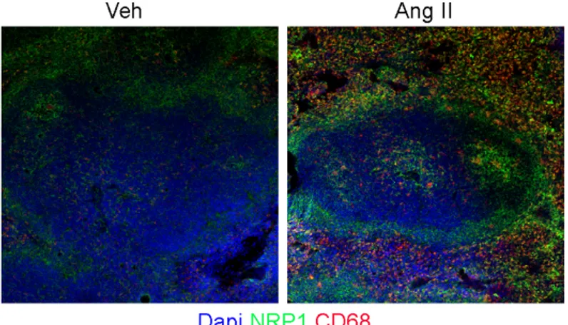
First of all, we evaluated the expression of Nrp1 in the spleen. The staining shows that Nrp1 is present in the spleen and is upregulated after infusion of Ang-II.
27
Figure 12: Nrp1 expression in the spleen.
Immunostaining of spleen from AngII and vehicle infused Nrp1fl/fl mice, showing Nrp1 co-localize in CD68+ MZ macrophages. Anti-CD169, anti-CD68 and anti-Nrp1 antibodies were used.
28 3.3.1 Cre-Lox Tissue-Specific Knockout
So to explore with better clarity Nrp1 and its probable PlGF receptor function in this AngII-induced hypertension model we generated a transgenic mouse with deletion for Nrp1.
Because the deletion of Nrp1 in germinal line is embryonically lethal, we generated a conditional knockout for Nrp1 using a mouse with a Cre recombinase under lysozyme M promoter. This Cre recombinase generates a conditional deletion of Nrp1 in monocyte-macrophage line when crossed with a mouse with Nrp1 gene flanked by LoxP sequences to allow the conditional cut of the sequence of our interest (Figure 15).
Figure 13: Mechanism of site-specific Cre recombination The loxP site consists of two inverted 13 bp Cre binding sites that surround a central 8 bp spacer. A light red arrow indicates the orientation of the loxP site. Cre recombinase is pictured in yellow. The model of the Cre-loxP recombination pathway consists of four Cre molecules form a synaptic tetramer. The tyrosines 324 from two of them cleave the DNA backbone. The released 5′ OH ends attack the partner strand to form a Holliday intermediate. A second round of cleavage and strand exchange results in the recombinant products C: If the two recombination sites are in the same orientation the strand exchange leads to excision or integration. If they are in the opposite orientation the outcome is an inversion Monocyte-macrophages Cre transgene Homozygous loxP “ floxed ” mouse Cre-lox mouse: homozigous for gene Nrp1 conditional knockout after 2 generations
29
In detail, we crossed mice LysMCre+/- heterozygous with Nrp1fl/fl.
Lys M Cre het Nrp1 fl/fl
Figure 14: Electrophoresis of PCR products.
Afterwards we crossed mice LysMCre+ Nrp1 heterozygous with mice LysM Cre-
Nrp1 heterozygous. Figure 15: Electrophoresis Electrophoresis showing PCR products from the mice used to ablate Nrp1 in monocytes/macrophages, allowing their genotype analysis.
We obtained in this way four useful genotypes LysMCre+ Nrp1fl/fl, LysMCre- Nrp1
fl/fl, LysMCre+ Nrp1wt/wt and LysMCre- Nrp1wt/wt.
700bp Mutant 350bp WT 398bp Mutant Mutant = ~700 bp Heterozygote = ~700 bp and ~350 bp Wild type = 350 bp Mutant = 398 bp Heterozygote = 183 bp and 398 bp Wild type = 183 bp
Lys M Cre het Nrp1 flox/+
Lys M +/+
30 3.3.2 Selective deletion of Nrp1 in macrophages
First of all we tested the efficiency of Cre recombinase in deleting Nrp1 in the monocyte/macrophage line by testing Nrp1 presence in monocyte-macrophage starting from total splenocytes by flow cytometry.
The FACS analysis shows that the signal isotype of Nrp1+ is suppressed in monocyte lineage in mouse LysM Cre+ Nrp1 fl/fl.
A. Nrp1+ Monocytes Nrp1+ Monocytes NRP1 WT/WT Lys M Cre -NRP1 WT/WT Lys M Cre+
31 B. Figure 16: FACS Analysys Outline of gating strategy for identification of Nrp1cells positive. Monocytes were selected by a gating on Cd11b+ cells. In this cell population, Nrp1+ monocytes were identified as a separated peak positive for the anti-Nrp1 antibody. In mice with deletion of Nrp1 in monocytes, the anti-Nrp1 peak was superimposed to the isotopic peak, indicating the Nrp1 was effectively deleted in LysM Nrp1 f/f Cre+ mice. (Unpublished data)
3.3.3 LysNrp1 fl/fl Cre+ mice were protected from hypertension
Once validated our model, we infused AngII for 28 days by osmotic pumps in the experimental groups showed above.
LysMCre- Nrp1fl/fl, LysMCre+ Nrp1wt/wt and LysMCre- Nrp1wt/wt mice respondend to the chronic infusion of AngII with an increase in blood pressure of about 30 mmHg, consistent with our previous observation in WT mice.
Nrp1+ Monocytes Deletion of Nrp1+ Monocytes NRP1 fl/fl Lys M Cre -NRP1 fl/fl Lys M Cre+
32
In contrast, LysM Cre+ Nrp1fl/fl mice, with selective deletion of Nrp1 in monocytes-macrophages, were unable to increase blood pressure in response to AngII.
These results strongly indicate that Nrp1 has a fundamental role in mediating the hypertensive response to AngII.
Figure 17: Noninvasive tail cuff. Noninvasive tail cuff monitoring of BP response to chronic AngII in LysMCre+ Nrp1 fl/fl versus WT mice (and respective vehicle infused mice). LysMCre+ Nrp1 fl/fl mice were protected from increase of blood pressure after chronic infusion of AngII (Unpublished data) -4 0 4 8 12 16 20 24 28 90 100 110 120 130 140 150 NRP1WT/WTLysMCre- AngII Ang II NRP1WT/WTLysMCre- Veh *** Time (days) SB P ( m m H g ) -4 0 4 8 12 16 20 24 28 90 100 110 120 130 140 150 NRP1WT/WTLysMCre+ AngII Ang II NRP1WT/WTLysMCre+ Veh *** Time (days) SB P ( m m H g ) -4 0 4 8 12 16 20 24 28 90 100 110 120 130 140 150 NRP1fl/flLysMCre- AngII NRP1fl/flLysMCre- Veh Ang II *** Time (days) SB P ( m m H g ) -4 0 4 8 12 16 20 24 28 90 100 110 120 130 140 150 NRP1fl/flLysMCre+ AngII NRP1fl/flLysMCre+ Veh Ang II Time (days) SB P ( m m H g )
33 3.3.4 Selective deletion of Neuropilin1 in macrophages inhibits
egression of T cells from spleen upon AngII.
Subsequently we analyzed the presence of immune cells in the white pulp (WP) area of the spleen, by evaluating staining for the CD3 antigen, marking lymphocytes, by immunohistochemistry, and by evaluating the signal of the CD4 and CD8 antigens, marking monocytes/macrophages, in WP by flow cytometry. Surprisingly the CD3 cells in the area of splenic white pulp (WP) were increased in LyMCre+ Nrp1fl/fl with AngII as compared to and LyMCre- Nrp1fl/fl treated in the same way (Fig. 18A).
Similarly, the flow cytometry analysis showed a reduction of
monocytes/macrophages in the WP of AngII-treated and LyMCre- Nrp1fl/fl, but
not in LyMCre+ Nrp1fl/fl mice (Fig.18 B-C).
Moreover, LyMCre- Nrp1fl/fl mice showed a reduced WP area after AngII infusion,
suggesting egression of lymphocyte cells from this region. In contrast, Nrp1-ablated mice lacked this response (Fig. 18A).
Overall, these data suggest impaired T-cells activation and egression from splenic reservoir toward inflamed target tissues.
These results indicate that the protection manifested by LyMCre+ Nrp1fl/fl in response to AngII is similar to the protection previously observed by the same research group in PlGF deficient mice (Carnevale et al., 2014).
A.
34 B. Nrp1 fl/fl LysMCre - Nrp1 fl/fl LysMCre + Veh AngII
35 C. Figure 18: Nrp1 is required for splenic T-cell egression.
(A)Immunostaining of spleen from LysMCre+Nrp1 fl/fl vs LysMCre- Nrp1 fl/fl , showing CD3+ white pulp, as delineated by counterstaining with B220 antigen recognizing red pulp. In LysMCre+Nrp1 fl/fl were a inhibiting the cell egression induced by AngII. (B) Flow cytometry analysis of CD8+ and CD4+ in splenocytes. (C) Graph showing the relative quantitative analysis of CD3+ area (Unpublished data)
Lymphocytes homing to target organs during hypertension has been shown to be a crucial step for both raise in BP and end organ damage (Guzik et al., 2007), suggesting that the hypertensive challenge involves innate and adaptive immune responses.
In the spleen, the innate and adaptive immune cells are organized to allow rapid sensing of challenges and consequently, molecular interactions that lead to differentiation and migration of T-cells (Mueller & Germain, 2009).
NRP1 fl/fl LysMCre - NRP1 fl/fl LysMCre + 0 30 60 90 120 150 180 210 VEH ANG II
**
***
Ar e a o f C D 3 + T *1 0 3 µm 2 *** P < 0.001 ** P < 0.0136 3.3.5 Selective deletion of neuropilin1 in macrophages protects from T-cells infiltration in kidneys upon AngII.
Immunohistochemistry for CD8+ and CD4+ T-cells in kidney, showed that
LyMCre+ Nrp1fl/fl mice were protected from infiltration of immune cells in target
organs in response to AngII supporting our previous observation.
A. NRP1 fl/fl LysMCre - NRP1 fl/fl LysMCre + 0 50 100 150 200
**
***
ANG II VEH CD8 + c ell s/ m m 237 B. Figure 19: Immunostaining and quantitative analysis of kidneys. (A)Immunostaining and quantitative analysis of CD8+ T-cells, (B) Immunostaining and quantitative analysis of CD4+ T-cells in kidneys from 4 weeks AngII. ***P<0.001 Cre- vs Cre+, **P<0.01 AngII vs. vehicle. (Unpublished data) NRP1 fl/fl LysMCre - NRP1 fl/fl LysMCre + 0 50 100 150 200
**
***
VEH ANG II CD4 + c ell s/ m m 238
Chapter IV:
Concluding Remarks
39 4.1 CONCLUDING REMARKS
The hypertension has a high incidence worldwide and it is one of the major risk factor for stroke, heart failure, kidney disease and other pathologies. In addition, the percentage of people who have a poorly controlled hypertension is very high and steadily increasing.
The focus of my PhD dissertation, a topic that has been arguing over the last few years, sees the immune system playing an important role in giving rise to high
blood pressure.
This thesis focuses on Placentar Growth Factor (PlGF) present in cardiovascular and immune system, which is known to be implicated in various pathologies, such as hypertension in pregnancy, the growth of blood vessels inside of tumors, in macular degeneration linked to age.
A study published on Immunity in 2014 by the research group in which I worked on the thesis has shown that PlGF is one of the main players in mediating the relationship in the immune system and hypertension and it is an important factor in the activation of T lymphocytes within the spleen and their subsequent migration to the blood vessels and to the organs that are typically damaged by hypertension. From this important premise my thesis starts.
We have set the goal of looking for the PlGF receptor involved in the interaction between immune system and arterial hypertension identified in the previous work. Our study focuses on the study of two possible PlGF receptors: the canonical receptor VEGFR1 and the non-canonical receptor Nrp1. Our results have led us to establish Nrp1 as possible receptor candidate.
In mice with conditional deletion of Nrp1 in the macrophage-monocyte line there is a protection against hypertension such as that observed in the
PlGF-/- model. We also observed, in this experimental group, a reduced egress of
the T-cells from the spleen and a reduced lymphocyte infiltrate in the target organs of pressure-induced injury.
40
In conclusion I can summarize by saying that VEGFR1-TK signaling is not required for the role of PlGF in AngII-induced hypertension; the expression of Nrp1 is indispensable in macrophages to allow blood pressure increase in response to AngII.
Moreover, the selective ablation of Nrp1 in monocytes-macrophages hampers T-cell egression from spleen and infiltration in kidney upon AngII.
41
Chapter V:
Material and Methods
42 5.1.1 Animals
All animal handling and experimental procedures were performed according to European Communities guidelines (EC Council Directive 2010/63) and the Italian legislation on animal experimentation (Decreto L.vo 26/2014). The protocol was approved by the Italian Ministry of Health (Permit number
58/2012-B). Mice of 12–15 weeks were used in all experiments. B6.129 (SJL)-Nrp1 tm2Ddg
/J mice (here indicated like Nrp1flox/flox ) were bought by The Jackson Laboratories
(Stock Number 005247). Mice of 8 weeks
B6.129P2-Lyz2tm1(cre)Ifo/J, B6.129(SJL)-Nrp1tm2Ddg/J and VEGFR1 tyrosine kinase knockout
mice (VEGFR1 TK−/− mice; 8 weeks old) with a C57BL/6 hybrid background. All
animals were provided with food and water ad libitum. According to our experience and current literature on the same experimental models (Carnevale & Lembo, 2012; Guzik et al., 2007; House, Potier, Bisaillon, Singer, & Trebak, 2008; Swirski et al., 2009; Vinh et al., 2010; Wenzel et al., 2011), the number of animals in each setting used was enough to determine whether there was a significant difference among groups, as tested by the software PS3. AngII (0.5 mg/kg/day) or vehicle (NaCl 0.9%) were delivered subcutaneously with osmotic mini-pumps (model 1007D, ALZET). At day 28, according to experimental design, mice were euthanized by overdose of sodium pentobarbital anesthesia.
43 5.1.2 Generation of transgenic mouse Nrp1flox/flox LysMCre
The Lyz2tm1(cre)Ifo/J homozygous mice were backcrossed with C57BL6J mice (wt
mice) to obtain mice bearing Cre recombinase in heterozygosis.
The mice born from these couplings were coded (see section 5.1.2.1) and a PCR was performed on the tail piece to check its genotype.
The primers used are:
Forward primer 5’ - CCC AGA AAT GCC AGA TTA CG - 3’; Reverse primer 5’- TTA CAG TCG GCC AGG CTG AC - 3’; Common primer 5’ - CTT GGG CTG CCA GAA TTT CTC - 3’. The PCR conditions used were:
The instrument used Proflex PCR system by Life Technologies.
20ul of PCR final product was loaded on 1.5% agarose gel with addition of 1ul of ethidium bromide and using the EZ Load 100bp as molecular marker. The electrophoretic run was performed for 30’ minutes and 90 Voltage in a chamber for electrophoretic run. The products obtained were visualized using the Biorad
Chemidoc ™ XRS+.
At the same time, we have crossed B6.129(SJL)-Nrp1tm2Ddg/J mice carrying the
loxP sites with C57BL6J mice to obtain Nrp1flox / + mice.
The primers used are:
Forward primer 5’ – AAG GAG TGG CAC AGC ATC TT- 3’;
44
The PCR conditions used were:
We selected the heterozygous LysM mice for Cre recombinase and the Nrp1 flox / +mice at two months of age were mated.
Lys M Cre het
Afterwards we crossed LysMCre+ Nrp1 heterozygous mice with LysMCre-
Nrp1 heterozygous mice. In this way, four useful genotypes (LysMCre+
Nrp1fl/fl, LysMCre- Nrp1 fl/fl, LysMCre+ Nrp1wt/wt and LysMCre- Nrp1wt/wt) were obtained.
5.1.2.1 Preparation of Genomic DNA from mouse tails.
DirectPCR Lysis Reagent (Tail): Cat # 101-T, 102-T 1. For 0.5 cm tail, add 200– 300 µl DirectPCR Lysis Reagent (Tail) containing freshly prepared 0.2-0.4 mg/ml Proteinase K (Sigma, cat # p6556, not included). Proteinase K is stable in DirectPCR reagents for ~24 hrs. If a small number of tails are processed, and
700bp Mutant 350bp WT
Mutant = ~700 bp
Heterozygote = ~700 bp and ~350 bp Wild type = 350 bp
45
therefore it is difficult to weigh Proteinase K powder, use genomic PCR-quality Proteinase K solution (Viagen, cat #501-PK) at 0.5-1.0 mg/ml (25-50 µl Proteinase K solution per 1 ml DirectPCR reagent). See Table 1 for starting conditions. NOTE: Although 200 µl DirectPCR is usually sufficient for complete lysis of 0.5 cm tail, application of 250-300 µl yields more reproducible results because of better mixing efficiency. Compare several different volumes of DirectPCR reagents for best performance. If tails are not mixed well with solutions, use 0.75 ml tubes. 2. Rotate the tubes in rotating hybridization oven at 55°C for 5-6 hrs or until no tissue clumps are observed. If necessary, rotation can be allowed overnight without loss of efficacy. Complete lysis is important. Since some tails may not be in contact with solutions, re-position once the tails by shaking the bottles containing tubes, preferentially after 2-3 hrs.
NOTE: Rotating hybridization oven performs better than rocking plate. Use 0.75
cm tubes for less than 200 µl of DirectPCR Reagent. DNA fragmentation by prolonged rotation will not influence significantly PCR performance. Use roughly proportional volume of DirectPCR Lysis Reagent for different sized samples. 3. Incubate crude lysates at 85°C for 45 min by floating the whole rack (containing tubes) on a water bath. (Optional) Precipitate hairs by centrifuging for 10 sec before step 4. Crude lysates may be stored at -20°C for 1 year (or at 4°C for 1 week) without losing efficacy. 4. Use 0.5-1.0 µl of lysate for 50 µl PCR reaction. Eppendorf Hotmaster Taq Polymerase (cat# 954-14-5018), Sigma JumpStart Taq DNA Polymerase (cat# D9307), or Qiagen HotStar Taq DNA polymerase (cat# 203203) is recommended for PCR. Rescue of DNA: DNA in crude lysates can be rescued for further analysis. Add NaCl to a final concentration of 250 mM, and then add 0.7 volume of isopropanol. DNA will form precipitates. Centrifuge at 4°C for 2 min, discard supernatant, wash DNA with 1 ml 70% EtOH, and dissolve DNA in 50 µl 10 mM Tris-HCl (8.0). Use 1 µl for PCR. Table 1. Suggested starting lysis conditions for mouse tails. Tails (cm) DirectPCR (µl) Dilution (fold) Lysates (µl) / 50 µl PCR rxn. 0.2-0.3 150-200 2 1.0-2.0 0.4 150-250 1 0.5-1.0 0.5 200-300 1 0.5-1.0 Related Products Products Description Cat # Price (US $) DirectPCR-Tail 500 mouse tails (100 ml) 102-T 139 DirectPCR-Ear 500 mouse ears (50 ml)
46
402-E 139 DirectPCR-Yolk sac Yolk sac (100 ml) 202-Y 139 DirectPCR-Cell Cultured cell (100 ml) 302-C 139 Proteinase K Solution All types of tissues (100 mg) 501-PK 99.
Important Technical Tips 1. Complete lysis. Big tissue clumps should not be observed after digestion. It is recommended to vigorously shake the bottle (containing microfuge tubes) for 2-3 sec anytime, once or twice, after tissues begin to partially dissolve. This will physically disperse partially digested tissues and reposition microfuge tube, in which tails are separated from lysis reagents, thereby facilitating overall lysis efficiency, 2. Proteinase K inactivation. Inactivation of proteinase K by incubating samples at 85C-86C for 45-50 min is critical to protect Taq polymerase from proteinase K. 3. Taq polymerase. We have tested many types of commercially available Taq polymerases. The listed enzymes are recommended for optimal results. 4. Tissue size. The size of tails should be 0.5 cm or slightly smaller. Use a minimal volume (0.5-1 µl for 50 µl PCR reaction) of crude lysates for PCR amplification. Too much DirectPCR reagents inhibit PCR efficiency. 5. Small tubes and evaporation. To minimize evaporation, use a 0.75 ml tube when the reagent volume is less than 100 µl. 6. Small samples and dilution. If the required DirectPCR reagent volume is less than 50 µl, dilute the reagent by up to 2-fold with water, while maintaining the same concentration of proteinase K. If the DirectPCR reagent is '2- fold' diluted, apply '2-fold' more crude lysates for PCR reaction. 7. PCR machine. PCR machines are occasionally a source of technical problems.
5.1.3 Blood-Pressure Measurements
Noninvasive blood pressure measurement was performed by tail-cuff plethysmography (BP-2000 Series II, Visitech Systems) in conscious mice daily for 2 hours (10 am–12 pm). Operators were blinded to the experimental group during blood pressure monitoring.
47 5.1.4 Isolation and Preparation of Single Cell Suspensions from Spleen, Kidney and Aorta
Mice were anesthetized with ketamine/xylazine and spleen was isolated. Then, mice were exsanguinated and both kidneys and aorta were collected. Spleen was mechanically disrupted and then passed through a 40 µm sterile filter (Falcon, BD) and centrifuged at 300× g for 10 min. After a treatment with Lysing Buffer (BD), the pellet was resuspended in PBS. Total splenic leukocytes were stained and analyzed using flow cytometry or used to isolate and enrich monocytes/macrophages by negative selection, using a commercially available monocyte enrichment kit (EasySep Kit and Purple EasySep Magnet, Stemcell Tech). Kidney cell suspension was obtained disrupting mechanically two decapsulated kidneys, and then passed through a 70 µm sterile filter (Falcon, BD). The resulted cell suspension was centrifuged at 300×g for 10 min. To isolate lymphocytes from cell suspension, the pellet was then suspended in 36% Percoll (Sigma), gently overlaid onto 72% Percoll, and centrifuged at 1,000× g for 30 min at RT. Aorta cell suspension was obtained disrupting mechanically the tissue and digesting the suspension in Digestion Cocktail, was then passed through a 70 µm sterile filter (Falcon, BD), and centrifuged at 300× g for 5 min to pellet the cells. Before proceeding with stainings for flow-cytometric analyses, lymphocytes from kidney and aorta total cells were enriched with Mouse T Lymphocyte Enrichment Set-DM (BD IMag). Samples were resuspended in PBS and then the number of the cells was assessed using trypan blue and an automated counter (Countess, Life Technologies).
5.1.5 Flow Cytometry Analysis
Splenic leukocytes (1 × 10⁶), enriched monocytes (2 × 10⁵), total kidney, and aorta lymphocytes were preincubated with anti-CD16/32 Fc receptor and then
48
incubated with various combinations of mAbs, washed with PBS, centrifuged at 300× g for 5 min at 4°C, and resuspended in FACS buffer. Immunofluorescence staining was analyzed using a C6 Accuri Flow Cytometer and BD FACSCanto (BD Biosciences). The data were analyzed using FlowJo Software.
5.1.5.1 Antibodies
The fluorochrome-conjugated mAbs to mouse antigens used for flow cytometry analysis were as follows: anti-CD16/CD32 (2.4G2), PE anti-CD86 (GL1), PE Isotype control Rat IgG2a, APC anti-CD11b (M1/70), PerCP-Cy5.5 anti-CD8a (53-6.7), FITC anti-CD8a (53-6.7), APC anti-CD3e (145-2C11), PerCP anti-CD45 (30-F11), FITC anti-CD4 (RM4-5), PE anti-CD4(RM4-5), APC anti-CD69 (H1.2F3), APC-Cy7 anti-CD45 (30-F11), FITC anti-F4/80 (FA-11) (AbD Serotec), and PerCP-Cy5.5 CD68 (FA-11) (BioLegend), APC anti-Neuropilin-1 (R&D).
5.1.6 Histological Analysis, Immunohistochemistry, and Immunofluorescence
Sections of kidney were deparaffinized, rehydrated, and then underwent antigen retrieval. Slides were processed for immunohistochemistry with the following primary antibodies: anti-CD8 and anti-CD4 (1:50; BD PharMingen). Appropriate biotinylated secondary antibodies (1:200; Vector) were used for successive processing with DAB (Vector). Hematoxylin (Sigma) was used as counterstaining. The number of CD8 and CD8-positive cells per mm2 was determined using the Image J software Cell Counter plugin analysis tool.
For immunofluorescence analysis, 25 µm sections from spleen were postfixed with 4% paraformaldehyde for 15 min and processed for staining with the following primary antibodies: rabbit anti-PlGF (Abcam) 1:250, rabbit-anti CD86 (Novus
49
Biologicals) 1:100, rat anti-mouse CD68 (AbD Serotec) 1:200, rat anti mouse CD169 (AbD Serotec) 1:100, Sheep-anti Tyrosine Hydroxylase (Millipore) 1:800, rat anti-mouse CD45R (B220, BD PharMingen) 1:50, anti-hamster CD3 (BD PharMingen) 1:50, rabbit anti VEGFR1 (R&D) 1:50, rabbit anti Nrp1 (R&D) 1:50 and rat anti-ER-TR7 (Acris) 1:200. Sections were then incubated with the respective secondary antibodies conjugated to Alexa Fluor 488, Alexa Fluor 647 or Cy3. Slides were then coverslipped with DAPI containing medium (Vector).
5.1.7 Confocal Microscopy Analysis
All coverslipped mounted tissue sections were scanned using a Zeiss 780 confocal laser scanning microscope with a Zeiss ECPLAN-NEOFLUAR 10x/0.30 M27, ECPLAN-NEOFLUAR 20x/0.50 M27, ECPLAN-NEOFLUAR 40×/1.30 M27 oil immersion objective or PLAN-APOCHROMAT 63×/1.4 oil DIC oil immersion objective (Carl Zeiss Microimaging Inc.). We used a 405 Diode laser to excite DAPI, a 488 nm argon laser to excite Alexa Fluor 488, and a 543 HeNe laser to excite Cy3. Pseudocoloring was performed using ZEN software (Carl Zeiss Microimaging)
5.1.8 Bright-field Microscopy Analysis
All images were captured using a DMI3000B Leica optical microscope provided of Leica cameras (Leica Microsystems) and processed with the Leica Application Suite (LAS V3.3).
50 5.1.9 Statistical Analysis
All the experiments were replicated within the laboratory. Sample size was pre-estimated from the previously published research and from pilot experiments performed in the laboratory. Data are presented as mean±s.e.m. Data distribution was assessed with the Shapiro–Wilk normality test and D'Agostino Pearson test, and assumption of homogeneity of variance was tested using Levene's test of equality of variances. For amplitude gain analysis, unequal variance between groups was observed in a minority of cases, and a Welch correction was performed for all comparisons. Statistical significance was assessed with the appropriate test according to each experimental design, as detailed in figure legends. After confirming that all data had normal distributions, we applied Student t-test for either independent samples or paired samples, according to the experimental design and as specified in the figure legends. Multiple group analysis was performed with two-way ANOVA followed by Bonferroni's post hoc. Analysis for repeated measures was applied when required by the experimental setting. P<0.05 was considered significant. Statistical analyses were performed with SPSS 23.0 (IBM Software) and graphs were made with GraphPad Software PRISM5.
51
Chapter V:
52
Achen, M. G., Gad, J. M., Stacker, S. A., & Wilks, A. F. (1997). Placenta growth factor and vascular endothelial growth factor are co-expressed during early embryonic development. Growth Factors (Chur, Switzerland), 15(1), 69–80. Retrieved from http://www.ncbi.nlm.nih.gov/pubmed/9401819
Ahmed, A., Dunk, C., Ahmad, S., & Khaliq, A. (2000). Regulation of placental vascular endothelial growth factor (VEGF) and placenta growth factor (PIGF) and soluble Flt-1 by oxygen - A review. Placenta, 21(SUPPL.1), S16-24. http://doi.org/10.1053/plac.1999.0524
Ahn, K. O., Lim, S. W., Li, C., Yang, H. J., Ghee, J. Y., Kim, J. Y., … Yang, C. W. (2007). Influence of angiotensin II on expression of toll-like receptor 2 and maturation of dendritic cells in chronic cyclosporine nephropathy.
Transplantation, 83(7), 938–47.
http://doi.org/10.1097/01.tp.0000258589.39006.94
Autiero, M., Luttun, A., Tjwa, M., & Carmeliet, P. (2003). Placental growth factor and its receptor, vascular endothelial growth factor receptor-1: novel targets for stimulation of ischemic tissue revascularization and inhibition of angiogenic and inflammatory disorders. Journal of Thrombosis and
Haemostasis : JTH, 1(7), 1356–70. Retrieved from http://www.ncbi.nlm.nih.gov/pubmed/12871269
Bagri, A., & Tessier-Lavigne, M. (2002). Neuropilins as Semaphorin receptors: in vivo functions in neuronal cell migration and axon guidance. Advances in
Experimental Medicine and Biology, 515, 13–31. Retrieved from
http://www.ncbi.nlm.nih.gov/pubmed/12613540
Barleon, B., Sozzani, S., Zhou, D., Weich, H. A., Mantovani, A., & Marmé, D. (1996). Migration of human monocytes in response to vascular endothelial growth factor (VEGF) is mediated via the VEGF receptor flt-1. Blood, 87(8), 3336–43. Retrieved from http://www.ncbi.nlm.nih.gov/pubmed/8605350 Bataillard, A., Freiche, J. C., Vincent, M., Sassard, J., & Touraine, J. L. (1986).
Antihypertensive effect of neonatal thymectomy in the genetically hypertensive LH rat. Thymus, 8(6), 321–30. Retrieved from http://www.ncbi.nlm.nih.gov/pubmed/3492791
53
Bielenberg, D., Pettaway, C., Takashima, S., & Klagsbrun, M. (2006). Neuropilins in neoplasms: Expression, regulation, and function ☆ . Experimental Cell
Research, 312(5), 584–593. http://doi.org/10.1016/j.yexcr.2005.11.024
Bomfim, G. F., Rodrigues, F. L., & Carneiro, F. S. (2017). Are the innate and adaptive immune systems setting hypertension on fire? Pharmacological
Research, 117, 377–393. http://doi.org/10.1016/j.phrs.2017.01.010
Bravo, Y., Quiroz, Y., Ferrebuz, A., Vaziri, N. D., & Rodríguez-Iturbe, B. (2007). Mycophenolate mofetil administration reduces renal inflammation, oxidative stress, and arterial pressure in rats with lead-induced hypertension. American
Journal of Physiology. Renal Physiology, 293(2), F616-23. http://doi.org/10.1152/ajprenal.00507.2006
Brentano, F., Kyburz, D., Schorr, O., Gay, R., & Gay, S. (2005). The role of Toll-like receptor signalling in the pathogenesis of arthritis. Cellular Immunology,
233(2), 90–96. http://doi.org/10.1016/j.cellimm.2005.04.018
Buford, T. W. (2016). Hypertension and aging. Ageing Research Reviews, 26, 96– 111. http://doi.org/10.1016/j.arr.2016.01.007
Carmeliet, P., Moons, L., Luttun, A., Vincenti, V., Compernolle, V., De Mol, M., … Persico, M. G. (2001). Synergism between vascular endothelial growth factor and placental growth factor contributes to angiogenesis and plasma extravasation in pathological conditions. Nature Medicine, 7(5), 575–583. http://doi.org/10.1038/87904
Carnevale, D., Cifelli, G., Mascio, G., Madonna, M., Sbroggiò, M., Perrino, C., … Lembo, G. (2011). Placental growth factor regulates cardiac inflammation through the tissue inhibitor of metalloproteinases-3/tumor necrosis factor-α-converting enzyme axis: crucial role for adaptive cardiac remodeling during
cardiac pressure overload. Circulation, 124(12), 1337–50.
http://doi.org/10.1161/CIRCULATIONAHA.111.050500
Carnevale, D., & Lembo, G. (2012). Placental growth factor and cardiac inflammation. Trends in Cardiovascular Medicine, 22(8), 209–12. http://doi.org/10.1016/j.tcm.2012.07.022
54
… Lembo, G. (2014). The angiogenic factor PlGF mediates a neuroimmune interaction in the spleen to allow the onset of hypertension. Immunity, 41(5), 737–52. http://doi.org/10.1016/j.immuni.2014.11.002
Chung, E. S., Packer, M., Lo, K. H., Fasanmade, A. A., & Willerson, J. T. (2003). Randomized, double-blind, placebo-controlled, pilot trial of infliximab, a chimeric monoclonal antibody to tumor necrosis factor-alpha, in patients with moderate-to-severe heart failure: results of the anti-TNF Therapy Against Congestive Heart Failure (AT. Circulation, 107(25), 3133–40. http://doi.org/10.1161/01.CIR.0000077913.60364.D2
Crowley, S. D., Song, Y.-S., Lin, E. E., Griffiths, R., Kim, H.-S., & Ruiz, P. (2010). Lymphocyte responses exacerbate angiotensin II-dependent hypertension. AJP: Regulatory, Integrative and Comparative Physiology,
298(4), R1089–R1097. http://doi.org/10.1152/ajpregu.00373.2009
Cui, J. Z., Hinz, B. J., Greve, M. D. J., Potter, M. J., Hornan, D., Samad, A., … Matsubara, J. A. (2003). Expression of neuropilin-1 in choroidal neovascular membranes. Canadian Journal of Ophthalmology. Journal Canadien
D’ophtalmologie, 38(1), 41–5. Retrieved from
http://www.ncbi.nlm.nih.gov/pubmed/12608516
De Batista, P. R., Palacios, R., Martín, A., Hernanz, R., Médici, C. T., Silva, M. A. S. C., … Alonso, M. J. (2014). Toll-like receptor 4 upregulation by angiotensin II contributes to hypertension and vascular dysfunction through reactive oxygen species production. PloS One, 9(8), e104020. http://doi.org/10.1371/journal.pone.0104020
De Ciuceis, C., Amiri, F., Brassard, P., Endemann, D. H., Touyz, R. M., & Schiffrin, E. L. (2005). Reduced vascular remodeling, endothelial dysfunction, and oxidative stress in resistance arteries of angiotensin II-infused macrophage colony-stimulating factor-deficient mice: evidence for a role in inflammation in angiotensin-induced vascular injury. Arteriosclerosis,
Thrombosis, and Vascular Biology, 25(10), 2106–13. http://doi.org/10.1161/01.ATV.0000181743.28028.57