Case Report
Laparoendoscopic Single-Site Surgery for Treatment
of Concomitant Ovarian Cystectomy and Cholecystectomy
Daniela Surico, MD
*
, Sergio Gentilli, MD, Alessandro Vigone, MD, Eleonora Paulli, MD,
Livio Leo, MD, and Nicola Surico, MD
From the Advanced Gynaecological Oncology Centre, Department of Obstetrics and Gynaecology (Drs. D. Surico, Vigone, Paulli, Leo, and N. Surico), and Department of Surgery (Dr. Gentilli), University of Eastern Piedmont, Novara, Italy.
ABSTRACT Since the first laparoscopic appendectomy was performed by Semm in 1983, laparoscopic surgery has become the criterion stan-dard surgical route for treatment of several pathologic conditions across disciplines. Attempts to minimize access-related injuries and complications resulted in development of laparoendoscopic single-site surgery (LESS), which, because of the decreased num-ber of ports used, may be the next generation of minimally invasive surgery. Laparoscopic single-site surgery has been reported in nephrectomy, pyeloplasty, radical prostatectomy, cholecystectomy, and colorectal, bariatric, and gynecologic surgery. This tech-nique may increase the benefits of traditional minimally invasive surgery such as decreased blood loss and postoperative pain, faster recovery time, fewer complications, and better cosmetic results, without increasing costs. Herein, we present a case report of single-port laparoscopic ovarian cystectomy and concomitant cholecystectomy performed with a multi-instrument access port (TriPort; Olympus America Inc., Center Valley, Pennsylvania). Single-port surgery eliminates the problem of multiple and different site placement for accessory ports, typical of these procedures when performed simultaneously at conventional laparos-copy. Journal of Minimally Invasive Gynecology (2010) 17, 656–659Ó 2010 AAGL. All rights reserved.
Keywords: Concomitant cholecystectomy; Laparoscopy; Ovarian cystectomy; Single port
Laparoscopy is now accepted as the criterion standard for treatment of benign ovarian cysts and benign gallbladder diseases, and compared with the traditional laparotomy approach, has several advantages including minimal skin in-cisions, short hospitalization, faster postoperative recovery, decreased hospital costs, and better cosmetic outcome.
Gynecologic surgery and cholecystectomy can be per-formed simultaneously using traditional laparoscopy, provid-ing the patient a sprovid-ingle hospitalization, reduced anesthesia exposure, and decreased total operative morbidity, and operative time is the same as the total time for the 2 procedures performed separately [1]. Common laparoscopic surgery requires skin incisions for multiple trocars, and for each
incision, there is risk of trocar-site bleeding, wound infection, de novo adhesion formation, and herniation[2,3].
Recent advances in surgical instrumentation have further minimized morbidity and improved cosmesis of laparoscopic surgery. These include minilaparoscopy, natural orifice trans-lumenal endoscopic surgery (NOTES), and laparoendoscopic single-site surgery (LESS). LESS has been reported for colo-rectal, bariatric, urologic, and gynecologic surgery[3,4].
Since the first single-wound laparoscopic cholecystectomy was reported in 1997, several published works have supported the feasibility of the technique[4,5]. In gynecology, LESS has been reported for tubal ligation, salpingectomy, removal of adnexal tumors, hysterectomy, and pelvic and paraaortic node dissection [2,4,6,7]. In both general and gynecologic endoscopy, during conventional laparoscopy, a 10-mm trocar is inserted through the umbilicus to pass the camera. In laparoscopic cholecystectomy, two 5-mm trocars are placed in a subxiphoid and right subcostal position, whereas in ovarian cystectomy two 5-mm ports are placed lateral to the inferior epigastric vessels bilaterally, as well as one 5-or 10-mm suprapubical port [8,9]. Because it is technically challenging for gynecologists to perform ovarian cystectomy using the ports positioned by surgeons for cholecystectomy,
The authors have no commercial, proprietary, or financial interest in the products or companies described in this article.
Corresponding author: Daniela Surico, MD, Department of Obstetrics and Gynaecology, ‘‘Maggiore della Carita`’’ Hospital, Viale Mazzini 18, 28100 Novara, Italy.
E-mail:[email protected]
Submitted February 2, 2010. Accepted for publication May 6, 2010. Available atwww.sciencedirect.comandwww.jmig.org
1553-4650/$ - see front matterÓ 2010 AAGL. All rights reserved. doi:10.1016/j.jmig.2010.05.005
and vice versa, when simultaneous interventions are required, more ports are often placed, with worse cosmetic outcome[1]. To our knowledge, this is the first clinical report of laparo-scopic ovarian cystectomy and concomitant cholecystectomy performed using a multi-instrument access port (TriPort; Olympus America Inc., Center Valley, PA), a recently developed commercially available single port that enables as many as 3 instruments to be inserted simultaneously through a single incision, with virtually scarless transumbilical access.
Case Report
A 26-year-old woman, gravida 1, para 1, with body mass index of 35.6 kg/m2, came to our emergency service with worsening dysmenorrhea and left lower quadrant pain. Her medical history was significant for a previous open appendectomy.
Pelvic examination revealed a left adnexal cyst mass. Labo-ratory investigations revealed negative findings of a pregnancy test, normal C-reactive protein concentration and white blood cell count, and increased CA125 concentration (38.8 U/mL). At transvaginal ultrasound examination, the left ovary was enlarged due to a unilocular cyst, with fine internal echoes (ground-glass appearance) and a maximum diameter of 83 mm. The patient reported a history of right upper quadrant ab-dominal pain radiating to the shoulder and associated with nausea and flatulence, with multiple previous visits to our surgical emergency service. Initial abdominal ultrasonogra-phy revealed the presence of multiple gallstones, with normal gallbladder wall thickness; absence of pericholecystic fluid; and normal sized common bile duct. As a result of this finding, she had already been scheduled to undergo elective laparoscopic cholecystectomy.
After discussion of the case with the general surgeon, it was agreed to perform concomitant cholecystectomy and ovarian cystectomy via single-port laparoscopic surgery. The operative approach was approved by the ethical commit-tee of the local hospital. Informed consent for the combined procedure was obtained from the patient after giving detailed information that risks, benefits, and indications for LESS are the same as for a traditional combined laparoscopic proce-dure except for possible better cosmetic outcome. The patient was also aware that she would be the first to undergo the specific combined procedure using the TriPort device.
With the patient under general anesthesia, a uterine manipulator was placed to optimize visualization of the ad-nexal cyst. The gynecologic surgeon and the assistant stood on the left and right sides of the patient, respectively, with the monitor in front of them. A 1.5-cm intraumbilical vertical skin incision and a 2-cm rectus fasciotomy were made to enter the peritoneal cavity and introduce the TriPort device. The Tri-Port is a sterile single-use device that consists of an introducer, a retractor, and a valve. Within the valve, 3 separate channels enable insertion of one 10-mm and two 5-mm (or three 5-mm) laparoscopic instruments. A separate port provides carbon dioxide insufflation. The retractor is formed by a distal ring,
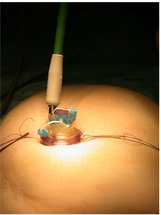
a removal external ring attached to the inner ring, a proximal ring, and a doubled-over cylindrical plastic sleeve. Once the distal ring is loaded into the introducer, the surgeon can insert the TriPort into the abdominal cavity through the skin-fascia incision. By pressing a button on the introducer, the distal ring is ejected, and the introducer can be removed. The surgeon just has to pull up on the sleeve and push down on the proximal ring to secure port fixation in the abdominal wall, which is completed by gently pulling the removal external ring. Excess sleeve is cut off, and the insufflation line is attached to the appropriate valve (Fig.1).
Pneumoperitoneum with pressure set at 12 mm Hg was created, and the patient was placed in the Trendelenburg po-sition. Laparoscopy was performed using a 0-degree 10-mm laparoscope (Olympus Medical Systems, Tokyo, Japan), and confirmed the presence of the left adnexal cyst with 90-mm diameter adherent to the ovarian fossa, without suspected peritoneal or abdominal organ lesions. To save time and reduce instrument crowding, the procedure was performed using at least 1 precurved instrument (prototype of Olympus Medical Systems) (Fig. 2).
Adhesions were released with a 5-mm precurved grasper used in the surgeon’s left hand and a rigid scissors. The lapa-roscopic cyst resection was performed using the classic tech-nique. A cortical incision was made, a cleavage plane identified, and the cyst enucleated and stripped from the normal ovarian tissue using bilateral traction[9]. In this phase, the surgeon operated using one 5-mm precurved grasper in the
Fig. 1. TriPort device (Olympus America Inc.) in situ with insufflation line attached.
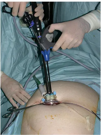
subordinate hand and a 5-mm rigid scissors or Manhes grasper in the right hand (Fig. 3). The ovarian fossa nodule adherent to the ovary was excised using the precurved grasper and a rigid monopolar scissors. Hemostasis was achieved with 5-mm conventional bipolar forceps applied on the ovarian parenchyma. The specimen was delivered through the umbi-licus, and the TriPort was replaced without difficulty.
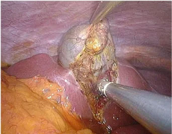
For the laparoscopic cholecystectomy, the patient was placed in a reverse Trendelenburg position with the left side rotated downward. The general surgeon stood between the opened legs, and the assistant stood on the left side of the patient; the monitor was placed on the opposite side of the patient. Dissection of the cystic pedicle and separation of the cystic duct from the cystic artery were achieved using a precurved grasper and 5-mm bipolar forceps. The cystic artery and the cystic duct were then clipped and divided using scissors. Dissection of the gallbladder from the liver bed was achieved using a unipolar electrode (Fig. 4). At the end of the procedure, the specimen was delivered through the umbilicus without the need for wound extension. The surgeon removed the port simply by pulling on the removal ribbon attached to the inner ring, and closed the fascia and the skin of the wound with sutures.
Single-port simultaneous ovarian cystectomy and chole-cystectomy was successfully completed without the need of extraumbilical skin incision or conversion to standard lapa-roscopy. Histologic evaluation confirmed the presence of cholelithiasis and an ovarian endometrioid cyst. Operative time was 132 minutes, estimated blood loss was 35 mL, and no intraoperative complications occurred. Postoperative recovery was uneventful, and pain control was achieved with a single dose of diclofenac sodium, 1 mg/kg. The visual an-alog scale scores for pain were 6/10 at 2 hours after surgery, 3/10 at 10:00PM, and 2/10 at discharge.
The patient was discharged to home 24 hours after sur-gery, and no subsequent complications were observed at follow-up 1 month after surgery. The cosmetic result was excellent (Fig. 5), and the patient was highly satisfied.
Discussion
Laparoscopy is considered the best surgical route for man-agement of benign adnexal masses and gallbladder diseases, and is typically performed using 4 ports. The development of technical expertise and advances in surgical instrumentation led to a search for less invasive alternatives that decrease the number of ports used and improve cosmetic outcome. These include minilaparoscopy, NOTES, and LESS[2,3].
In 2005, Ghezzi et al [10]reported that 3-mm ancillary trocars could replace traditional-sized equipment without a negative effect on surgeon ability to perform gynecologic laparoscopy. In that study, the authors did not address patient satisfaction with cosmetic appearance; however, there are articles in the literature that point out how the cosmetic result is better in patients undergoing minilaparoscopy compared with standard laparoscopy [11]. Because minilaparoscopy downsizes the port incisions but cannot eliminate them, dur-ing the last few years there has been increasdur-ing enthusiasm for NOTES, which eliminates abdominal incisions and related complications. However, NOTES involves viscerot-omy that is often difficult to close, with potential major complications, and requires many special instruments[3].
In contrast with NOTES, in LESS, no deliberate viscerot-omy is created, and the location of the single port in the umbi-licus is virtually as scarless as with NOTES. LESS has been reported for nephrectomy, pyeloplasty, radical prostatectomy, cholecystectomy, and colorectal, bariatric, and gynecologic surgery[4–7]. In 2009, LESS was demonstrated to be a safe
Fig. 2. Precurved grasper.
Fig. 3. External view of instruments during cyst enucleation. The surgeon uses a precurved grasper and a rigid scissors.
and feasible approach, with better cosmetic outcome, for cholecystectomy[2]and resection of adnexal tumors[5].
To perform conventional laparoscopic cholecystectomy, general surgeons place the primary trocar through the umbi-licus, and two 5-mm trocars in a right and left subcostal position, respectively, to enable retraction of the gallbladder, preparation of the Calot triangle, and dissection of the cystic duct and artery, with attention to possible anatomical abnor-malities. In contrast, the position of accessory ports preferred by gynecologists is different; two 5-mm ports are usually inserted lateral to the inferior epigastric vessels bilaterally, and one 5- or 10-mm port suprapubically.
In interventions performed simultaneously by a surgeon and a gynecologist, there is need for an increased number of ports, as many as 6, to enable safe and ergonomic comple-tion of each procedure. Addicomple-tional skin incisions involve the risk of wound complications such as bleeding, herniation, and infection, and poorer cosmetic outcome, and may increase postoperative pain and analgesic requirements.
The present case demonstrates that use of the TriPort system for concomitant ovarian cystectomy and cholecystec-tomy is not only safe and feasible but also highly cosmetic be-cause it can overcome the problem of multiple-port placement characteristic of such procedures when performed at conven-tional laparoscopy. These advantages are amplified in obese patients, as suggested by Langwieler et al[5], who hypothe-sized that LESS is especially suitable in these patients because of fewer incision-related postoperative complications. The body mass index in our patient was 35.6 kg/m2.
However, LESS is difficult to perform because of clashing of instruments and laparoscope, and loss of triangulation. To offset this problem, we suggest the following. (1) Use 1 curved and 1 straight instrument, which enables triangulation of work-ing instruments. (2) If the surgeon operates standwork-ing on a 20-cm footboard using instruments of different lengths, external work-ing space is optimized. (3) Internal crowdwork-ing of instruments can be minimized by using the EndoEYE (Olympus America Inc.) and curved instruments that can be crossed during dissection.
Conclusion
Use of a single-port device when performing simulta-neous cholecystectomy and gynecologic interventions may have important advantages in terms of risk of complications, postoperative pain, and cosmetic appearance. Comparisons with similar cases performed using traditional laparoscopy and case series are needed to better define the advantages of LESS in concomitant procedures.
References
1. Wadhwa A, Chowbey PK, Sharma A, Khullar R, Soni V, Baijal M. Combined procedures in laparoscopic surgery.Surg Laparosc Endosc Percutan Tech. 2003;13:382–386.
2. Lim MC, Kim TJ, Kang S, Bae DS, Park SY, Seo SS. Embryonic natural orifice transumbilical endoscopic surgery (E-NOTES) for adnexal tu-mors [published online ahead of print April 3, 2009]. Surg Endosc. 2009;23:2445–2449.
3. Desai MM, Rao PP, Aron M, et al. Scarless single port transubilical nephrectomy and pyeloplasty: first clinical report.BJU Int. 2008;101:83–88. 4. Romanelli JR, Earle DB. Single-port laparoscopic surgery: an overview.
Surg Endosc. 2009;23:1419–1427.
5. Langwieler TE, Nimmesgern T, Back M. Single-port access in laparo-scopic cholecystectomy.Surg Endosc. 2009;23:1138–1141.
6. Fader AN, Escobar PF. Laparoendoscopic single-site surgery (LESS) in gynaecologic oncology: technique and initial report.Gynecol Oncol. 2009;114:157–161.
7. Rettenmeier MA, Abaid LN, Erwin MR, et al. A retrospective review of the GelPort system in single-port access pelvic surgery. J Minim Invasive Gynecol. 2009;16:743–747.
8. Leggett PL, Bissell CD, Churchman-Winn R, Ahn C. Three-port microla-paroscopic cholecystectomy in 159 patients.Surg Endosc. 2001;15:293–296. 9. Muzii L, Bellati F, Palaia I, et al. Laparoscopic stripping of endometrio-mas: a randomized trial on different surgical techniques; part I, clinical results.Hum Reprod. 2005;20:1981–1986.
10. Ghezzi F, Cromi A, Columbo G, et al. Minimizing ancillary ports size in gynecologic laparoscopy: a randomized trial.J Minim Invasive Gynecol. 2005;12:480–485.
11. Bisgaard T, Klarskov B, Trap R, Kehlet H, Rosenberg J. Microlaparo-scopic vs conventional laparoMicrolaparo-scopic cholecystectomy: a prospective ran-domized double-blind trial [published online ahead of print November 16, 2001].Surg Endosc. 2002;16:458–464.
Fig. 4. Dissection of gallbladder from liver bed.
Fig. 5. Photograph obtained at 4-weeks follow-up.