Correlation between Specific Bacterial Groups in the Oral Cavity and the
Severity of Halitosis: Any Possible Beneficial Role for Selected Lactobacilli?
Mario Del Piano1*, Marco Balzarini1, Michela Pagliarulo1, Mario Migliario2, Filomena Sforza3, Luca Mogna4 and Giovanni Mogna51Gastroenterology Unit, Maggiore Della Carità Hospital, Novara, Italy 2Dentistry Specialist, Maggiore Della Carità Hospital, Novara, Italy 3Private Hospital “I Cedri”, Fara Novarese, Italy
4Biolab Research Ltd., Novara, Italy 5Probiotical SpA, Novara, Italy
*Corresponding author: Mario Del Piano, Gastroenterology Department, Maggiore della Carità Hospital, Novara Corso Mazzini, 18-28100 Novara, Italy, Tel: +39 335 6225686; Fax: +39 0321 32466; E-mail: [email protected]
Received date: May 22, 2014, Accepted date: July 08, 2014, Published date: July 15, 2014
Copyright: © 2014 Del Piano M, et al. This is an open-access article distributed under the terms of the Creative Commons Attribution License, which permits unrestricted use, distribution, and reproduction in any medium, provided the original author and source are credited.
Abstract
Objective: Halitosis is a widespread problem, normally attributable to specific volatile sulphur compounds (VSC)
in the breath. The aim of this study was to first relate halitosis with possible gastric infection by Helicobacter pylori and secondly to quantify specific bacterial groups in the oral cavity flora, thus correlating them with VSC concentrations and Proton Pump Inhibitors (PPIs) intake. Four selected lactobacilli were then assessed in the possible reduction of halitosis in subjects with a total salivary bacterial concentration higher than 105 CFU/ml.
Methods: Specific bacterial groups, namely total bacteria, total coliforms, sulphite-reducing bacteria (SRB) and
lactobacilli, were quantified in samples of saliva from 29 subjects taking PPIs compared with 36 control subjects. The amount of the three VSC hydrogen sulfide (H2S), methyl mercaptan (CH3SH) and dimethyl sulfide (CH3)2S in the breath and the presence of H. pylori were determined.
Results: No significant correlation was found between H. pylori and halitosis as well as with PPIs intake. The
baseline bacterial groups quantification (log10 CFU/ml of saliva, PPI group vs. control) showed: total bacteria 8.44 vs. 4.47 (p=0.001); total coliforms 4.95 vs. 2.82 (p=0.001); sulfite-reducing bacteria 5.47 vs. 2.58 (p=0.052); total lactobacilli 4.00 vs. 2.36 (p=0.016). After 15 days of lactobacilli supplementation, the same parameters (d15 vs baseline) gave: total bacteria 7.92 vs. 8.44 (p=0.019); total coliforms 3.13 vs. 4.95 (p=0.001); sulfite-reducing bacteria 4.69 vs. 5.47 (p=0.047); total lactobacilli 7.86 vs. 4.00 (p=0.048). No statistically significant differences were noted in VSC concentrations at any time.
Conclusions: The intake of PPIs directly correlated with the overgrowth of specific bacterial groups in the oral
cavity, but there was no correlation with H. pylori or with VSC concentration. The significant reduction in all the bacterial groups analysed after two weeks suggested the improvement of the overall oral flora in subjects chronically treated with PPIs.
Keywords: Proton Pump Inhibitors (PPIs); Halitosis; Helicobacter pylori; Bacterial overgrowth; Lactobacilli; Oral flora; Volatile Sulphur Compounds (VSC).
Introduction
Halitosis, or bad breath, is a really widespread problem and apprehension for it is valued to be the third most frequent reason for people to seek dental care, following dental caries (tooth decay) and periodontal disease. It is reported that about 25% of the general population suffers from it to some extent [1]. In any case, the amount of epidemiological research on bad breath is limited, since this topic is still a large but underestimated taboo. A public investigation in 2005 in The Netherlands showed that halitosis was one of the 100 biggest human overall exasperations (TNS-NIPO).
Men and women seem to suffer in the same proportions, whereas women seem to seek faster for professional help than men [2].
Miyazaki found that there is a clear correlation between age and oral malodour: the older one gets, the more intense the odour will become [3].
Of those people who have halitosis, about 90% of the time the odour is caused by something in the mouth, commonly odours released by bacteria present below the gumline and on the back of the tongue [4]. The remaining overall 10% is accounted for by many different conditions, including disorders in the nose, sinuses, throat, lungs, oesophagus, stomach or elsewhere [5]. Since halitosis is a social taboo, psychological or social problems could often develop, such as anxiety and depression, low self-esteem or other mood disorders.
Diagnostics of halitosis includes subjective methods (examiner’s sense of smell) and objective methods (instrumental analysis of specific molecules in the breath). Simple, subjective examination is regarded as a “golden standard” in clinical practice. In case of pathological halitosis recognizing the direct cause is crucial. After
excluding, or after successful treatment, of all oral pathologies, in case of remaining fetor ex ore identification and treatment of bad breath often requires multidisciplinary approach [4].
There is an extensive list of possible causes of halitosis in the mouth alone, however by far the most prevalent reasons reported are halitogenic biofilm on the posterior dorsal tongue and within gingival crevices and periodontal pockets [6]. The dorsum of the tongue, which is irregular and has a surface of 25 cm2 is an ideal niche for oral
bacteria [7]. Since desquamating epithelial cells and food remnants are available, putrefaction occurs. Hence, the tongue surface seems to be an important reservoir in the recolonisation of tooth surfaces [8]. Poor oral hygiene, dental plaque, dental caries, accumulation and putrefaction of food remnants and unclean acrylic dentures (worn at night or not regularly cleaned or with rough surfaces) contribute to bad breath [9].
The putrefactive activity in the mouth may be attributable to the proteolysis of sulphur containing amino acids in dietary and salivary proteins by mostly anaerobic, Gram-negative bacterial species [5,10]. There are over 600 types of bacteria documented in the average mouth. The odors are generated mainly by the hydrolysis of proteins into individual amino acids, followed by the additional breakdown of some of them to produce measurable foul gases. Due to this progression, volatile sulphur compounds (VSCs) are formed. The most important VSCs involved in halitosis are hydrogen sulfide (H2S),
methyl mercaptan (CH3SH) and dimethyl sulfide (CH3)2S. It is well
known, for example, that cysteine and methionine can produce H2S
and CH3SH, respectively [11]. VSCs have been shown to be
statistically associated with oral malodor levels, and usually decrease following successful treatment.
Other molecules involved in this bacterial degradation process are: diamines (indole and skatole) or polyamines (cadaverine and putrescine). They seem to play a less important role in the expression of bad breath. The main substrate for skatole and indole production is tryptophan, whereas lysine and ornithine are the basis for the putrescine/cadaverine production [5].
Most of the responsible microorganisms in halitosis are involved in periodontitis. In this way, there is a positive correlation between bad breath and periodontitis [12].
It could be hypothesized also a correlation between the composition of oral cavity microbiota and possible bacterial overgrowth in the stomach and/or upper intestine, especially in subjects presenting a Gastro-esophageal Reflux (GERD). It is well known, in fact, that a prolonged gastric acid suppression is associated with a larger risk of bacterial proliferation and a higher incidence of faecal-type bacteria [13]. Proton Pump Inhibitors (PPIs) are among the most widely used drugs in the world and represent the most potent inhibitors of gastric acid secretion available today.
In subjects chronically treated with acid-suppressant drugs, the risk of Helicobacter pylori infection is increased. In turn, colonization of the gastric mucosa by H. pylori can cause peptic ulcers. There is no 100% clear correlation found between these ulcers and halitosis [14,15]. In vitro studies show significant VSC production by H. pylori
[16]. Moreover, it is suggested that H. pylori was detected in subjects with periodontitis, suggesting that progression of periodontal pocket and inflammation may favour colonization by this species and that H. pylori infection may be indirectly associated with oral pathological halitosis following periodontitis [17]. Kinberg et al. [18] showed that halitosis has often been described among the symptoms related to H.
pylori infection and gastroesophageal reflux disease. When gastrointestinal pathology was treated, most of the halitosis complaints disappeared. Eradication treatment was found beneficial in the treatment of children with halitosis and positive H. pylori stool antigen test [19].
Since the oral causes of bad breath are related to microorganisms, the therapy can consist of: (i) mechanical reduction of the intra-oral nutrients and microorganisms; (ii) chemical reduction of microorganisms; (iii) inverting volatile fragrant gasses into non-volatile components or (iv) masking of the malodour [20].
Recently, several studies were performed to replace bacteria responsible for halitosis with specific probiotics, which are live microorganisms thought to be beneficial to the host organism and, when administered in adequate amounts, to confer a health benefit on the host. Lactic acid bacteria and bifidobacteria are the most common types of microbes used as probiotics. Probiotics strengthen the immune system to combat allergies, stress, exposure to toxic substances and other diseases [21].
The potential application of probiotics for oral health has recently attracted the attention of several teams of researchers. The objective is to prevent re-establishment of non-desirable bacteria and thereby limit the re-occurrence of oral malodour over a prolonged period. However, only a few clinical studies have been conducted so far, and the results to date suggest that probiotics could be useful in preventing and treating oral infections, including dental caries, periodontal disease and halitosis [22,23]. The oral administration of specific lactobacilli not only seemed to improve the physiologic halitosis, but also showed beneficial effects on bleeding from the periodontal pockets [24].
The aim of this study was to first correlate halitosis with the presence of Helicobacter pylori in the stomach, a well-known problem especially in subjects having an impaired intragastric acidity, and secondly to quantify particular bacterial groups in the oral cavity flora, thus correlating them with VSC concentrations in the breath and Proton Pump Inhibitors (PPIs) intake. The second part of the study was focused on quantifying the possible beneficial effects of a formulation containing the four selected lactobacilli L. rhamnosus LR06 (DSM 21981), L. pentosus LPS01 (DSM 21980), L. plantarum LP01 (LMG P-21021), and L. delbrueckii subsp. delbrueckii LDD01 (DSM 22106) in the reduction of halitosis as well as in the restoration of a healthy oral flora.
Patients and methods
Study design: A total of 65 subjects (25 males, 40 females) has voluntarily adhered to the study, with the enrollment taking place between March and June 2013 among the people who underwent a gastroscopy at the Gastroenterology Unit.
Eligible subjects were selected according to the following exclusion criteria: age younger than 18 years, ongoing pregnancy or lactation, severe chronic degenerative diseases, severe cognitive deficits, previous abdominal surgery, diverticulitis, immunodeficiency states, intestinal infections, concomitant organic bowel disease, use of mouthwash or other products for oral hygiene, treatment with antibacterial agents, glucocorticoids or other products containing lactobacilli or bifidobacteria in the previous two months [25].
Informed written consent was obtained from each subject. This study was carried out in accordance with the Declaration of Helsinki (2000) of the World Medical Association.
Subjects were then divided into two groups. In detail, 29 subjects were being treated with PPIs for at least 3 months (Group A), while 36 subjects were enrolled as control population (Group B).
Subjects of Group A were directed to continue therapy with their specific PPI drug at the same dose throughout the duration of the study.
Quantification of Volatile Sulphur Compounds (VSCs) in the breath: The concentrations of the three VSC hydrogen sulfide (H2S),
methyl mercaptan (CH3SH) and dimethyl sulfide ((CH3)2S) in the
breath were determined by gas chromatography (OralChroma). The OralChroma is a portable gas chromatograph offering lower cost, higher performance and more user-friendly operations than conventional gas chromatographs by limiting the target gases to three types: H2S, CH3SH and (CH3)2S [26].
Detection of Helicobacter pylori infection: The presence of H. pylori was also measured using a histopathology evaluation of antrum biopsies. Histopathology could be regarded as accurate as the PCR of biopsy. In detail, gastric biopsy specimens were immersed in 10% formalin and fixed in paraffin. Sections were stained by haematoxylin and eosin, and modified Giemsa [27,28]. Odontostomatological evaluation: An odontostomatological visit was performed after enrolment as well as the registration of different oro-dental health indices. In particular, the protocol included the plaque index (according to Dababneh) [29], the detection of the simplified index of oral hygiene, debris and tartar (OHI-S), the gingival index (G.I. according to Silness and Loe) [30], the bleeding index (P.B.I. according to Saxer and Muhlemann) [31], the Winkel tongue coating index (WTCI) [32].
Quantification of specific bacterial groups in the oral cavity: Saliva of each subject was sampled over a 5 minute time in a sterile test tube previously filled with 10 ml of Amies transport liquid (BD Italia, 212225). All material was stored at 4°C and delivered to the laboratory
(Biolab Research Ltd., Novara, Italy) within 24 hours after collection. Samples were processed as soon as received and in any case within 24 hours after collection. Samples were weighed, diluted with Amies liquid to achieve 1:10 wt/vol, homogenised and then decimally diluted using a sterile saline. 1 ml of each appropriate dilution was plated on specific cultural agarized media.
For total viable cells the non-selective medium LAPTg was used [33], while the selective count of total Lactobacillus was performed on Rogosa Acetate Agar medium (Oxoid, CM0627) [34]. Total coliforms were selectively counted on Petrifilm CC (3M, 6410) [35], while SRB were enumerated using tryptose-sulfite-cycloserine (TSC) (Sigma-Aldrich, 93745) [36]. All plates seeded with lactobacilli were incubated for 48 to 72 hours at 37°C under anaerobic conditions (GasPak system) with Anaerocult A kits (Merck Millipore, 1138290001), while LAPTg plates were incubated in aerobic conditions for 24 to 48 hours at 37°C. Total coliforms were incubated in aerobic conditions at 37°C for 24 hours, while SRB were incubated in anaerobiosis at 46°C for 24 hours.
Assessment of the efficacy of a formulation containing lactobacilli: The subjects in the group treated with PPIs with a concentration of total bacteria in the saliva higher than 105 cells/ml have been selected
for the intervention study with the four specific lactobacilli L. rhamnosus LR06 (DSM 21981), L. pentosus LPS01 (DSM 21980), L. plantarum LP01 (LMG P-21021), and L. delbrueckii subsp. delbrueckii LDD01 (DSM 22106) delivered in the form of a chewable tablet. In particular, the formulation contained 15 mg each of the three strains L.
rhamnosus LR06, L. pentosus LPS01, and L. plantarum LP01, corresponding to 1.5×10⁹ CFU/strain/dose, 5 mg of the
microorganism L. delbrueckii subsp. delbrueckii LDD01, corresponding to 0.5×10⁹ CFU/dose, and 1.15 grams of maltodextrin.
The total number of viable cells per chewable tablet was 5 billion (5×10⁹ CFU).
Subjects were directed to take one chewable tablet every day before sleeping. The duration of such supplementation was 15 days.
At d15 saliva samples for the enumeration of the same microbial
groups as baseline were collected and analysed as previously described. The same three VSC hydrogen sulfide (H2S), methyl mercaptan
(CH3SH) and dimethyl sulfide ((CH3)2S) were quantified in the breath
of each subject employing the OralChroma instrument, as formerly detailed.
Lactobacilli strains in the final formulation used in this study were kindly manufactured and provided by Probiotical Ltd., Novara, Italy.
Statistical analysis. Volatile sulphur compounds (VSC) concentration in the saliva are expressed as mean parts per billion (ppb) ± standard error of the mean (m ± SEM). Results of the Helicobacter pylori detection are expressed as positive (pos), negative (neg), not evaluated (n.e.), or uncertain. All values relating to the concentration of total bacterial population and specific microbial groups or genera in salivary samples are expressed as mean number of viable cells/ml of sample ± standard deviation (m ± SD).
Paired and unpaired t-test statistical analyses were used to weigh the results and compare them between d0 and d15 in Group A (paired) and at d0 between the two groups (unpaired). Differences were considered significant at p ≤ 0.05.
Results
Detection of Helicobacter pylori infection: All 65 subjects underwent gastroscopy at baseline (d0) with the aim of detecting
possible H. pylori infection. 27 out of 29 subjects had a total bacterial population in the mouth higher than 105 colony-forming units (CFU)/ml and were enrolled for the 15 day supplementation with lactobacilli. No drop out was recorded, as the preparation was very well tolerated and accepted by each participant in Group A enrolled for the second part of the trial.
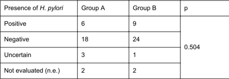
No significant correlation was found between the presence of H. pylori and PPIs intake (Table 1). In Group A the number of positive results was 6 at baseline, while in the control group 9 subjects had a positive output for what concerns this parameter. 18 patients were negative in Group A compared with 24 subjects in the control group (p=0.504).
Presence of H. pylori Group A Group B p
Positive 6 9
0.504
Negative 18 24
Uncertain 3 1
Not evaluated (n.e.) 2 2
Table 1: Assessment of Helicobacter pylori infection in Group A and B at baseline. Results are expressed as positive, negative, uncertain, or not evaluated.
Quantification of specific bacterial groups in the oral cavity: The baseline bacterial groups quantification (log10 CFU/ml of saliva, PPI group vs. control) highlighted significant differences as regards total bacteria (8.44 vs. 4.47, p=0.001), total coliforms (4.95 vs. 2.82, p=0.001), and total lactobacilli (4.00 vs. 2.36, p=0.016). Sulfite-reducing bacteria (SRB), despite an almost 3 log difference between the PPIs group and the control, showed no statistical significance (5.47 vs. 2.58, p=0.052) (Table 2a). Parameters evaluated Group A Group B p log10 CFU/ml log10 CFU/ml Total bacteria 8.44 ± 1.15 4.47 ± 1.31 0.001 Total coliforms 4.95 ± 0.80 2.82 ± 0.96 0.001 Sulfite-reducing bacteria (SRB) 5.47 ± 1.15 2.58 ± 1.05 0.052 Lactobacilli 4.00 ± 0.93 2.36 ± 1.00 0.016
Table 2: Quantification of total bacteria, total coliforms, sulfite-reducing bacteria (SRB), and total lactobacilli in saliva samples at d0
(both groups) (a) and d15 (Group A) (b). The data are expressed as
mean ± SD values (log10 CFU/ml of saliva) of 3 independent analysis.
p values are calculated using Student’s t-test and considered significant if ≤ 0.05. a) comparison between the two groups at d0
After 15 days of supplementation with the four lactobacilli the quantification of salivary bacterial groups provided the following results: total bacteria 7.92 vs. 8.44 at time zero (p=0.019); total coliforms 3.13 vs. 4.95 at time zero (p=0.001); sulfite-reducing bacteria 4.69 vs. 5.47 at baseline (p=0.047); total lactobacilli 7.86 vs. 4.00 at baseline (p=0.048) (Table 2b and 2c).
Time Group A log10 CFU/ml p d0 Total bacteria 8.44 ± 1.15 ** Total coliforms 4.95 ± 0.80 Sulfite-reducing bacteria (SRB) 5.47 ± 1.15 Lactobacilli 4.00 ± 0.93 d15 Total bacteria 7.92 ± 1.40 0.019 Total coliforms 3.13 ± 0.94 0.001 Sulfite-reducing bacteria (SRB) 4.69 ± 1.10 0.047 Lactobacilli 7.86 ± 1.63 0.048
Table 2b: Comparison between time zero (d0) and d15 in Group A; **
comparison reference time (d0)
Time Group A Group B % % d0 Total Lactobacillus 0.27 4.04 d15 Total Lactobacillus 64.96 /
Table 2c: Percentage of total lactobacilli at d0 in both groups and d15 in
Group A
Quantification of Volatile Sulphur Compounds (VSCs) in the breath: No statistically significant differences were found in hydrogen sulfide, methyl mercaptan and dimethyl sulfide concentrations, expressed as parts per billion (ppb), in the two groups, neither at baseline (comparison between the control population and Group A) nor at the end of supplementation with lactobacilli compared to baseline in subjects treated with PPIs. The threshold values of VSC concentration normally considered as discriminants for the presence of halitosis are as follows: 112 ppb for hydrogen sulfide, 26 ppb for methyl mercaptan and 8 ppb for dimethyl sulfide (Table 3).
VSC evaluated Group A Group B p ppb ppb Hydrogen sulfide 7.69 ± 6.33 2.67 ± 0.73 0.444 Methyl mercaptan 289.6 ± 81.8 222.2 ± 61.6 0.549 Dimethyl sulfide 60.4 ± 10.1 62.5 ± 8.2 0.796
Table 3: Quantification of the three VSC hydrogen sulfide, methyl mercaptan and dimethyl sulfide in the breath of subjects taking PPIs since at least 3 months compared with control group at baseline (a) and at the end of treatment in Group A (b). The data are expressed in ppb as means ± Standard Error of the Mean (m ± SEM). P values are calculated using Student’s t-test. comparison between the two groups at d0 a). comparison between time zero (d0) and d15 in Group A
Time Group A ppb p d0 Hydrogen sulfide 7.69 ± 6.33 ** Methyl mercaptan 289.6 ± 81.8 ** Dimethyl sulfide 60.4 ± 10.1 ** d15 Hydrogen sulfide 4.19 ± 2.68 0.337 Methyl mercaptan 258.9 ± 85.6 0.498 Dimethyl sulfide 65.5 ± 11.2 0.804
b) comparison between time zero (d0) and d15 in Group A; **
Discussion
Halitosis is a really widespread issue, commonly attributed to the presence of Volatile Sulphur Compounds (VSC) in the breath [4]. In any case, the present pilot study suggests that bacterial genera in the oral flora other than SRB, not directly enumerated in our work, may contribute to the synthesis and secretion of VSC. In fact, SRB showed almost a 3 log difference between subjects taking PPIs and subjects with a normal intragastric acidity, even if the statistical comparison gave no significance, probably due to the very high standard deviation of the values recorded especially in Group A. However, in Group B the concentrations of methyl mercaptan and dimethyl sulfide were only slightly lower, if not completely overlapping, compared with individuals treated with PPIs, and in any case consistently higher than the respective thresholds (26 ppb and 8 ppb, respectively) in all the conditions tested, thus suggesting an objective assessment of halitosis.
The treatment with four selected lactobacilli, given by means of oral chewable tablets, was able to significantly decrease each bacterial group concentration after 2 weeks, even if this had no influence on VSC levels, as already discussed. Nonetheless, it is interesting to point out the ability of this specific association of beneficial bacteria to help the restoration of a plausibly healthier oral flora in subjects chronically treated with PPIs. As it could be reasonably expected, the majority of the oral cavity flora at the end of the supplementation period was represented by lactobacilli (64.96%), thus demonstrating the effective ability of such strains to integrate into the autochthonous microbiota and to modify its composition.
There are some interesting papers demonstrating the usefulness of lactobacilli, or other beneficial bacteria, in the amelioration or attenuation of periodontal disease or other unfavorable conditions that could affect the oral cavity [37-40]. A randomized controlled trial confirmed the plaque inhibition, anti-inflammatory, and antimicrobial effects of Lactobacillus reuteri Prodentis, actually an association of two different microorganisms, given by means of lozenges at a daily dose of 108 viable cells of each strain [37].
It is well known that probiotics may affect the oral ecology by specifically preventing the adherence of other bacteria and by modifying the protein composition of salivary pellicle. Probiotic microorganisms could modify the protein composition of the pellicle by two different ways, namely binding to and the degradation of salivary proteins [41]. Most probiotics lower the pH so that other potentially harmful microorganisms cannot form dental plaque and calculus that causes oral inflammation.
Other mechanisms considered to be responsible for the beneficial clinical effects of probiotics include a direct interaction with pathogenic bacteria [42], an increase of the host immune response [43] and a production of antimicrobial substances such as organic acids, hydrogen peroxide and bacteriocins [44].
Our study, even if not directly focused on specific oro-dental health indexes, demonstrated the ability of four selected lactobacilli to reduce the concentration of coliforms and sulfite-reducing bacteria (SRB) in the oral cavity in subjects treated with acid suppressive drugs.
In fact, it is pretty consolidated that a prolonged intake of Proton Pump Inhibitors (PPIs) significantly causes a bacterial overgrowth in the stomach and the duodenum [45], thus suggesting a possible role also as concerns the composition of the oral cavity flora. Our present results confirm that the intake of PPIs directly correlates with the overgrowth of specific bacterial groups in the mouth, even if there is
no association with possible H. pylori gastric infection since no differences were recorded in terms of prevalence of H. pylori detection in the control group compared with subjects with an impaired gastric acidity.
A previous pilot study by Del Piano et al. [45] assessed the extent of gastric bacterial overgrowth in subjects taking PPIs either since 3 to 12 months (short-term) or since more than 12 months (long-term).
Considering the overall results collected to date on this association of lactobacilli, it could be hypothesized their prospective usefulness in subjects with an unbalanced oral flora composition, especially if ascribed to the chronic intake of a PPI. In any case, future evaluations will be needed to confirm this preliminary indications and to deeper investigate the microbial groups and the pathways underlying the onset and maintenance of halitosis both in subjects taking PPIs and in control population. In fact, our findings seem to slightly reduce the role attributable solely to SRB in the production of VSC and consequent bad breath.
References
1. Chomyszyn-Gajewska M, Skrzypek A (2013) Halitosis-diagnosis and treatment. Przegl Lek 70: 65-68.
2. Rosenberg M, Kulkarni GV, Bosy A, McCulloch CA (1991) Reproducibility and sensitivity of oral malodor measurements with a portable sulphide monitor. J Dent Res 70: 1436-1440.
3. Miyazaki H, Sakao S, Katoh Y, Takehara T (1995) Correlation between volatile sulphur compounds and certain oral health measurements in the general population. J Periodontol 66: 679-684.
4. Zalewska A, Zatoński M, Jabłonka-Strom A, Paradowska A, Kawala B, et al. (2012) Halitosis--a common medical and social problem. A review on pathology, diagnosis and treatment. Acta Gastroenterol Belg 75: 300-309.
5. Bollen CM, Beikler T (2012) Halitosis: the multidisciplinary approach. Int J Oral Sci 4: 55-63.
6. Quirynen M, Dadamio J, Van den Velde S, De Smit M, Dekeyser C, et al. (2009) Characteristics of 2000 patients who visited a halitosis clinic. J Clin Periodontol 36: 970-975.
7. Collins LM, Dawes C (1987) The surface area of the adult human mouth and thickness of the salivary film covering the teeth and oral mucosa. J Dent Res 66: 1300-1302.
8. Faveri M, Feres M, Shibli JA, Hayacibara RF, Hayacibara MM, et al. (2006) Microbiota of the dorsum of the tongue after plaque accumulation: an experimental study in humans. J Periodontol 77: 1539-1546.
9. de Souza RF, de Freitas Oliveira Paranhos H, Lovato da Silva CH, Abu-Naba'a L, Fedorowicz Z, et al. (2009) Interventions for cleaning dentures in adults. Cochrane Database Syst Rev : CD007395.
10. Krespi YP, Shrime MG, Kacker A (2006) The relationship between oral malodor and volatile sulfur compound-producing bacteria. Otolaryngol Head Neck Surg 135: 671-676.
11. Goldberg S, Kozlovsky A, Gordon D, Gelernter I, Sintov A, et al. (1994) Cadaverine as a putative component of oral malodor. J Dent Res 73: 1168-1172.
12. Calil C, Liberato FL, Pereira AC, de Castro Meneghim M, Goodson JM, et al. (2009) The relationship between volatile sulphur compounds, tongue coating and periodontal disease. Int J Dent Hyg 7: 251-255.
13. Sandeanu S, Jonkers D, De Bruine A, Hameeteman W, Stockbrugger RW (2001) Non-Helicobacter pylori bacterial flora during acid suppressive therapy: differential findings in gastric juice and gastric mucosa. Aliment Pharmacol Ther 15: 379-388.
14. Werdmuller BF, van der Putten TB, Balk TG, Lamers CB, Loffeld RJ (2000) Clinical presentation of Helicobacter pylori-positive and -negative functional dyspepsia. J Gastroenterol Hepatol 15: 498-502.
15. Moshkowitz M, Horowitz N, Leshno M, Halpern Z (2007) Halitosis and gastroesophageal reflux disease: a possible association. Oral Dis 13: 581-585.
16. Hoshi K, Yamano Y, Mitsunaga A, Shimizu S, Kagawa J, et al. (2002) Gastrointestinal diseases and halitosis: association of gastric Helicobacter pylori infection. Int Dent J 52 Suppl 3: 207-211.
17. Suzuki N, Yoneda M, Naito T, Iwamoto T, Masuo Y, et al. (2008) Detection of Helicobacter pylori DNA in the saliva of patients complaining of halitosis. J Med Microbiol 57: 1553-1559.
18. Kinberg S, Stein M, Zion N, Shaoul R (2010) The gastrointestinal aspects of halitosis. Can J Gastroenterol 24: 552-556.
19. Yilmaz AE, Bilici M, Tonbul A, Karabel M, Dogan G, et al. (2012) Paediatric Halitosis and Helicobacter pylori Infection. J Coll Physicians Surg Pak 22: 27-30.
20. Bradshaw DJ, Perring KD, Cawkill PM, Provan AF, McNulty DA, et al. (2005) Creation of oral care flavours to deliver breath-freshening benefits. Oral Dis 11 Suppl 1: 75-79.
21. Rao Y, Lingamneni B, Reddy D (2012) Probiotics in oral health--a review. J N J Dent Assoc 83: 28-32.
22. Anilkumar K, Monisha AL (2012) Role of friendly bacteria in oral health - a short review. Oral Health Prev Dent 10: 3-8.
23. Burton JP, Chilcott CN, Moore CJ, Speiser G, Tagg JR (2006) A preliminary study of the effect of probiotic Streptococcus salivarius K12 on oral malodour parameters. J Appl Microbiol 100: 754-764.
24. Iwamoto T, Suzuki N, Tanabe K, Takeshita T, Hirofuji T (2010) Effects of probiotic Lactobacillus salivarius WB21 on halitosis and oral health: an open-label pilot trial. Oral Surg Oral Med Oral Pathol Oral Radiol Endod 110: 201-208.
25. Bouhnik Y, Pochart P, Marteau P, Arlet G, Goderel I, et al. (1992) Fecal recovery in humans of viable Bifidobacterium sp ingested in fermented milk. Gastroenterology 102: 875-878.
26. Laleman I, Dadamio J, De Geest S, Dekeyser C, Quirynen M (2014) Instrumental assessment of halitosis for the general dental practitioner. J Breath Res 8: 017103.
27. Pourakbari B, Ghazi M, Mahmoudi S, Mamishi S, Azhdarkosh H, et al. (2013) Diagnosis of Helicobacter pylori infection by invasive and noninvasive tests. Braz J Microbiol 44: 795-798.
28. Nogueira C, Figueiredo C, Carneiro F, Gomes AT, Barreira R, et al. (2001) Helicobacter pylori genotypes may determine gastric histopathology. Am J Pathol 158: 647-654.
29. Dababneh RH, Khouri AT, Smith RG, Addy M (2002) A new method of plaque scoring: a laboratory comparison with other plaque indices. J Clin Periodontol 29: 832-837.
30. McClanahan SF, Bartizek RD, Biesbrock AR (2001) Identification and consequences of distinct Löe-Silness gingival index examiner styles for the clinical assessment of gingivitis. J Periodontol 72: 383-392.
31. Saxer UP, Mühlemann HR (1975) Motivation and education. SSO Schweiz Monatsschr Zahnheilkd 85: 905-919.
32. Winkel EG, Roldan S, Van Winkelhoff AJ, Herrera D, Sanz M (2003) Clinical effects of a new mouthrinse containing chlorhexidine,
cetylpyridinium chloride and zinc-lactate on oral halitosis. A dual-center, double-blind placebo-controlled study. J Clin Periodontol 30: 300-306.
33. Raibaud P, Caulet M, Galpin JV, Mocquot G (1961) Studies on the microbial flora of the alimentary tract of pigs. II. Streptococci: selective enumeration and differentiation of the dominant group. J Appl Bacteriol 24: 285-291.
34. ROGOSA M, MITCHELL JA, WISEMAN RF (1951) A selective medium for the isolation and enumeration of oral and fecal lactobacilli. J Bacteriol 62: 132-133.
35. Line JE, Stern NJ, Oakley BB, Seal BS (2011) Comparison of an automated most-probable-number technique with traditional plating methods for estimating populations of total aerobes, coliforms, and Escherichia coli associated with freshly processed broiler chickens. J Food Prot 74: 1558-1563.
36. Hauschild AH, Hilsheimer R, Griffith DW (1974) Enumeration of fecal Clostridium perfringens spores in egg yolk-free tryptose-sulfite-cycloserine agar. Appl Microbiol 27: 527-530.
37. Vivekananda MR, Vandana KL, Bhat KG (2010) Effect of the probiotic Lactobacilli reuteri (Prodentis) in the management of periodontal disease: a preliminary randomized clinical trial. J Oral Microbiol 2.
38. Staab B, Eick S, Knöfler G, Jentsch H (2009) The influence of a probiotic milk drink on the development of gingivitis: a pilot study. J Clin Periodontol 36: 850-856.
39. Karuppaiah RM, Shankar S, Raj SK, Ramesh K, Prakash R, et al. (2013) Evaluation of the efficacy of probiotics in plaque reduction and gingival health maintenance among school children - A Randomized Control Trial. J Int Oral Health 5: 33-37.
40. Slawik S, Staufenbiel I, Schilke R, Nicksch S, Weinspach K, et al. (2011) Probiotics affect the clinical inflammatory parameters of experimental gingivitis in humans. Eur J Clin Nutr 65: 857-863.
41. VINCENT JG, VEOMETT RC, RILEY RF (1959) Antibacterial activity associated with Lactobacillus acidophilus. J Bacteriol 78: 477-484.
42. Patzer SI, Baquero MR, Bravo D, Moreno F, Hantke K (2003) The colicin G, H and X determinants encode microcins M and H47, which might utilize the catecholate siderophore receptors FepA, Cir, Fiu and IroN. Microbiology 149: 2557-2570.
43. Schlee M, Harder J, Köten B, Stange EF, Wehkamp J, et al. (2008) Probiotic lactobacilli and VSL#3 induce enterocyte beta-defensin 2. Clin Exp Immunol 151: 528-535.
44. Reid G, Jass J, Sebulsky MT, McCormick JK (2003) Potential uses of probiotics in clinical practice. Clin Microbiol Rev 16: 658-672.
45. Del Piano M, Anderloni A, Balzarini M, Ballare M, Carmagnola S, et al. (2012) The innovative potential of Lactobacillus rhamnosus LR06, Lactobacillus pentosus LPS01, Lactobacillus plantarum LP01, and Lactobacillus delbrueckii subsp. delbrueckii LDD01 to restore the “gastric barrier effect” in patients chronically treated with PPI: a pilot study. J Clin Gastroenterol 46: S18-26.