gain direct access to mandibular condylar head fractures; they can be divided into 2 groups: intraoral and extraoral. In 2005, Neff et al (Mund Kiefer Gesichtschir 2005;9:80), supported by a previous ex-perimental work, reported a successful clinical study of condylar head fractures treated by a retroauricular approach; this article is in German, and the later English-language literature does not mention about this approach to open reduction and internal fixation of man-dibular condylar fractures. The retroauricular transmeatal access, se-lected and performed by the senior author to treat 14 patients affected by highly located condylar head fracture, is illustrated in details. Methods: We collected data of 14 consecutive adult patients who, after the discussion about all options, had consented to have 16 man-dibular condylar head fractures treated with open reduction and inter-nal fixation by miniplates and screws via a retroauricular transmeatal approach. We exposed the temporomandibular joint area easily and better by dissecting via a retroauricular route with identification, liga-tion, and transection of the retromandibular vein; because of the posterior access, the frontal branch of the facial nerve and the auric-ulotemporal nerve are located and protected within the substance of the anteriorly retracted flap, superficial to the retromandibular vein. The follow-up clinical examination showed temporary weakness of the frontal branch of the facial nerve in 1 case with a recovery to nor-mal function of 1.6 months; no patients had permanent weakness of the facial nerve or injury of the auriculotemporal nerve. There was absence of any salivary fistula, sialocele, and Frey syndrome; hearing was preserved in all cases, without any auditory stenosis or aesthetic deformity, and there was absence of any infections, hematoma, or scarring.
Conclusions: Retroauricular approach provides good exposure of the temporomandibular joint and satisfactory protection from nerve injuries and vascular lesions, allowing an adequate osteosynthesis. The scar is hidden behind the ear, and the morbidity is low in terms of auditory stenosis, aesthetic deformity, and salivary fistulas.
(J Craniofac Surg 2011;22: 641Y647)
T
here is a multitude of reported surgical approaches and tech-nical variants to gain direct access to mandibular condylar fractures, and they can be divided into 2 groups: intraoral and extraoral.1The first group includes the endoscopic2 and the endo-scopically assisted approaches3; some authors prefer this method, stating that it is advantageous because it minimizes the risks of facial nerve injury and leaves a hidden scar. The disadvantages are that it necessitates specific instruments and an additional training of the surgeon.4
Moreover, an intraoral approach can be technically demand-ing when treatdemand-ing high and medially displaced fractures, and it can lead to unsatisfactory results in terms of anatomic reduction and functionality. Some authors have described for this technique a high percentage of complications such as myofascial pain, condylar head resorption, incorrect reduction, and malocclusions.5
The extraoral approaches are preauricular,6 modified pre-auricular,7retromandibular,8miniretromandibular,9transmasseteric anteroparotid,10submandibular,11and facial rhytidectomy.12
Extraoral approaches simplify the management of condylar fractures, but they are associated with some unresolved technical problems such as facial nerve, vascular structures, skin incisions, and parotid gland; the preauricular approach is indicated to manage high condylar neck fractures but can have a disadvantage when treating lower fractures such as subcondylar. By contrast, the sub-mandibular and retrosub-mandibular accesses allow for a comfortable management of lower fractures, but they become troublesome in treating higher fractures.13Y18
In 2005, Neff et al,19supported by a previous experimental work,20 reported a successful clinical study of condylar fractures treated by a retroauricular approach; this article is in German, and the later English-language literature does not mention about this approach to open reduction and internal fixation of mandibular condylar fractures.
In the Maxillofacial Unit at the Novara Major Hospital, we choose open treatment for 14 patients affected by highly located con-dylar head fracture using the retroauricular transmeatal approach.21,22
The retroauricular transmeatal access, selected and performed by the senior author to treat these cases, is introduced. This method is a relatively simple route to gain direct access to fractures of the mandibular condyle, allowing an easy anatomic reduction of the stumps and a proper osteosynthesis with miniplates and screws.
This approach permits an easy and fast management of highly located condylar fracture while minimizing the risks of facial nerve injuries and leaving a hidden scar in a barely noticeable region.
From the Department of Maxillo-Facial Surgery, Azienda Ospedaliera Maggiore della Carita`, University of Piemonte Orientale BAmedeo Avogadro,[ Novara, Italy.
Received September 2, 2010.
Accepted for publication September 24, 2010.
Address correspondence and reprint request to Francesco Arcuri, MD, SCDU di Chirurgia Maxillo-Facciale, Ospedale Maggiore della Carita`, Corso Mazzini 18, 28100 Novara, Italy; E-mail: [email protected] The authors report no conflicts of interest.
Copyright* 2011 by Mutaz B. Habal, MD ISSN: 1049-2275
Most surgeons use only screws for fixation because the seg-ments of the condylar fractures are small and difficult to reduce and fix23Y26; we found this technique unstable for our patients, so our methods of fixation used to improve stability were miniplates and screws.
We will not discuss which is the ideal surgical approach, which fractures should be managed with open reduction and inter-nal fixation, or which type of osteosynthesis is most suitable bio-mechanically; these issues have already been addressed by other authors, and it is beyond the scope of this work27Y31: we will entirely focus on the retroauricular transmeatal approach that is herein-after illustrated step by step.
PATIENTS AND METHODS
We collected data of 14 consecutive adult patients (from January 2006 to December 2008; 7 white male and 2 North African
male patients, 1 South American male patient, 3 white female patients, and 1 North African female patient; mean age, 33 years; age range, 17Y64 years ) who underwent open reduction and inter-nal fixation for 16 condylar head fractures.
Twelve patients presented with unilateral fractures and 2 with bilateral fractures; the 16 condylar fractures consisted of 11 intracapsular condylar fractures and 5 extracapsular highly located condylar head fractures (Fig. 1). A total of 5 patients presented with associated fractures of the facial bones (3 mandibular body and 2 panfacial). The average duration of surgery was 43 minutes (range, 29Y67) for each surgical procedure.
The diagnosis was performed by clinical examination, con-ventional radiography, and computed tomography (CT) scanning; after the discussion about other surgical and nonsurgical alterna-tive, all patients had consented to have the mandibular condylar frac-tures treated by osteosynthesis with miniplates and screws via a retroauricular transmeatal approach.
SURGICAL TECHNIQUE
The operation is performed with the patient under general an-esthesia with nasotracheal intubation; if other mandibular fractures are concomitant, these are treated first with a standard intraoral approach to recreate the continuity of the mandibular arch. Consequently, a new FIGURE 1. AYC, Preoperative conventional radiographic
examinations showing 3 cases of highly located condylar head fractures.
FIGURE 2. Surgical field at the beginning of the procedure.
FIGURE 3. Amount of infiltration in the retroauricular region.
FIGURE 4. Skin and soft-tissue flaps dissected with the direct exposure of the concha.
sterile surgical field is made, and it is separated from the oral and nasal cavities using a sterile adhesive film to prevent bacterial contamination at the condylar site (Fig. 2).
Afterward, a preoperative drawing is made, marking the angle and the body of the mandible, the zygomatic arch, the glenoid fossa, and the fracture site. The ear, the lateral canthus of the eye, and the mandible are prepared and draped as pertinent landmarks of the face to properly expose the surgical field and to mentally visualize the fracture; placing a cotton soaked in the external auditory canal and shaving the preauricular hair are optional.
The retroauricular area is quite vascular, so it is preferable to inject a vasoconstrictor to decrease the amount of bleeding during the incision and the dissection. The posterior surface of the auricle and the occipital area are slowly infiltrated with 4 to 8 mL of 1% lidocaine with 1:100,000 epinephrine; although local anesthesia enhances hemostasis and demarcates the plane of dissection, it is preferable to inject a small amount of infiltration to avoid distortion of the tissues (Fig. 3).
A simple 25- to 30-mm vertical incision is made in the poste-rior occipital area, approximately 10 to 15 mm medial to the sulcus in a retroauricular crease, through skin and subcutaneous connective tissues. Normally, the edges of the incision are 0.5 to 10 mm cephalad to the radix of the auricular lobule and 0.5 to 10 mm caudal to the
insertion of the helix; any bleeding superficial vessels are cauterized before proceeding with deeper dissection.
The anterior and posterior edges of the skin and soft-tissue flaps are elevated to gain a direct exposure of the concha and the mastoid fascia. A clean surgical dissection is kept during the flap elevation; muscle fibers and fat can be cleanly excised off the perichondrium and the mastoid fascia (Fig. 4).
Attention is now focused on the concha: the flap is retracted anteriorly, and a first incision with a scalpel is made through the posterior aspect of the cartilaginous meatus, sectioning the cartilage and the skin (Fig. 5); then a second incision is carried out through the anterior wall of the canal at a wide portion to prevent stenosis, transecting conclusively the external auditory meatus (Fig. 6).
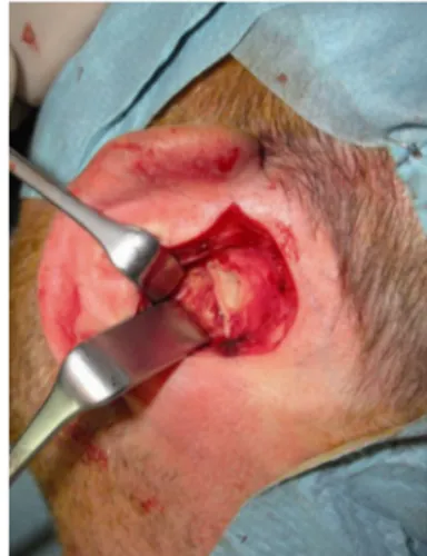
At this depth, retraction of the external ear anteriorly permits the exposure of the retromandibular space with the parotid gland (Fig. 7), and blunt dissection commonly reveals the retromandibular vein that is ligated and transected (Fig. 8). Because of the posterior access, the frontal branch of the facial nerve and the auriculotem-poral nerve are located and gently protected by a retractor within the substance of the anteriorly retracted flap, superficial to the retroman-dibular vein (Figs. 7 and 8).
At this point, the mandible is manipulated open and closed to help determine the location of the ramus and the condyle; when FIGURE 5. The posterior aspect of the cartilaginous meatus
transected.
FIGURE 6. Incision of the anterior wall of the canal and consequently the entire external auditory canal transected.
FIGURE 7. The retromandibular space with the parotid gland.
the bone surface is reached, the mandibular periosteum is incised, dissected, and elevated, revealing the fracture fragments.
The manipulation screw (386.902) and the handle for ma-nipulation screw (386.903) of the endoscopic subcondylar/ramus fixation set (Synthes, West Chester, PA) are used similar to a Bjoystick[ to facilitate the reduction of the condylar head; after a caudal distraction of the distal stump by intraoral pressure to the last mandibular molars with a finger, while taking great care to preserve sterility over the condylar surgical field by changing the surgical gloves, anatomic reduction has been completed (Fig. 9). Finally, after temporary maxillomandibular fixation, the osteosyn-thesis is performed with miniplates and screws (Fig. 10).
Attention is finally paid to close the access: the surgical site is generously irrigated with hydrogen peroxide, and any hemorrhage is meticulously controlled; afterward, the parotid capsule is closed tightly with running, slowly resorbing, horizontal mattress suture to prevent salivary fistula, and the auricular canal is reconstructed with interrupted, slowly resorbing suture to prevent stenosis. Sub-cutaneous and skin tissues are closed again with interrupted, slowly resorbing suture (Fig. 11).
A petrolatum gauze is inserted in the external auditory me-atus, and it is left in place for 10 days; a compressive dressing is applied for 7 days (Fig. 12). In the postoperative period, the wound is kept moist twice a day for 1 week with hydrogen peroxide and
antibiotic ointment; the ear is checked daily for hematoma and infection.
RESULTS
We have used 2 weeks of maxillomandibular fixation with elastics for all patients; postoperative conventional radiographic or CT scans were taken to check fracture osteosynthesis, and a soft diet was ordered for 1 month. After that, if a patient had mandibular hypomobility, mouth-opening exercises were taught: clinical follow-up was performed at 1 week; at 1, 3, and 6 months; and at 1 year; radiologic controls were scheduled in the immediate postoperative period, at 6 months, and at 1 year.
The follow-up clinical examination showed satisfactory results were achieved in all patients; temporary weakness of the frontal branch of the facial nerve was detected in 1 case, with a recovery to normal function after 1.6 months; no patients had permanent weakness of the facial nerve or injury of the auriculotemporal nerve.
There was absence of any salivary fistula, sialocele, and Frey syndrome; hearing was preserved in all cases without any auditory stenosis or aesthetic deformity, and there was absence of any in-fections, hematoma, or scarring.
The mean maximal interincisal mouth opening was 38 mm, and in every patient, good occlusion without dysfunctional FIGURE 9. The anatomic reduction.
FIGURE 10. Rigid internal fixation.
FIGURE 11. Closure of the retroauricular skin with interrupted slowly resorbing suture.
FIGURE 12. A petrolatum gauze is inserted in the external auditory meatus.
amination on the basis of conventional radiography in 10 of the 14 patients and axial, coronal, and three-dimensional CT scanning in 4 of the 14 patients showed an acceptable osteosynthesis, except in 1 patient with a failure of fixation but with a satisfactory occlusion: no patient needed to return to the operating room for adjustment because of malocclusion (Fig. 14).
DISCUSSION
Condylar fractures are very common in craniomaxillofacial traumas, and they account for 20% to 52% of all mandibular frac-tures32; properly managing condylar fractures is of priority impor-tance because a mistake during the diagnosis or the treatment can lead to severe anatomic and functional impairment.33
These fractures can be classified in several ways; moreover, the patient’s age, the fracture’ site, and the degree of displacement seem to be of critical importance when deciding the indications for surgical management.34
Historically, highly located condylar fractures are treated closed because of 3 main reasons: the difficulty to reduce and fix the small segments of the fracture, the concern about any facial nerve injuries,
tive treatment during long-term follow-up.35
After the development of radiologic examinations, such as CT scanning and magnetic resonance imaging, surgeons analyzed the unsatisfactory results of the closed treatment and began to con-sider a more aggressive approach.36,37
One of the most important anatomic findings observed after the treatment of condylar fractures by functional therapy is the re-duction of mandibular ramus height and severe functional impair-ment including poor occlusion, reduced opening, deviation, and limited lateral mandibular movement38; moreover, other possible complications such as temporomandibular joint (TMJ) ankylosis, TMJ dysfunction, and craniofacial pain need to be evaluated before considering a closed treatment.39
The surgical treatment of condylar fractures has been strongly debated, and various investigators have previously favored a non-operative approach40; a number of reports have now suggested that the treatment of condylar fractures by open reduction and rigid fixation leads to much better anatomic and functional results com-pared with closed and functional treatment.41Y43
Nonoperative treatment is still indicated for some cases such as pediatric patients and comminuted intracapsular fractures;
FIGURE 14. AYD, Postoperative three-dimensional CT scanning (A) and conventional radiographic examination (BYD) of 3 cases of highly located condylar head fractures demonstrating successful anatomic mandibular reduction and adequate osteosynthesis.
fracture treatment but require long skin incisions and present some risk for the facial nerve fibers and vascular lesions.6Y12
Furthermore, these methods are not equivalent for treating all condylar fractures; the preauricular access is well indicated only for treating fractures at higher levels, whereas the standard sub-mandibular and retrosub-mandibular approaches do not allow comfort-able management of the same type of lesion.13Y18
The authors exposed the condyle easily and better by dis-secting via a retroauricular transmeatal route with identification, li-gation, and transection of the retromandibular vein; because of the posterior access, the frontal branch of the facial nerve and the auriculotemporal nerve are located and protected within the sub-stance of the anteriorly retracted flap, superficial to the retroman-dibular vein.
This approach is not technically demanding; it provides ex-cellent access, allowing direct visualization of highly located con-dylar fracture, and it ensures that the plates can be well adapted, and the screws placed at 90 degrees to the bony surface, giving maxi-mum mechanical advantage, which is not always possible with the more traditional preauricular, retromandibular, and submandibular accesses.
Moreover, once the flap is retracted anteriorly, the surgical field is always perpendicular to the fractured stumps; this simplifies the treatment of even difficult fractures (anteromedial luxation of the condylar head, delayed treatment) and their rigid fixation.
The ideal surgical approach should be the one that allows fracture management while minimizing the risk of potential pitfalls; we believe that the retroauricular transmeatal approach can be a valid alternative to the common transfacial accesses in selected highly located condylar head fracture such as (1) patients with intracap-sular condylar head fractures; (2) overweight patients with redun-dant cheek soft tissues; (3) patients with a genetic predisposition to develop hypertrophic keloids; (4) patients who do not accept preop-eratively any potential nerve injuries or noticeable facial scars; (5) patients who refer during the clinical examination previous surgical procedures in the preauricular area (facelift, parotid surgery).
CONCLUSIONS
Retroauricular transmeatal access provides good exposure of condylar fractures permitting an adequate osteosynthesis; it is relatively easy and fast to perform; it presents an extremely low risk to injure the facial nerve, and it leaves a unnoticeable scar in a very hidden region behind the ear. The morbidity is extremely low in terms of auditory stenosis, aesthetic deformity, facial nerve lesions, vascular injuries, salivary fistulas, sialocele, and Frey syndrome.
The TMJ area frequently requires exposure for a multitude of surgical procedures such as internal derangements, arthritis, trau-ma, developmental disorders, and neoplasia; several approaches have been proposed and used clinically; up to date, the ideal access has not yet been found.
We have successfully performed this approach for 14 trauma cases, and we suggest this access to selected patients owing to its ease, speed, versatility, and freedom from the complications that are common with other extraoral approaches.
velopmental disorders and neoplasia of the TMJ area.
REFERENCES
1. Ellis E III, Moos K, El-Attar A. Ten years of mandibular fractures: an analysis of 2137 cases. J Oral Surg 1985;59:120
2. Kellman RM, Cienfuegos R. Endoscopic approaches to subcondylar fractures of the mandible [published online ahead of print February 10, 2009]. Facial Plast Surg 2009;25:23Y28
3. Loukota RA. Endoscopically assisted reduction and fixation of condylar neck/base fractures The learning curve. Br J Oral Maxillofac Surg 2006;44:480
4. Jacobovicz J, Lee C, Trabulsy PP. Endoscopic repair of mandibular subcondylar fractures. Plast Reconstr Surg 1998;101:437
5. Jensen T, Jensen J, Norholt SE, et al. Open reduction and rigid internal fixation of mandibular condylar fractures by an intraoral approach: a long term follow up study of 15 patients. J Oral Maxillofac Surg 2006;64:1771
6. Kermer Ch, Undt G, Rasse M. Surgical reduction and fixation of intracapsular condylar fractures. A follow up study. Int J Oral Maxillofac Surg 1998;28:191
7. He D, Yang C, Chen M, et al. Modified preauricular approach and rigid internal fixation for intracapsular condyle fracture of the mandible. J Oral Maxillofac Surg 2010;68:1578Y1584
8. Narayanan V, Kannan R, Sreekumar K. Retromandibular
approach for reduction and fixation of mandibular condylar fractures: a clinical experience. Int J Oral Maxillofac Surg 2009;38:
835Y839
9. Biglioli F, Colletti G. Mini-retromandibular approach to condylar fractures [published online ahead of print July 2, 2008]. J Craniomaxillofac Surg 2008;36:378Y383
10. Wilson AW, Ethunandan M, Brennan PA. Transmasseteric antero- parotid approach for open reduction and internal fixation of condylar fractures. Br J Oral Maxillofac Surg 2005;43:57 11. Malkin M, Kresberg H, Mandel l. Submandibular approach for open
reduction of condylar fracture. Oral Surg Oral med Oral Pathol 1964;17:152Y157
12. Anastassov GE, Rodriguez ED, Schwimmer AM, et al. Facial rhytidectomy approach for treatment of posterior mandibular fractures.
J Craniomaxillofac Surg 1997;25:9Y14
13. Tasanen A, Lamberg MA. Transosseous wiring in the treatment of condylar fractures of the mandible. J Maxillofac Surg 1976;4:200 14. Kallela I, Soderholm AL, Paukku P, et al. Lag screw osteosynthesis
of mandibular condyle fractures: a clinical and radiological study. J Oral Maxillofac Surg 1995;53:1397
15. Ellis E III, McFadden D, Simon P, et al. Surgical complications with open treatment of mandibular condylar process fractures. J Oral Maxillofac Surg 2000;58:950
16. Manisali M, Amin M, Aghabeigi B, et al. Retromandibular approach to the mandibular condyle: a clinical and cadaveric study. Int J Oral Maxillofac Surg 2003;32:253
17. Vesnaver A, Gorjanc M, Eberlinc A, et al. The periauricular transparotid approach for open reduction and internal fixation of condylar fractures. J Craniomaxillofac Surg 2005;33:169
18. Blair VP. Operative treatment of ankylosis of the mandible. South Surg Gynecol 1913;26:436
19. Neff A, Kolk A, Meschke F, et al. Small fragment screws vs. plate osteosynthesis in condylar head fractures [in German]. Mund Kiefer Gesichtschir 2005;9:80
of the mandibular condyle with a headless bone screw. Br J Oral Maxillofac Surg 2007;45:399
26. Bos RR, Ward Booth RP, de Bont LG. Mandibular condyle fractures: a consensus. Br J Oral Maxillofac Surg 1999;37:87
27. Eckelt U, Rasse M. Clinical, radiographic and axiographic control after traction-screw osteosynthesis of fractures of the mandibular condyle region. Rev Stomatol Chir Maxillofac 1995;96:158Y165 28. Ellis E III, Dean J. Rigid fixation of mandibular condyle fractures.
Oral Surg Oral Med Oral Pathol 1993;76:6Y15
29. Ziccardi VB, Schneider RE, Kummer FJ. Wurzburg lag screw plate versus four-hole miniplate for the treatment of condylar process fractures. J Oral Maxillofac Surg 1997;55:602Y609
30. Schoen R, Fakler O, Metzger MC, et al. Preliminary functional results of endoscope-assisted transoral treatment of displaced bilateral condylar mandible fractures. Int J Oral Maxillofac Surg
2008;37:111Y116
31. Martin M, Lee C. Endoscopic mandibular condyle fracture repair. Atlas Oral Maxillofac Surg Clin North Am 2003;11:169Y178 32. Haug R, Prather J, Indresano A. An epidemiologic survey of facial
fractures and concomitant injuries. J Oral Maxillofac Surg 1990;48:926 33. Silvennoinen U, Iizuka T, Lindqvist C, et al. Different patterns
of condylar fractures: an analysis of 382 patients in a 3-year period. J Oral Maxillofac Surg 1992;50:1032
34. Eulert S, Proff P, Bokan I, et al. Study on treatment of condylar process fracture of the mandible. Ann Anat 2007;189:377
35. Ellis E. Condylar process fractures of the mandible. Facial Plast Surg 2000;16:193
36. Hlawitschka M, Loukota R, Echelt U. Functional and
radiological results of open and closed treatment of intracapsular
42. Palmieri C, Ellis E III, Throckmorton GS. Mandibular motion after closed and open treatment of unilateral mandibular condylar process fractures. J Oral Maxillofac Surg 1999;57:764
43. Ellis E III, Simon P, Throckmorton GS. Occlusal results after open or closed treatment of fractures of the mandibular condylar process. J Oral Maxillofac Surg 2000;58:260
44. Ellis E III, Throckmorton GS. Bite forces after open or closed treatment of mandibular condylar process fractures. J Oral Maxillofac Surg 2001;59:389
45. Throckmorton GS, Ellis E III. Recovery of mandibular motion after closed and open treatment of unilateral mandibular condylar process fractures. Int J Oral Maxillofac Surg 2000;29:421 46. Throckmorton GS, Ellis E III, Hayasaki H. Masticatory motion
after surgical or nonsurgical treatment for unilateral fractures of the mandibular condylar process. J Oral Maxillofac Surg 2004;62:127 47. Eckelt U, Schneider M, Erasmus F, et al. Open versus closed
treatment of fractures of the mandibular condylar processVa prospective randomized multi-centre study. J Craniomaxillofac Surg 2006;34:306
48. Schneider M, Lauer G, Eckelt U. Surgical treatment of fractures of the mandibular condyle: a comparison of long term results following different approachesVfunctional, axiographical, and radiological findings. J Cranio-Maxillofacial Surg 2007;35:151
49. Hovinga J, Boering G, Stegenga B. Long term results of nonsurgical management of condylar fractures in children. Int J Oral
Maxillofac Surg 1999;28:429
50. Gerbino G, Boffano P, Tosco P, et al. Long-term clinical and radiological outcomes for the surgical treatment of mandibular condylar fractures. J Oral Maxillofac Surg 2009;67:1009Y1014