Review
Calcium regulates cell death in cancer: Roles of the mitochondria and
mitochondria-associated membranes (MAMs)
☆
Alberto Danese
a, Simone Patergnani
a, Massimo Bonora
a, Mariusz R. Wieckowski
b, Maurizio Previati
c,
Carlotta Giorgi
a,⁎
, Paolo Pinton
a,⁎
a
Dept. of Morphology, Surgery and Experimental Medicine, Section of Pathology, Oncology and Experimental Biology, Laboratory for Technologies of Advanced Therapies (LTTA), University of Ferrara, Ferrara, Italy
b
Dept. of Biochemistry, Nencki Institute of Experimental Biology, Warsaw, Poland
c
Department of Morphology, Surgery and Experimental Medicine, Section of Human Anatomy and Histology, Laboratory for Technologies of Advanced Therapies (LTTA), University of Ferrara, Ferrara, Italy
a b s t r a c t
a r t i c l e i n f o
Article history:
Received 24 October 2016
Received in revised form 30 December 2016 Accepted 8 January 2017
Available online 10 January 2017
Until 1972, the term‘apoptosis’ was used to differentiate the programmed cell death that naturally occurs in or-ganismal development from the acute tissue death referred to as necrosis.
Many studies on cell death and programmed cell death have been published and most are, at least to some de-gree, related to cancer. Some key proteins and molecular pathways implicated in cell death have been analyzed, whereas others are still being actively researched; therefore, an increasing number of cellular compartments and organelles are being implicated in cell death and cancer. Here, we discuss the mitochondria and subdomains of the endoplasmic reticulum (ER) that interact with mitochondria, the mitochondria-associated membranes (MAMs), which have been identified as critical hubs in the regulation of cell death and tumor growth. MAMs-de-pendent calcium (Ca2+) release from the ER allows selective Ca2+uptake by the mitochondria. The perturbation
of Ca2+homeostasis in cancer cells is correlated with sustained cell proliferation and the inhibition of cell death
through the modulation of Ca2+signaling. This article is part of a Special Issue entitled Mitochondria in Cancer,
edited by Giuseppe Gasparre, Rodrigue Rossignol and Pierre Sonveaux.
© 2017 Elsevier B.V. All rights reserved. Keywords:
Calcium (Ca2+)
Apoptosis Autophagy
Mitochondria associated membranes (MAMs) Endoplasmic reticulum
Tumor
1. Introduction
Cell death is a crucial and essential aspect of life. Although this
state-ment may be contradictory, cell death itself is directly connected to cell
proliferation and cell survival
[1]
. (See
Fig. 1
)
Programmed cell death has been established as an anti-cancer
de-fense mechanism; therefore, any modi
fication to the related pathways
leads to uncontrolled cell proliferation and oncogenesis.
Early classi
fications of cell death were based on morphological
as-says, and apoptosis was one of the
first processes to be described. The
initial morphological observations were described as the rounding-up
of the cell, a reduction in cellular volume, chromatin condensation,
cyto-plasmic shrinkage, the retraction of pseudopods, nuclear fragmentation,
and a particular boiling-like process termed blebbing
[2]
.
Apoptosis is the most important and well-studied mechanism of cell
death; approximately 10 million cells undergo the apoptotic process in
an adult human under physiological conditions each day
[3]
. The
prima-ry proteins involved in apoptotic cell death and their respective
activi-ties will be discussed in the subsequent sections.
The apoptotic process is classi
fied as type I cell death. Type II cell
death is classi
fied as autophagy, a pro-survival process that also acts
as a pro-death pathway, as it is involved in several biological events,
such as aging, development, protein turnover, neurodegeneration and
cancer
[4]
. In addition to the canonical proteins mammalian target of
rapamycin (mTOR) and AMP-activated protein kinase (AMPK), the
most well-studied proteins that act as initiation sensors, BECLIN1 and
autophagy and BECLIN1 regulator 1 (AMBRA1) are also involved in
the initial steps of autophagosome formation
[5]
.
Necrosis is de
fined as type III cell death and has long been
consid-ered as an accidental and unscheduled form of cell death. Nonetheless,
according to several recent studies, the execution of the necrotic process
may be regulated by a set of catabolic mechanisms and signal
transduc-tion pathways
[6]
. The Bcl-interacting protein 3 (BNIP3),
Bcl-2-modify-ing factor (BMF) (pro-death proteins and members of the Bcl-2 family),
NIP3-like protein X (Nix), the kinase receptor-interacting protein 1
☆ This article is part of a Special Issue entitled Mitochondria in Cancer, edited by Giuseppe Gasparre, Rodrigue Rossignol and Pierre Sonveaux.
⁎ Corresponding authors.
E-mail addresses:[email protected](C. Giorgi),[email protected](P. Pinton).
http://dx.doi.org/10.1016/j.bbabio.2017.01.003
0005-2728/© 2017 Elsevier B.V. All rights reserved.
Contents lists available at
ScienceDirect
Biochimica et Biophysica Acta
j o u r n a l h o m e p a g e :
w w w . e l s e v i e r . c o m / l o c a t e / b b a b i o
(RIP1) and RIP3 proteins may be involved in this process, as they are
speculated to be key signaling molecules involved in necrosis and, in
turn, are regulated by caspases and ubiquitination
[7
–10]
.
The aforementioned cell death types are considered the main
path-ways involved in cell death. For a complete description, we must
men-tion atypical cell death modalities, such as anoikis, a particular type of
apoptosis induced by the loss of attachment to other cells or matrix.
Anoikis involves Bid and Bim, which are activated following the
detach-ment of cells from the extracellular matrix (ECM) and rapidly promote
the assembly of Bax
–Bak oligomers within the outer mitochondrial
membrane (OMM)
[11]
.
Pyroptosis is a cell death mechanism induced by caspase-1
activa-tion, leading to interleukin (IL)-1
β and IL-18 release. NLRP3-dependent
caspase-1 activation plays a key role in this process. Indeed,
mitochon-dria-associated adapter molecules, MAVS, are required for optimal
NLRP3 in
flammasome activity
[12]
.
Paraptosis is a type of cell death triggered by the expression of the
insulin-like growth factor receptor I
[13]
. Mitochondrial Ca
2+has an
ex-tremely important role in hesperidin-induced paraptotic cell death
[14]
.
Other cell death mechanisms that are not yet well characterized
include cell death preceded by multinucleation and entosis (a
phenomenon that occurs when a cell engulfs one of its live
neighbor-ing cells)
[15]
.
All these cell death mechanisms are
finely regulated by a complex
network of proteins, whose transcription and degradation have
pro-found effects on malignant cancer phenotypes. Some oncogenic
muta-tions disrupt programmed cell death, leading to tumor initiation,
progression or metastasis (e.g., mutations in the Bcl-2 protein family
de-regulate cell death).
Bcl-2 does not behave like a typical oncogene; it promotes cell
survival by blocking programmed cell death (i.e., by having a direct
effect on endoplasmic reticulum Ca
2+handling) instead of
disrupting normal proliferation checkpoints
[16]
. Other examples
in-clude p53-like proteins, including TP53 itself, which was the
first
gene linked to apoptosis. p53 has tumor suppression proprieties,
and this gene is mutated in the majority of human tumors
[17]
.
Dis-ruption of the Fas/CD95 receptor pathway, which regulates cell
num-ber in the immune system, leads to lymphoproliferative disorders
and cancer
[18]
. Ras activation and phosphatase and tensin
homo-logue (PTEN) loss are common in human tumors; phosphoinositide
3-kinase (PI3K) is activated by Ras and downregulated by the
tumor suppressor PTEN.
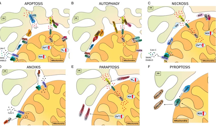
Fig. 1. Summary of the mitochondrial, ER and MAM proteins involved in primary cell death mechanisms. Representation of proteins at the mitochondria-ER interface that play active roles in cell death. Proteins that prevent or promote cell death affect intracellular Ca2+
dynamics and homeostasis by binding Ca2+
and modulating intracellular Ca2+
uptake and release mechanisms. Ca2+overload-induced mitochondrial damage and ROS production display a cause-effect relationship, resulting in a decreased mitochondrial membrane potential (Ψ
m).
Calcium channels play essential roles in Ca2+homeostasis, and modifications in their activities are potentially fatal to the cell. SERCA is a Ca2+ATPase that transfers Ca2+from the
cytosol to the ER lumen at the expense of ATP hydrolysis. IP3R consists of 4 subunits of approximately 310 kDa each and is essential for efficient Ca2+transfer between the ER and
mitochondria. VDAC is the major permeability pathway in the OMM; Ca2+
flux across the outer membrane occurs mainly through VDAC. The MCU allows the passage of calcium ions into the mitochondrial matrix. A) MAM proteins involved in apoptosis. The Bcl-2-protein family includes numerous anti-apoptotic (i.e., Bcl-XL) and pro-apoptotic (i.e., Bak, Bax, and Bad) members. H-Ras reduces Ca2+
transfer from the ER to mitochondria and blocks the apoptotic program. The oncosuppressor PML regulates Ca2+
-dependent apoptosis. PTEN interacts with IP3R to prevent Akt from phosphorylating the receptors. p53 modulates apoptosis by controlling Ca2+flux into the mitochondria. np66Shc and the putative oncogene
PKC-β cooperate to preserve the physiological levels of apoptosis and B) autophagy. Autophagy is a self-degradative process that recruits a double membrane-bound vesicle, termed the autophagosome, which then fuses with a lysosome to form an autolysosome. BECLIN1 and AMBRA1 are involved in the initial steps of autophagosome formation. AMPK is an energy sensor that is activated during nutrient deprivation to inhibit the activity of mTOR, a negative regulator of autophagy. The mitochondria-shaping proteins MFN-1/-2 modulate interactions between the mitochondria and ER; their ubiquitination precedes the removal of damaged mitochondria and thus is an early event in autophagy. Concerning necrosis C), recent evidence has implicated Bax, Bmf, BNIP3, and Nix as part of the necrotic program. The kinases RIP1 and RIP3 are key signaling molecules in necrosis and are regulated by caspases and ubiquitination. Anoikis D) involves Bid and Bim, which are activated by the detachment of cells from the ECM and rapidly promote the assembly of Bax–Bak oligomers within the OMM. Hesperidin E) induces paraptosis-like cell death by activating ERK1/2. Pyroptosis F) is induced by NLRP3-dependent caspase-1 activation; MAVS is required for optimal NLRP3 inflammasome activity.
These mechanisms are only some of the many mechanisms that
reg-ulate cell death, all of which induce different morphological phenotypes
by regulating or directly controlling the involvement of downstream
molecular pathways. In this review, we will focus on Ca
2+-dependent
cell death mechanisms. Ca
2+transients have been implicated in most
aspects of cell physiology and play important roles in regulating cell
death
[19]
. In particular, we analyzed the ER and mitochondrial
com-partments and the intimate interactions that physically occur through
the mitochondria-associated ER membranes (MAMs) that play
impor-tant roles in cellular physiology and participate in the mechanism by
which cancer cells resist apoptotic stimuli
[20]
.
2. General concepts, facts, hypothesis and controversy related
to Ca
2+The evolution of biological complexity arising from unicellular
or-ganisms to pluricellular structures was mediated by the development
of dedicated messengers capable of synchronizing the activities of
dif-ferent cells and producing advantageous cooperation. These types of
communication required cells to be able to produce an extracellular
sig-nal that bound a dedicated receptor on the cell membrane. This
messen-ger, in turn, allowed the transduction of information across the
membrane to produce an intracellular second messenger that bound
different intracellular ligands after traveling across the cytoplasm and
stimulated multiple activities located in different cellular regions.
Considering its availability at the time the
first multicellular
organ-isms evolved and its chemical properties, Ca
2+became one of the
most important (and most studied) intracellular second messengers.
Within cells, the average Ca
2+concentration is far above the
μM
range, but its heterogeneous distribution re
flects the importance of its
tight regulation. Indeed, Ca
2+is present at a high concentrations in
the so-called intracellular stores, mainly the endo/sarcoplasmic
reticu-lum (ER/SR) and the Golgi apparatus, where its concentrations range
between 300 and 1000
μM
[21,22]
. In contrast, very low Ca
2+concen-trations are maintained in the cytoplasm, mitochondrial matrix and
per-oxisomal lumen, where it is expected to exert signaling effects (ranging
from 100 to 500 nM)
[23]
.
In non-excitable cells, binding of an extracellular messenger to a
speci
fic G protein-coupled receptor allows the generation of
intracellu-lar inositol phosphate 3 (IP3), which binds the IP3 receptor (IP3R)
locat-ed in the ER membrane. This receptor, an ion channel-linklocat-ed receptor
that opens after binding IP3, leads to the selective passage of Ca
2+from the ER lumen to the cytosol. Due to the dramatic difference in
the Ca
2+concentrations between these two compartments, the
open-ing of IP3R leads to a fast and dramatic increase in the cytosolic Ca
2+concentration ([Ca
2+]
c
), which easily reaches its targets and promotes
events such as transcriptional regulation, protein synthesis, metabolic
control, hormone secretion, cytoskeletal remodeling, and cell motility.
In excitable cells, voltage-gated channels allow the generation of Ca
2+signals by importing extracellular Ca
2+(usually in the mM range) into
the cytosol. These signals, which are reinforced by Ca
2+-induced
cyto-plasmic release of intraluminal ER/SR Ca
2+, promote Ca
2+-mediated
regulation of contractility and vesicle release, supporting the
involve-ment of Ca
2+in a plethora of cellular functions
[24]
.
A fascinating aspect of Ca
2+signaling that highlights its dramatic
importance in the evolution of multicellular organisms is its capacity
to regulate cell death. Indeed, multicellular organisms require tight
control of the cellular Ca
2+concentrations to allow proper tissue
ho-meostasis; loss of this control results in excess proliferation (which
may lead to malignancies) or loss of tissue function
(neurodegener-ative pathologies).
One of the earliest contexts that allowed researchers to comprehend
how Ca
2+regulates cell death was the activation of the T-cell receptor
(TCR) on immature lymphocytes. TCR activation generates IP3 and
leads to IP3R opening. Short-term TCR activation has been proposed to
lead to short and synchronized Ca
2+waves that activate nuclear factor
of activated T cells (NFAT), subsequently stimulating IL-2 production
and cell survival. In contrast, prolonged TCR activation induces wide
and persistent elevation of the cytosolic free calcium concentrations
([Ca
2+]
c
), resulting in apoptosis. These results provide the foundation
for the mechanism by which T cells undergo positive selection, which
remains one of the prototypical examples of the duality of Ca
2+signal-ing
[25]
.
Many other molecules have been proposed to induce elevated
[Ca
2+]
cand subsequent apoptosis, including glucocorticoids in
thymocytes, angiotensin II in cardiomyocytes, and testosterone in
cardiomyocytes, T cells and neurons.
In addition, some pharmacological compounds yield the same result,
including compounds that induce ER stress (e.g., thapsigargin
[26]
) and
oxidative stress (e.g., hydrogen peroxide or menadione
[27]
), as well as
prostaglandin intermediates, sphingolipids (e.g., arachidonic acid and
c2-ceramide
[28]
), cisplatin and staurosporine. Indeed, all these
com-pounds share the capacity to induce IP3R-mediated elevations of
[Ca
2+]
c
followed by apoptosis.
Furthermore, the involvement of IP3R in apoptosis has been
report-ed in different cell types through IP3R isoform-speci
fic silencing in
re-sponse to several apoptotic stimuli
[29]
.
Based on its messenger nature, Ca
2+is able to regulate the induction
of apoptosis at different sites, presumably to maximize its effect or avoid
the possibility that the alteration of one site would compromise
mes-sage transmission.
Within the cytoplasm, elevated [Ca
2+] is able to activate a class of
cysteine proteases called calpains. These enzymes are capable of
in-ducing proteolytic activation of caspase-12 that, in turn, induces
the cascade activation of caspase-9 and -3, resulting in the execution
of the apoptotic program. This program has primarily been
associat-ed with ER stress and cisplatin exposure and depends on IP3R
[30]
.
One of the most well-studied Ca
2+-induced cell death pathways
is the cross-talk between the ER and mitochondria. These
compart-ments communicate through selective signals in regions called
MAMs (see following chapter). At these sites, Ca
2+released from
the ER is directly taken up by the mitochondria through specialized
microdomains. The main physiological role of Ca
2+uptake involves
the control of mitochondrial metabolic activity, as revealed by the
ATP production rate. Indeed, the Ca
2+-sensitive mitochondrial
dehy-drogenases (i.e., pyruvate-,
α-ketoglutarate- and
isocitrate-dehy-drogenases) are activated by Ca
2+. These three enzymes represent
the rate-limiting steps of the Krebs cycle and thus control the supply
of electrons into the respiratory chain and the generation of the
pro-ton gradient across the inner membrane, which is necessary for Ca
2+uptake and ATP production.
In contrast to these physiological parameters, prolonged
accumula-tion of mitochondrial Ca
2+may lead to a phenomenon known as the
mitochondrial permeability transition (MPT).
Induction of the MPT leads to the loss of inner mitochondrial
mem-brane (IMM) permeability, with a rapid breakdown of mitochondrial
membrane potential (
ΔΨ
mt), loss of ATP, osmotic shock to the organelle
and rupture of the OMM
[2]
. Loss of ATP then decreases ion homeostasis
and cell integrity, ultimately resulting in necrosis
[31]
.
This mechanism has been widely investigated in pathological cell
death, particularly in cell death associated with ischemia and
reperfu-sion injury.
Most of the current literature is consistent with the notion that the
Ca
2+-induced MPT is primarily related to necrotic cell death.
Nonetheless, some reports have proposed that the MPT is involved
in the regulation of apoptosis. Indeed, the rupture of the OMM during
mitochondrial swelling can lead to the release of mitochondrial
pro-ap-optotic factors, including cytochrome C, apoptosis-inducing factor (AIF),
SMAC/DIABLO and EndoG, which are required for the intrinsic apoptosis
pathway. Isolated mitochondria exposed to MPT-inducing stimuli are
able to induce apoptotic-like morphological rearrangements when
mixed with isolated nuclei. In addition, the pro-apoptotic protein Bax
can induce the loss of
ΔΨ
mtthrough a pathway distinct from the Ca
2+-inducible, cyclosporin A-sensitive PTP pathway.
Because ATP is a critical component of apoptosis, one could argue
that the loss of mitochondrial ATP synthesis due to the MPT would not
be permissive to MPT-induced apoptosis. Thus, the MPT may not
in-volve the entire mitochondrial network within the cell, but instead it
may appear as
“flickering” at the level of a single mitochondrion. This
flickering could generate localized and multi-phasic release of
pro-apo-ptotic factors from the mitochondria, leading to apoptosis.
ER-to-mitochondria Ca
2+transfer has also been recently linked to
type II programmed cell death
[32]
. Autophagy is usually activated
dur-ing metabolic energy stress, a condition in which the process promotes
the recycling of intracellular contents to produce metabolic
intermedi-ates
[33]
. As mentioned above, mitochondrial Ca
2+uptake stimulates
ATP production, and blocking this signaling by IP3R knockdown or
pharmacological inhibition (i.e., using xestospongin B) stimulates
au-tophagy
[34]
. This stimulation is mediated by the activation of AMPK,
an energy sensor that is activated during nutrient deprivation to inhibit
the activity of the mTOR, a negative regulator of autophagy
[35]
.
Recent-ly, contradictory reports have highlighted the complex role of Ca
2+in
the activation of autophagy. Reports from both Missiroli et al.
[36]
and
Cardenas et al.
[37]
were focused on the role of autophagy in tumor
pro-gression, the former by knockout of the master oncosuppressor PML
(promyelocytic leukemia protein) and the latter by pharmacological
in-hibition and siRNAs targeting the Ca
2+machinery. Both reports
con-firmed the inhibition of ER-to-mitochondria Ca
2+transfer, although
the authors reported contrasting outcomes related to cell survival. The
report from Missiroli et al.
[36]
proposed that inhibition of Ca
2+trans-fer, a mechanism typical of several pro-tumor conditions (see the
fol-lowing chapters), stimulates pro-survival autophagy, which is only
shifted to pro-cell death autophagy when cells were further stressed
(i.e., by the administration of chemotherapeutics and pro-autophagic
compounds). In contrast, the report from Cardenas et al. indicated that
the induction of autophagy was not suf
ficient to compensate for the
en-ergetic crisis in cancer cells, leading to cell death.
[37]
. Several
explana-tions may justify these different observaexplana-tions, including the differences
in experimental procedures, but the most likely explanation is the
ex-tent to which Ca
2+signaling was inhibited. Indeed, the former report
observed a milder Ca
2+transfer inhibition that could have resulted in
milder autophagic stimulation, leading to a pro-survival state compared
with that of the latter report.
Overall, in addition to the many regulatory aspects that should be
further investigated, Ca
2+is clearly an important intracellular
messen-ger that participates in a complex system of cell functions, with cell
death being one of the most relevant functions.
3. Effector system for elevated Ca
2+concentrations
A plethora of stimuli in
fluence the increase in the cytosolic Ca
2+con-centrations ([Ca
2+]
i
) and the release of Ca
2+from the ER; therefore,
cells are constantly working to maintain the correct concentration
gradient.
Under physiological conditions, stimuli generally promote low and
transient increases in [Ca
2+]
i
; in contrast, under pathological conditions,
variations in [Ca
2+]
i
induced by these stimuli are pronounced and
sustained. In particular, during programmed cell death, especially
apoptosis, [Ca
2+]
i
is dramatically increased. Consequently, the
mitochondria take up large amounts of Ca
2+, leading to the induction
of apoptosis.
These stimuli, termed apoptotic inducers, are physiological (e.g.,
cor-ticosteroids or nitric oxide enzymes, NOS) or pharmacological (e.g.,
chemodrugs, such as cisplatin) stimuli. Corticosteroids are widely used
to treat cancer and other diseases, such as autoimmunity, by
counteracting TCR activation. Interestingly, Ca
2+has been shown to
be an indispensable factor for T lymphocyte activation and proliferation;
moreover, short-term corticosteroid treatments attenuate the
TCR-mediated Ca
2+elevations necessary for T-cell activation. Prolonged
treatments cause thymocyte apoptosis mediated by persistently
elevat-ed cytosolic Ca
2+levels. However, the exact mechanism of
corticoste-roid action on Ca
2+handling is not well understood. The primary
hypothesis is that these hormones inhibit the Src family kinase Lck,
which normally regulates IP3R activity
[38]
.
The relationship between Ca
2+and hormones is not only restricted
to autoimmunity and cancer but is also involved in heart disease. For
ex-ample, angiotensin activates the apoptotic program by modulating Ca
2+signaling. In particular, angiotensin, which is released in response to
glucocorticoids and estrogens, generates IP3 and diacylglycerol (DAG)
by binding to AT-1 receptor (AT-1R), a G-protein-coupled receptor
that activates phospholipase C
[39]
. As a result, overall Ca
2+signaling
is activated and apoptosis may be triggered. Interestingly, angiotensin
may also function by opening the L-type voltage-dependent Ca
2+chan-nel (L-VDCC). In fact, administration of an L-type Ca
2+channel blocker
inhibits angiotensin-induced apoptosis
[40]
.
Other factors have been shown to be involved in heart disease
fol-lowing alterations in Ca
2+signaling. Nitric oxide synthase (NOS)
en-zymes widely regulate Ca
2+homeostasis by inhibiting L-type channel
activity and Ca
2+-release from the SR. As a result, the apoptotic program
is blocked due to a reduction in mitochondrial Ca
2+uptake, which
pre-vents mitochondrial fragmentation and cytochrome C release. Overall,
the NOS family seems to exert bene
ficial effects that counteract some
pathologies, including ischemia and reperfusion injury. In fact, these
en-zymes have also been described as the primary causes of several
pathol-ogies, particularly cancer. First, alterations in the expression of NOS
enzymes have been observed in several human cancers
[41]
. High NO
levels were suf
ficient to activate anti-apoptotic proteins, such as Akt,
Bcl-2 and Ras
[42,43]
. Interestingly, all these proteins are important
me-diators of apoptosis, as they regulate Ca
2+signaling.
Finally, intracellular Ca
2+homeostasis and Ca
2+release from the ER
may be modulated by various cytotoxic agents. Generally, these
com-pounds are used to promote apoptotic cell death by disrupting Ca
2+ho-meostasis. Notably, most of these compounds are widely used as
anti-cancer treatments. For example, cisplatin, one of the most widely used
chemotherapeutic agents, induces Ca
2+leakage from the ER, causing a
subsequent increase in intracellular Ca
2+levels and apoptosis
[44]
.
4. The mitochondrial calcium uniporter (Mcu) complex and the role
of its components in tumorigenesis (
Fig. 2
)
As described above, Ca
2+homeostasis is responsible for controlling a
vast number of cellular functions. Mitochondrial Ca
2+uptake plays an
essential role in the maintenance of homeostasis and participates in
cel-lular metabolism, cytosolic Ca
2+buffering, secretory functions, cell
sur-vival, proliferation, migration and cell death
[45]
. For many years,
mitochondrial Ca
2+uptake was ascribed to a single transport
mecha-nism mediated by an individual protein that functions as a uniporter;
however, the uniporter was recently shown to be a macromolecular
complex consisting of pore-forming and regulatory subunits, rather
than a single protein
[46]
.
The pore is physically formed by oligomers of MCU, a protein located
in the IMM. MCU has two putative transmembrane domains, with
C-and N-terminal domains spanning the mitochondrial matrix
[47]
.
Mitochondrial calcium uptake protein 1 (MICU1) is an important
regulatory subunit of the complex; its discovery preceded the identi
fica-tion of MCU
[48]
. MICU1 performs a gatekeeping function, stabilizing
the closed state of the MCU complex and cooperating to allow Ca
2+to
accumulate inside the mitochondria
[49]
.
MICU2 shares 25% sequence identity with MICU1 and interacts with
both MICU1 and MCU. The structure and function of this protein are still
subjects of debate
[50]
.
A 10-kDa single-pass membrane protein named ef
flux-multidrug
re-sistance protein (EMRE) interacts with MICU1 and MCU oligomers.
Thus, EMRE acts as a bridge between MICU1 activity and the channel
proprieties of MCU; its loss drives the reduction of mitochondrial Ca
2+uptake to the same extent as MCU depletion
[51]
.
MCUb is an MCU paralogue/isogene that acts as an endogenous
dominant-negative isoform
[52]
.
Mitochondrial Ca
2+overload has been associated with apoptosis
and necrosis in many pathological conditions
[17]
, and as a
mitochon-drial Ca
2+uniporter, MCU has many pathophysiological implications.
When a pro-apoptotic stimulus occurs, MCU-expressing cells display
an enhanced sensitivity to apoptosis
[53]
. Moreover, inhibition of the
pathway that activates the MCU complex by phosphorylating the
uniporter through Ca
2+/calmodulin-dependent protein kinase II
(CaMKII) and protein tyrosine kinase 2 beta (Pyk2) prevents Ca
2+over-load in mitochondria, reactive oxygen species (ROS) production, and
cell death
[54]
. Therefore, modulation of the expression of MCU
complex subunits could improve our understanding of the possible
pathogenic role of the uniporter. Below, we discuss its potential
tumor-igenic, apoptosis-modulating functions.
As shown in our previous study, microRNA-25 (miR-25) is
up-regu-lated in human prostate and colon cancers and targets the MCU gene.
miR-25 decreases MCU expression and, consequently, reduces
mitochon-drial Ca
2+uptake and resistance to Ca
2+-dependent apoptotic death
[55]
.
Elevated mitochondrial [Ca
2+] and ROS accumulation via MCU
activ-ity may induce cell death by increasing OMM permeabilactiv-ity and the
opening of the mitochondrial permeability transition pore (mPTP)
[31]
. MCU overexpression increases mitochondrial ROS generation and
accumulation and, conversely, silences MCU or inhibits its activity
through the introduction of the dominant-negative subunit MCUb,
de-creasing mitochondrial ROS generation induced by various stimuli
[56]
.
MICU1 knockout cells lose their MCU complex gatekeeping function
and are highly susceptible to apoptotic cell death under stress
condi-tions because of the increased basal ROS levels during mitochondrial
Ca
2+uptake
[57]
.
A recent study from the University of Padua highlights a possible role
of MCU in the regulation of breast cancer progression via hypoxia
inducible factor-1
α (HIF-1α). MCU and HIF-1α expression are
sug-gested to be directly related; moreover, HIF-1
α-regulated genes are
expressed in human breast cancer samples, which is suf
ficient to
con-sider MCU as a novel marker of cancer progression
[58]
.
5. Roles of other mitochondrial proteins involved in mitochondrial
Ca
2+homeostasis in tumorigenesis
To date, a consensus supports the theory that the MCU complex is
responsible for mitochondrial Ca
2+in
flux. However, other proteins
have been proposed to promote mitochondrial Ca
2+uptake. The most
relevant of these proteins is the leucine zipper-EF hand-containing
transmembrane protein 1 channel (LETM1), which seems to function
as an electrogenic Ca
2+/H
+exchanger with a dual role
[59]
. Indeed, at
high mitochondrial [Ca
2+] or at low cytosolic pH, LETM1 functions
through an extrusion mechanism. In the presence of low mitochondrial
[Ca
2+], it regulates the entry of this cation
[60]
. Nevertheless, the true
role of LETM1 is not well understood. In fact, many research groups
have proposed that LETM1 acts as a mitochondrial K
+/H
+exchanger
[61]
. As shown in the study by the Demaurex group using LETM1 and
NCLX overexpression and the redox-sensitive probe roGFP, NCLX, but
not LETM1, mediates Ca
2+extrusion from mitochondria
[62]
. However,
this channel seems to be involved in tumorigenesis. For instance,
changes in LETM1 expression have been detected in human
malignan-cies, including triple-negative breast cancer (TNBC) and head and
neck squamous cell carcinoma
[63,64]
.
Under physiological conditions, mitochondrial Ca
2+in
flux must be
equal to mitochondrial Ca
2+ef
flux. This action is principally achieved
by the Na
+-Ca
2+-Li
+exchanger (NCLX), which exchanges 3 Na
+ions
for 1 Ca
2+ion
[65]
. This
flux is primarily achieved because the
mito-chondrial Na
+concentration is less than the cytosolic Na
+concentra-tion. This Na
+gradient, coupled with the large negative mitochondrial
membrane potential, provides a huge driving force for Ca
2+extrusion.
Despite its importance in Ca
2+extrusion, the actual link between this
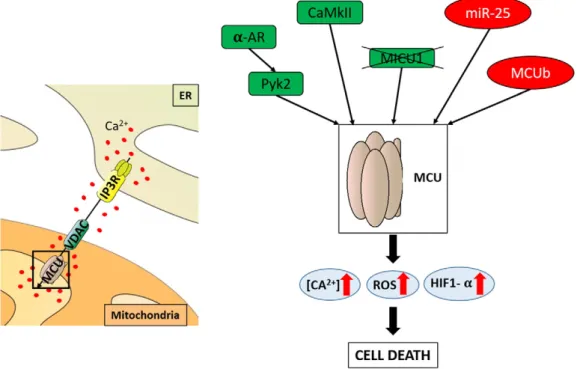
Fig. 2. Proteins that modulate the activity of the MCU complex and cell death. The MCU located in the IMM is responsible for Ca2+
uptake. Modulation of MCU complex subunits and function could increase the probability that cells will undergo apoptotic cell death under stress conditions because of the increased basal ROS levels present during mitochondrial Ca2+
uptake. Proteins that promote Ca2+entry into the mitochondria are shown in green, and proteins that decrease mitochondrial Ca2+uptake and ROS generation are shown in red. The
pathway that activates the MCU complex is inhibited by CaMKII-dependent phosphorylation of the uniporter, and Pyk2 prevents Ca2+overload in the mitochondria, ROS production
and subsequent cell death, which are important in tumor progression. MICU1 knockout cells have increased basal ROS levels during mitochondrial Ca2+
uptake, leading to apoptotic cell death under stress conditions. In contrast, miR-25 (which decreases MCU expression) and MCUb (which acts as an endogenous dominant-negative isoform of MCU) reduce mitochondrial Ca2+
uptake and, consequently, drive resistance to Ca2+
exchanger and tumorigenesis has not been well described.
Neverthe-less, the mitochondrial Ca
2+levels have been increased with the
benzo-diazepine CGP37157, a speci
fic inhibitor of NCLX, to promote
subsequent mitochondrial damage and to induce apoptosis
[66]
;
inter-estingly, these effects have also been observed in a prostate cancer cell
line
[67]
.
As reported above, mitochondrial Ca
2+is primarily transported into
the matrix by MCU. However, Ca
2+must pass through the OMM. The
voltage-dependent anion channel (VDAC) exerts this function. VDAC
is a large channel (2.5
–3 nm) that represents the primary permeability
pathway through which solutes enter the OMM
[68]
. The VDAC channel
has often been referred to as a
“general diffusion pore,” although this
appellation is not accurate. In fact, VDAC
finely regulates several cellular
processes, particularly apoptosis, due to its capacity to allow the
“pas-sage
” of Ca
2+and thus to amplify or diminish the apoptotic response.
In the 2012 study by the Van Remmen group, mitochondrial superoxide
release occurred through VDAC
[69]
.
Because the absence of apoptosis is recognized as one of the
hall-marks of cancer, the mPTP may play a pivotal role in cancer.
Accordingly, the role of the mPTP in cell death has been investigated
in several cancer types, including colon cancer, osteosarcomas and
leu-kemia. However, despite the great potential of this target as a cancer
treatment, the use of mPTP modulators during tumorigenesis has not
shown great ef
ficacy, likely because the molecular structure of mPTP is
not well understood and not all of its components have been identi
fied.
The C subunit of mitochondrial F1/FO ATP synthase was recently shown
to be a fundamental regulator of mPTP activity
[70-72]
. Indeed, upon its
pharmacological inhibition, the induction of the MPT by Ca
2+is
inhibited. Thus, this mPTP member may be a novel target for promising
new anti-cancer therapies
[73]
.
6. Oncogene- and oncosuppressor-mediated modulation of
mito-chondrial Ca
2+homeostasis
As reported above, only a few proteins regulate the proper in
flux and
ef
flux of Ca
2+from the mitochondria. In contrast, several proteins
reg-ulate Ca
2+flux towards this organelle with the ultimate purpose of
ac-tivating/inhibiting apoptosis. Not surprisingly, most of these proteins
are oncogenes and tumor suppressors.
Historically, the proto-oncogene Bcl-2 was the
first protein to be
identi
fied as an anti-apoptotic protein capable of preventing apoptosis
in a Ca
2+-dependent manner
[28,74]
. For example, alterations of Bcl-2
function have been identi
fied in several leukemias and carcinomas
[75]
. Bcl-2 is a member of the large Bcl-2-protein family, which contains
numerous anti-apoptotic and pro-apoptotic members
[76]
. Notably, the
anti-apoptotic protein Bcl-XL, which is deregulated in several cancer
types, blocks the apoptosis pathway by neutralizing pro-apoptotic
Bcl-2 members, such as Bak, Bax, Bid and Bim
[77]
. In addition, Bcl-XL exerts
its anti-apoptotic functions by regulating the activity of Ca
2+channels,
including IP3Rs and VDAC isoforms
[78]
.
Furthermore, the protein kinase B (PKB)/Akt protein regulates Ca
2+flux inside the mitochondria. In fact, this well-known oncogene reduces
Ca
2+release from the ER by modulating IP3R activity, thus protecting
cells from apoptotic stimuli
[79]
. Furthermore, the PKB/Akt signaling
pathway is regulated by PTEN
[80]
. Notably, PTEN is a tumor suppressor
whose expression is lost in a plethora of human malignancies (e.g.,
breast and prostate cancer). A lack of PTEN increases PKB/Akt activity,
which in turn regulates the reduction of mitochondrial Ca
2+accumula-tion due to a decrease in Ca
2+release from the ER and interferes with
the apoptotic machinery
[81]
.
In addition to its transcriptional role, the tumor suppressor p53
modulates apoptosis by controlling Ca
2+flux into the mitochondria. In
fact, p53 was shown to cooperate with sarco/endoplasmic reticulum
Ca
2+-ATPase (SERCA) pumps at the ER-MAM compartment
[17]
. As a
result, the activity of these pumps increases to increase the amount of
Ca
2+released from the ER. Thus, mitochondria are overloaded with
Ca
2+, and the apoptotic program is triggered. However, p53 not only
co-operates with SERCA pumps at the MAM level but also acts as a bridge to
maintain the correct PML localization at ER-mitochondria sites
[36]
.
No-tably, the oncosuppressor PML, which is deregulated in several human
cancers
[82,83]
, regulates Ca
2+-dependent apoptosis
[84]
. In the
ab-sence of p53, PML is no longer localized at the juxtapositions between
mitochondria and MAMs, and its pro-apoptotic functions are lost. In
ad-dition, as shown in the study by Missiroli et al.
[36]
,
PML-p53-depen-dent regulation of mitochondrial homeostasis is also a crucial element
in the autophagic pathway, thus highlighting the possibility of creating
pioneering therapeutic strategies against malignancies characterized by
the absence/mutations of PML and p53.
Likewise, p66Shc and the putative oncogene protein kinase C-
β
(PKC-
β) cooperate to preserve the physiological levels of apoptosis
and autophagy. Indeed, upon activation by PKC-
β, p66Shc becomes
lo-calized to the mitochondrial compartment, where it in
fluences
apopto-sis and autophagy by regulating the mitochondrial Ca
2+levels and
bioenergetics
[85,86]
.
Ca
2+-dependent apoptosis may also be regulated by at least two
members of the Ras family. In fact, altered expression of K- and H-Ras
is suf
ficient to reduce Ca
2+transfer from the ER to the mitochondria,
thus blocking the apoptotic program
[87]
. Remarkably, this oncogene
is mutated in 33% of cancers, including pancreatic, colorectal and lung
cancers
[88]
.
7. MAMs: structure and composition
Speci
fic organization of the intracellular organelles enables direct
communication between various compartments within the cell.
Among the different direct interactions or
“close contacts” between
cel-lular organelles, MAMs have recently attracted the attention of many
re-searchers, as represented by the growing number of publications
describing the important roles of MAMs in physiology and pathology.
MAMs consist of regions of the ER involved in direct interactions with
the mitochondria. However, proteins from other cellular compartments
have also been found in MAMs, suggesting that MAMs also form close
contacts with other intracellular structures in addition to the ER. For
in-stance, plasma membrane (PM) proteins are observed in MAMs,
indicat-ing the presence of close contacts between the mitochondria and the PM
[89]
. According to numerous studies, mitochondria-ER contact sites are
dynamic structures. However, because we are able to isolate these
struc-tures, the interactions between these membranes are strong, and they
are not destroyed during isolation procedures. Plasma
membrane-associated membranes (PAMs)
[90]
and ER-mitochondria encounter
structures (ERMES)
[91]
are other examples of the physical and
func-tional contacts that are isolated during cellular subfractionation.
Most researchers believe that the
first evidence showing that the
mi-tochondria and ER are closely positioned at some regions comes from
the early 1970s in the studies by Franke and Kartenbeck
[92]
and
Morre et al.
[93]
. However, the
first reports on the direct association
be-tween the mitochondria and ER date back to as early as the late 1950s
[94]
. Almost simultaneously with the aforementioned observations
from the 1970s, Lewis and Tata
[95]
observed that a fraction of the ER
was observed in low-speed centrifugation pellets containing the
mito-chondrial fraction during subfractionation of rat liver homogenates.
Based on this observation, we acknowledge this paper as the
first report
to describe a MAM isolation procedure. Almost twenty years later, in the
early 1990s, the Vance group made great progress in the MAM
field by
presenting a detailed protocol describing the isolation of pure MAM
fractions in a series of articles published in J. Biol. Chem.
[96]
, which
was improved upon by Meier et al.
[97]
ten years later. Over the years,
the MAM isolation method was improved and optimized to enable the
isolation of MAMs from different animal tissues and cell cultures
[98]
.
The existence of the MAM fraction is not an exclusive characteristic of
mammalian cells; close interactions between mitochondria and the ER
have also been described in yeast
[99]
. Interestingly, similar contacts
between mitochondria and the ER have been described for chloroplasts
and the ER in plants
[100]
.
In addition to the development of more re
fined protocols for
isolat-ing pure MAM fractions, the list of proteins present at mitochondria-ER
contact sites increases every year. Although many proteins localized at
the MAM have been identi
fied, we have not determined which proteins
can be used as universal MAM markers because some MAM proteins are
only present in certain organs, tissues or cell types. Another problem is
the observation that no protein is exclusively localized to the MAM
frac-tion. Instead, the localization of a speci
fic protein at the MAM is only
ap-propriately termed as enriched because these proteins are also present
in other cellular compartments. Regarding the molecular composition
of the MAM fraction, an article by Poston et al.
[89]
presents a detailed
proteomic analysis of the MAM. These authors detected and classi
fied
approximately 1200 proteins from the MAM fraction isolated from a
mouse brain and con
firmed that the MAM fraction contains proteins
characteristic of the PM and the Golgi apparatus (24% and 6%,
respec-tively, of the total proteins detected in MAMs)
[89]
. Detailed, systematic
lists of proteins present in the MAM fraction grouped based on their
function have been presented by several groups, including Schon and
Area-Gomez
[101]
, Poston et al.
[89]
, Vance
[102]
, Marchi et al.
[103]
,
Patergnani et al.
[104]
, Raturi and Simmen
[105]
, and Giorgi et al.
[20]
.
Based on the long list of proteins found in the MAM fraction or that
translocate to the MAMs under certain conditions, MAMs seem to play
important roles in various processes. Originally, the MAM fraction was
considered important for lipid synthesis and traf
ficking (long-chain
fatty acid coenzyme A ligase-1 (FACL-1) and -4, phosphatidylserine
synthase-1 (PSS-1) and -2, serine active site containing 1 (SERAC1),
fatty acid transport protein 4 (FATP4), acyl-CoA desaturase,
phosphati-dylethanolamine N-methyltransferase 2 (PEMT2) and many other
pro-teins present in MAM involved in this process are reviewed in
[101]
)
and Ca
2+handling (e.g., IP3R, ryanodine receptor, sigma-1 receptor
(SIG1R), and promyelocytic leukemia protein (PML)). MAMs were
later linked to the modulation of mitochondrial morphology
(mitochon-dria-shaping proteins and chaperone proteins (MFN-1 and -2)),
apopto-sis (Bcl-2, hematopoietic cell-speci
fic Lyn substrate1 (HCLS1)-binding
protein 3 (HS1BP3)), mitochondrial contact site formation (VDAC and
adenine nucleotide translocase (ANT)), protein folding (calnexin
(CNX)) and sorting (phosphofurin acidic cluster sorting protein 2
(PACS-2)), ER stress (glucose-regulated protein 75-kDa (GRP75) and
endoplasmic reticulum resident protein 44 (ERp44)), in
flammation
(in
flammasome components: NALP3, adaptor ASC and thioredoxin
interacting protein (TXNIP)), autophagy (pre-autophagosome/
autophagosome markers (ATG14 and ATG5), and p66Shc) and the
cellular response to oxidative stress (p66Shc protein, an Ero1
α). The
presence of these important proteins involved in crucial cellular
pro-cesses explains why alterations in MAM composition are related to
the pathogenesis of different disorders
[105]
, including type-2 diabetes
(mTOR complex 2 (mTORC2) and MAM-associated Akt), and several
neuronal-based diseases, such as Parkinson's disease and Huntington's
disease, and neurodegenerative diseases, such as schizophrenia,
dementia and seizures. Moreover, MAMs have been proposed to be
involved in familial Alzheimer's disease (FAD)
[106,107]
and
GM1-gangliosidosis
[108]
.
8. MAM proteins modulate Ca
2+homeostasis, cell death and
tumorigenesis (
Table 1
)
The ER and mitochondrial networks control different aspects of
cellular metabolism, and, through their both dynamic and close
interactions, are also involved in the transmission of physiological
and pathological Ca
2+and ROS signals directly from the ER to the
mitochondria.
Table 1
MAM proteins involved in tumorigenesis and tumor progression. A = amplification; M = mutation; D = deletion.
Protein Gene expression in cancer Relation to Ca2+ References
Akt Pancreas (A, D), Breast (M), Prostate (A) Inhibition of Ca2+
release from the ER [79,124]
AMBRA1 Breast (A), Prostate (A, M) Involved in regulating TPC-dependent calcium release [5]
AMPK Skin (M), Prostate (A), Pancreas (A, D) Chronic calcium exposure decreases AMPK activity [35]
Bad Prostate (A), Pancreas (A, D), Uterus (A, D) Sensitizes the mitochondria to Ca2+, making them more susceptible to
Ca2+release from the ER
[125]
Bak Breast (A), Prostate (A), Skin (A, M) Regulate calcium leakage from the endoplasmic reticulum [126]
Bax Prostate (A), Pancreas (A, D), Breast (A), Stomach (M) Regulate calcium leakage from the endoplasmic reticulum [126]
Bcl-XL Uterus (A, M), Breast (A), Prostate (A), Colon (A), Nervous System (D, M)
Acts on Bax inhibitor-1 (BI-1) to increase Ca2+
leakage from the ER, with BI-1 acting as a Ca2+
channel or as an IP3R sensitizer
[74,127]
Bcl-2 B-cell (M), Central Nervous System (A, M), Pancreas (D), Breast (A) Induction of Ca2+
release from the ER [74]
Bid Prostate (A), Nervous System (A) Regulates calcium concentrations and homeostasis in the ER [74]
Bim Prostate (A, M, D), Breast (A, M), Uterus (M, A) Bim-deficient cells exhibit severe defects in calcium release [8,75,128]
BECLIN1 Prostate (A), Breast (A) Activated by increased [Ca2+
]iand may induce autophagy [5]
BMF Uterus (B, M, A), Prostate (A), Supports Bim in some cell death processes [8]
BNIP3 Prostate (A, D), Pancreas (A, D) Induces atypical cell death with features of both apoptosis and necrosis [7]
MAVS Breast (A), Prostate (A), Ovary (A, D), Stomach (M, A) Lead to defects in mitochondrial calcium [12]
MFN-1 Lung (A, M), Ovary (A), Esophagus (A, M), Breast (A), Head (A, M) Important as a mediator of mitochondrial fusion [129,130]
MFN-2 Pancreas (A, D), Esophagus (M, A, D), Prostate (M, A, D) Facilitates calcium cross-talk between the ER and mitochondria [129,130]
mTOR Skin (M), Uterus (M, A), Prostate (M, A, D) Intracellular Ca2+
signaling is a crucial component in the canonical mTOR-dependent autophagy pathway
[33,35]
NIX Prostate (D), Ovary (D, A) Increase ER/SR calcium stores in cardiac myocytes [9]
NLRP3 Breast (A), Prostate (A), Skin (M), Lung (M, A) Localization of PML at ER/MAM contact sites is required for its pro-apoptotic activity via a calcium (Ca2+
)-mediated pathway
[12]
PKC-β Breast (A), Prostate (A), Lung (M), Skin (M) Binds Ca2+
through its C2 domain [131]
PML Prostate (M), Colon (M), Breast (A), Lung (M, A), Uterus (A, M) Regulates apoptosis in the ER by modulating calcium release, negative regulator of Akt
[36,83,84]
PP2a Prostate (A), Central Nervous System (M), Pancreas (A, D) Regulates calcium transients in cardiomyocytes [106,132]
PTEN Uterus (M), Prostate (M, D), Head (M, D), Stomach (M), Breast (A, M), Pancreas (M)
Regulates Ca2+
release via IP3R3 [80,81]
p53 Almost all Interacts with the C-terminal portion of the SERCA pump, increasing ER Ca2+loading
[17,19,35]
p66 Prostate (A), Breast (A), Esophagus (A, M, D) Involved in the cellular response to oxidative stress [85,121]
HRAS Pancreas (A, D), Breast (A), Urinary Tract (M), Head (M) Regulation of Ca2+
signaling [87]
RIP1 Skin (A), Prostate (A), Breast (A) Phosphorylated by increased cytoplasmic calcium concentrations [133]
Thus, their contact sites are considered specialized microdomains for
the transfer of Ca
2+signals. Ca
2+ions released from the ER by IP3Rs
cross the OMM
[2]
, which is freely permeable through VDACs, move
to the IMM and accumulate in the matrix via the MCU complex in a
pro-cess mediated by local Ca
2+sites of accumulation, which overcome the
apparent low Ca
2+af
finity of the MCU
[109]
. Nevertheless, if excessive
Ca
2+in
flux occurs, pro-apoptotic factors, such SMAC/DIABLO,
cyto-chrome C, and AIF may be released into the cytosol, resulting in
apopto-sis triggered by the opening of the mPTP
[110,111]
.
MAMs represent platforms for the anchoring of many pro- and
anti-apoptotic factors involved in tumor regulation.
This role was previously de
fined for the serine/threonine kinase Akt
[112]
, which is physically and functionally linked to both the ER and
mi-tochondria. Akt phosphorylates numerous proteins, including members
of the Bcl-2 family (i.e., Bad and Bax), to activate its anti-apoptotic
func-tion, and hexokinase 2 (HK2), which phosphorylates VDAC1 to prevent
Ca
2+-dependent opening of the mPTP and the release of pro-apoptotic
proteins
[113]
.
Bcl-2 is an oncoprotein that has a central role in the regulation of
ap-optosis because of its capacity to delay or block the programmed death
pathway in different cell types. Bcl-2 overexpresssion reduces the
steady state Ca
2+levels within the ER, resulting in reduced
mitochon-drial fragmentation and the initiation of apoptosis
[28]
. Bax and Bak
are members of the Bcl-2 family that control apoptosis from the ER
and mitochondria in response to Ca
2+stimulation
[114]
.
As shown in a recent study by our group, myeloid cell leukemia
pro-tein 1-long isoform (Mcl-1L) is highly expressed in human malignancies
and is localized to the mitochondrial membrane. This localization is
con-sistent with its role in the control of key mitochondrial events during
apoptosis to counteract the activity of the pro-apoptotic proteins Bak
and Bax
[115]
.
The tumor suppressor PTEN, one of the most commonly lost or
mu-tated tumor suppressor genes in human cancers, also localizes to MAMs.
PTEN is a dual-speci
fic phosphatase for lipids and proteins that exhibits
enhanced ER localization during Ca
2+-dependent apoptosis. In this case,
PTEN interacts with IP3R3 to counteract Akt-mediated phosphorylation
of the receptors and induce a subsequent increase in Ca
2+transfer from
the ER to the mitochondria to trigger apoptosis in a protein
phospha-tase-dependent manner
[81]
.
IP3R3 is the favorite target of another important tumor suppressor,
PML, which is localized to the nucleus and cytosol and is associated
with the ER and MAMs
[84]
. Indeed, PML forms a multiprotein complex
with IP3R3, Akt and the protein phosphatase PP2a to modulate the
ap-optotic pathway triggered by Ca
2+accumulation in the matrix. The
sup-pression of this protein blocks PP2a localization at MAMs, which in turn
does not prevent Akt-mediated IP3R3 phosphorylation. Therefore,
IP3R3 hyperphosphorylation inhibits Ca
2+transfer from the ER to the
mitochondria, thereby inhibiting apoptosis
[116]
.
Based on recent studies, fetal and adult testis expressed 1 (FATE1), a
cancer-testis antigen that has a role in regulating the ER-mitochondria
distance and Ca
2+uptake by the mitochondria,
[117]
inhibits
pro-apo-ptotic signaling in many cancer cell lines by destabilizing BIK, a
pro-ap-optotic protein
[118]
.
The interactions between the mitochondria and the ER are
modulat-ed by different proteins, including the mitochondria-shaping proteins
MFN-1/-2 (mitofusin-1/-2). In particular, the absence of MFN-2 changes
the morphology of the ER and decreases the interactions between the
mitochondria and ER by 40%, causing altered transfer of Ca
2+signals
to the mitochondria
[119]
.
Moreover, the mitochondria and the ER are the major sites of ROS
production, and many regulators of the oxidative state of the cell are
lo-calized at the MAM
[120]
. p66Shc plays a key role in this process, as it is
a cytosolic protein involved in the cellular response to oxidative stress
and tumorigenicity that has recently been identi
fied at
mitochondria-ER association sites. The increase in p66Shc induced by elevated
intra-cellular ROS levels is, in turn, triggered by steroid-induced signaling,
which promotes cell proliferation in prostate cancer cells
[121]
. Instead,
the oxidative cellular environment promotes the phosphorylation of
p66Shc at Ser36 that migrates to the mitochondrial matrix after being
recognized by the prolyl-isomerase Pin1 and dephosphorylated by
phosphatase 2A. Here, the protein perturbs mitochondrial function
that ultimately leads to apoptosis
[85]
. Based on other data from the
Lebiedzinska group, a signi
ficant portion of p66Shc is also present in
MAM fractions and its levels increase in an age-dependent manner
[122]
.
Moreover, according to the recent
findings by Anelli et al.
[123]
,
Ero1
α, a quality controller of oxidative protein folding in the ER, is
enriched in MAMs and regulates Ca
2+flux. The levels of redox-active
Ero1
α impact Ca
2+storage and IP3R-dependent
flux. Its silencing
inhibits Ca
2+re-uptake by the mitochondria, likely by modifying MCU
activity. The overexpression of redox-active Ero1
α increases passive
Ca
2+ef
flux from the ER, thus reducing the [Ca
2+]
ER