Università degli Studi del Piemonte Orientale
“Amedeo Avogadro”
Dipartimento di Scienze e Innovazione Tecnologica
Dottorato di Ricerca in Chemistry & Biology
curriculum: Drug Discovery and Development
XXXII ciclo a.a. 2018-2019
SSD: CHIM/03
Anticancer activity of multifunctional Pt(IV)
prodrugs
Beatrice Rangone
Supervised by Prof. Domenico Osella
PhD program co-ordinator Prof. Luigi Panza
Università degli Studi del Piemonte Orientale
“Amedeo Avogadro”
Dipartimento di Scienze e Innovazione Tecnologica
Dottorato di Ricerca in Chemistry & Biology
curriculum: Drug Discovery and Development
XXXII ciclo a.a. 2018-2019
SSD: CHIM/03
Anticancer activity of multifunctional Pt(IV)
prodrugs
Beatrice Rangone
Supervised by Prof. Domenico Osella
Omnia venenum sunt: nec sine veneno quicquam existit.
Dosis sola facit, ut venenum non fit. PARACELSO
Contents
Chapter 1
Introduction 1.1 The tumorigenesis 1 1.2 Cell cycle 3 1.3 Cell death 5 1.3.1 Apoptosis 5 1.3.2 Autophagy 8 1.3.3 Necrosis 111.3.4 Non-lethal processes: cell senescence and mitotic catastrophe 11
1.4 The anticancer chemotherapy 13
1.5 Platinum-based anticancer drugs 15
1.5.1 History of cisplatin 15
1.5.2 Cisplatin mechanism of action 16
1.5.3 Cisplatin side effects and chemoresistance 19
1.5.4 Second and third generation of platinum anticancer drugs 22
1.5.5 Platinum(IV) prodrugs 25
References 28
Chapter 2
Outline of the thesis 33Chapter 3
Unsymmetric cisplatin-based Pt(IV) derivative containing a novel Histone DeACetylase inhibitor (HDACi): a very efficient multi-action antitumor prodrug candidate
3.1 Introduction 36
3.2 Material and methods 38
3.2.1 Cell culture 38
3.2.2 Compounds and drug candidates 39
3.2.3 Antiproliferative activity 40
3.2.4 Cellular accumulation and DNA platination 41
3.2.5 HDAC assay 43
3.2.6 Chromatin staining 44
3.2.7 Quantitative PCR (RT-qPCR) 44
3.2.8 Caspase 3/7 activity 47
3.2.9 Experiments with animals 47
3.2.10 In vivo antitumor activity 48
3.2.11 Statistical analysis 49
3.3 Results and discussion 49
3.3.1 Antiproliferative activity 49
3.3.2 Cellular uptake 52
3.3.3 Epigenetic activity 55
3.3.4 Cell death 58
3.3.5 In vivo tumor growth inhibition 59
3.4 Conclusions 62
Chapter 4
Pt(IV) multifunctional 1R,2R-diamine-based Pt(IV) prodrug containing the novel HDACi, POA: induction of immunogenic cell death on colon cancer
4.1 Introduction 66
4.2 Material and methods 68
4.2.1 Cell culture 68
4.2.2 Compounds and drug candidates 68
4.2.3 Antiproliferative activity 69
4.2.4 Cellular accumulation 69
4.2.5 Chromatin staining 70
4.2.6 In vivo experiments 70
4.2.7 Statistical analysis 72
4.3 Results and discussion 72
4.3.1 Antiproliferative activity 72
4.3.2 Cellular accumulation 74
4.3.3 Epigenetic activity 75
4.3.4 In vivo activity 76
4.3.5 Pt accumulation and organ histology 80
4.4 Conclusions 89
References 90
Chapter 5
Selective activity against human colon cancer cell lines of cyclohexane- 1R,2R-diamine-based Pt(IV) dicarboxylato anticancer prodrugs 5.1 Introduction 945.2 Material and methods 95
5.2.2 Compounds and drug candidates 96
5.2.3 Antiproliferative activity 96
5.2.4 Cellular uptake 97
5.2.5 3D-spheroids model 98
5.2.6 Statistical analysis 98
5.3 Results and discussion 99
5.3.1 Antiproliferative activity 99
5.3.2 Cellular uptake 102
5.3.3 Prolonged antitumor effect using 3D spheroids model 104
5.4 Conclusions 109
References 110
Chapter 6
Antiproliferative Activity of Pt(IV) Conjugates Containing the Non-Steroidal Anti-Inflammatory Drugs (NSAIDs) Ketoprofen and Naproxen 6.1 Introduction 1126.2 Material and methods 114
6.2.1 Cell culture 114
6.2.2 Compounds and drug candidates 115
6.2.3 Antiproliferative activity and combination index 116
6.2.4 Cellular uptake 116
6.2.5 Quantitative PCR (RT-qPCR) 117
6.2.6 Statistical analysis 119
6.3 Results and discussion 119
6.3.1 Antiproliferative activity and combination index 119
6.3.2 Cellular uptake 125
6.4 Conclusions 129 References 130
Chapter 7
Concluding Remarks and Perspectives 133
List of publications
1381
Chapter 1
Introduction
1.1 The tumorigenesis
Most tumors do not result from a single phenomenon, hence tumorigenesis is a complex and multifactorial process characterized by the acquisition of several abnormal functionalities, needed by the cells to evolve through different premalignant steps into invasive cancers. So, they originate from normal cells that have experienced a transformation process as a result of mutations induced by errors in cell replication or by exogenous factors, as X-rays, UV light, chemicals and oncoviruses (Figure 1.1).
Figure 1.1: The transformation process of normal cells into malignant ones. The picture is a modification of that reported in literature [1].
These alterations confer to mutant cells a selective growth advantage, allowing them to proliferate out of control. This process, called evolutionary clonal selection, makes the cell to acquire malignant traits, which are the hallmarks of tumor [1]. The normal cell that acquire the initial cancer-promoting mutation is considered the “cell of origin”. On the contrary, the subset of self-renewing cancer cells that uniquely
2
sustain tumor malignant growth are known as cancer stem cells (CSC). These cells have proven to be a common component in most tumors and several hypotheses have been proposed about the origin of CSC, either from stem or differentiated cells; however, until now little is known about them [2].
Tumorigenesis is the result of mutations in protein-encoding genes, which can be classified into two classes: proto-oncogenes and tumor suppressor genes. Proto-oncogenes encode for proteins which promote cell growth and proliferation, including kinases, tyrosine kinase receptors, transcription and growth factors and GTPase regulators. Their mutated form are known as oncogenes. A representative example is the RAS protein family, chronically active in 30% of cancers, due to inactivating mutations in one of its negative regulators or missense mutations in its gene. The components of downstream cascades mediating RAS functions could be mutated in similar way in different type of cancers. Thus, mutations in RAS, in its regulatory proteins and in its downstream pathways result in a panel of effects which enhance growth and proliferation [3]. On the other hand, tumor suppressor genes encode for proteins which operate in different ways in order to limit cell growth and proliferation. The most commonly mutated (in over 50% of sequenced malignancies) tumor suppressor gene encodes for P53. This protein acts as a detector of a variety of stresses, arresting further progression of cell cycle, being involved in repair mechanism, or eventually triggering apoptosis if the degree of damage is excessive [4]. Another key tumor suppressor gene encodes for retinoblastoma (RB) protein, which is commonly inactivated in a great number of cancers. It is a critical regulator of cell-cycle progression and its absence leads to persistent cell proliferation [5]. Thus, mutations, which affect both oncogenes and tumor suppressor genes, cause uncontrolled cell proliferation and, consequently, alterations in tissue homeostasis. Despite cancer is not a single disease, but exhibit a strong heterogeneity in cellular morphology, proliferative activity, genetic lesions and response to therapy, generally malignant cells show the following features:
3
• selective growth and proliferative advantage, due to self-sufficiency in growth signals and insensitivity to antigrowth pathways;
• altered stress response leading to overall cell survival, for example defects in DNA repair machinery, escape of cell death (both apoptosis and autophagy) and loss of senescence ability;
• sustained angiogenesis and other modes of tumor neo-vascularization; • acquisition of the capacity to invade surrounding tissue and to form
secondary sites of growth (metastasis);
• metabolic rewiring that represents a selective advantage during initiation and progression of cancer, as the switch from aerobic to anaerobic metabolism, phenomenon known as “Warburg effect”;
• creation of dynamic and rich microenvironment;
• ability to escape immune surveillance, supported by the fact that primary and acquired immunodeficiency in mice and humans are related with greater susceptibility to carcinogenesis [1].
1.2 Cell cycle
Cellular replication is regulated by the cell cycle, a tightly ruled process with multifactorial and very complex control mechanisms. It is divided into several stages, each of them with very typical functional properties. Upon receiving the mitogenic stimulus the eukaryotic cell starts its cell cycle in G1 (gap 1) phase: the cell shows a rise in the expression of protein and in its metabolism, in order to set the conditions needed for the following S (synthesis) phase, which is the step involving DNA replication. This is followed by the G2 (gap 2) phase, during which the accuracy of chromosome replication is checked and further synthesis of the proteins occurs. G1, S, and G2 form the interphase. Then, the M (mitotic) phase is characterized by the separation of chromosomes in the nucleus and, finally, by the division of cytoplasm, called cytokinesis (Figure 1.2).
4
Figure 1.2: The cell cycle events. The picture is a modification of that reported in literature [6].
After M phase, cells can enter into a new G1 phase or proceed to a quiescent state, known as G0, which can persist for a long time, or even for the rest of cell life in the case of some somatic cells. The strictly correlation between a step and the correct completion of the preceding one allows the successful progression of cell cycle. The interdependency is related to surveillance mechanisms, namely checkpoints, which prevent the propagation of genetic abnormalities. If cell damage cannot be repaired, checkpoint signaling may induce apoptosis. This control system is based on two groups of proteins: the family of cyclin-dependent kinases (CdKs) and cyclins. CdKs bind cyclins and check their ability to phosphorylate appropriate targets. There are several classes of CdKs and cyclins, depending on the step of the cell cycle where they are activated (Table 1).
Loss of cell cycle regulation leads to uncontrolled cell proliferation, which is an hallmark of cancer [7].
5 CYCLIN-CDK COMPLEX VERTEBRATES CYCLIN CDK PARTNER BUDDING YEAST CYCLIN CDK PARTNER G1-Cdk G1/S-Cdk S-Cdk M-Cdk cyclin D* Cdk4, CdK6 cyclin E Cdk2 cyclin A Cdk2, Cdk1** cyclin B Cdk1 Cln 3 Cdk1** Cln1, 2 Cdk1 Clb5, 6 Cdk1 Clb1, 2, 3, 4 Cdk1
Table 1: The main classes of cyclins and CdKs, referred to vertebrate and budding yeast [6].
1.3 Cell death
Like cell growth and proliferation, cell death plays a crucial role in tissue homeostasis. Defects in the regulation of cell death are related to a variety of human disease, including tumor. In addition, the main purpose of anticancer drugs is selectively inducing cell death in malignant cells only. Different kinds of cell death can be distinguished, according to biochemical and morphological parameters, but they are strictly interconnected and may co-exist, hence often it is difficult to discriminate them [8].
1.3.1 Apoptosis
Apoptosis is a type of programmed cell death and it is a key component of cell growth control. It is fundamental for various processes involved in tissue homeostasis, as embryonic development and organogenesis. Thus, abnormal regulation of apoptosis leads to pathogenesis of several human disorders, particularly its suppression is strictly related to tumorigenesis.
This kind of cell death is characterized by a series of morphological changes, such as cell shrinkage and reduction of cell volume (pyknosis), disruption of organelles membrane, chromatin condensation, nuclear cleavage (karyorhexis) and finally cell fragmentation into apoptotic bodies, which can be engulfed by macrophages.
6
Apoptosis may be triggered by a plethora of signals and it is induced by two major pathways: the intrinsic and extrinsic one (Figure 1.3). Both pathways converge in the activation of a specific series of cysteine aspartyl-protease, known as caspases, which are both the initiators (like caspase 2, -8 , -9 and -10), showing autocatalytic activity, and the downstream effectors (as caspase 3, -6 and -7), activated by proteolytic cleavage by the previous ones. The effector caspases can further cleave other nuclear and cytosolic substrates, progressively activating proteolytic cascades that lead to the biochemical and morphological features of apoptosis.
The intrinsic apoptotic pathway is triggered by intracellular signals, such as DNA damage. Upon these stimuli the activation of pro-apoptotic proteins, as BAX, BAK, BAD and BID, leads to mitochondrial outer membrane permeabilization (MOMP), resulting in the release of proteins from the mitochondrial intermembrane space. Among them cytochrome c is the most important; indeed, once released in cytoplasm, it transiently binds the apoptotic protease-activating factor 1 (Apaf-1), triggering Apaf-1 oligomerization into an heptamer, which exposes the caspase activation and recruitment domain (CARD). This domain binds to pro-caspase 9, forming a multi-protein complex, namely apoptosome, in which pro-caspase 9 auto-activates and subsequently recruits the executioner caspases 3 and -7 to orchestrate apoptosis. This pathway can also be modulate by anti-apoptotic proteins (e.g BCL-2 and IAPs).
On the contrary, the extrinsic pathway is activated by cell transmembrane receptors in the presence of extracellular signals. These death receptors include a subset of tumor necrosis factor (TNF) superfamily, characterized by different protein motifs, called death domains (DD) and death effectors domains (DED). There are three main receptor-systems involved in this apoptotic pathway: Fas, activated by Fas ligand (Fas L); tumor necrosis factor receptor 1 (TNFR1), activated by tumor necrosis factor a (TNF-a); and death receptors 4 and 5 (DR4, DR5), activated by TNF-related apoptosis inducing ligand (TRAIL). After the activation, these receptors attract DD-containing molecules, such as Fas-associated death domain protein (FADD), thus
7
triggering pro-apoptotic pathways. FADD recruits other DD/DED-containing factors, including pro-caspases 8 and -10, promoting the formation of the death inducing complex (DISC) in the cytoplasm. The recruitment of these proteins in DISC favors their self-cleavage and once activated, caspases 8 and -10 directly engage the effector caspases 3, -6 and -7 to perpetrate apoptotic cell death. Alternatively, caspase 8 can activate BID, promoting the intrinsic apoptotic pathway [9] [10].
Figure 1.3: Key step in apoptotic intrinsic and extrinsic signaling pathways. The picture is a modification of that reported in literature [11].
8
1.3.2 Autophagy
Autophagy is another type of programmed cell death. It contributes to cell homeostasis by degrading several intracellular materials, particularly dysfunctional organelles, protein aggregates, and invading pathogens. Similarly to apoptosis, aberrant autophagy seems to be linked to a wide range of human diseases, including cancer. It has been reported that autophagy plays contradictory roles in tumor initiation and progression. Hence, both stimulation and repression of autophagy should be explored as therapeutic approaches, due to the several factors that contribute to tumorigenesis.
The general term autophagy describes the process by which cytoplasmic components are delivered to lysosomes for degradation. Three main autophagy pathways have been elucidated, which may be distinguished by the delivery method of substrates: macroautophagy, microautophagy and chaperone-mediated autophagy (CMA) [12] (Figure 1.4).
Figure 1.4: Three main autophagic pathways. The picture is a modification of that reported in literature [13].
9
Among the three autophagic pathways, macroautophagy represents the main and the most studied one. During this process, an isolation membrane, called phagophore, encloses a small part of unnecessary or dysfunctional cytoplasmic components, such as damaged organelles, forming a double-membraned structure, namely autophagosome. Then, its outer membrane fuses with lysosomes, to evolve into an autolysosome, in which lysosomal enzymes degrade the cytoplasmic substrates (Figure 1.4). Macroautophagy consists of different steps: initiation and nucleation, elongation, maturation, fusion and degradation (Figure 1.5). The machinery is finely regulated by a series of factors, the Atg proteins, which can assemble into many different multimolecular complexes to form autophagosome. Among them, the Unc-51 like kinase 1 (ULK1) complex plays a key role in the initiation of the process. In fact, in response to several stresses, like starvation, the serine-threonine kinase mammalian target of rapamycin (mTOR) is inhibited and dissociates from the ULK1 complex, leading to its activation and to the initiation. The activation of this complex promotes the recruitment of the class III phosphatidylinositol 3 kinase (PI3K), which is essential for the nucleation and assembly of phagophore membrane, through the phosphorylation of key factors, like activating molecule in Beclin-1-regulated protein autophagy 1 (AMBRA) and Beclin-1. The subsequent elongation process involves two conjugation reactions leading to Atg5-Atg12 and microtubule associated protein-1 light chain (LC3)-phosphatidylethanolamine (PE). The product of the second conjugation is essential for the closure of the autophagosome membrane. Finally, in the fusion stage, in which autophagosome fuses with lysosome to form an autolysosome, are involved monomeric GTPases of the Rab family. This process is followed by the lysosomal degradation of the autophagic substrate [9].
10
Figure 1.5: The macroautophagy steps. The picture is a modification of that reported in literature [14].
Conversely, microautophagy is the less studied. It is characterized by the direct engulfment of cytoplasmic portions by the lysosomes (Figure 1.4). Thus, the lysosomal membrane randomly undergoes invagination and differentiation into autophagic tubes, in order to enclose substrates for degradation. The main functions of microautophagy are the maintenance of membrane homeostasis, organellar sizes, and cell survival under nitrogen starvation [15].
The third type of autophagy, CMA, is a process in which the substrates are proteins containing a specific pentapeptide motif which are selectively recognized by a chaperone protein, belonging to the family of Heat Shock Proteins 70 (HSP70), the HSPA8/HSP70. The resulting chaperone-substrate complex binds the lysosomal receptor lysosomal-associated membrane protein type 2A (LAMP-2A), leading to its multimerization, with the help of the lysosomal protein HSP90. Then, the substrates are unfolded and delivered to the lysosome lumen, through the LAMP-2A-enriched translocation complex, where lysosomal proteases degrade them (Figure 1.4). Subsequently, LAMP-2A multimers undergo disassembly and degradation for the next cycle of CMA. This pathway is tightly regulated and can be activated under
11
protracted starvation and a variety of stresses, and it represents a quality control aimed to remove unfolded or damage proteins [16].
1.3.3 Necrosis
Necrosis is another type of cell death without the typical features of apoptosis (cell shrinkage, pyknosis, karyorhexis and formation of apoptotic bodies) and without abundant autophagic vacuolization. On the contrary, the main characteristics of necrosis include an increase of cell volume (oncosis), which finally leads to a disruption of plasma membrane and to disorganized dismantling of swollen organelles. Thus, this type of cell death lacks typical biomarkers, except for the plasma membrane permeabilization. Necrosis is usually regarded as harmful, due to its association with pathological cell loss and to the capacity of necrotic cells to favor local inflammation, which can support tumor growth [17].
1.3.4 Non-lethal processes: cell senescence and mitotic
catastrophe
The molecular machinery involved in cell death is also related to other processes that cannot be considered as cell death per se, including cell senescence and mitotic catastrophe.
Cell senescence is a pathophysiological state during which the cells persistently lose their proliferative ability while remaining metabolically active and viable. Senescent cells show typical morphological features, such as intracellular vacuolization, altered chromatin structure, flattening and cellular or nuclear enlargement. The biochemical markers are: increased lysosomal galactosidase beta (GLB1) activity, inhibition of several CDKs and subsequent dephosphorylation of multiple members of the RB protein family, absence of proliferation markers, activation of the DNA damage response (DDR) machinery, and presence of senescent-associated heterochromatic
12
foci (SAHF). Moreover, senescent cells secrete the so-called senescent-associated secretory phenotype (SAPS), composed by mitogenic and immunomodulatory chemokines, cytokines and growth factors, which is involved in the immunological clearance of these cells, and also modulate the activity of other cells with an intact proliferative capacity. Hence, cell senescence seems to contribute to embryogenesis, as well as to several adult pathophysiological processes, such as tissue regeneration and repair, immunity and tumor suppression. Particularly, cell senescence has been reported to occur in response to potentially tumorigenic events (as tumor suppressor genes inactivation or oncogenes activation) and various sublethal cell insults (including DNA damage, telomere shortening, mitochondrial dysfunctions). However senescent cells are also implicated in the side effects of some chemotherapeutic treatments and in the recurrence of specific cancers, thus senescence plays a controversial role in cancer progression, acting both as a tumor suppressor and promoter.
Mitotic catastrophe is a regulated mechanism, hampering the proliferation and the survival of cells that cannot complete mitosis, due to extensive DNA damage, defects in the mitotic machinery and failure of cell cycle checkpoints. Mitotic defects can originate both from exogenous and endogenous sources, and the main alterations driving aberrant mitosis can derive from other cell cycle phases, especially the S phase. This process is morphologically characterized by typical nuclear alterations, as multinucleation, macro- and micronucleation, while the molecular mechanisms triggering mitotic catastrophe cascade are still unclear, but probably involve p53 in many cell types. The destiny of cells undergoing mitotic catastrophe appears to be related to the period spent under mitotic arrest, and their ability to escape from abnormal mitosis. Hence, the arrested cells may undergo cell death (most often intrinsic apoptosis) or enter cell senescence, after activating some cell cycle checkpoints. Consequently, the abrogation of mitotic catastrophe constitutes a fundamental event during tumorigenic transformation and progression [8] [17].
13
1.4 The anticancer chemotherapy
The oldest case of cancer was described around 3000 BC in an Egyptian papyrus, in which eight cases of breast tumors were recorded along with their treatment by cauterization. Thus, surgical resections were historically the main way of cancer treatment. Later, in 1896 X-rays began to be used for recurrent breast carcinoma therapy, and since then radiotherapy and surgery represented the main treatments. Only since 1950 chemotherapy has become a valid therapeutic option, with the discovery of nitrogen mustard (now regarded as the first chemotherapeutic agent) and its application for lymphomas and solid tumors therapy. This event led to a rapid development of various chemotherapeutics. However, despite the advances in this field, nowadays cancer remains one of the major cause of death worldwide, partly due to an ageing global population [18].
The ultimate goal of conventional anticancer chemotherapeutics is to induce malignant cells to die or arrest their proliferation. Hence, they mainly modify, directly or indirectly, the cell cycle. Anticancer agents may act as cytostatic or cytotoxic drugs. Cytostatic drugs stop the cancer cells proliferation, by inhibiting signaling pathways related to processes as cancer growth, angiogenesis, invasion and metastasis. For example the current therapies include: receptor antagonists, antigrowth factors antibodies, small tyrosine kinase inhibitors and anti-receptor monoclonal antibodies [19].
Conversely, cytotoxic agents target rapidly proliferating cells, as cancer cells, leading to cell death. These represent most of anticancer drugs and, according to their main mechanism of action, they can be divided into:
• alkylating agents, which react with DNA, chemically modifying its structure; • antimetabolites, which inhibits DNA and RNA synthesis;
• topoisomerase inhibitors, which interfere with the correct DNA unwinding during replication and transcription;
14
• microtubular poisons, which hamper the polymerization and depolymerization of tubulin, thus inhibiting mitosis;
• cytotoxic antibiotics, which exert anticancer effects by several mechanisms, such as DNA intercalation and over production of reactive oxygen species (ROS) [20].
Nowadays, immunostimulating- and targeted-therapies are considered among the most promising strategies to get high therapeutic antitumor effects with mild systemic toxicity. However, traditional chemotherapy is still one of the treatments of choice for several highly aggressive cancers. Most of such chemotherapeutic schedules consist of the so-called combination therapy (i.e. the administration of two or more additive, or better synergistic, drugs together). In order to improve patient compliance, the different drugs are usually incorporated into the same formulation. However, this is not a simple task because of pharmacokinetic differences between drugs; moreover, possible interactions between the individual components can occur. Thus, a more effective strategy to reach such a goal may involve the preparation of a single drug containing different agents, reversibly bonded, also called “combo” [21].
15
1.5 Platinum-based anticancer drugs
1.5.1 History of cisplatin
Cisplatin, cis-diamminedichloridoplatinum(II), is a square-planar complex of Pt(II) coordinated to two chloride ligands (leaving groups) and two ammine ligands (carrier groups). The prefix cis indicate the geometry of this complex, where two identical ligands are on the same side with respect to the metal, differentiating this from its trans isomer (Figure 1.6).
Figure 1.6: Geometric isomers of diamminedichloridoplatinum(II). a) Cisplatin and b) Transplatin.
Cisplatin was synthesized for the first time in 1845 by Michele Peyrone, and, therefore, it was known as Peyrone’s chloride since the end of the nineteenth century. Then Alfred Werner elucidated its chemical structure for the first time in 1893. However, its anticancer properties were serendipitously discovered in 1965 by the physicist Barnett Rosenberg at the Michigan State University, during the investigation of the role of electric or magnetic field in Escherichia Coli cell division. He observed an inhibition of cell division, not due to the electric field, but to the complexes produced by the electrolysis from the platinum electrodes in contact with a growth medium containing ammonium chloride. Among the possible species, the cis-isomer of [PtIICl
2(NH3)2], i.e cisplatin, was identified as the major bioactive
species. The results suggested that, since this molecule was able to stop bacterial cell growth, it might be active also against proliferation of cancer cells. As a result, in 1968 cisplatin showed the ability to cause a strong tumor regression when
16
administered intraperitoneally to mice bearing sarcoma-180 [22] [23]. After further in vivo tests, this compound entered clinical trials and the first patient was treated in 1971. However cisplatin was approved by the Food and Drug Administration (FDA) as anticancer agents in 1978. Since then, cisplatin was employed for the treatment of a wide range of solid tumors, including bladder, head and neck, lung, ovarian and testicular cancers (in this case cure rates have overcome 95%) [24].
The trans isomer is much less active than the cis homologue and so it is not used as an anticancer agent.
1.5.2 Cisplatin mechanism of action
Cisplatin is administered to patients by intravenous infusion over a period of six to eight hours. In the bloodstream the compound does not undergo appreciable aquation reaction (hydrolysis) due to the high concentration of chloride ions (about 100 mM), although cisplatin can react with plasmatic proteins, as human serum albumin, and extracellular enzymes.
The molecular mechanism by which cisplatin enters cells is not completely clarified yet. Being a neutral molecule with moderate molecular weight, cisplatin can cross the cell membrane by passive diffusion, although active transporters, like copper transporter-1 (Ctr1), and organic cationic transporters (OCT), are also involved in cisplatin uptake [25] [26].
Once inside the cytoplasm, where the concentration of chloride ions drops to 2-10 mM, cisplatin undergoes aquation reaction with the replacement of one (monoaqua species) or two (diaqua species) chlorido groups with water molecules. The rate of the aquation of the first chloride ion at 37°C is 7 x 10⁻³ min-1 (half-life time is about
120 min) while the substitution of the second chloride ligand is two times slower [27].
Once inside the cell, cisplatin interacts with different biomolecules but its main pharmacological target is the DNA. The mono- and di-aqua species, positively
17
charged, represent the active forms of the complex and covalently bind to nucleophilic sites on the DNA strand. In particular platinum binds nitrogen in position 7 (N7) of the imidazole ring of the purine bases, preferentially guanine (G) and, to a lesser extent, adenine (A). Cisplatin is able to form monofunctional or bifunctional adducts. The most frequent lesions occur on the same DNA strand and are known as intrastrand adducts, involving adjacent bases as GpG 1,2 intrastrand (60-65% of DNA binding) and ApG 1,2 intrastrand (20-25%). Other binding modes, produced on the same strand, includes GpXpG 1,3 intrastrand crosslink, involving a third nucleotide X between the two platinated guanines (2%) and monofunctional adducts on guanines (2%). Finally, approximately 2% of adducts occur on opposite DNA strand (G-G “interstrand crosslinks”). These adducts cause severe distortions in the structure of the DNA, including unwinding and bending, thus preventing DNA replication and transcription [28] (Figure 1.7).
Figure 1.7: Formation and effects of cisplatin-DNA adducts. The picture is a modification of that reported in literature [29].
Cisplatin-induced DNA damage leads to cell cycle arrest in G2 phase and activation of DNA repair pathways, as nucleotide excision repair (NER), mismatch repair (MMR) and DNA-dependent protein kinase (DNA PK). Among these, NER is the main system known to remove cisplatin lesions from DNA and it is involved in repair
18
of intrastrand Pt-DNA crosslinks. Also the high-mobility group (HMG) proteins, a family of non-histone chromatin-associated proteins, are able to identify and bind Pt-DNA adducts, particularly 1-2 intrastrand crosslinks, but these elements have a controversial role. Indeed, on the one hand they are implicated in repair efforts, but on the other hand they may interfere with the activity of DNA repair enzyme, allowing the damage to persist.
The failure of the DNA-repair ability of the cells and the consequently persistence of DNA-adducts activate cell death pathways. Especially, cells treated with high concentrations of the drug show a necrotic-like phenotype, while an apoptotic one occurs after exposition to (and the pharmacologically optimal) lower concentrations. Cisplatin-induced DNA damage activates several proteins, including ATM and ATR sensor kinases, p21 and p53, leading to intrinsic apoptosis. Also the extrinsic apoptotic pathway may be activated [30].
Cisplatin also stresses different cellular organelles, including mitochondria, endoplasmic reticulum (ER) and lysosomes. In addition to genomic DNA, the platinum complex can bind mitochondrial DNA (mtDNA) with high efficiency, inducing characteristic mitochondrial alterations which lead to the activation of the intrinsic apoptotic pathway, due to the low efficiency of mtDNA repair. Moreover, the drug causes calcium-dependent mitochondrial swelling and depolarization, calcium release and decrease in NAD(P)H levels [31]. The mitochondrial permeabilization is also linked to ROS overproduction; at the same time cisplatin inhibits antioxidant enzymes, such as catalase, superoxide dismutase (SOD), glutathione peroxidase, glutathione S-transferase and glutathione reductase, leading to mitochondrial and cellular oxidative stress.
Furthermore, cisplatin induces the activation of ER stress pathways, due to deregulation of calcium homeostasis and accumulation of misfolded protein, which may lead to apoptosis [30]. Lysosomal alterations and injuries are also involved in cell death; cisplatin treatment results in lysosomal membrane permeabilization and
19
consequently release in the cytosol of proteases of the cathepsin family, which are involved in the activation of apoptotic pathways [32].
Additionally, cisplatin interacts with plasma membrane phospholipids, inducing plasmalemma destabilization and changes in fluidity, alterations in cholesterol metabolism and FAS receptor aggregation and stimulation, which activates the extrinsic pathway of apoptosis [33].
Finally, as already mentioned above, cisplatin is able to react with different peptides and polypeptides, including endogenous nucleophiles containing sulphur functionalities, such as methionine, cysteine-rich proteins as metallothioneins and reduced glutathione (GSH). However this tripeptide seems to have a controversial role toward the cytotoxicity of cisplatin. Indeed, on the one hand GSH is involved in cisplatin detoxification, but on the other hand the platinum-GSH conjugate has been shown to interfere with transcription factors, leading to the arrest of the protein synthesis. Moreover the GSH depletion alters the redox state of the cells, increasing the levels of ROS.
The formation of the Pt-DNA adducts may also interfere with the activity of DNA polymerase, preventing the DNA recognition by this enzyme. Otherwise cisplatin might directly bind the enzyme, causing its structural changes or loss of active sites [34].
1.5.3 Cisplatin side effects and chemoresistance
The antitumor activity of cisplatin is associated to severe dose-limiting toxicities. Among these, nephrotoxicity represents one of the major side effects during chemotherapy. Indeed, about 20% of patients treated with high dose of cisplatin show serious renal injury and most of them experience kidney dysfunction shortly after initial administration. The pathophysiological phenomena involved in this kind of toxicity include induction of renal vasoconstriction, sequentially reduction in renal plasma flow, decrease of glomerular filtration rate and alterations of serum
20
creatinine, as well as of potassium and magnesium levels, which may lead to a permanent kidney dysfunction. The cisplatin-induced nephrotoxicity is mainly related to a high uptake of the platinum complex by the proximal tubular cells, due to a high renal expression of OCT2. Then, the renal injury may spread from this site to other tubular areas, such as collecting and distal tubules. However, this side effect can be largely controlled by administration of diuretics and adequate pre-hydration of the patients [35].
Also peripheral neurotoxicity is associated with cisplatin treatment. Early effects include tingling, numbness, or paresthesia and a reduction of distal vibratory sensitivity, while long term treatment may affect proprioception. The pathophysiology of this phenomenon is not completely elucidated, but it is known that the dorsal root ganglia (DRG) and peripheral neurons are the main target of the drug [36].
Another common type of cisplatin-related side effect is ototoxicity, due to injury of outer hair cells of the cochlea in the inner ear, causing functional deficits as hearing loss [37].
Moreover, hepatotoxicity, cardiotoxicity and disorders of the gastrointestinal tract (controlled by additional therapies with antiemetics) may be related to cisplatin treatment [38].
In addition to this plethora of toxicities, the clinical use of cisplatin is limited by induction of chemoresistance, which can be inherent, when the drug is inefficient from the initial treatment, or acquired, if the drug is effective at the beginning of the therapy but it loses activity over time. Resistance to cisplatin is considered a multifactorial event (Figure 1.8), which may be described as:
1) pre-target resistance: it involves processes that precede the interaction between cisplatin and its target, hampering DNA binding. This kind of chemoresistance consists of reduced influx or increased efflux or both, leading to a minor cellular accumulation of cisplatin. Thus, it includes alterations in functionality, expression level or subcellular localization of
21
Ctr1, which play an important role in cisplatin uptake, or of copper P-type adenosine triphosphate transporters (ATP7A and ATP7B), which significantly mediate the drug extrusion. Other plasma membrane transporters, like multidrug resistance-associated protein 2 (MRP2) and phospholipid transporting ATPase 11B (ATP11B), participate to cisplatin efflux, hence promoting this kind of chemoresistance. Moreover, pre-target resistance is ascribed to increased levels of cytoplasmic scavenger containing thiols, like GSH and metallothioneins, which contribute to drug sequestration and inactivation;
2) on-target resistance: it is directly related to cisplatin-induced molecular damage. The response of cancer cells to cisplatin is limited by the presence of an efficient DNA repair system. Thus, components of NER and MMR machinery are implicated in cisplatin resistance.
3) post-target resistance: it is ascribed to defects in the cell death signaling pathways triggered by molecular lesions caused by cisplatin. Indeed, resistance is associated with p53 mutations but also with alterations in many other pro-apoptotic factors, as mitogen-activated protein kinase 14 (MAPK14 or p38) and c-Jun N-terminal kinase 1 (JNK1), leading to a defective apoptosis. This process is also significantly affected by changes in the expression levels and functionality of BCL-2 family proteins and caspases;
4) off-target resistance: it involves the activation of compensatory survival signals, but not directly triggered by cisplatin. For example, v-erb-b2 avian erythroblastic leukemia viral oncogene homolog 2 (ERBB2) overexpression seems to sustain cisplatin resistance in ovarian and breast carcinomas, delivering strong pro-survival signals through the v-akt murine thymoma viral oncogene homolog 1 (AKT1) axis, and regulating cell cycle arrest that is necessary for the repair of cisplatin-induce DNA damage. Moreover, Y-phosphorylation-regulated kinase 1B (DYRK1B) has been suggested to
22
promote resistance by favoring the expression of several antioxidants enzymes. Finally, various unspecific adaptive stress responses, like macroautophagy and HSPs activity, are also implicated in cisplatin resistance [39].
Figure 1.8: Molecular mechanisms of cisplatin chemoresistance. The picture is a modification of that reported in literature [39].
1.5.4 Second and third generation of platinum
anticancer drugs
Although cisplatin is a very effective anticancer drug, its benefits are limited by a panel of severe side effects and by inherent and acquired resistance. Thus, the research has moved toward the development of less toxic analogues without affecting success in cancer treatment. These efforts have led to the discovery of a second generation platinum complex, carboplatin, cis-diammine(1,1¢- cyclobutanedicarboxylato)platinum(II), which was approved by FDA in 1989. Compared to cisplatin, this drug contains the same carrier groups, so it forms the same adducts on DNA after acquation, maintaining the same efficacy. However, the
23
replacement of the chloride leaving groups with a cyclobutanedicarboxylate makes carboplatin about hundred times more inert to hydrolysis reactions, allowing the compound to have a higher life time. As a consequence, the presence of a more stable ligand has contributed to reduction of nephro-, neuro- and gastrointestinal tract toxicity. By contrast, myelosuppression (especially thrombocytopenia) and anemia are dose-limiting for carboplatin [28].
Later, oxaliplatin, cis-(1R,2R-cyclohexanediammine)oxalatoplatinum(II), a third generation of Pt(II) compound, has proved to be effective in treatment of intrinsic platinum-resistant tumor, as colorectal cancer. The different pharmacological activity is ascribed to the modification of the carrier groups: 1R,2R-diaminocyclohexane (1R,2R-DACH) replaces the two monodentate ammine ligands of cisplatin. Moreover MMR system and HMG domain proteins recognize less efficiently oxaliplatin-induced DNA adducts. Oxaliplatin treatment is frequently associated with neutropenia, but the most common dose limiting toxicity is represented by a purely sensory peripheral neuropathy. This complex became the third platinum drug approved by FDA in 2002 [28].
Cisplatin, carboplatin and oxaliplatin are employed worldwide. On the contrary, the approval of other Pt(II)-based compounds is locally restricted: lobaplatin (1,2-di(aminomethyl)cyclobutane)lactatoplatinum(II) approved in China, nedaplatin (cis-diammineglycolatoplatinum(II)) in Japan, and heptaplatin cis-malonato[(4R,5R)]-4,5-bis(aminomethyl)-2-isopropyl-1,3-dioxolane] platinum(II) in Korea [40] (Figure 1.9).
24
Figure 1.9: Main platinum complexes of second and third generation.
In order to overcome cisplatin inactivation by thiol containing species, like GSH and metallothioneins, a new Pt(II)-based complex providing a steric bulk around the platinum core, known as picoplatin, was designed (Figure 1.10). Its efficacy against a wide range of cisplatin- and oxaliplatin-resistant tumor has been investigated, and it has been considered a promising drug candidate for small cell lung cancer therapy. However, unfortunately, it seems not to have a considerable antitumor activity. Clinical studies with picoplatin have shown dose-limiting side effects similar to those of carboplatin, including thrombocytopenia and neutropenia, but no marked neurotoxicity and nephrotoxicity have been observed [41].
Figure 1.10: Picoplatin. NH2 Pt NH2 O O O O O O Heptaplatin O Pt O H3N H3N O O Carboplatin Pt H2 N O N H2 O O O Oxaliplatin Pt NH2 O NH2 O O Lobaplatin Pt H3N O H3N O O Nedaplatin
25
1.5.5 Platinum(IV) prodrugs
Nowadays, about 50% of all chemotherapy regimens, include a platinum drug. Despite their success in cancer treatment, Pt(II)-based compounds show two main limitations: a panel of severe side effects and the induction of chemoresistance. Moreover all platinum drugs used in clinic are intravenously administrated, via debilitating perfusion cycles, as their oral administration is hampered by acute emesis, toxicity to gastrointestinal tract and poor bioavailability. Thus, a large amount of Pt(II) complexes is lost before arriving at their final target, due to protein bindings in the bloodstream, giving rise to undesirable side reactions [42].
One strategy to overcome Pt(II) complexes limits is the use of Pt(IV) compounds. They have coordination number 6 associated to an octahedral geometry, so they are quite inert to substitution, and consequently amenable to oral administration, improving patients compliance.
Their inertness increase their lifetime in the bloodstream and so the chances of reaching the tumor target intact. Indeed, Pt(IV) agents act as prodrugs, because they are activated by reduction in hypoxic conditions, typical of cancer microenvironment, by intracellular reducing agents, such as GSH, ascorbic acid, cytochrome c and other bioreductants. Pt(IV) complexes release, upon reduction, the corresponding cytotoxic square-planar Pt(II) drug and two axial ligands, which may be identical or different [43] (Figure 1.11).
Figure 1.11: The so called “activation by reduction” of the inert octahedral Pt(IV) prodrug to square planar Pt(II) compound, with loss of the axial ligands X. The picture is a modification of
26
The choice of the six ligands (axial and equatorial) around the Pt(IV) core allows to modulate the pharmacokinetic profile of these prodrugs, which is related to two critical parameters: lipophilicity and the rate of reduction. The former affects the capacity to enter cancer cells by passive diffusion and the absorption by the gastro-intestinal tract, whereas the latter refers to the kinetic of the reduction in biological conditions [44].
Up to now, some Pt(IV) compounds reached clinical trials. The most studied are tetraplatin, iproplatin and satraplatin (JM216) (Figure 1.12).
Tetraplatin (or [PtCl4(DACH)], where DACH is 1R,2R-diminocyclohexane) and
iproplatin (JM9 or cis,trans,cis-[PtCl2(ipa)2OH2], where ipa is isopropylamine) were
abandoned, due to a severe neurotoxicity induced by the former and the minor efficacy than cisplatin exhibited by the latter [45].
Satraplatin (JM126 or cis,trans,cis-[Pt(c-C6H11NH2)Cl2(NH2)(OCOCH3)2]) is still
undergoing clinical trials. It was evaluated in phase III in combination with prednisone for hormone refractory prostate cancer treatment. This Pt(IV) prodrug exhibits a milder toxicity and the advantage of its possible oral administration, compared to cisplatin [46].
Figure 1.12: The most studied Pt(IV) compounds.
Moreover suitable axial ligands can be carefully selected to improve the pharmacological properties of the Pt(IV) complexes, as active tumor targeting agents. These ligands may selectively bind to receptors on the surface of cancer cells. However, some proteins, as essential nutrient receptors required for tumor growth,
27
are overexpressed in cancer cells, but are not specific for them, providing only a pseudo-active targeting. Alternatively, the targeting of other receptors, which may be unique to cancer cell, represents a very selective drug delivery method.
Furthermore Pt(IV) complexes can be bound to different macromolecules, or nanoparticles, including micelles and liposomes, polymers, carbon nanotubes and other delivery systems. Such passive drug targeting formulations exploit the enhanced permeability and retention (EPR) effect, in order to better target tumor tissue. This effect is due to the rapid growth of solid tumors, which show large fenestrations between the endothelial cells of blood vessels and an absent or inefficient lymphatic drainage. The combination of these two phenomena allows the tumor to trap and retain circulating macromolecules [47].
Finally, bioactive molecules per se (enzyme inhibitors, epigenetic agents or complementary anticancer drugs), can be coordinated to the Pt(IV) core, resulting in a synergistic activity with Pt(II) drug. Thus, the resulting Pt(IV) species can act on multiple targets with higher potency and fewer side effects compared to single-target drugs, being considered multi-action agents (combo) [44]. Since different mechanisms of action have been pointed out in studies about pharmacological activity, toxicity and chemoresistance of cisplatin, this drug itself may be regarded as a multi-target drug. Indeed, although genomic DNA is considered as its main cellular target, several other interactions with cisplatin are suggested by multiple disciplines, as previously described.
Moreover, Pt(IV) complexes are more or less potent oxidizing agents per se, which can alter the cellular redox homeostasis, increasing the ROS level and reducing the mitochondrial membrane potential. This creates a vicious cycle, inducing oxidative stress, which concurs to bringing cells to apoptosis. As previously reported, also cisplatin itself, albeit to a lesser extent, can generate ROS. This effect is especially evident for Pt(IV) compounds bearing efficient ROS-amplifying modulators. This may be a further advantage, since cancer cells are more sensitive to oxidative stress, having a higher basal level of ROS compared to their healthy counterparts [48] [49].
28
References
[1] YA. Fouad, C. Aanei. Revisiting the hallmarks of cancer. Am J Cancer Res (2017) 7(5), 1016-1036.
[2] SM. Afify, M. Seno. Conversion of Stem Cells to Cancer Stem Cells: Undercurrent of Cancer Initiation. Cancers (2019) 11, 345.
[3] P. Csermely, T. Korcsmáros, R. Nussinov. Intracellular and intercellular signaling networks in cancer initiation, development and precision anti-cancer therapy: RAS acts as contextual signaling hub. Semin Cell Dev Biol (2016) 58, 55-9.
[4] G. Stracquadanio, X. Wang, MD. Wallace, AM. Grawenda, P. Zhang, J. Hewitt, J. Zeron-Medina, F. Castro-Giner, IP. Tomlinson, CR. Goding, KJ. Cygan, WG. Fairbrother, LF. Thomas, P. Saetrom, F. Gemignani, S. Landi, B. Schuster-Böckler, DA. Bell, GL. Bond. The importance of p53 pathway genetics in inherited and somatic cancer genomes. Nat Rev Cancer (2016) 16(4), 251-65.
[5] FA. Dick, SM. Rubin. Molecular mechanisms underlying RB protein function. Nat Rev Mol Cell Biol (2013) 14(5), 297-306.
[6] B. Alberts, A. Johnson, J. Lewis, et al. Molecular Biology of the Cell. 4th edition. New York: Garland Science, (2002).
[7] LH. Hartwell, TA. Weinert. Checkpoints: controls that ensure the order of cell cycle events. Science (1989) 246, 629 – 634.
[8] L. Galluzzi, I. Vitale, et al. Molecular mechanisms of cell death: recommendations of the Nomenclature Committee on Cell Death 2018. Cell Death Differ (2018) 25(3), 486–541.
[9] H. Wu, X. Che, Q. Zheng, A. Wu, K. Pan, A. Shao, Q. Wu, J. Zhang, Y. Hong. Caspases: a molecular switch node in the crosstalk between autophagy and apoptosis. Int J Biol Sci (2014) 10(9), 1072-83.
[10] JL. Koff, S. Ramachandiran, L. Bernal-Mizrachi. A time to kill: targeting apoptosis in cancer. Int J Mol Sci (2015) 16(2), 2942-55.
29
[11] A. Ashkenazi. Directing cancer cells to self-destruct with pro-apoptotic receptor agonists. Nat Rev Drug Discov (2008) 7(12), 1001-12.
[12] L. Lin, EH. Baehrecke. Autophagy, cell death, and cancer. Mol Cell Oncol (2015) 2(3), e985913.
[13] MA. Lynch-Da, DJ. Klionsky. The Cvt pathway as a model for selective autophagy. FEBS Lett (2010) 584(7), 1359-66.
[14] TT. Wu, WM. Li, YM. Yao. Interactions between Autophagy and Inhibitory Cytokines. Int J Biol Sci (2016) 12(7), 884-97.
[15] WW. Li, J. Li, JK. Bao. Microautophagy: lesser-known self-eating. Cell Mol Life Sci (2012) 69(7), 1125-36.
[16] AM. Cuervo, E. Wong. Chaperone-mediated autophagy: roles in disease and aging. Cell Res (2014) 24(1), 92-104.
[17] L. Galluzzi, MC. Maiuri, I. Vitale, H. Zischka, M. Castedo, L. Zitvogel, G. Kroemer. Cell death modalities: classification and pathophysiological implications. Cell Death Differ (2007) 14(7), 1237-43.
[18] A. Ediriwickrema, WM. Saltzman. Nanotherapy for Cancer: Targeting and Multifunctionality in the Future of Cancer Therapies. ACS Biomater Sci Eng (2015) 1(2), 64-78.
[19] NJ. Serkova, SG. Eckhardt. Metabolic Imaging to Assess Treatment Response to Cytotoxic and Cytostatic Agents. Front Oncol (2016) 6, 152.
[20] L. Galluzzi, A. Buqué, O. Kepp, L. Zitvogel, G. Kroemer. Immunological Effects of Conventional Chemotherapy and Targeted Anticancer Agents. Cancer Cell (2015) 28(6), 690-714.
[21] M. Ravera, E. Gabano, MJ. McGlinchey, D. Osella. A view on multi-action Pt(IV) antitumor prodrugs. Inorg Chim Acta (2019) 492, 32-47.
[22] B. Rosenberg, L. VanCamp, T. Krigas. Inhibition of cell division in Escherichia coli by electrolysis products from a platinum electrode. Nature (1965) 205, 698–699. [23] B. Rosenberg, L. VanCamp, JE Trosko, VH. Mansour. Platinum compounds: a new class of potent antitumour agents. Nature (1969) 222, 385–386.
30
[24] M Galanski. Recent developments in the field of anticancer platinum complexes. Recent Pat Anticancer Drug Discov (2006) 1, 285–295.
[25] S. Ishida, J. Lee, D. J. Thiele, I. Herskowitz, Uptake of the anticancer drug cisplatin mediated by the copper transporter Ctr1 in yeast and mammals. Proc Natl Acad Sci USA (2002) 99, 14298–14302.
[26] MD. Hall, M. Okabe, DW. Shen, XJ. Liang, MM. Gottesman. The role of cellular accumulation in determining sensitivity to platinum-based chemotherapy. Annu Rev Pharmacol Toxicol (2008) 48, 495-535.
[27] RJ. Knox, F. Friedlos, DA. Lydall, JJ. Roberts. Mechanism of cytotoxicity of anticancer platinum drugs: evidence that diamminedichloroplatinum(II) and cis-diammine-(1,1-cyclobutanedicarboxylato)platinum(II) differ only in the kinetics of their interaction with DNA. Cancer Res (1986) 46 (4 Pt 2), 1972-1979.
[28] L. Kelland. The resurgence of platinum-based cancer chemotherapy. Nat Rev Cancer (2007) 7(8), 573-584.
[29] D. Wang, SJ. Lippard. Cellular processing of platinum anticancer drugs. Nat Rev Drug Discov (2005) 4(4), 307-20.
[30] SM. Sancho-Martínez, L. Prieto-García, M. Prieto, JM López-Novoa, FJ. López-Hernández. Subcellular targets of cisplatin cytotoxicity: an integrated view. Pharmacol Ther (2012) 136(1), 35-55.
[31] Z. Yang, LM. Schumaker, MJ. Egorin, EG. Zuhowski, Z. Guo, KJ. Cullen. Cisplatin preferentially binds mitochondrial DNA and voltage-dependent anion channel protein in the mitochondrial membrane of head and neck squamous cell carcinoma: possible role in apoptosis. Clin Cancer Res (2006) 12(19), 5817-25. [32] H. Appelqvist, P. Wäste, K. Kågedal, K. Öllinger. The lysosome: from waste bag to potential therapeutic target. J Mol Cell Biol (2013) 5(4), 214-26.
[33] S. Lacour, A. Hammann, S. Grazide, D. Lagadic-Gossmann, A. Athias, O. Sergen, G. Laurent, P. Gambert, E. Solary, MT. Dimanche-Boitrel. Cisplatin-induced CD95 redistribution into membrane lipid rafts of HT29 human colon cancer cells. Cancer Res (2004) 64(10), 3593-8.
31
[34] RN. Bose. Biomolecular targets for platinum antitumor drugs. Mini Rev Med Chem (2002) 2(2), 103-11.
[35] X. Yao, K. Panichpisal, N. Kurtzman, K. Nugent. Cisplatin nephrotoxicity: a review. Am J Med Sci (2007) 334(2), 115-24.
[36] A. Avan, TJ. Postma, C. Ceresa, A. Avan, G. Cavaletti, E. Giovannetti, GJ. Peters. Platinum-induced neurotoxicity and preventive strategies: past, present, and future, The Oncologist (2015) 20(4), 411-32.
[37] LP. Rybak, CA. Whitworth, D. Mukherjea, V. Ramkumar. Mechanisms of cisplatin-induced ototoxicity and prevention. Hear Res (2007) 226(1-2), 157-67. [38] JT. Hartmann, HP. Lipp. Toxicity of platinum compounds. Expert Opin Pharmacother (2003) 4(6), 889-901.
[39] L. Galluzzi, I. Vitale, J. Michels, C. Brenner, G. Szabadkai, A. Harel-Bellan, M. Castedo, G. Kroemer. Systems biology of cisplatin resistance: past, present and future. Cell Death Dis (2014) 5, e1257.
[40] NJ. Wheate, S. Walker, GE. Craig, R. Oun. The status of platinum anticancer drugs in the clinic and in clinical trials. Dalton Trans (2010) 39(35), 8113-27. [41] BA. Chan, JI. Coward. Chemotherapy advances in small-cell lung cancer. J Thorac Dis (2013) 5 Suppl 5, S565-78.
[42] MD. Hall, HR. Mellor, R. Callaghan, TW. Hambley. Basis for design and development of platinum(IV) anticancer complexes. J Med Chem (2007) 50(15), 3403-11.
[43] TC. Johnstone, K. Suntharalingam, SJ. Lippard. The Next Generation of Platinum Drugs: Targeted Pt(II) Agents, Nanoparticle Delivery, and Pt(IV) Prodrugs. Chem Rev (2016) 116(5), 3436-86.
[44] E. Gabano, M. Ravera, D. Osella. Pros and cons of bifunctional platinum(IV) antitumor prodrugs: two are (not always) better than one. Dalton Trans (2014) 43(26):9813-20.
[45] MR. Reithofer, AK. Bytzek, SM. Valiahdi, CR. Kowol, M. Groessl, CG. Hartinger, MA. Jakupec, M. Galanski, BK. Keppler. Tuning of lipophilicity and
32
cytotoxic potency by structural variation of anticancer platinum(IV) complexes. J Inorg Biochem (2011) 105(1), 46-51.
[46] S. Akshintala, L. Marcus, KE. Warren, RF. Murphy, TM. Sissung, A. Srivastava, WJ. Goodspeed, A. Goodwin, CC. Brewer, C. Zalewski, KA. King, A. Kim, WD. Figg, BC. Widemann. Phase 1 trial and pharmacokinetic study of the oral platinum analog satraplatin in children and young adults with refractory solid tumors including brain tumors. Pediatr Blood Cancer (2015) 62(4), 603-10.
[47] MG. Apps, EH. Choi, NJ. Wheate. The state-of-play and future of platinum drugs. Endocr Relat Cancer (2015) 22(4), R219-33.
[48] D. Tolan, V. Gandin, L. Morrison, A. El-Nahas, C. Marzano, D. Montagner, A. Erxleben. Oxidative stress induced by Pt(IV) pro-drugs based on the cisplatin scaffold and indole carboxylic acids in axial position. Sci Rep 6 (2016) 29367. [49] V. Reshetnikov, A. Arkhypov, P.R. Julakanti, A. Mokhir. A cancer specific oxaliplatin-releasing Pt(IV)-prodrug. Dalton Trans 47 (2018) 6679–6682.
33
Chapter 2
Outline of the thesis
Nowadays cancer remains one of the major causes of death. In the field of the anticancer therapy, the FDA approved Pt(II)-based drugs, cisplatin, carboplatin and oxaliplatin, play an important role. They are DNA-damaging chemotherapeutic agents used worldwide, especially in combination therapy, being administered in 50% of chemotherapeutic regimens in the clinic. However, the antitumor activity of Pt(II) drugs is limited by severe systemic toxicity, induction of inherent or acquired chemoresistance, and poor water solubility and pharmacokinetic profile. The opportunity to overcome some of these limits is offered by octahedral Pt(IV)-based complexes. They are quite inert to substitution and reach intact cancer cells, minimizing the side effects and the off-target reactions typical of the more reactive Pt(II) counterparts. Indeed, Pt(IV) agents act as prodrugs, since they are reduced in the hypoxic cancer milieu to the corresponding cytotoxic Pt(II) metabolite (the so-called activation by reduction) with the simultaneous release of two ligands from the axial positions. Thus, suitable axial ligands can be carefully selected to improve the pharmacological properties of such Pt(IV) complexes. Additionally, these ligands can also be bioactive molecules per se, providing a synergistic or adjuvant activity with that of the Pt(II) drugs, hence the resulting Pt(IV) conjugates could be considered dual- or multi-action agents, with a great advantage in the combinatory approach.
The aim of this Ph.D. work is focused on the evaluation of the biological properties of several newly designed, synthetized and characterized bifunctional, or better multifunctional, Pt(IV) prodrug candidates.
34
In particular, the anticancer activity of such molecules has been investigated as reported below.
1) Cisplatin-based Pt(IV) complexes containing the histone deacetylase inhibitor 2-(2-propynyl)octanoate (POA):
¨ In vitro studies of the antiproliferative activity, cellular accumulation, DNA platination, and epigenetic activity on human cancer cell lines with different sensitivity.
¨ Evaluation of the in vivo activity of this multi-action prodrug in a model of solid tumor (murine Lewis lung carcinoma).
2) Pt(IV) complexes based on the oxaliplatin-analogue cis-dichlorido(cyclohexane-1R,2R-diamine)platinum(II) (or [PtCl2(dach)]) and
containing POA:
¨ Evaluation of in vitro activity (antiproliferative activity, cellular uptake and epigenetic properties) on human and mouse colon cancer cell lines.
¨ The in vivo activity of this compound was investigated on a pool of immunocompetent BALB/c mice bearing the highly aggressive syngeneic CT26 mouse colon carcinoma. Particularly, this part of the work is focused on the study of immunogenic cell death induction.
3) Comparisons of two series of asymmetric Pt(IV) complexes, cisplatin- or dach-based, containing the bioactive axial MCFA ligands clofibrate (2-(p-chlorophenoxy)-2-methyl- propionic acid, or heptanoate or octanoate:
¨ Investigation of the in vitro biological properties of couples of complexes. In order to demonstrate the selectivity towards human colon cancer cell lines conferred by the dach carrier ligand, the antiproliferative activity, as well as the cellular uptake, of the two series of Pt(IV) complexes was evaluated. ¨ Moreover, the long-term effects of the Pt(IV) treatment were studied on 3D
multicellular tumor models, known as spheroids, which more accurately resemble the conditions of in vivo tumor tissue.
35
4) Cisplatin-based Pt(IV) complexes containing Non-Steroidal Anti-inflammatory Drugs (NSAIDs), ketoprofen or naproxen, acting as inhibitors of both cyclooxygenases, COX-1 and COX-2:
¨ Evaluation of the in vitro biological activity on a panel of human cancer cell lines. Particularly, antiproliferative activity, synergism between cisplatin and NSAIDs, cellular accumulation and modulation of the expression of NSAIDs target genes were studied.
36
Chapter 3
Unsymmetric cisplatin-based Pt(IV)
derivative containing a novel Histone
DeACetylase inhibitor (HDACi): a very
efficient multi-action antitumor prodrug
candidate
3.1 Introduction
In order to increase antitumor efficacy of chemotherapy, usually, clinicians combine the FDA approved Pt(II) drugs with adjuvant agents. As previously mentioned (Chapter 1), the Pt(IV) derivatives are ideally suited for such a combination therapy since bioactive molecules can be coordinated as axial ligands in the octahedral assembly of the complexes, generating an ideally synergistic anticancer action when released along with the Pt(II) drug upon reduction [1].
There is growing interest in the co-administration of cisplatin with Histone DeACetylase inhibitors (HDACi) [2] [3]. They increase the acetylation level of histones (especially of their lysine residues) in chromatin, thus weakening the histone-DNA interactions and exposing nuclear DNA to the action of DNA-damaging chemotherapeutics as cisplatin. These features enhance tumor growth suppression and induce apoptosis [4] [5]. Moreover, HDACi, being epigenetic agents, alter in the meantime the expression of several genes, thus affecting the cell fate. In particular, HDACi can modulate inter alia the expression and functions of
37
DNA-repair proteins, further increasing the persistence and efficacy of the Pt-DNA adducts [6] [7].
HDACi consist of two main chemical families: hydroxamic acids (e.g. suberoylanilide hydroxamic acid, SAHA or Vorinostat) and medium-chain fatty acids (MCFAs) or, more precisely (at physiological pH), their corresponding anions. Unfortunately, the highly effective SAHA requires heavy chemical modifications to coordinate the metal core [8]. On the contrary, MCFAs (e.g. 2-propylpentanoate or valproate, VPA, clinically employed as anticonvulsant agent, and phenylbutyrate, PhB, orphan drug used to treat urea cycle disorders) [9] [10], have been easily conjugated to the metal core via “esterification” reaction of hydroxido Pt(IV) synthons [11] [12]. These derivatives, based on the square-plane arrangement of cisplatin or oxaliplatin and containing one or two MCFA molecules linked in axial position, have been actively studied in the context of the antitumor dual-, or better multi-action, strategy [13].
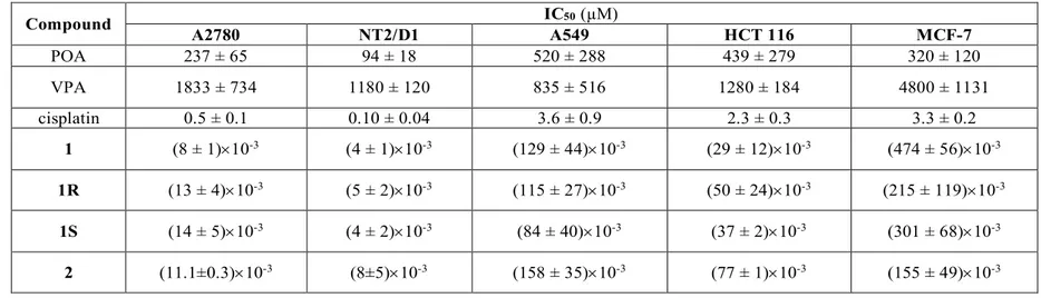
The biological properties of a new cisplatin-based Pt(IV) prodrug, containing an inert acetato and the bioactive 2-(2-propynyl)octanoato (POA) as axial ligands (1) are here reported. Indeed, it has been demonstrated that POA is more potent than VPA in inducing histone hyperacetylation in cerebellar granule cells, and in exhibiting antiproliferative and neurotrophic activity [14]. Since POA is a chiral molecule, the activity of the racemate (1), as well as that of the corresponding isomers (1R and
1S), was investigated and compared to that of cisplatin and of compound 2, a
cisplatin-based Pt(IV) complex with two VPA molecules as axial ligands (Figure 3.1).
38
Figure 3.1: Sketch of the Pt(IV) complexes under investigation.
3.2 Material and methods
3.2.1 Cell culture
Human cancer cell lines were purchased from European Collection of Cell Cultures (ECACC, UK) or Interlab Cell Line Collection (ICLC, Genova, Italy): ovarian endometrioid adenocarcinoma A2780 (ICLC HTL98008), colon carcinoma HCT 116 (ECACC 91091005), breast ductal carcinoma MCF-7 (ECACC 86012803), embryonal carcinoma of the testis NTERA-2 clone D1 (also known as NT2/D1, ICLC HTL97025), and lung adenocarcinoma A549 (ICLC HTL03001). A2780 and A549 cells were grown in RPMI 1640, whereas McCoy’s 5A was used for HCT 116, Dulbecco’s Modified Eagle’s Medium (DMEM) for NT2/D1, and Minimum Essential Medium (MEM) supplemented with non-essential amino acids for MCF-7. All media were supplemented with L-glutamine (2 mM), penicillin 100 IU mL-1,
streptomycin (100 mg L-1) and 10% heat inactivated fetal bovine serum (FBS).
Media and supplements were purchased by Sigma-Aldrich or Life Technologies. Cell culture and treatment were carried out at 37 °C in a 5% CO2 humidified
![Figure 1.1: The transformation process of normal cells into malignant ones. The picture is a modification of that reported in literature [1]](https://thumb-eu.123doks.com/thumbv2/123dokorg/4806999.49643/12.774.157.621.613.745/figure-transformation-process-malignant-picture-modification-reported-literature.webp)
![Figure 1.2: The cell cycle events. The picture is a modification of that reported in literature [6]](https://thumb-eu.123doks.com/thumbv2/123dokorg/4806999.49643/15.774.200.574.112.385/figure-cell-cycle-events-picture-modification-reported-literature.webp)
![Table 1: The main classes of cyclins and CdKs, referred to vertebrate and budding yeast [6]](https://thumb-eu.123doks.com/thumbv2/123dokorg/4806999.49643/16.774.116.660.106.296/table-classes-cyclins-cdks-referred-vertebrate-budding-yeast.webp)
![Figure 1.3: Key step in apoptotic intrinsic and extrinsic signaling pathways. The picture is a modification of that reported in literature [11]](https://thumb-eu.123doks.com/thumbv2/123dokorg/4806999.49643/18.774.223.554.352.741/apoptotic-intrinsic-extrinsic-signaling-pathways-modification-reported-literature.webp)
![Figure 1.4: Three main autophagic pathways. The picture is a modification of that reported in literature [13]](https://thumb-eu.123doks.com/thumbv2/123dokorg/4806999.49643/19.774.218.554.596.936/figure-main-autophagic-pathways-picture-modification-reported-literature.webp)
![Figure 1.7: Formation and effects of cisplatin-DNA adducts. The picture is a modification of that reported in literature [29]](https://thumb-eu.123doks.com/thumbv2/123dokorg/4806999.49643/28.774.169.611.539.782/figure-formation-effects-cisplatin-adducts-modification-reported-literature.webp)
![Figure 1.8: Molecular mechanisms of cisplatin chemoresistance. The picture is a modification of that reported in literature [39]](https://thumb-eu.123doks.com/thumbv2/123dokorg/4806999.49643/33.774.230.567.247.564/figure-molecular-mechanisms-cisplatin-chemoresistance-modification-reported-literature.webp)