"Dilige, et quod vis fac: sive taceas, dilectione taceas; sive clames, dilectione clames; sive emendes, dilectione emendes; sive parcas, dilectione parcas: radix sit intus dilectionis, non potest de ista radice nisi bonum existere".
Abstract
This thesis has investigated diversity and distribution of "pathogenic" and "non-pathogenic" fungal endophyte communities in twigs, leaves and buds of Fagus sylvatica in beech Italian forest into Monte Cimino area. In last decade European beech has been subject to a number of biotic and abiotic stresses included the increasing impact of drought and, as a consequence, the impact of native parasites that attack weakened hosts. Among these, Biscogniauxia nummularia (Bull.) Kuntze, associated with severe beech-declines, has been studied for its importance as latent pathogen of healthy beech tissues. Understanding how fungal endophyte communities differ in abundance, diversity and taxonomic composition is key to understanding the ecology and evolutionary context of endophyte-plant associations. Fungal ITS sequences were used for validation of morphological identification and phylogenetic analysis. According to the ITS phylogeny, endophytes studied belong to the main non-parasitic and non-lichenised orders of Pezizomycotina. The effects of native tissue, site exposure, and season on endophyte assemblages were investigated. Native tissue was the major factor shaping endophyte assemblages. Exposure and seasonal differences played a significant but lesser role. Several recently developed molecular methods offer new tools for determining the presence and diversity of fungi in complex microbial communities. Terminal restriction fragment (TRF) pattern analysis was tested as a method for assessing the molecular diversity of endophyte fungal community within asymptomatic beech tissues. ITS ribosomal region of communities DNA isolated from ten beech twigs was amplified and then digested with MspI enzyme. The ITS region showed high degree of specificity in matching observed TRF profiles to those generated from GenBank sequence data for species identification. These data suggest that ITS rDNA TRF pattern analysis has great potential as a rapid and specific method for fungal community analysis and species identification. Results of quantitative Real-time PCR (Rt PCR) assay using DNA of B. nummularia isolated from different geographic areas were compared. The assay confirmed higher risk associated with our beech forest in Central Italy, compared to the other equal phyto-coenosis located at most latitudes, in terms of declining damages and loss of biodiversity.
Keywords: Fagus sylvatica, endophyte communities, Biscogniauxia nummularia, ITS phylogenetic
analysis, TRFLP, quantitative Real-time PCR.
Riassunto
Questa tesi ha studiato diversità e distribuzione delle comunità degli endofiti fungini "patogeni" e "non patogeni" in rami, foglie e gemme di Fagus sylvatica in una faggeta italiana nella zona del Monte Cimino. Negli ultimi dieci anni il faggio è stato oggetto di una serie di stress biotici ed abiotici inclusi il crescente impatto della siccità e, per conseguenza, l'impatto di parassiti nativi che attaccano ospiti indeboliti. Tra questi, Biscogniauxia nummularia (Bull.) Kuntze, associato a gravi deperimenti di faggio, è stato studiato per la sua importanza come agente patogeno latente in tessuti sani faggio. Capire come le comunità fungine endofite differiscono in abbondanza, diversità e composizione tassonomica è la chiave per comprendere l'ecologia e il contesto evolutivo delle associazioni endofita-impianto. Le sequenze ITS sono state utilizzate a conferma dell’identificazione morfologica e per l’analisi filogenetica. In base alla filogenesi ITS, gli endofiti studiati appartengono ai principali ordini non parassiti e non lichenici dei Pezizomycotina. Sono stati studiati gli effetti del tessuto d’origine, dell'esposizione e della stagione sui gruppi endofiti. Il tessuto è stato il principale fattore di formazione dei gruppi endofiti. L'esposizione e le differenze stagionali hanno giocato un ruolo significativo, ma minore. Diversi metodi molecolari recentemente sviluppati offrono nuovi strumenti per determinare la presenza e la diversità dei funghi in comunità microbiche complesse. L’analisi dei frammenti terminali di restrizione (TRF) è stata verificata come metodo per la valutazione della diversità molecolare della comunità endofitica fungina all'interno di tessuti asintomatici di faggio. La regione ITS dell’rDNA delle comunità isolate da dieci rami di faggio, è stata amplificata e quindi digerita con l'enzima
MspI. La regione ITS ha mostrato un alto grado di specificità, nella corrispondenza tra profili di TRF
osservati e quelli generati da sequenze in banche dati ufficiali, per l'identificazione delle specie. Questi dati suggeriscono che il modello di analisi TRF basato sulle regioni ITS ha un grande potenziale come metodo rapido e specifico per l'analisi delle comunità fungine e l'identificazione delle specie. Sono stati confrontati i risultati dell’analisi quantitativa Real time PCR (Rt-PCR) del DNA di B. nummularia isolato da diverse aree geografiche. L’analisi ha confermato un più alto rischio associato alla nostra faggeta nel Centro Italia, rispetto ad altre simili fitocenosi a maggiori latitudini, in termini di deperimento e perdita di biodiversità.
Contents
Preface...1
1. Background...1
1.1 Global climate is changing...1
1.1.1 Shift of species ranges as response to global warming...3
1.1.2 Plant responses to elevated carbon dioxide and temperature...3
1.1.3 Increasing water stress and loss of biodiversity...4
1.1.4 Evolutionary risk for European beech forests...6
1.2 The European beech (Fagus sylvatica L.)....6
1.2.1 Main damages...9
1.3 Endophytism: an age-old phenomenon...12
1.4 The genus Biscogniauxia Kuntze...14
1.4.1 Biscogniauxia nummularia (Bull.:Fr.) O. Kuntze...16
1.4.2 B. nummularia a latent invaders with higher damage potential due to climate change in Mediterranean Basin...18
2. Aims...20
3. Materials and methods: objectives 1...21
3.1 Study sites....21
3.2. Plant sampling...23
3.3 Preparation of culture media...26
3.4 Isolation, cultivation and identification....26
3.5 DNA extraction, amplification and sequencing...26
3.6 Alignment and phylogenetic analyses...28
3.7 Statistical analysis of data...28
4. Results: objective 1...29
4.1 Taxon richness at species and order level...29
4.2 Isolation frequencies of endophytic fungi from terminal buds...35
5. Discussion: objective 1...37
5.1 Fungal endophyte community composition...37
5.2 Detection of fungal endophytes in buds...41
5.3 Phylogenetic diversity of beech endophytes...42
6. Conclusion: objective 1...43
7. Materials and methods: objectives 2...46
7.1 Endophyte fungi in twigs and leaves...46
7.2 Differences between native tissues...52
7.3 Differences between exposures...53
7.4 Differences between seasons....55
7.5 Differences between seasons....57
8. Discussion: objective 2...59
8.1 Effectiveness of sampling effort...59
8.2 Ecological diversity of beech endophytes...59
9. Conclusion: objective 2...62
10. Materials and methods: objective 3...63
10.1 TRFLP analysis technic...63
10.2 DNA extraction, amplification and quantification...64
10.3 Choice of restriction enzymes...65
10.4 Ethanol Precipitation of Digest...65
10.5 Capillary Electrophoresis...65
10.6 Developing a TRFLP database....66
10.7 Diversity of fungi in TRFLP profiles...66
10.8 Comparison between TRFLP and isolation methods...67
11. Results: objective 3...68
11.1 Database predictions...68
11.2 Identification of fungi using TRF patterns...69
11.3 Comparison between TRFLP and isolation methods...70
12. Discussion: objective 3...73
13. Conclusion: objective 3...76
14. Material and methods: objective 4...78
14.1 Detection of B. nummularia by Real-time PCR....78
14.2 Genomic DNA extractio...78
14.3 Coparison of Isolation with Real-time detection....79
14.4 Coparison of two different forest sites...79
15. Results: objective 4...80
15.1 Coparison of Isolation with Real-time detection....80
15.2 Coparison of two different forest sites...80
16. Discussion: objective 4...82
17. Conclusion: objective 4...83
Preface
Concerning forest preserve large economic and environmental importance is recognized to Fagus sylvatica in Italy with an important ecological and hydrogeological role. On the Apennines, beech forests represent the most widespread mountain climax association (Scoppola, 1999). Cause of its frequency, ability to grow on sites of wide ecological variability and its longevity, beech is well used to evaluate the growth and climate relationships in different bioclimatological units, and to estimate future prospects and possible ecological risks associated with “Global Climate Change” (Eckstein et al., 1984; Gutierrez, 1988; Biondi, 1992; Rozas, 2001; Dittmar et al., 2003; Piovesan et al., 2005; Lebourgeois et al., 2005; Di Filippo et al., 2007; Biondi, 2008). Currently, among European regions, the Mediterranean Basin, that is at the southern limit of beech geographical range, is consedered an “hot spot” of both climate change and biodiversity and could be most affected by global warming in term of habitat loss and extinction (Brook et al., 2002). In ecological and evolutionary time the preserving of so sensitive biocenosis as beech forests, is only possible after understanding of interactions complex existing in these ecosistems. In this context, “patogenic” and “non- pathogenic” endophyte communities, which living in healthy beech tissues, could show differential response to physiological changes in trees. In the Mediterranean Basin beech is able to provide a unique opportunity to explore connection between tree physiological condition and long-term drought stress that promotes aggressiveness of weak parasites, among which Biscogniauxia nummularia, responsible of beech decline (Luchi et al., 2006).
1 Background
1.1 Global climate is changing
The latest assessment report of the Intergovernmental Panel on Climate Change (IPCC, 2007) on the impacts of human-induced climate change reconfirms that global atmospheric concentrations of greenhouse gases have increased markedly since 1750 as a result of human activities, and cause radiative forcing. The warming of our climate system is unequivocal, as is now evident from observations of increases in global average air and ocean temperatures, widespread melting of snow and ice, and rising global average sea level (Haeberli and Beneston, 1998; Joughin et al., 2004; Rignot, 2006; IPCC, 2007). During the last 100 years the earth has heated up by 0.74°C; with
the linear warming trend over the last 50 years nearly twice as much that for the last 100 years (IPCC, 2007).
The frequency of extreme temperature events has changed: cold days, cold nights and frost have become less frequent, while hot days, hot nights, and heat waves have become more frequent. Changes in precipitation patterns are at least as divergent as in temperature patterns: rainfall increased in eastern parts of North and South America, northern Europe and parts of northern and Central Asia, while drying has been observed in southern Europe, southern Africa and parts of southern Asia. Droughts are linked to higher temperatures, reduced precipitation, changes in sea surface temperatures, wind patterns, and decreased snow pack and snow cover. Meanwhile, the frequency of heavy precipitation events has increased over most land areas, consistent with warming and accompanying increases of atmospheric water vapour.
Projections for the future predict a warming of about 0.2°C per decade, analogous to a temperature change of 1.8°C (1.1-2.9°C; B1 scenario: environmental sustainability) to 4.0°C (2.4-6.4°C; A1FI scenario: fossil intensive, very rapid growth) until the end of the century (IPCC, 2007) (Fig. 1).
Fig. 1. Regional Climate Change Index (RCCI) over 26 land regions of the World calculated from 20 coupled AOGCMs and 3IPCC emission scenarios (A1B, A2, B1).
Even if all radiative forcing agents are held constant at year 2000 levels, a further warming trend would occur in the next two decades at a rate of about 0.1°C per decade,
mainly due to the slow response of the oceans, and would continue for centuries. Since the world’s energy needs and consequently the emission of carbon dioxide will continue to grow for at least the next three decades (IEA, 2005), constancy at 2000 levels is not very likely. Continued greenhouse gas emissions will cause further warming and induce many changes much larger than those observed during the 20th century.
Global warming will influence the global hydrological cycle. Projections indicate decreasing water availability and increasing drought risk in many regions of the world (Gerten et al., 2007), manifest as reductions in river discharge (e.g., Chalecki and Gleick, 1999), ground water resources (e.g., Sandstrom, 1995), or soil moisture (Gregory et al., 1997; Wetherald and Manabe, 2002). Even though, rate and distribution of precipitation strongly depend on a variety of parameters, e.g., topography, vegetation structure, and land use, and therefore strongly differ in their spatial and temporal distribution, hot extremes and summer heat waves like in summer 2003 will continue to become more frequent in particular in central and southern Europe (EEA, 2004, Kundzewicz et al., 2006; Rowell and Jones, 2006).
1.1.1 Shift of species ranges as response to global warming
In the past, migration has been the most common response of plants to Quaternary climate change (Huntley et al., 1991). Palynological data indicate that plant species tracked during Quaternary climate changes favourable conditions with estimated migration rates of 150-500 m year-1 (Huntley et al., 1991). Global analyses document significant range shifts of present vegetation toward the poles (or metres per decade upward) of 600 m year-1 (Grabherr et al., 1994; Meshinev et al., 2000; Kullman, 2001; Parmesan and Yohe, 2003; Peñuelas and Boada, 2003; Meier and Leuschner, 2008). These maximum rates of migration are one or two orders of magnitude too slow to track the predicted climatic changes in the next century and species may not be able to avoid the severest effects of global warming. Climate-induced ecological change is also expected to outpace the rates at which successional processes could occur in forests. The understanding of the plasticity of plant responses towards changing climatic conditions therefore becomes increasingly important.
1.1.2 Plant responses to elevated carbon dioxide and temperature
The predicted rapid and simultaneous changes in several environmental factors controlling forest ecosystem functions are raising concerns about future terrestrial
ecosystem productivity. Among the most critical of these concerns are the increase of atmospheric concentrations of CO2 and extreme temperature, and changes in
precipitation distribution (Bazzazz, 1990; Mooney et al., 1991). The primary effect of elevated CO2 in most ecosystems is through a direct positive effect on photosynthetic
C-fixation that increases net primary production (NPP) (Lindroth et al., 1993; Hamilton et
al., 2002; Norby et al., 2002). However, long-term growth in elevated CO2
concentrations has caused in forest species like beech reduced sensitivity and acclimation of stomatal conductance to vapour pressure deficit (Heath, 1998), reduced sensitivity to drought (Heath and Kerstiens, 1997), and reduced sensitivity to atmospheric CO2 concentrations (Santrucek and Sage, 1996; Idso, 1999). Increasing
temperatures and vapour pressure deficits additionally increase the rate of evapotranspiration (Norby and Luo, 2004). Photosynthetic acclimatization to elevated CO2 and temperature reduces the water use efficiency. Thus, actual water use per
individual tree may increase, if the stomatal response to CO2 and temperature is weak.
Further, CO2 and temperature can also influence ecosystem processes via their effects
on soil moisture (Hungate et al., 1997; Yang et al. 2003). Rising temperature and CO2
concentration will impair the water supply for plants that will be already diminished due to lower precipitation amounts in summer. Thus, water deficiencies might become more hazarding for ecosystems than altered CO2 or temperature regimes, indeed, without
commensurate increases in precipitation, water stress can result even at constant rainfall levels.
1.1.3 Increasing water stress and loss of biodiversity
In many regions of the world, net primary production is in the first instance limited by available soil water and only secondly by temperature, CO2, or radiation (Nemani et
al., 2002; Xiao and Moody, 2004). The geobiosphere as a whole appears to be currently
in a state of water deficiency (Lee and Veizer, 2003). Precipitation is often used as a proxy of soil water availability; therefore declining precipitation can be linked to a decrease in soil moisture. If soil water content falls below some species-specific level, plants experience drought stress that alters both soil-root and leaf-atmosphere interfaces and threatens the integrity of the liquid phase continuum from soil to leaves (Bréda et
al., 2006). In this so critical state,drought-induced physiological disorders dramatically
can change the distribution of potential forest types increasing tree vulnerability to secondary stresses like frost or another drought, insect damage (Tuomi et al., 1988;
Docherty et al., 1997; Rouault et al., 2006) and pathogens (Dale et al., 2001; Desprez- Loustau et al., 2006). Such cumulated stress factors rapidly may end either with partial recovery of tree growth, or with a final shift into decline and eventual death (Law et al., 2002; Sturrock, 2007) with a final consequence of the dramatic loss of biodiversity.
In spite of this, the effects of climate change in the next few years will hardly be perceptible as measured by changes in tree health and species composition, and conservationists are far from able to assist all species under threat (Myers et al., 2000). One effective way to support the most species threatened with extinction is to identify “biodiversity hotspots” where exceptional concentrations of endemic species are undergoing exceptional loss of habitat. As many as 44% of all species of vascular plants are confined to 25 hotspots comprising only 1.4% of the land surface of the Earth. Concentrating a large proportion of conservation support on these areas would go far to stem the loss of biodiverity that is now underway (Myers et al., 2000). According to Myers et al. (2000) one of the hottest under threat area in term of rate of habitat loss is the Mediterranean Basin, where, in recent decades, on several Fagaceae species the impact of forest plant pathogen has clearly increased in connection with exceptionally dry years (Desprez-Loustau et al., 2006). In particular, beech trees under water stress can become affected by Biscogniauxia nummularia (Bull.: Fr:) O. Kuntze., which causes necrosis and cankers on branches and stems (Nugent et al. 2005). This specific beech pathogen is able to spend most of its life cycle in latent form in symptomless aerial plant organs as endophyte to switch pathogenic face once the host becomes subjected to physiological stresses, in particular water stress (Granata and Whalley, 1994; Paoletti et al., 1996; Capretti et al., 2003; Granata and Sidoti, 2004; Nugent et al., 2005; Luchi et al., 2006).
In conclusion, to better understand the whole complex of forest ecosystem functions is necessary to well know its biodiversity in terms not only of plant species diversity but also of microbial diversity and place each species into a trophic level or into its niche dimension within the host plant. Indeed, recent studies have shown that physiological changes in trees may also lead to differential response of those organisms that normally colonize beech tissues (Hendry et al., 1998), that suggests the importance of role of endophytic fungi associated with woody perennials, which can be complex and labile both in ecological and evolutionary time (Saikkonen, 2007).
1.1.4 Evolutionary risk for European beech forests
Concerning Forest Preserve large economic and environmental importance is recognized to European beech (Fagus sylvatica L.) that is a characteristic feature of central and southern Europe with a wide natural distribution range (Fig. 2), and also plays an important ecological and hydrogeological role in the Italian mountain landscapes. Timber quality, landscape value and role in preventing soil erosion justify the economic and environmental importance attached to this tree in the Italian mountain areas. On the Apennines, beech forests represent the most widespread mountain climax association (Scoppola, 1999). Cause of its frequency, ability to grow on sites of wide ecological variability and its longevity, beech is well used to evaluate the growth and climate relationships in different bioclimatological units, and to estimate future prospects and possible ecological risks associated with climate change (Eckstein et al., 1984; Gutierrez, 1988; Biondi, 1992; Rozas, 2001; Dittmar et al., 2003; Piovesan et al., 2005; Lebourgeois et al., 2005; Di Filippo et al., 2007; Biondi, 2008). In this century, in the Mediterranean Basin, that is southern limit of beech geographical range, beech forests are able to work as bio-indicator and provide a unique opportunity to explore connection between tree physiological conditions and long-term drought stress (Piovesan et al., 2003-2008) European beech either will have to respond to rapidly changing climatic conditions or will face local extinction if not sufficiently adapted to altered drought and temperature conditions, that can promotes aggressiveness of weak parasites living inside plant tissues (Schröter et al., 2005). Rapid climate change as predicted requires a better understanding of structure and diversity of "pathogenic" and "non-pathogenic" fungal endophyte community of Fagus sylvatica in the Mediterranean Basin.
1.2 The European beech (Fagus sylvatica L.)
The European beech (Fagus sylvatica L.) belongs to the family Fagaceae, order Fagales, which includes woody plants with flowers pollinated by wind and characterized by a particular structure called “cupule”, partially or entirely enclosing the fruit. Beech is the most frequent deciduous forest tree species in Central Europe, it predominates at all natural or near-natural forest sites that are not extremely dry, wet or acidic (Thomas and Sporns, 2008).
Fig.2. Natural distribution of beech forests in Europe (according to Bohn 1992)
With a slow growth rate F. sylvatica is a large, deciduous tree, capable of reaching heights up to 40–50 m, with thin, cylindrical trunk and conical, later wide-branched treetop. Root system is heart-shaped with strong many-sides roots. Bark is smooth, grey to white-grey, rarely fissured. Annual shoots are red-brown, primarily white-hairs, later glabrous. The buds are alternately double-rowed, thin fusiform, 10–25 mm long, cinnamon brown, with scales whitish on the top. Leaves are smooth, shining above, lighter beneath, with short petiole; lamina is elliptic to oval-elliptic, 3–12 cm long, entire to shallow crenate, undulate at the edges, ciliate. Leaf primary vein is whitish hairy, in the axils of the marginal veins are long-haired. Secondary veins are in 5–9 pairs. Petiole is 5–10 mm long, pinnate. Stipules are narrowly lanceolar, light brown, smooth, and soon deciduous. Male flowers are in the axils of the leaves on long pedunculated bundle, female flower are 2–3 in reddish cup with hairy nodulation. The seeds are about 1 cm long, brown, smooth, triangular, at the edges with winged achenes – beechnuts. The beechnuts borne singly or in pairs in soft-spined husks 1.5–2.5 cm long, known as cupules are closed by 3-4 lobels in brown, ligneous, echinate, pedunculated cup which is about 2 cm long and dehiscent by four lappets (Pignatti, 1982). Raw beechnuts can be slightly toxic for some people. You can lower the toxicity
by thermal treatment, for example roasting (Leugnerová, 2007). Beech flowers in late April and May. Seeds ripen in the autumn and coloration of leaves comes from yellow over the reddish until dark brown. European beech has lifespan of 200 to 400 years.
European beech is an oceanic and sub oceanic climate woody species (De Philippis, 1937) with need of 800–1000 mm precipitation. This tree creates multi-storeyed forest, mostly unmixed, because of its huge its shading.European beech prefers freshly wet, well aerated soils, often calcific and rich in humus and minerals. It dislikes waterlogged but also dry and sandy soils. It is sensitive to drought and late frost too. European beech is diagnostic species for Fagion alliance (Leugnerová, 2007).
Beech forests characterise the landscape of many mountain areas in Italy, from the Alps down to the southern regions of Campania, Basilicata, Calabria and Sicily in the Mediterranean area, except Sardinia (Fig. 2). According to the National Forest Inventory (INFC, 2005), the total area covered by beech in Italy is 1.042.129 hectares, which corresponds to 9.4% of the country’s total forest area. In the Alps, beech generally forms pure stands above 1000 m a.s.l. altitude in areas with relatively low rainfall, while it grows at around 600-700 m a.s.l. in more humid areas. On the Apennine mountains beech usually grows above 900-1000 m a.s.l. In the central Apennines sporadic beech or beech stands can also be found at lower altitudes (< 700-800 m asl) associated with chestnut or mesophilous mixed oak forests; these beech spots are considered relict sites and proof of a much wider diffusion of beech in the area (Montelucci, 1956; Anzalone, 1961; Anzalone, 1980; Scoppola and Caporali, 1997; Scoppola, 1999). Beech forests are more widespread on the northern slopes and where rain and fog maintain moist air conditions. On the sunnier and warmer southern slopes, the lower vegetation limit for beech tends to move higher (Hofmann, 1991;Pignatti, 1998). On the northern slope of the Mount Etna in Sicily beech reaches an altitude of 2000 m (Hofmann, 1960; Del Favero, 2008). In the southern regions, in areas with high air moisture conditions, beech can descend to an altitude of 400-500 m, where it comes into contact with evergreen oak (Quercus ilex L.). In some valleys on the Aspromonte mountain range at the southern most tip of Calabria, there is an inversion of the vegetation planes, with beech occurring at lower elevations compared to evergreen oak. In the “Gargano” peninsula (Puglia) beech grows at an altitude of 200-300 m a.s.l. (Hofmann, 1961; Fenaroli, 1966; Pignatti, 1998). Beech forests also host other mountain hardwoods, such as helm, linden, cherry, sycamore and Norway maple
(Pignatti, 1998). Silver fir (Abies alba Mill.) is found in beech forests along the Apennine mountains and, especially, in the Alps (Nocentini, 2009).
Beech, with plenty of cultivars, is often setting out as solitaire wood species in larger gardens and parks; it is also usable in hedge (Leugnerová 2007).Wide use has European beech, which are different by its habitat, shape or colour of leaves in gardens.
European beech is one of the most important economic leafy woody species in Europe. It has interspersed porous wood, skin-pink to pink-brown colour, without marked true heartwood. Its wood is hard, heavy and little flexible. However, false heartwood with red-brown colour that occurs in old trees lowers the quality of wood. Beechwood can be used in production of bent-wood furniture (beechwood contains a huge amount of lignin, which softens at high temperatures, so than is wood very shapable), for veneer, plywood, parquet. In the Middle Ages was using in production of wood coal.
1.2.1 Main damages
Usually none diseases cause death of plants provided soil is not compacted and is well-drained. Several fungi cause leaf spots but are generally not serious to warrant chemical control. Among these Apiognomonia errabunda (Roberge ex Desm.) Höhn causes leaf anthracnose with discrete, irregular, brown necroses distributed in patches on leaf blade. Infected areas are often found along the veins and midrib of the leaf (Fig. 3).
Fig. 3. A. errabunda symptoms on beech leaves
Fruit honey-brown bodies are formed on the undersides of the leaves. Many studies have reported A. errabunda living in symptomless tissues as endophyte in beech trees (Sieber and Hugentobler, 1987; Morelet, 1989; Hämmerli et al., 1992; Toti, 1993; Viret
et al., 1993; Kowalski and Kehr, 1996; Danti et al., 2002).
Similar necroses are cuased in the spring by the larva of the beech leaf miner,
Rhynchaenus fagi L., an curculionidea insect, but these can be distinguished by the
presence of the mined area which consists of a pale brown patch linked by a narrow tunnel to the midrib of the leaf (Butin, 1995).
Another diseases are: powdery mildew most common late in the season, by fungi belong to Erisyphaceae family, causes a white coating on the leaves; bleeding canker forms small or large patches of dying bark on the stems or branches from which were scattered drops of rusty-red, yellow-brown or almost black, gummy liquid ooze. Crown symptoms include leaves of smaller size and lighter green color than normal. In severe cases the leaves wilt and the branches die. Avoid feeding with high nitrogen fertilizers as it seems to worsen the condition of infected trees. Beech bark disease occurs when the feeding site of woolly beech scale is invaded by a fungus. The fungus kills the bark and in the process favoured the attacks of insects, such as bark beetles, and aphids. There are no satisfactory controls for the fungus. Cankers infect, girdle, and occasionally kill branches. (Michigan State University Extension Ornamental Plants 1999).
Beech root system can be strongly damaged after infection of soil pathogens which cause root rot in host plants. Among these were remembered, for their virulence and geographic distribution and diffusion, differents type of species in the oomycete genus
Phytophthora. These root rot fungi produce motile, flagellate zoosperes as dispersal
units with which move through soil water. The species most virulance at our latitudes are P. cambivora (Petri) Buism (Jung et al., 2005), P. citricola Sawada (Wang et al., 2003) e P. cactorum (Lebert & Cohn) J. Schröt., while most spread in UK are P.
kernoviae (Brasier et al. 2005) e P. ramorum Werres (Heungens et al., 2006) (Fig. 4)
Fig. 4. Bleeding canker by P. ramorum on beech trunk
(http://www.ilvo.vlaanderen.be)
Serious threat of F. sylvatica is Heterobasidion annosum (Fr.) Bref.. This fungus is a wood-decay fungus widespread in the coniferous forests of temperate and boreal regions of the northern Hemisphere. It causes great financial losses due to root and butt rot on several conifers, especially in managed forests (Stambaugh, 1989; La Porta et al., 1997). H. annosum kills resinous hosts directly by decaying roots and killing the cambium around the root collar. Trees are often predisposed to bark beetle infestation and usually die standing. Some non-resinous hosts are affected in the same way, but others develop extensive butt decay rather than being killed outright. The most reliable way to identify the disease in the forest involves finding its fruiting bodies, called “conks” (Fig. 5).
Fig. 5. Fruit bodies of H. annosum on trunk of F. sylvatica.(www.unipd.it)
Advanced decay caused by the fungus is often laminated with pitting and it can also be stringy and moist with white streaks and scattered black flecks (USDA Forest Service Gen. Tech. Rep. PSW-GTR-165. 1998).
Stressed and damage hosts were attacked by weak parasites, among these, some are
xylariaceous fungi, such as Xylaria polymorpha Pers. ex St. Amans, Kretzchmaria
deusta Fr. [ex Ustulina deusta] and Biscogniauxia nummularia (Prljincevic, 1982; Sinclair et al., 1987; Schwarze et al., 1995; Hendry et al., 1998; Luchi et al., 2006). Over the last 20 years decline of european beech in Sicily and Calabria (Italy) was observed to be associated with the ascomycete B. nummularia. This fungus is naturally present in beech stands and to date has not been considered a primary pathogenic agent (Granata and Whalley, 1994; Paoletti et al., 1996; Moriondo et al., 1999; Capretti et al., 2003; Granata and Sidoti, 2004). Many studies reported B. nummularia living in asymptomatic tissues and being able to cause strip-canker and wood decay in trees that suffer severe water stress during the growing season (Granata and Whalley, 1994; Hendry et al., 1998; Hendry et al., 2002; Capretti et al., 2003; Nugent et al., 2005; Luchi et al., 2006). According to the redefinition proposed by Petrini (1991), it could thus be considered an endophyte during the latent period.
1.3 Endophytism: an age-old phenomenon
Endophyte ownes its origin to De Barry (1866) who first coined the term. Since then it has deeply embedded in the literature. Within the last decade, different authors have proposed a range of similar, but more complex definitions (e.g., Carroll, 1986; Petrini, 1991; Wilson, 1993). However, “endophyte” does not stand alone as a term whose definition has changed over time, or as a term whose definition biologists might not agree over. At the most basic level, endophyte could simply refer to the location of the organism: endo means within and phyte means plant. This is contrasted to “epiphyte” which refers to organisms living on the outside of the plant (Kogel et al., 2006). The organisms commonly associated with the term endophyte are fungus and bacteria (Fahey et al., 1991) and this restricted definition (Wennström, 1994) has evolved into a definition which describe not only the location, but also the nature of the association between one organism with its host (Kogel et al., 2006).
Symbiosis between a fungus and a plant is a widespread phenomenon in nature. Most, if not all, plants studied in natural ecosystems are infested by fungi that live in intercellular space or inside cells of host plant without cause any kind of disease
(Saikkonen et al., 1998; Kogel et al., 2006) or apparent damage, if by disease we mean the result of an interaction between a host (susceptible), a pathogen (virulent) and an environment (favourable to the pathogen). During the long co-evolution term of endophyte and plant, equilibrium between these organisms have been established (Gao
et al., 2010). The true endophyte will exist once equilibrium between fungal activity
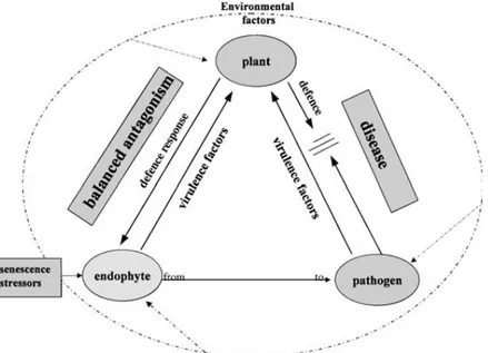
and the plant reaction is achieved and is maintained over time (Giménez et al., 2007), therefore a mutualistic interaction does not mean the absence of plant defence but is a finely tuned balance of antagonisms that keeps the host–microbe interaction in a stable state that disadvantages neither partner (Kogel et al., 2006) (Fig. 6).
Fig. 6. Hypothesis: a balance of antagonisms between endophytic virulence and plant defence response results in asymptomatic colonisation.
Commensalism and mutualism represent the balanced stages plant–endophyte interactions. Commensalism provides benefit to the endophyte by enabling an undisturbed existence and nutrient supply without affecting the host. Mutualism, by contrast, is defined as an interaction that is beneficial for both partners (Kogel et al., 2006). In addition to the providing benefits for the endophyte, mutualism can frequently result in promoted growth of the host (Dai et al., 2008), improved tolerance to abiotic stress (Redman et al., 2002, Lewis 2004; Malinowski et al., 2004) and increased resistance against pathogens (Colditz et al., 2005; Tanaka et al., 2005; Vega et al., 2008). Direct effect of the potential mechanisms of endophytes inhibition of plant
pathogen is characterized by producing of antifungal and antibacterial secondary metabolites like antibiotic, terpenoids, alkaloids, aromatic compounds and polypeptides (Gunatilaka, 2006), and by releasing of lytic enzymes that can hydrolyze a wide variety of polymeric compounds, including chitin, proteins, cellulose, hemicellu-lose and DNA (Tripathi et al., 2008). When endophytes colonize the plant surface, they produce enzymes to hydrolyze plant cell walls and to start the growth within host tissues. These enzymes also have the function to suppress plant pathogen activities directly by degrading of cell walls of fungi and oomycetes eventually present on the surface of host tissues (Gao et al., 2010). However, the direct interactions between fungal endophytes and pathogens are complex and sensitive to species-specific antagonism (Arnold et al., 2000).
Under stress conditions, the plant-endophyte interaction can become unbalanced and disease symptoms can appear or the fungus can be excluded by induced host defence reactions (Clay and Schardl, 2002; Schulz and Boyle, 2005). Actually, some fungal endophytes are patogens of plant host and may evolve from plant pathogenic to non-pathogenic state go through an endophytic or latent face of its lifecycle (Carroll, 1988; Freeman and Rodriguez, 1993; Saikkonen et al., 1998; Kogel et al., 2006) (Fig. 6). These pathogens are known to be symptomless colonizers of healthy forest trees in an unstable equilibrium between transient mutualism or neutralism and latent pathogenesis. Climatic factors can change the endophytic nature of these fungi turning it into a weak pathogen or an opportunistic invader of senescing trees (Müller and Krauss 2005, Schulz and Boyle 2005, Moricca and Ragazzi 2008, La Porta et al. 2008, Gao et al. 2010). The host–microbe interactions can range from mutualism through commensalism to parasitism in a continuous manner (Johnson et al. 1997, Redman et al. 2001) (Fig. 6). Symbioses of plants with beneficial or neutral endophytes share many common attributes with plant interactions with pathogens. The molecular and biochemical basis for the switch from endophyte to parasite is still to be elucidated, but recent findings of compatible plant–microbe interactions have enhanced our understanding of what factors determine endophytic and parasitic lifestyles(Kogel et al., 2006).
1.4 The genus Biscogniauxia Kuntze
The genus Biscogniauxia, a member of the family Xylariaceae, has a worldwide distribution with over 50 taxa recognised (Nugent et al., 2005).
Biscogniauxia has long been known as Nummularia Tul. & C. Tul., until Miller
(1961) placed most of its members in his section Applanata of Hypoxylon. The current concept of Biscognauxia was defined by Pouzar (1979, 1986) and resumed by Ju, Rogers, San Martin and Granmo (1998).
This genus is represented in Europe by ten known species, five of which having applanate stromata and low margins, i.e., B. anceps, B. cinereolilacina, B. granmoi, B.
mediterranea and B. nummularia, and thus being likely to be confused with Hypoxylon.
The five other species B. dennisii, B. marginata, B. querna, B. repanda and B.
simplicior differ in having more or less cupulate (concave) stromata with conspicuously
raised margins.
Biscogniauxia shares with Hypoxylon discoid ascal apical rings and
Nodulisporium-like anamorphs as defined in Ju and Rogers (1996). It is separated from Hypoxylon primarily in having bipartite stromata with an outer stromatal layer which disappears on mature stromata, and in lacking KOH-extractable pigments. Moreover, the stromatal carbonization is important in Biscogniauxia while it is weak in Hypoxylon section
Annulata and usually absent in Hypoxylon section Hypoxylon (Ju and Rogers, 1996; Ju et al., 1998). The outer dehiscent stromatal layer is often overlooked as it is only present
on young stromata, remnants of this layer may be found at margins of mature stromata, but are usually inconspicuous. It is noteworthy that in Biscogniauxia, the natural anamorph develops on young stromata either between the outer layer and the stromatal surface, before the outer layer dehisces (Ju et al., 1998), or on the upper side of the dehiscing layer and on stromatal margins.
In the field, applanate stromata of Biscogniauxia also are easily confused with those of Diatrype Fr. (Diatrypaceae) and Graphostroma Piroz. (Graphostromataceae). In
Diatrype, stromata differ in lacking carbonaceous tissue and in having a well-developed
interperithecial tissue (entostroma) of usually white granules; moreover, asci are typically long-stipitate, ascospores are yellowish and allantoid, and conidia are scolecosporous (anamorph Libertella Desm.). Graphostroma is somewhat intermediate between the Xylariaceae and the Diatrypaceae through a Nodulisporium-like anamorph recalling the Xylariaceae and light-coloured suballantoid ascospores recalling the Diatrypaceae.
Biscogniauxia species develop in bark of trees and shrubs, especially on dead or
dying branches, more rarely on trunks, and are suspected to be at least weak pathogens (Ju et al., 1998). Therefore they are to be searched for on dead branches still attached to
the trunk, and specimens of Biscogniauxia found on dead branches lying on the ground are usually old and their perithecia are empty, unless the branches have broken off recently. It is also probable that many of these species occur “unseen” in healthy host tissue but only become apparent when stromata develop following senescence or death of the host material in which they occur. Whether they are classed as endophytes or are seen as “sneaky” fungi sensu Rogers (2000) is open to debate.
Identification of Biscogniauxia mainly relies on stromatal characters and ascospore size and morphology. Identification of host is a decisive feature in several species which are apparently host-specific, i.e., B. cinereolilacina specific to Tilia sp., B. granmoi specific to Prunus sp., B. nummularia specific to F. sylvatica and B. simplicior specific to Rhamnus catharticus.
The stromatal surface may be plane to slightly convex and then with low margins to more or less concave or cupulate, and then with thick raised margins, (B. marginata). The stromata are considered carbonaceous when, sectionned with a razor blade, they turn out to be broken rather than properly cut. The dehiscing outer layer is only seen on very young stromata, and therefore is rarely observed. It can be thin and torn up, operculum-like to crust-like.
Ostioles may be umbilicate or discoid when opening lower than stromatal surface, to papillate or coarsely papillate when opening higher than stromatal surface.
Perithecia may be obovoid to tubular, but in some species this character may be variable within a same collection.
Ascospores may be one-celled to two-celled, ellipsoid to subglobose, with a germ slit that can be straight, sigmoid or bilateral.
The germ slit morphology is frequently a diagnostic character and its observation is made easier by the use of mounting media such as lactic acid or lactophenol which improve the clearness. Permanent mounts of ascospores in Polyvinylic alcohol (Rhodoviol®: Rhodoviol 50g, water 150g, lactic acid 80g, phenol 40g.) dissolved in lactophenol (Van Brummelen, 1967) make the germ slits particularly conspicuous. Bilateral germ slits are best observed on ascospores that are not exactly in front view, when focusing alternately on upper and lower side.
1.4.1 Biscogniauxia nummularia (Bull.:Fr.) O. Kuntze
B. nummularia is known to cause the Beech Tarcrust. The Latin “nummus” meaning
1981). This fungus causes strip-cankers and wood decay in consequence of severe dry spell (Hendry et al., 1998) These striking lesions, often many metres long, typically spiralled around the trunks of affected trees, terminating at a dead branch or root. Yet despite their great size, such strip-cankers were formed in a single growing season (Lonsdale, 1983; Rayner, 1986; Hendry et al., 1998). The disease symptoms also include yellowing and leaf drop (Granata and Sidoti, 2004). The necrotic bark on the faces of cankers bore fungal fruiting structures (Hendry et al., 1998). The fruiting body forms a thick and shiny black crust, on beech bark and is found at all times of the year (Hendry et al., 1998). It is not edible (Phillips, 1981). Young specimens are covered by a light brown outer layer. The spores are black to dark brown.
Taxonomic classification of this fungus following: Kingdom: Fungi
Division: Ascomycota Class: Ascomycetes Order: Xylariales Family: Xylariaceae
Genus: Biscogniauxia Kuntze
Species: Biscogniauxia nummularia (Bull.) Kuntze
B. nummularia is distinctive among other members of genus with applanate
stromata in the combination of black stromatal surface with slightly papillate ostioles, blackish brown ellipsoid ascospores 10-13 x 7.5-8.5 µm and specificity for F. sylvatica.
The closest taxon is B. granmoi, thoroughly described by Læssøe et al. (1999). They pointed out the main differences between these two species: in B. granmoi, ostioles are pitted and not papillate, stromatal margin is slightly raised, ascospores are medium brown, narrower (x 5-6.5 µm) and all records were made on Prunus padus (in Europe) and P. padus var. pubescens (in Far Eastern Russia).
Morphological characters are used in description and for identification of this type species:
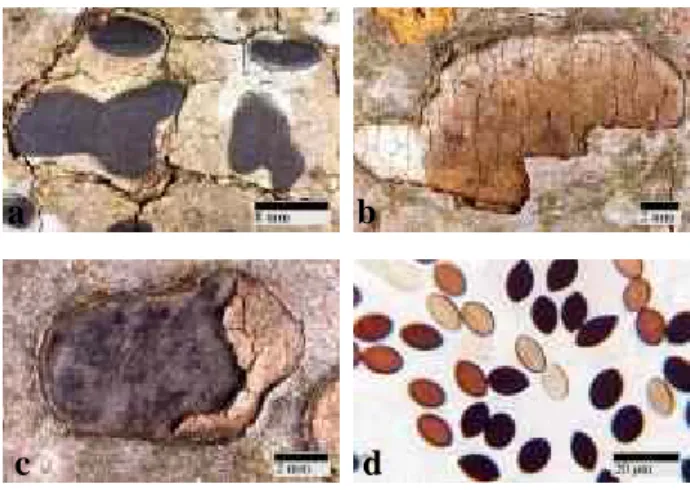
Stromata applanate, discoid 5-20 mm diam to irregularly ellipsoid 15-50(-120) mm long x 10-22(-60) mm broad x 0.6-0.8 mm thick; surface black, carbonaceous(Fig. 7a-c), when young covered by a dehiscing light brown outer layer; tissue beneath perithecia inconspicuous (Fig. 7b).
Ostioles discoid, slightly papillate to nearly at the same level as the stromatal surface.
Asci short-stipitate, with a discoid amyloid apical ring.
Ascospores dark brown to blackish, ellipsoid, frequently with narrowly rounded ends, 11.5 x 13.5 µm, with straight, inconspicuous germ slit spore-length (Fig. 7d).
1.4.2 B. nummularia a latent invaders with higher damage potential due to climate change in Mediterranean Basin
In Italy a serious decline of F. sylvatica was reported by the Forestry Commission of Messina (Sicily) in the lower region of a beech wood on the Nebrodi mountain range in 1990, and a few years later, in old beech coppice in the Ferdinandea forest, on the jonic side of the Calabrian Serre. Furthermore was observed that B. nummularia was capable of rapid growth at high temperatures, and that it was able to colonize large volumes of wood within host under water stress (Granata and Whalley 1994, Paoletti et al. 1996, Moriondo et al. 1999).
Currently, the presence of B. nummularia as endophyte was been also reported in Italian beech forests at major latitude (Capretti et al. 2003, Luchi et al. 2006). Given that, how was already suggested for other pathogens like B. mediterranea on mediterranean oaks (Vannini and Valentini 1994; Vannini et al. 1996; Collado et al. 2001, Anselmi et al. 2000, 2004, Vannini et al. 2009), Sphaeropsis sapinea on Pinus
resinosa (Stanosz et al. 2001) and Sclerotinia pseudotuberosa on chestnut (Vettraino et al. 2005), the potential ecological risk predicts a spread of damages caused by B.
Fig. 7. a-c: Stroma(ta) at maturity . b: immature stroma(ta). d: Ascospore of B. nummularia. Herbarium: JF99047-JF00114 JF99047 c a b d a b d c
nummularia infection more and more along north, just as result of drought increasing
due to Global warming. Thus, the potential spread of disease is not due to the movement of pathogen but to "climate shift" linked to global climate change in progress. On the basis of this possibility is necessary well full prevention of these critical evens, that can be possible just after full knowledge of composition and dynamics of fungal endophytic community associated with F. sylvatica.
2. Aims
Fungal endophytes that colonize forest trees are widespread phenomenon, but relations between fungal endophytes and forest trees have begun to be the subject of survey more recently. The few studies on endophytes in trees mainly concern the tropical areas and the most northern latitudes, whereas similar investigations in the Mediterranean region have so far been scarce and sparce. Actually endophytes are studied mostly in economically important forests suffering from diseases such as oak forests, but now, the discovery of old-growth beech forests is very interesting to undrestanding the long-term ecosystem dynamics in relation to natural disturbances. Within forest ecosystem dynamics new informantions about the endophytic distribution patterns, in term of possible effects of location, geographical and seasonal factors can provides an opportunity to develop future guess concerning beech decline in relation to climate changes.
This present study was undertaken in order to :
1. Evaluate the composition of fungal endophyte community of Fagus sylvatica in Center Italy (twigs, leaves and buds);
2. Analyze the endophytic distribution patterns within tissues, and the dynamics of symbiotic relationship over time and in reference to the effects of station (twigs and leaves);
3. Compare two approaches used to address description of fungal endophyte community: traditional isolation techniques and DNA-based, cultivation-independent techniques (TRFLP, Real-time PCR);
4. Assess the incidence of B. nummularia in asymptomatic beech tissues from two different climatic stations (in collaboration with Prof. Capretti, University of Florence).
3. Materials and methods: objectives 1
To evaluate how fungal endophyte communities differ in taxonomic composition and abundance is key to understanding the ecology and evolutionary context of endophyte–plant associations.
In this study were examined endophytes associated with healthy beech leaves, twigs and buds in different stations.
3.1 Study sites
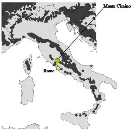
The sampling site is an old-growth beech forest called “La Faggeta” characterized by minimal human disturbance during the last 50 years located in Central Italy (Fig. 8) inland Soriano town on Monte Cimino (Viterbo Province, Latium), between 950 and 1050 m a.s.l. (Fig. 9A).
Fig. 8. Geographic distribution of F. sylvatica in Italy (dark area; Von Wuehlisch 2006), together with the position of study sites in Monte Cimino area.
“La Faggeta” is an oldgrowth secondary forest (58 ha) in the demographic transition stage (Frelich 2002), passing from an impressive single-layer canopy, where trees can reach >40m in height, to an old, yet multiaged, structure (Piovesan, 1998; Di Filippo et
al., 2005) (Fig. 9B). It is dominated by a widespread even-aged cohort (120–150 years
old) that has suffered mortality in consequence of windthrows and glaze storms (Piovesan, 1998; Di Filippo et al., 2005); the last timber logging occurred in 1947– 1949.
A B
Fig. 9. A) Study site tracking on Monte Cimino area (referent system UTM33-ED50). It’s possible see Viterbo city on the East and Vico lake on the South side. B) “La Faggeta” beech stand.
“La Faggeta” are characterized by the occurrence of European Beech (F. sylvatica) and Sweet Chestnut (Castanea sativa Mill.), at lower vegetation limit of beech stand, as main tree species with a group of least species as Sycamore Maple (Acer
pseudoplatanus L.) and European Hornbeam (Carpinus betulus L.) and more rarely
Wild Cherry (Prunus avium L.), Turkey Oak (Quercus cerris L.) and Downy Oak (Quercus pubescens Wild.). According to the phytosociological analysis “La Faggeta” belongs to the Aquifolio-Fagetum unit, where the limestone rocks are dominant (Pignatti, 1998) and has been managed following high-forest silvicultural systems so that trees older than a century are not uncommon over the landscape.
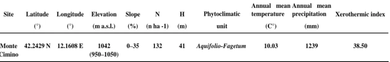
The geographic and environmental characteristics of sampling area are summarized in table 1.
Table 1 Geographic and structural features of sampled beech forest. Site Latitude Longitude Elevation Slope N H Phytoclimatic
Annual mean temperature
Annual mean
precipitation Xerothermic index (°) (°) (m a.s.l.) (%) (n ha -1) (m) unit (C°) (mm)
Monte 42.2429 N 12.1608 E 1042 0–35 132 41 10.03 1239 38.50 Cimino (950–1050)
Aquifolio-Fagetum
Soil has developed from a volcanic bedrock, are generally deep (41 m), and was classified as Vitrandic Hapludalf (Lorenzoni et al., 1995) following the Soil Taxonomy of the US Soil Survey Staff (1992). Site topography is gently sloping.
The pluviometry and mean temperature values of the stand were obtained from meteorological station located in Caprarola (Viterbo Province) about 10 km from trial site, at 42°19' of latitude, 12°10' of longitude (reference system UTM33-ED50) and 650m of altitude, the station has been activated since 1992/10/1 and climatic data were provided by the UCEA service (www.ucea.it).
Sampling was carried out in two different forest sites, called A and B, with North and South facing aspect respectively (Fig. 10).
Fig. 10. Sampling sites into “La Faggeta” beech stand with different exposure. For each area five symptomless plants are randomly chosen.
3.2. Plant sampling
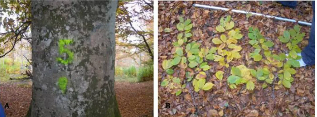
In beech forest stand five plants from each site with different exposure are randomly chosen for a total of ten apparently healthy beech trees. Each tree was marked with coloured paint and identified with a progressive number from 1 to 10 (Fig. 11A). Sampling collection was carried out in five different periods: November (2007),
February (2008), June (2008), September (2008) and January (2009). During which stems were always picked up, whereas leaves were presented only three times (Febuary, June and September 2008). From the basal and outer portion of the crown 3 twigs, 30 cm long, and 15 leaves per plant were collected (Fig. 11B).
Fig. 11. A) Plant n. 5 marked with coloured paint. B) Plant materials picked up from beech plants.
During last sampling in January 2009 were also collected buds of beech trees. From each tree 3 branches from the basal and outer portion of the crown carrying at least 30-40 buds were cut, labeled and taken to the laboratory. Five buds with bud-carrying twig segments were picked from each branch (Fig. 12).
Fig. 12. Fifteen buds per plant were picked during the last sapling period (January 2009).
Once collected tissues samples were taken to laboratory and were stored immediately at 4°C into plastic shoppers to avoid umidity loss until further processing within 24 h.
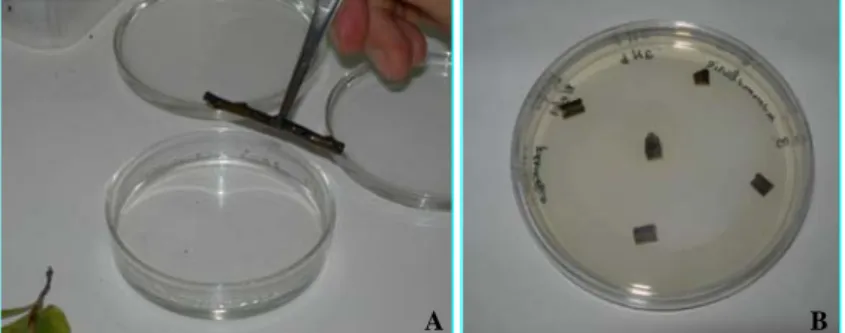
Shoot portions 30 mm longs, leaves and buds were surface-sterilized with 75% ethanol (1 min), 3% NaClO (3 min) and 75% ethanol (30 s) according to Luchi et al. (2006) and rinsed three times with sterile water. Each shoot, leaf and bud was split longitudinally into two parts: one was used for conventional isolation on PDA medium (PDA - MERCK KGaA, Darmstadt, Germany) added with Streptomycin (0,06 g/l) (PDS medium) and the second for DNA extraction.
The portion of shoot and leaf used for fungal isolation was cut into 5-6 small fragments (5 mm each), placed in Petri dishes on PDS and incubated in darkness at 20°C for 10 days (Fig. 13).
A B
Fig. 13. A) Sterilization of plant materials. B) 5-6 small fragments were cut for each tissue and placed in Petri dishes on PDS medium.
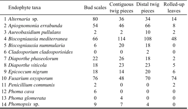
Each bud was divided into its different component tissues: scales, rolled up leaves and current-twigs. Contiguous twig tissues were separated into two pieces: twig pieces directly underneath the buds were called contiguous, as opposed to the distal ones. Bud scales, rolled up and twig pieces were placed in serial order in PDS medium and incubated in darkness at 20°C for 10 days.
3.3 Preparation of culture media.
The following components were added for every liter of PDS medium to be prepared: 39g of Potato Dextrose Agar (PDA - MERCK KGaA, Darmstadt, Germany) powder in 1000 ml of distilled water. Solution was autoclaved at 115°C for 15 minutes. After cooling to 55°C Steptomycin was added to solution (0,06 g/l) to suppress bacterial growth and the medium was dispensed to the Petri plates.
3.4 Isolation, cultivation and identification
The mycelia growing out of the fragments were individually subcultured on potato-dextrose agar (PDA). Pure cultures were identified according to their morphological and molecular features.
Cultures were grouped into morphotypes according to cultural and morphological characteristics (e.g. growth rate, habit and colour of colonies) and colonies were identified morphologically at the genus or species level under light microscopes (Zeiss Axioscope). Taxonomy followed dicotomic keys in standard mycological manuals (Watanabe, 2002) and descriptions of "Index Fungorum"
(http://www.indexfungorum.org).
3.5 DNA extraction, amplification and sequencing
As control, at least one isolate was taken from each morphotype for sequence analysis.
Mycelial cultures were frozen and lyophilized. Genomic DNA was extracted with DNeasy® Plant Mini Kit (Qiagen, Hilden, Germany) according to manufacturer’s instructions. Dry mycelium was transferred into steril ceramic mortar and pulverized with steril ceramic pestle. After, a portion (20 mg of dry weight) was trasferred to 2-ml microfuge tubes with 0,4-ml lysis buffer AP1 and 0,004 ml of RNase (Dneasy Plant Minikit; Qiagen) and ground with a Vortex (2 min at max rpm) to homogenize mixture. After incubation at 65°C for 10 min, 0,13 ml of buffer AP2 was added to the lysis mixture and incubated for 5 min on ice to precipitate detergents, proteins and polysaccharides. After centrifugation for 5 min at 14.000 rpm (Beckman Cuolter ™ Microfuge® 22R Centrifuge), the supernatant was loaded into a QIAshredder™ (Qiagen, Hilden, Germany) spin column and centrifuged for 2 min at 14.000 rpm to remove all precipitates and cell debris. The flow-through fraction was then mixed with
buffer AP3, which contained ethanol, to create conditions for optimal binding to the DNeasy mini-spin column. An aliquot (0,65 ml) of the mixture was applied to the Dneasy mini-spin column and centrifuged at 8.000 rpm for 1 min. After one washing step with buffer AW to remove potential PCR inhibitors, DNA was eluted in 0,1-ml of distilled water and was then ready for PCR processing. To ensure a successful extraction, DNA was separated by gel electrophoresis on 1.5% agarose gels (Agarose D-1 low eeo, Eppendorf Italy) with ethidium bromide (1.2%) in TBE buffer (45mM Tris-borate, 1mM EDTA, pH 8.0) for 25 minutes (70 V). Metagenomic DNA was detected spectrophotometrically under UV lightwave.
The ITS1, 5.8s, and ITS2 regions of rDNA were amplified using the primers ITS1 5′-TCC GTA GGT GAA CCT GCG G-3′ and ITS4 5′-TCC TCC GCT TAT TGA TAT GC-3′ (White et al., 1990). Polymerase chain reactions (PCR) were performed using a PCR- BeadsTM (Amersham Pharmacia Biotech) that included:
- 1,5 U/ml Taq Polymerase; - 10 mM Tris- HCl;
- 50 mM KCl; - 1,5 mM MgCl2 ;
- 200 mM for each dNTP;
to this misture was added 1μl of each primer ITS1 and ITS4 [10 μM], 1μl DNA template [20ng/μl] in 25 μl total reaction volume. Cycling reactions were run on DNA Thermal Cycler (Mastercycler personal, Eppendorf) with the following protocol: 95°C for 2 min; 35 cycles of 94°C for 1 min, 50°C for 30 s, and 72°C for 1 min, with a final elongation step at 72°C for 7 min after cycling. The PCR amplicons were initially scored for successful amplification on 1.5% agarose gels and were purified using Ultra Clean-Up DNA Purification Kit (MoBio Laboratories, Inc., Solano Beach, CA) according to manufacturer protocols. Sequencing PCRs were produced by Macrogen DNA sequencing service under Big Dyes™ terminator cycling conditions. Consensus sequences were computed from forward and reverse sequences using BioEdit software (BioEdit version 7.0.0) and the identity of the amplicon sequence was determined with the standard nucleotide–nucleotide BLAST (blast n) of the NCBI GeneBanck (http://www.ncbi.nlm.nih.gov/).
3.6 Alignment and phylogenetic analyses
For each identified species one sequence generated for this study and two sequence available from official GeneBank was retained for phylogenetic analysis. Sequences were aligned with the program Clustal X (Thompson et al., 1997). Distance analysis was performed using Neighbour-joining method based on K2P distances. A consensus tree was calculated from 1000-fold bootstrapped analyses. Phylogenetic analyses were performed in PHYLIP 3.6 (Felsenstein, 1993).
3.7 Statistical analysis of data
Frequency of species occurrence was calculated as a percentage of the number of samples with the species to the total number of samples tested. The percentage of twig, leaf and bud pieces with single and multiple infections was computed in Microsoft Excel. The colonization frequency (CF) of an individual endophytic taxon was calculated with the formula
CF=Ni/Nt*100
where Ni= number of tissue units from which the fungus were isolated and Nt= total number of units examined. The CF divergences between different plant tissues (twigs, leaves and buds) and between plant samples from different stations (A with North facing and B with South facing) were evaluated by analysis of variance (ANOVA) of percentages.
When the frequency of a species on any tissue type was significantly (P<0.05) higher than zero by Fisher’s exact probability test, the species was regarded as frequent. The chi-square test was used when comparing the frequency of a species among different bud component tissues: rolled-up leaves, bud scales, and twigs.
A statistical interpretation and characterization of the isolated fungal populations in different bud tissues such as diversity and distribution were analyzed for ecological indices (Maria and Sridhar, 2002) computed with software PAST v. 2.04. in particular were considered: Margalef index (DMg); Shannon’s Diversity Index (H′) and Shannon’s
equitability or evenness index (J).
The differences among tissues were evaluated by analysis of variance (ANOVA) and Tukey test (p<0.05) for number taxa and ecological indices and were performed using the GraphPad Prism 4 Istat ® 3.5 ( GraphPad Software inc., San Diego, California, USA).
4. Results: objective 1
4.1 Taxon richness at species and order level
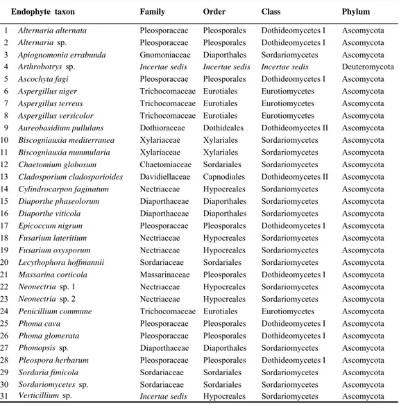
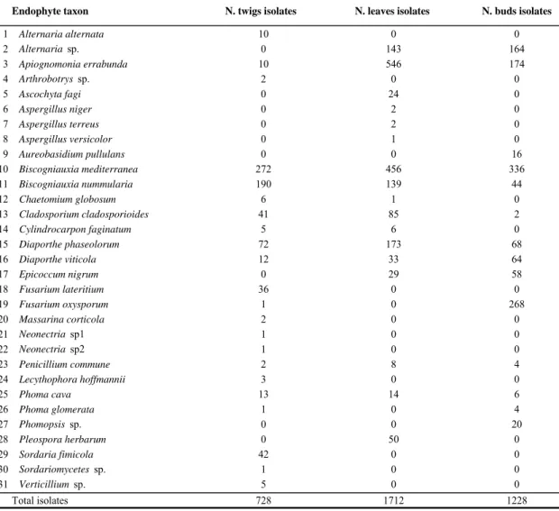
3668 isolates from 150 twigs, 450 leaves and 150 buds collected from 10 symptomless beech trees in 5 successive samplings were used for the assessment of species richness. According to molecular analysis morphotype grouping was evaluated by BLAST searches using sequence data from GeneBanck NCBI(Tab. 2).
Taxa Reference Code
1 Alternaria alternata This study FF32F5
2 Alternaria alternata This study FF3F5
3 Alternaria alternata Wicklow and Poling, 2009 GQ221851*
4 Alternaria alternata Ghosta, 2002 AY154682*
5 Alternaria sp. This study FF3F9
6 Apiognomonia errabunda This study RF5G1
7 Apiognomonia errabunda This study FF3F12
8 Apiognomonia errabunda Sogonov (unpublished) DQ313530*
9 Apiognomonia errabunda Bahnweg (unpublished) AJ888477*
10 Arthrobotrys sp. This study R1R9
11 Arthrobotrys sp. Hagedorn and Scholler, 1999 AF106536*
12 Arthrobotrys sp. Arhipova (unpublished) GU062287*
13 Ascochyta fagi This study F2R2
14 Ascochyta fagi Hashizume (unpublished) AB472195*
15 Ascochyta fagi Hashizume (unpublished) AB472196*
16 Aspergillus niger This study F2FF
17 Aspergillus niger Kausar (unpublished) HM438947*
18 Aspergillus niger Espino del Castello (unpublished) EU833208*
19 Aspergillus terreus This study F4F16
20 Aspergillus terreus Peterson, 2008 EF669580*
21 Aspergillus terreus Chang, 2009 FJ842764*
22 Aspergillus versicolor This study F4F26
23 Aspergillus versicolor Li (unpublished) EU497952*
24 Aspergillus versicolor Espino del Castello (unpublished) EU833210*
25 Aureobasidium pullulans This study F5G1
26 Aureobasidium pullulans Manitchotpisit, 2009 EU719541*
27 Aureobasidium pullulans Manitchotpisit, 2009 EU719545*
28 Biscogniauxia mediterranea This study RF3R6
29 Biscogniauxia mediterranea This study RF3R10
30 Biscogniauxia mediterranea Sanchez Marquez (unpublished) FN394711* 31 Biscogniauxia mediterranea Collado et al. , 2001 AF326482*
32 Biscogniauxia nummularia This study FF3F4
33 Biscogniauxia nummularia Volkenant (unpublished) EF155488*
34 Biscogniauxia nummularia Pinto-Sherer, 1999 AF201706*
35 Chaetomium globosum This study RF3R18
36 Chaetomium globosum Zhou (unpublished) EU301639*
37 Chaetomium globosum Tian, 2009 AB470915*
(*)= Accession number of GeneBank source sequence
Tab. 2. Taxa list of beech tissue (twigs, leaves and buds) endophytes in alphabetical order and references of avalaible ITS sequences in NCBI GeneBank using to complete identification of taxon (BLAST searches and ITS phylogeny).
Tab. 2. (continued)
Taxa Reference Code
38 Cladosporium cladosporioides This study RF3R17
39 Cladosporium cladosporioides Braun et al. , 2003 AY251074*
40 Cladosporium cladosporioides Arteau, 2010 GQ458030*
41 Cylindrocarpon faginatum This study R41
42 Cylindrocarpon faginatum Hallen et al. , 2004 AY677277*
43 Diaporthe phaseolorum This study FF3F15
44 Diaporthe phaseolorum Restrepo (unpublished) EU436686*
45 Diaporthe phaseolorum Lopera (unpublished) EU821481*
46 Diaporthe viticola This study FF3F14
47 Diaporthe viticola This study FF3F13
48 Diaporthe viticola Sanchez Marquez (unpublished) FN386282*
49 Diaporthe viticola Bakys et al. , 2009 FJ228188*
50 Epicoccum nigrum Zhou (unpublished) HM776421*
51 Epicoccum nigrum This study R7
52 Epicoccum nigrum Gorfer (unpublished) HQ115657*
53 Fusarium lateritium Wu (unpublished) HM061323*
54 Fusarium lateritium This study M1RF
55 Fusarium lateritium Santori et al. , 2010 FN547420*
56 Fusarium oxysporum Zheng (unpublished) EU364842*
57 Fusarium oxysporum This study RF5R3
58 Fusarium oxysporum Soca-Chafre (unpublished) EU715659*
59 Lecythophora hoffmannii Vasiliauskas et al. , 2005 AY781227*
60 Lecythophora hoffmannii This study R14R
61 Lecythophora hoffmannii Menkis et al. , 2004 AY805566*
62 Massarina corticola This study RF3R3
63 Massarina corticola Liew et al. , 2002 AF383957*
64 Neonectria sp. 1 This study RF3R5
65 Neonectria sp. 2 This study RF3R4
66 Penicillium commune This study F4R5
67 Penicillium commune Huang (unpublished) GU183158*
68 Penicillium commune Huang (unpublished) GU183165*
69 Phoma cava This study FF3 2F8
70 Phoma glomerata This study F5GF1
71 Phoma glomerata Lindqvist-Kreuze (unpublished) AJ428533*
72 Phoma sp. Bitter (unpublisher) HQ130716*
73 Phomopsis sp. This study FF3F13
74 Phomopsis sp. Kacergius (unpublished) EU571102*
75 Phomopsis sp. Qi et al. , 2009 FJ176469*
76 Pleospora herbarum This study FF3F3
77 Pleospora herbarum Saito, 1999 AB026164*
78 Pleospora herbarum Saito, 1999 AB026165*
79 Pyrenochaeta cava Merhautova (unpublished) FJ379832*
80 Pyrenochaeta cava Gorfer (unpublished) HQ115698*
81 Sordaria fimicola This study R1R19
82 Sordaria fimicola Rotella, 2010 FN868475*
83 Sordariomycetes sp. This study RF3R14
84 Sordariomycetes sp. Hoffman and Arnold, 2010 GQ153239*
85 Sordariomycetes sp. Arnold (unpublished) GQ153034*
86 Verticillium sp. This study R2R2
87 Verticillium sp. Li (unpublished) HM346536*
88 Verticillium sp. Hoffman and Arnold, 2010 GU062214*