Tuscia University of Viterbo
Faculty of Agriculture
Department of Plant Protection
P
HD
INP
LANTP
ROTECTION AGR/12
-
XXIII
CYCLE-
C
URRICULUM:
“Plant parasite control with minimal environmental impact”
TITLE
Antagonistic and plant growth promoting bacteria from
organic growing substrates for controlling some soil borne
fungal pathogens of melon
Ph.D. Dissertation of:
Claudine Sebaaly
Coordinator
Supervisor
Prof. Leonardo Varvaro
Prof. Leonardo Varvaro
Tutors:
Prof. Leonardo Varvaro
Dr. Anna Maria D’Onghia
Dr. Mariagrazia Antonelli
The PHD thesis “Antagonistic and plant growth promoting bacteria from organic
growing substrates for controlling some soil borne fungal pathogens of melon” has been
defended by Claudine Sebaaly (Lebanon) in February 2, 2011. The thesis committee members are:
Prof. Antonio BELCARI, Università degli Studi di Firenze, Italy. Prof. Lucio MONTECCHIO, Università di Padova, Italy.
Dott. Giorgio Mariano BALESTRA, Università degli Studi della Tuscia, Italy.
Tutors: Prof. Leonardo Varvaro Dr. Anna Maria D’Onghia
Dedications
I will offer this work,
To my husband Chadi Adwan,
To my parents Ishaak & Najat,
To my brother and my sisters,
To all my family,
And
To all my friends and my colleagues,…
Acknowledgments
I would like to express my cordial thanks and great gratitude to
my supervisor Prof. Leonardo VARVARO and my tutors Dr.
Anna Maria D’ONGUIA and Dr. Mariagrazia
ANTONELLI for their helpful and precious advices, suggestions
and guidance for the achievement of my work, and all the time that
they dedicated me, but especially for their kindness, their
encouragements and for the confidence that they had in me during
all these months.
My sincere thanks and gratitude to MAI-Bari staff, with
special mention to the Director Dr. C. LACIRIGNOLA, the
Deputy Director Dr. M. RAELI and the Dr. Anna Maria
D’ONGUIA for their financial support, for their suggestions,
advices and help during the three years of studies.
My special recognition and deep thanks to Dr. Alfredo Fabi
for his helpful advising for my thesis work.
And I would like to express my thanks to my colleagues in Tuscia
University for their loving care, patience and support.
Finally, I would like to address my thanks to my friends who
were always sustaining me by their encouraging, supporting and
helping, also for their kindness and friendship.
The PhD thesis has been co-financed by the Mediterranean
Agronomic Institute of Bari (MAIB).
TABLE OF CONTENTS
Page List of abbreviations i List of figures ii List of tables viChapter 1: INTRODUCTION
……… 11.1. Melon cultivation
……….………... 11.2. Phytosanitary problems of melon plants
…………..………... 31.2.1. Monosporascus cannonballus (MC)
………... 41.2.1.1. History of Monosporascus root rot……… 4
1.2.1.2. Classification of Monosporascus cannonballus (Pollack & Uecker, 1974)...………... 4 1.2.1.3. Morphological features……….. 5 1.2.1.4. Symptoms………... 5 1.2.1.5. Life cycle……… 6 1.2.1.6. Epidemiology………... 7 1.2.1.7. Disease dissemination ………... 8
1.2.2. Fusarium oxysporum f. sp. melonis (FOM)
………... 81.2.2.1. History of Fusarium Wilt………... 8
1.2.2.2. Classification of Fusarium oxysporum f. sp. melonis………... 9
1.2.2.3. Morphological features……….. 9
1.2.2.4. Symptoms………... 10
1.2.2.5. Disease life cycle………... 12
1.2.2.6. Epidemiology………. 12
1.2.2.7. Disease dissemination……… 13
1.3. Disease management
……….………... 131.3.1. Physical control………. 13
1.3.2. Chemical control……… 14
1.3.3. Resistant cultivars and rootstocks………... 15
1.3.4. Biological control……….. 16
1.3.4.1. Plant growth promoting bacteria……… 16
1.3.4.2. Fungi………... 17
1.3.4.3. Organic amendment………... 18
1.3.4.3.1. Nitrogenous soil amendments……… 18
1.3.4.3.2. Green Manure effects………. 19
1.3.4.3.3. Compost……….……... 19
Chapter 2: OBJECTIVES
………...………. 21Chapter 3: MATERIALS AND METHODS
………. 233.1. Isolation and quantification of bacteria from compost (ECOS),
organic growing substrate (MAIB-ECOS), and infected field by
FOM
………... 233.1.1. Collection of samples………... 23
3.1.1.1. Commercial compost (ECOS)……….... 23
3.1.1.2. Organic growing substrate (MAIB-ECOS)……… 23
3.1.1.3. Infected field by FOM……… 24
3.1.2. Isolation of microorganisms and colony counting from soil samples………... 24
3.1.4. Physiological and biochemical test……… 26 3.1.4.1. KOH test………. 26 3.1.4.2. Levan production………... 26 3.1.4.3. Liquefaction of gelatin………... 27 3.1.4.4. Lecithinase production………... 27 3.1.4.5. Growth at 41 °C………... 27
3.2. Antagonistic effect of the matured compost (ECOS)
……... 273.2.1. The indicator fungi………. 27
3.2.2. In situ suppressive effect of ECOS compost………... 28
3.2.3. In vitro antagonism assay for bacterial extraction from water suspension of ECOS compost………... 28
3.3. In vitro antagonism assays against two fungal pathogens
………... 293.3.1. Dual culture-plate method on PDA………. ………….. 29
3.3.2. Inhibition of fungal mycelium growth………... 30
3.3.3. Assay for bacterial volatile inhibitors……… 32
3.3.4. Hydrocyanic acid (HCN) production………... 33
3.4. In vitro detection of bacterial plant
growth promoting
features
………. 343.4.1. Siderophore production………... 34
3.4.2. Production of Indole-3-acetic acid (IAA)……….. 35
3.4.3. Mineral phosphate solubilization assays……… 36
3.5. Standardization
of
organic
growing
media
for
melon
crops
………. 373.5.1. Experimental site……… 37
3.5.2. Media ingredients………... 38
3.5.2.1. Inert materials………. 38
3.5.2.2. Organic fertilizer……… 39
3.5.2.3. Coconut fiber and commercial compost………. 39
3.5.2.4. Micronutrients……… 39
3.5.3. Parameters and measurements……… 39
3.5.3.1. Plant growth parameters………... 40
3.5.3.1.1. Germination percentage (G) and mean germination time (MGT)... 40
3.5.3.1.2. Chlorophyll level………... 40
3.5.3.1.3. Plant height (H)………... 40
3.5.3.1.4. Root length (L)………... 40
3.5.3.1.5. Fresh and dry weight of the plant………... 41
3.5.3.2. Colonization of bulk soil, rhizosphere and rhizoplane by bacterial communities………... 41
3.6. In vivo antagonism assay against Fusarium oxysporum f. sp.
melonis
………. 413.6.1. Experimental site………….………... 41
3.6.2. Production of spore forming bacteria (SFB)………... 43
3.6.3. Production of fluorescent pseudomonads bacteria………. 44
3.6.4. Seed preparation………. 44
3.6.5. Preparation of inocula (FOM)……… 45
3.6.6. Data analysis………... 45
3.6.6.1. Disease severity (DS) and area under the disease progress curve (AUDPC)…..………... 45
3.6.6.2. Growth measures……… 46
3.7. Molecular characterization of antagonistic bacteria
………...49
3.7.1. DNA extraction……….. 49
3.7.2. DNA quantification……… 50
3.7.3. Identification by polymerase chain reaction (PCR) technique………... 50
3.7.4. DNA sequencing and phylogenetic analysis………... 51
Chapter 4: RESULTS
……… 524.1. Isolation and quantification of bacteria from compost (ECOS),
organic growing substrate (MAIB-ECOS), and infected field by
FOM
………... 524.1.1. Analysis of bacterial population density isolated from the bulk soil of ECOS compost substrate………... 52
4.1.2. Analysis of bacterial population density isolated from Bulk soil and rhizosphere of MAIB-ECOS substrate and infected field by FOM………... 53
4.1.3. Analysis of bacterial population density isolated from rhizoplane “a” and “b” of MAIB-ECOS substrate and infected field by FOM………... 55
4.2. Antagonistic effect of the matured compost (ECOS)
………... 574.2.1. In situ antagonistic effect of matured compost……….. 57
4.2.2. In vitro antagonism assay for bacterial extraction from water suspension of ECOS compost………... 58
4.2.3. Physiological and biochemical test for bacterial identification……... 59
4.3. In vitro antagonism assays against two fungal pathogens
………... 614.3.1. Dual culture-plate method on PDA……… 61
4.3.2. Inhibition of fungal mycelium growth………... 64
4.3.3. Assay for bacterial volatile inhibitors and HCN production………... 66
4.4. In vitro detection of bacterial plant
growth promoting
features
………... 674.4.1. Quantitative screening of plant growth promoting rhizobacteria………... 67
4.4.2. Correlation among plant growth promoting features and antagonism activities………... 71
4.5. Standardization
of
organic
growing
media
for
melon
crops
………...………... 734.5.1. Plant growth parameters………... 73
4.5.1.1. Germination percentage (G) and mean germination time (MGT)………..……… 73
4.5.1.2. Chlorophyll level……..……….………. 74
4.5.1.3. Plant height (H)……..……….... 76
4.5.1.4. Root length (L)…..………. 77
4.5.1.5. Fresh and dry shoot weight…………..……….. 78
4.5.1.6. Fresh and dry root weight………..………. 80
4.5.2. Colonization of bulk soil, rhizosphere and rhizoplane by bacterial communities………... 81
4.5.2.1. Bacterial communities isolated from bulk soil and rhizosphere... 81
4.5.2.2. Bacterial communities isolated from rhizoplane “a” and “b”……… 84
4.6. In vivo antagonism assay against Fusarium oxysporum f. sp.
melonis
………. 864.6.1. Disease severity (DS) and area under the disease progress curve
(AUDPC)……… 86
4.6.2. Growth measures………...………. 90
4.6.2.1. Plant height………...……….. 90
4.6.2.2. Root length………..…... 92
4.6.2.3. Fresh and dry shoot weight……… 93
4.6.2.4. Fresh and dry root weight………... 96
4.7. Molecular identification of antagonistic bacteria
…………... 98Chapter 5: DISCUSSION
………...……….. 99Chapter 6: CONCLUSIONS
………... 107List of abbreviations
°C Degree CelsiusCfu Colonies forming units Cm Centimeter
DNA Deoxyribonucleic acid
EDTA Ethylene Diamine Tetra-acetic
G Gram H Hour ha Hectare L Litre M Mole m2 Scare meter Mg Milligram Min Minute Ml Millilitre Mm Millimetre mM Millimole Nb Number Ng Nanogram
PCR Polymerase Chain Reaction Rpm Rotation per minute
Sec Second
List of figures
Page 1. Yellowing and death of the melon leaves……… 6
2. Necrosis, lesions and loss of feeder melon roots………... 6
3. Yellowing and wilting of leaves in melon plants...………. 11
4. Necrotic lesion and red gumming on stems near the crown of the melon plant………... 11
5. External lesions on the melon roots………... 11
6. Dual culture plate method on PDA medium. The left and right plate show respectively the FOM F30 and MC-R isolates……… 29
7. Qualitative evaluation of antagonism by recording the mycelial growth of FOM F30 (a) and MC-R (b) isolates………... 31
8. Co-cultivation of antagonsitic bacteria with pathogenic fungi in the presence or absence of charcoal. Nutrient broth (control, left plate) and overnight bacterial cultures were plated in one compartment (left panels) of a tripartite Petri dish. After 2 days of incubation, a fungal mycelium plug (FOM F30 [A] or MC-R [B]) with (right plate) or without charcoal (middle plate) were placed in the other compartments (middle and right panels, respectively)……. 32
9. HCN production with a change in color from yellow (left plate) to reddish brown (right plate)………... 33
10. In vitro detection of bacterial Plant Growth Promoting (PGP) features: Siderophores production (a), Indole-3-acetic acid (IAA) production (b), Phosphate-solubilization (c)……… 37
11. Fusarium wilt caused by FOM F30 isolate on melon in greenhouse experiment (A). The bacterial isolates were tested separately by applying two different techniques of treatment (B) melon seeds coated by the bacterial isolates, and (C) drenching a bacterial suspension in the soil around the melon plants. Plants at the end of the experiment (D)………... 46
12. Total numbers of viable bacteria, fluorescent pseudomonads (FPB) and aerobic spore-forming bacteria (SFB) isolated from bulk soil of ECOS compost substrate……… 52
13. Total numbers of viable bacteria, fluorescent pseudomonads (FPB) and aerobic spore-forming bacteria (SFB) isolated from bulk soil of MAIB-ECOS and rhizosphere of melon seedlings grown in MAIB-ECOS substrate………... 54
14. Total numbers of viable bacteria, fluorescent pseudomonads (FPB) and aerobic spore-forming bacteria (SFB) isolated from bulk soil of infected field and rhizosphere of melon seedlings grown in infected field by FOM………… 54
15. Total numbers of viable bacteria, fluorescent pseudomonads (FPB) and aerobic spore-forming bacteria (SFB) isolated from rhizoplane (“a” and “b”) of melon seedlings grown in MAIB-ECOS substrate………... 56
16. Total numbers of viable bacteria, fluorescent pseudomonads (FPB) and aerobic spore-forming bacteria (SFB) isolated from rhizoplane “a” and “b” of melon seedlings grown in infected field by FOM………... 56
17. The suppression effect of ECOS compost on the hyphal growth of MC-R (a) and of FOM F30 (b). The left and the right plate in each photo, represent respectively the unautoclaved and autoclaved compost. The unautoclaved plates showed clear suppressive zone and weak fungal growth... 57
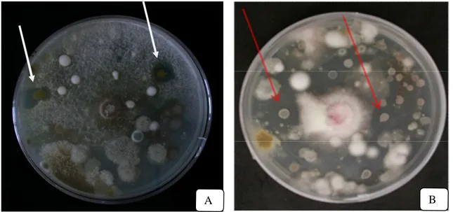
18. The arrows indicate the bacterial colonies present in the water solution of ECOS compost possessing an antagonistic effect (shown as inhibition zone) against MC-R (A) and FOM F30 (B)……….. 58
19. Antifungal activities of fluorescent pseudomonads and aerobic spore forming bacteria, isolated from the four different sites of MAIB-ECOS substrate, against Fusarium oxysporum f. sp. melonis (race 1-2) isolate (FOM F30)……. 62
20. Antifungal activities of fluorescent pseudomonads and aerobic spore forming bacteria, isolated from the four different sites of MAIB-ECOS substrate, against Monosporascus cannonballus isolate (MC-R)……….... 62
21. Antifungal activities of fluorescent pseudomonads and aerobic spore forming bacteria, isolated from the four different sites of infected field by FOM, against Fusarium oxysporum f. sp. melonis (race 1-2) isolate (FOM F30)……. 63
22. Antifungal activities of fluorescent pseudomonads and aerobic spore forming bacteria, isolated from the four different sites of infected field by FOM, against Monosporascus cannonballus isolate (MC-R)……….... 63
23. Percent of fluorescent pseudomonads bacteria (FPB) and spore forming bacteria (SFB) isolated from bulk soil of ECOS compost showing the plant growth promoting activities: phosphate solubilisation, IAA test and siderophore production……… 68
24. Percent of fluorescent pseudomonads bacteria (FPB) isolated from four different sites of MAIB-ECOS substrate samples showing the plant growth promoting activities: phosphate solubilisation, IAA test and siderophore production……….... 69
25. Percent of aerobic spore forming bacteria (SFB) isolated from four different sites of MAIB-ECOS substrate samples showing the plant growth promoting activities: phosphate solubilisation, IAA test and siderophore production……. 69
26. Percent of fluorescent pseudomonads bacteria (FPB) isolated from four different sites of infected field samples showing the plant growth promoting activities: phosphate solubilisation, IAA test and siderophore production…... 70
27. Percent of aerobic spore forming bacteria (SFB) isolated from four different sites of infected field samples showing the plant growth promoting activities: phosphate solubilisation, IAA test and siderophore production………... 70
28. Germination percentage (G) and mean germination time (MGT) of melon plant seedlings grown in the six different organic media (T1 – T2 – T3 – T4 – T5 – T6) during June – July 2009………... 73
29. Germination percentage (G) and mean germination time (MGT) of melon plant seedlings grown in the six different organic media (T1 – T2 – T3 – T4 – T5 – T6) during April – May 2010……….. 74
30. Effect of the six tested organic growing media (T1 – T2 – T3 – T4 – T5 – T6) on seedling chlorophyll level of melon after 22, 24, 26, 28 days of sowing during June – July 2009………... 75
31. Effect of the six tested organic growing media (T1 – T2 – T3 – T4 – T5 – T6) on seedling chlorophyll level of melon after 22, 24, 26, 28 days of sowing during April – May 2010………. 76
32. Plant height (H) of melon seedlings grown in the six different based-organic substrates (T1 – T2 – T3 – T4 – T5 – T6) during two different seasons June – July 2009 and April – May 2010………. 77
33. Root length (L) of melon seedlings grown in the six different based-organic substrates (T1 – T2 – T3 – T4 – T5 – T6) during two different seasons June – July 2009 and April – May 2010………. 78
34. Shoot fresh weight (SFW) of melon seedlings grown in the six different based-organic substrates (T1 – T2 – T3 – T4 – T5 – T6) during two different seasons June – July 2009 and April – May 2010……….... 79
35. Shoot dry weight (SDW) of melon seedlings grown in the six different based-organic substrates (T1 – T2 – T3 – T4 – T5 – T6) during two different seasons June – July 2009 and April - May 2010……….. 79
36. Root fresh weight (RFW) of melon seedlings grown in the six different based-organic substrates (T1 – T2 – T3 – T4 – T5 – T6) during two different seasons June – July 2009 and April - May 2010……….. 80
37. Root dry weight (RDW) of melon seedlings grown in the six different based-organic substrates (T1 – T2 – T3 – T4 – T5 – T6) during two different seasons June – July 2009 and April - May 2010……….. 81
38. Total numbers of viable bacteria isolated from bulk soil of the six studied substrates and rhizosphere of melon seedlings grown in these substrates………... 82
39. Total numbers of fluorescent pseudomonads bacteria isolated from bulk soil of the six studied substrates and rhizosphere of melon seedlings grown in these substrates……… 83
40. Total numbers of aerobic spore-forming bacteria isolated from bulk soil of the six studied substrates and rhizosphere of melon seedlings grown in these substrates………. 83
41. Total numbers of viable bacteria isolated from rhizoplane “a” and rhizoplane “b” of melon seedlings grown in the six studied substrates……….... 85
42. numbers of fluorescent pseudomonads bacteria isolated from rhizoplane “a” and rhizoplane “b” of melon seedlings grown in the six studied substrates………. 85
43. Total numbers of aerobic spore-forming bacteria isolated from rhizoplane “a” and rhizoplane “b” of melon seedlings grown in the six studied substrates………. 86
44. Severity of Fusarium wilt caused by FOM on melon plants treated with four bacterial isolates (B1, B2, B3, C6) collected from ECOS compost by using two techniques of treatment………. 88
45. Severity of Fusarium wilt caused by FOM on melon plants treated with two bacterial isolates (S.2.2, R.2.1) collected from MAIB-ECOS substrate and two bacterial isolates (P8, P18) collected from infected field by FOM by using two techniques of treatment………. 90
46. Plant height of melon seedlings treated with four spore forming bacterial isolates “B1” (D, H), “B2” (E, I), “B3” (F, J) and “C6” (G, K) by using control and two techniques of treatment: seed bacterization and drenching bacterial suspension, in the presence of FOM F30………... 91
47. Plant height of melon seedlings treated with two pseudomonad bacterial isolates “S.2.2” (IV, VIII) “R.2.1” (V, IX) and two spore forming bacterial isolates “P8” (VI, X) “P18” (VII, XI) by using control and two techniques of treatment: seed bacterization and drenching bacterial suspension, in the presence of FOM F30……….. 91
48. Root length of melon seedlings treated with four spore forming bacterial isolates “B1” (D, H), “B2” (E, I), “B3” (F, J) and “C6” (G, K) by using control and two techniques of treatment: seed bacterization and drenching bacterial suspension, in the presence of FOM F30……….. 92
49. Root length of melon seedlings treated with two pseudomonad bacterial isolates “S.2.2” (IV, VIII) “R.2.1” (V, IX) and two spore forming bacterial isolates “P8” (VI, X) “P18” (VII, XI) by using control and two techniques of treatment: seed bacterization and drenching bacterial suspension, in the presence of FOM F30……….. 93
50. Shoot fresh weight of melon seedlings treated with four spore forming bacterial isolates “B1” (D, H), “B2” (E, I), “B3” (F, J) and “C6” (G, K) by using and two techniques of treatment: seed bacterization and drenching bacterial suspension, in the presence of FOM F30………. 94
51. Shoot dry weight of melon seedlings treated with four spore forming bacterial isolates “B1” (D, H), “B2” (E, I), “B3” (F, J) and “C6” (G, K) by using control and two techniques of treatment: seed bacterization and drenching bacterial suspension, in the presence of FOM F30……….. 94
52. Shoot fresh weight of melon seedlings treated with two pseudomonad bacterial isolates “S.2.2” (IV, VIII) “R.2.1” (V, IX) and two spore forming bacterial isolates “P8” (VI, X) “P18” (VII, XI) by using control and two techniques of treatment: seed bacterization and drenching bacterial suspension, in the presence of FOM F30……… 95
53. Shoot dry weight of melon seedlings treated with two pseudomonad bacterial isolates “S.2.2” (IV, VIII) “R.2.1” (V, IX) and two spore forming bacterial isolates “P8” (VI, X) “P18” (VII, XI) by using control and two techniques of treatment: seed bacterization and drenching bacterial suspension, in the presence of FOM F30……….. 95
54. Root fresh weight of melon seedlings treated with four spore forming bacterial isolates “B1” (D, H), “B2” (E, I), “B3” (F, J) and “C6” (G, K) by using control and two techniques of treatment: seed bacterization and drenching bacterial suspension, in the presence of FOM F30………. 96
55. Root dry weight of melon seedlings treated with four spore forming bacterial isolates “B1” (D, H), “B2” (E, I), “B3” (F, J) and “C6” (G, K) by using control and two techniques of treatment: seed bacterization and drenching bacterial suspension, in the presence of FOM F30……….. 97
56. Root fresh weight of melon seedlings treated with two pseudomonad bacterial isolates “S.2.2” (IV, VIII) “R.2.1” (V, IX) and two spore forming bacterial isolates “P8” (VI, X) “P18” (VII, XI) by using control and two techniques of treatment: seed bacterization and drenching bacterial suspension, in the presence of FOM F30……….. 97
57. Root dry weight of melon seedlings treated with two pseudomonad bacterial isolates “S.2.2” (IV, VIII) “R.2.1” (V, IX) and two spore forming bacterial isolates “P8” (VI, X) “P18” (VII, XI) by using controls and two techniques of treatment: seed bacterization and drenching bacterial suspension, in the presence of FOM F30……….. 98
List of tables
Page 1. Melon cultivated area (hectares) and production quantity (tons) in the entire
world and in the continents………... 2
2. The main characteristics of ECOS commercial product………... 23
3. The number of bacterial isolates selected from the different ecological sites of the collected samples (ECOS-compost, Organic growing substrate (MAIB-ECOS) and infected field by FOM)……….. 30
4. The bacterial isolates that were selected to be tested for their antagonistic effect against the pathogenic fungi (FOM F30 and MC-R isolates)…………. 31
5. The bacterial isolates that were selected to identify their volatile fungistatic activity………... 33
6. The main components of MAIB growing media………... 38
7. Characteristics of perlite……… 38
8. Characteristics of Guanito………. 39
9. The components of micronutrients commercial product (Micro-Z G)……….. 39
10. The characteristics of the microorganisms selected through a previous in vitro assay………... 42
11. Characteristics of the bacterial and pathogen inoculation techniques of the first experiment……….. 47
12. Characteristics of the bacterial and pathogen inoculation techniques of the second experiment………. 47
13. Severity of symptoms was assessed using the following empirical scale with five classes of severity………... 48
14. Antifungal activities of spore forming bacterial isolates collected from MC-R and FOM F30 plates against F. oxysporum f.sp. melonis (FOM F30) and Monosporascus cannonballus (MC-R)…... 59
15. General characteristics of fluorescent pseudomonad bacteria isolated from the four different sites of the organic growing substrate (MAIB-ECOS)……… 60
16. Differential characteristics of spore forming bacteria isolated from the water solution of ECOS compost and from three different sites of infected field by FOM………... 60
17. In vitro effect of fluorescent bacteria (FPB) isolated from four different sites of MAIB-ECOS substrate on mycelial growth of F. oxysporum f. sp. melonis (FOM F30) and Monosporascus cannonbalus (MC-R)……… 65
18. In vitro effect of spore forming bacteria, isolated from three different sites of the infected filed and from the water suspension of ECOS compost, on mycelial growth of F. oxysporum f. sp. melonis (FOM F30) and Monosporascus cannonbalus (MC-R)………... 65
19. The intensity of antifungal activity by volatiles produced by the selected bacteria on inhibition of mycelial growth of F. oxysporum f. sp. melonis (FOM F30) and Monosporascus cannonbalus (MC-R) and the hydrogen cyanide (HCN) production……… 66
20. Detection of Plant Growth Promoting properties and antifungal activities of some fluorescent bacteria isolated from four different sites of MAIB-ECOS substrate………... 72
21. Detection of Plant Growth Promoting properties and antifungal activities of some spore forming bacteria isolated from three different sites of infected field by FOM and from the water suspension of ECOS compost………. 72
22. Effect of four bacterial isolates (B1, B2, B3, C6) collected from water suspension of ECOS compost on the area under the disease progress curve (AUDPC) of Fusarium wilt caused by F. oxysporum f. sp. melonis (FOM F30) in melon plants in greenhouse experiment……… 87
23. Effect of two FPB isolates (S.2.2, R.2.1) collected from MAIB-ECOS substrate and of two SFB isolates (P8, P18) collected from infected field by FOM on the area under the disease progress curve (AUDPC) of Fusarium wilt caused by F. oxysporum f. sp. melonis (FOM F30) in melon plants in greenhouse experiment……….. 89
1) Introduction
1.1. Melon cultivation
Melon is generally thought to have originated in western Africa (Bailey, 1976; Purseglove, 1976; Whitaker and Bemis, 1976; Zeven and Zhukovsky, 1975), with China or India as possible secondary centers of diversity. Wild melons growing in natural habitats have been reported in desert and savanna zones of Africa, Arabia, southwestern Asia, and Australia. As Sauer (1993) notes, it is unclear where melon was domesticated and "it is conceivable that it was independently domesticated from different wild populations in Africa and southwestern Asia". Melon was an important food crop in ancient China, where archaeological data suggest that it has been cultivated for over 5,000 years (Robinson and Decker-Walters, 1997). Archaeological evidence also suggests that melon was cultivated in Iran some 5,000 years ago and in Greece and Egypt about 4,000 years ago (Zohary and Hopf, 1988). As with cucumbers, melons were cultivated in the Roman Empire and diffused throughout Europe by the Middle Ages where the "variety and quality of melon cultivars were evidently greatly increased by selection in Medieval gardens" (Sauer, 1993). As with cucumbers and watermelons, melons were introduced to the New World by Spanish colonial settlers in the late fifteenth and early sixteenth centuries and subsequently spread very rapidly among Native American horticultural groups. Later during the eighteenth century they reached the Pacific Islanders via British explorers.
Melon belongs to the Kingdom Plantae, and is classified as a fruit. Its genus is Cucumis and its species is Cucumis melo L. Within these groups, they are also distinguished by their Latin names into two groups, Cucumis (muskmelons) and Citrullus (watermelons). Aside from these classifications, they are also separated into many varieties. Exactly how many varieties are unknown. Some biblical accounts say that there are thousands, and others far fewer. The three most common are the watermelon, cantaloupe, and honeydew. The cantaloupe, also called a muskmelon, it has beige, netted skin and sweet orange flesh that is high in beta carotene. Melons are mostly andromonoecious and have annual trailing vines with nearly round stems bearing tendrils and circular to oval leaves with shallow lobes. Staminate flowers are borne in axillary clusters on the main stem, and perfect flowers are borne at the first node of lateral branches. Fruits vary in size, shape, rind characteristics, and flesh color depending on variety. Fruit quality is related to external appearance, thick, well-colored interior flesh with high (>10 percent) soluble solids, and a pleasant aroma and taste
(Maynard and Elmstrom, 1991). It is a common misconception that poor-quality melon fruit results from cross-pollination with cucumber because these species are incompatible. Rather, the poor-quality melon fruit sometimes encountered is due to unfavorable weather or growing conditions that restrict photosynthetic activity and, thereby, sugar content of the fruit. Seeds are cream-colored, oval, and on average 10 mm long.
Among horticultural species, melons (Cucumis melo L.) have been produced in large quantity, especially in countries with a temperate-warm climate. The main producers are China, Turkey, Iran, Mexico, Romania and Spain; also North Africa is a very important area, where the climatic conditions lead to early productions (Bignami, 2002). Asia dominates the world production of melon by producing more than 71% of the total tonnage (FAO, 2004) (table 1). Cultivation areas in Europe account for about 11% of the world areas (161,000 ha) with a production of 3.16 million of tons: about 70% of these are produced in E.U. and particularly Spain (39,000 ha), Italy (23,000 ha), France (17,500 ha) and Greece (7,900 ha).
Table 1. Melon cultivated area (hectares) and production quantity (tons) in the entire world
and in the continents.
Raking Continent/Country Production (tons) Cultivated Area (hectares)
World 27,371,268 1,313,272
By continent
1 Asia 19,537,759 902,582
2 Europe 3,160,000 161,210
3 North and central America 2,440,099 125,081
4 Africa 1,601,325 79,250 5 South America 565,400 42,800 6 Australia 64,150 2,635 By Nation 2 Turkey 1,700,000 115,000 3 U.S.A 1,240,000 46,000 4 Iran 1,000,000 70,000 5 Romania 1,000,000 52,000 6 Spain 1,000,000 39,100
Cucurbits, in their complex, cover more than 40,000 ha in Italy; melon is the most diffused one, with two different kind of cultivation: melon in the open field mainly diffused in the South and islands; sheltered cultivation, forced (greenhouse or tunnel) or semi-forced (little tunnel), in the North and for early production in every area. In the last ten years, melon cultivation expanded constantly, and in the last five years the cultivated surface overcame 27,000 ha: 3,000 ha in greenhouse and about 24,000 in the field (Chilosi et al., 2008). In northern Italy are cultivated about 11,000 ha (in Lombardia, Emilia-Romagna and Veneto), in the South 8,000 ha and more than 1,000 ha on islands, in the central part more than 6,000 ha (Lazio, Toscana, Marche and Umbria); in particular, in the province of Viterbo the cultivation of melon (ISTAT, data last five years) increase to 256 ha.
1.2. Phytosanitary problems of melon plants
Like most other plants - ornamental and edible - melons are susceptible to a number of pests, viruses, phytoplasma, bacteria, nematodes and fungi, some of which may be more prevalent in one area of the country than another. A lot of parasitic adversities can attack cucurbits' crop both in sheltered cultivation and in the field. Some pests of Italian environments affect plant aerial part (Pseudoperonospora cubensis, Erysiphe cichoracearum, Sphaerotheca fuliginea, Colletotrichum lagenarium, Alternaria cucumerina, A. Alternata), while others develop on roots; these are particularly diffused and are very difficult to control. The main are: Sclerotinia sclerotiorum, Didymella bryoniae, Fusarium oxysporum f. sp. melonis, Verticillium dahliae and V. albo-atrum, Rhizoctonia solani. Fusarium solani, F. oxysporum f. sp. radici-lycopeci, Macrophomina phaseolina, Pythium ultimum, P. aphanidermatum, P. myriotylum and Pyrenochaeta lycopersici, can be also considered.
In recent years, the two main soil born fungal pathogens of melon causing a critical problem and economic losses in central Italy are Monosporascus cannonballus (MC) and Fusarium oxysporum f. sp. melonis (FOM) (Chilosi et al., 2004; 2008). These pathogens are agents of collapse and vascular wilts of the melon plants, respectively. Thus, the suppression of these plant pathogens is considered an urgent need of present agriculture. The use of organic amendment to suppress soil borne pathogens of melon plant has been extensively reviewed, and several microorganisms, have been studied in different organic amended substrates (Antonelli et al., 2009; Sebaaly et al., 2010).
1.2.1. Monosporascus cannonballus (MC)
1.2.1.1. History of Monosporascus root rot
Monosporascus root rot and vine decline is an emergent disease all over the world (Cohen et al., 2000; Martyn and Miller, 1996) and one that only recently has gained attention among plant pathologists. The causal agent, Monosporascus cannonballus, was described as novel genus and species by Pollack and Uecker (1974) on the base of some melon necrotic roots isolated in Arizona. At that time no pathogenic tests were performed; later, the pathogenicity of this pest has been determined in Israel, in 1983, when it was correlated with adult plant collapse (Dias et al., 2004; Martyn and Miller, 1996). Pathogenicity of isolates from the United States was first reported in 1991 (Mertely et al.) in Texas, and the disease was named Monosporascus root rot and vine decline (MRR/VD). In 1996, the disease was identified in Arizona and California (USA) and in other thirteen countries: Mexico, Guatemala, Honduras, Spain, Israel, Iran, Libia, Tunisia, Pakistan, India, Saudi Arabia, Japan and Taiwan (Martyn, 2002). It has since been reported in four additional countries (Korea, Italy, Brazil, Iraq) bringing the total number of countries reporting this disease in 2009 to seventeen. It is highly probable it will be detected in additional countries in the near future. In Italy it was signaled on water-melon near Bologna (Gennari et al., 1999) and on melon in Lazio and Puglia (Infantino et al., 2002; Stravato and Vagnozzi, 2003), later on melon and cucumber near Imola and Ravenna (Montuschi, 2002).
This pathogen was cancelled from the EPPO Alert List in 2001, because of the repeated signallings. In consideration of the aspecific disease symptoms, it cannot be excluded that this pathogen could be more diffused than known.
1.2.1.2. Classification of Monosporascus cannonballus (Pollack & Uecker, 1974)
Kingdom: Fungi Phylum: Ascomycota Class: Sordariomycetes Subclass: Sordariomycetidae Order: Sordariales
Family: Incertae sedis Genus: Monosporascus Species: M. cannonballus
1.2.1.3. Morphological features
M. cannonballus is an Ascomycetes pirenomycetes; it's a homothallic fungus that produces abundant typical reproductive structures, perithecia, both on infected roots and on lab's artificial substrate. Perithecia contain a lot of ascus, mainly with only one ascospore. It's not known the production of asexual spores by M. cannonballus; therefore, pathogen survival and diffusion are ensured by ascospores (Waugh et al., 2003) and, maybe, by mycelium present on infected crop residual.
1.2.1.4. Symptoms
Aboveground symptoms: The symptoms are manifested at the end of the vegetative cycle,
about 2-3 weeks before harvest. Affected plants initially show a state of suffering with extended yellow leaves followed, very soon, by drying and withering (figure 1). Fruits of these plants have a reduced dimension and they do not reach commercial maturation, therefore they have not suitable organolectic requirements for bestowal (lower degree sweetens and less resistant peel) (Mirotti et al., 2003). Moreover, collapsed plants expose fruits to solar radiation, rendering them subject to burns.
Belowground symptoms: The most representative symptoms of the disease are localized on
roots, with necrosis and lesions of different extension, reduced development and rots. Root lesions, root rot, loss of feeder roots and, in severely dry conditions, death of taproot are results of Monosporascus root rot and vine decline (figure 2). Lesions first develop as small areas of necrosis at the joints between secondary and tertiary roots or at the tips of young roots. These lesions are typically dry, however in the event of abundant soil moisture they may appear as a wet rot. Lesions are tan to red-brown. In severe cases of M. cannonballus infection, most of the root system may become necrotic and result in death of the plant. Large, black perithecia form on dead roots and are visible to the naked eye. The perithecia first appear on smaller feeder roots in the first few centimeters of soil and typically appear late in the season.
Researchers conducted in Spain on collapsed plant showed that the described symptomatology can be attributed also to Acremonium cucurbitacearum and Plectosporium tabacinum (Abad et al., 2000; Garcìa-Jiménez, 1994), while in USA Farr et al. (1997) have isolated Rhyzopycnis vagum from roots of a collapsed melon plant that in pathogenetic tests reproduced disease symptoms. Recent studies conducted in Italy on collapsed plant grown in
different regions showed the constant presence of M. cannonballus, A. cucurbitacearum, P. tabacinum and R. vagum (Montuschi, 2002; Infantino et al., 2002a, 2002b).
Fig 1. Yellowing and death of the melon leaves
Fig 2. Necrosis. Lesions and loss of feeder melon roots
1.2.1.5. Life cycle
The disease cycle of M. cannonballus appears to be relatively simple. Infection of the roots can occur from either mycelium that survived in the soil or plant debris or from germinating ascospores. The fungus invades the fine feeder roots, colonizes the cortex and xylem, and kills the rootlet. The infection advances to the next root via the root junction or perhaps from new infections. Smaller roots die while lesions are formed predominantly on larger roots. The fungus continues to colonize the root tissue and invades the xylem causing the plant to form tyloses. While the fungus invades the xylem, it becomes systemic in the plant however it is not a true vascular wilt pathogen, such as Fusarium oxysporum or Verticillium spp. Infection is favored by warm soil temperatures (25 to 35°C). In climates where double
cropping of melons occurs (two crops per year), disease typically is more severe in the late-summer or autumn-planted crop than the spring or winter-planted crop. Perithecia are formed on the roots most abundantly late in the season but may form throughout the life cycle. When mature, the perithecia rupture, discharging the ascospores into the soil. Several hundred ascospores may form in a single perithecium and a single mature melon plant root system is capable of producing 400,000 or more ascospores.
1.2.1.6. Epidemiology
No aerial stage of the disease is known, and no asexual spore stage of the fungus has been observed. Thus, ascospores are considered the primary inoculum and can survive in the soil or plant debris until the next planting season or, in the absence of a host, may survive for many years in a dormant state. Mycelium from infected crop debris can serve as a source of inoculum, although it is not known how long mycelium survives in the soil. The disease is considered monocyclic.
Ascospore germination is favorites by the presence of exudates produced by roots. M. cannonballus adapted at temperate-warm climate, it has an optimum of development at 25-30°C (Pivonia et al., 2002), with pH of 6-7; its development is completely inhibited at pH 4. Moreover this pathogen seems favored by moderately alkaline and salt soil. Studies on variability of M. cannonballus are few and no one includes Italian isolates. Isolates coming from USA, Spain and Japan seem to be characterized by a low variability of morphology, cultural characters, virulence on melon and also in some sequences of DNA. Vegetative compatibility between isolates was not deeply studied, but it seems that there are meaningful differences between the American one and the others.
Ascospores can build up to high numbers in fields cropped successively to melons; however, the minimum threshold level for disease development is not known. Severe disease has occurred in fields with as few as one to two ascospores per gram of dry soil and as high as 15 spores per gram of dry soil. In the few cases studied, ascospores appear uniformly distributed throughout the fields, with the highest concentration in the upper 10 inches (25 cm). Thus, the epidemiology of this disease may be more complex than we currently understand.
1.2.1.7. Disease dissemination
The collapse has assumed a greater importance in the last 15 years, but the reasons are not completely known. Probably, several factors contributed to the increasing spread of the disease. The spread of the pathogen can be by any method that displaces contaminated soil or crop debris (e.g., cultivation, flooding, wind, erosion, etc.). Airborne spread of the pathogen is not likely because of the large size of the ascospores and the lack of a known conidial stage. There is no evidence that the pathogen is seedborne or systemic in the plant. Colonization appears to be limited to the roots. Fruit and foliage are not infected directly, although they manifest symptoms related to root infection, water stress and plant decline. Another aspect regards the changes of the tillage techniques recorded in the last 15-20 years, just contemporaneously with disease spreading. A strong variety change has been recorded, passing from cultivar with a wide genetic base, to hybrids that have plants with more premature and concentrated production. Moreover, the technique of the transplant in place of the direct sown, employing plastic film and localized irrigation diffused. These techniques carried remarkable agronomic and economic advantages, but they produced plants with superficial and little developed roots. Finally, growth of the investments and the production costs induced many farmer to a shorten crop alternation and, in extreme cases, to cultivate cucurbits on the same field for two or three years. This technique inevitably increased the inoculum presence in the soil. For this reason it's necessary to consider the fact that M. cannonballus produces a huge amount of ascospores, which are equipped of a very thick wall and can survive in the soil for several years. Moreover, the pathogen seems to be able to colonize the roots of several vegetable species, both dicotyledonous and monocotyledon, cultivated in succession to the cucurbits without causing noticeable disease, providing another means of survival and reproduction. Moreover, ascospores were found in soils never subjected to cucurbit cultivation and in native desert soils in Texas and Arizona in the U.S. and in Brazil; therefore, it can be argued that M. cannonballus is an indigenous fungus.
1.2.2. Fusarium oxysporum f. sp. melonis (FOM)
1.2.2.1. History of Fusarium Wilt
Separate pathogens or formae speciales (f. sp.) are responsible for Fusarium wilts of melon and cucumber. There are seven formae speciales attacking cucurbits, with Fusarium oxysporum f. sp. melonis is one of the most important pathogen on the major melon growing areas (Dutky et al., 1986; Gonzalez et al., 1988; Gubler and Grogan, 1976; Katan et al.,
1994, 1996; Martyn and Amador, 1987; Smith et al., 1992;) by producing serious economic problems. Fusarium wilt of melon (Fusarium oxysporum f. sp. melonis abbreviated as FOM) was first reported as a disease of melon in the United States in New York in 1930, although its pathogenicity was not confirmed until the disease was described in Minnesota in 1933 (Thomas, 1998). By 1945, the disease was described from states as far west as California and north into Ontario, Canada. Although the pathogen is not uniformly distributed throughout the melon producing regions of the world, it is very widespread.
Investigations over the years on pathogen variability (called races) have led to the description of as many as four races. The four races described for FOM are called races 0, 1, 2, and 1,2. Until recently, the only described race of FOM in the United States was race 2. In 1985, race 1 was reported from Maryland, and in 1987, race 0 was discovered in Texas. In 1992, race 1 was recovered from collapsed melon fields in New York. But race 2 still remains the most widely distributed race in the United States. In contrast, race 1 is the most common race in Europe and the Middle East. Until recently, race 1,2 had only been reported from France, but is now known to occur in Maryland (Villeneuve and Maignien, 2008).
1.2.2.2. Classification of Fusarium oxysporum f. sp. melonis
Kingdom: Fungi Subkingdom: Dikarya Phylum: Ascomycota Subphylum: Pezizomycotina Class: Sordariomycetes Subclass: Hypocreomycetidae Order: Hypocreales Family: Nectriaceae Genus: Fusarium
Specific descriptor: oxysporum
Scientific name: Fusarium oxysporum f. sp. melonis (W.C. Snyder & H.N. Hansen).
1.2.2.3. Morphological features
Fusarium wilt of melon is caused by a seed and soil borne fungus that is specific to melon. F. oxysporum produce three types of asexual spores. These include the macroconidia, microconidia, and chlamydospores. Macroconidia spores have between three and five cells,
septate, that are larger, slightly curved at the ends. These spores are often found on the surface of the plants killed by F. oxysporum. Microconidia spores have between one or two cells, small, colorless, oval to elliptical. They are the type most often produced by the pathogen and are located within the actual vessel of infected plants. Chlamydospores are round spores that have between one and two cells. They have thick walls that are created in either older mycelium (vegetative part of the fungus) or in macroconidia.
1.2.2.4. Symptoms
Plants may be affected in any stage of development; those affected early in their development suffer greater injury than those infected later in the season. Infected plants are often stunted and yellowed. In seedlings, the cotyledons or seed leaves lose their healthy lustre and wilt, followed by the complete collapse of the plant. If affected at an early enough stage, plants will rot (or damp-off) at the soil line and die. In older plants, there is marginal yellowing progressing to a general yellowing of the older leaves, and wilting of one or more runners. In some cases, sudden collapse occurs without any yellowing of the foliage. Fruit from affected vines (if any is produced) tend to be small, with poor flavour and colour. Mature plants often wilt severely (collapse) late in the season because of the fruit load stress. Fusarium wilt collapse should not be confused, however, with sudden wilt of melon. Sudden wilt is a complex disease associated with plant stress brought on by heavy fruit set, cool evening soil temperatures followed by warm and sunny days, feeder root loss caused by soil borne fungi (may include Verticillium and other species), and virus infection (primarily cucumber mosaic and watermelon mosaic, but also papaya ring spot W strain and zucchini yellow mosaic virus).
Leaves: Older plants can show wilting and yellowing of leaves near the crown (figure 3). As
the disease progresses, the leaves show tip-browning. Leaves often have dead areas which can mimic nutrient deficiencies.In some cases, plants exhibit a temporary wilt which appears repeatedly in the middle of the day. Some recovery may occur at night, but the plant finally dies.
Stems and runners: On stems near the crown of the plant, a linear, necrotic lesion may
develop (figure 4), extending up the plant and usually on one side of the vine. One runner on a plant may wilt and collapse, with the rest of the runners remaining healthy, and if environmental conditions change to those which do not favour the fungus, the plant may
recover and fruit normally. However, if conditions continue to favour FOM infection, the whole plant will eventually die.
Roots and vascular system: External lesions may develop on roots, accompanied by red
gumming at or just below the soil surface, similar to that seen on stems (figure 5). Vascular discoloration should be evident and is very diagnostic. If the taproot and stem are split open, an orange-brown discoloration of the water conducting tissues can be seen.
Fig 3. Yellowing and wilting of leaves in melon plants
Fig 4. Necrotic lesion and red gumming
on stems near the crown of the melon plant
1.2.2.5. Disease life cycle
Once introduced into a field, F. oxysporum f. sp. melonis can remain indefinitely, even after rotation to nonsusceptible crops (Gordon et al., 1989). These pathogens can exist time among susceptible crops (Burgess, 1981; Garret, 1970) with their persistence mainly attributed to the production of long-lived chlamydospores (Garret, 1970; McKeen and Wensley, 1961; Newcombe, 1960), which are thick-walled modifications of the mycelium. FOM may also be seed borne, both externally and internally. Internal infection is limited to the area immediately beneath the seed coat and does not infect the embryo. Infection of the host can also occurs by penetration the host through root tips, primarily in the elongation area, and aided by wounding (Martyn and Gordon, 1996). It can also invade the plant with its sporangial germ tube or the mycelium. After penetration, once inside the plant, the mycelium grows within the cells, through the root cortex. When the mycelium reaches the xylem, it invades the vessels. It remains in the vessels and moves up toward the stem and the top of the plant. Then the mycelium branches and produces microconidia, which is carried to the plant's sap stream. The mycelium can penetrate the upper wall of the xylem vessel when the microconidia germinate and produce the next vessel. After infected plants wilt, the Fusarium fungi produce masses of microscopic spores on and in dead vines. The conidia may be splashed and blown from diseased vines resulting in expansion of areas infested by the fungi. Disease development is favored by high nitrogen, low calcium, and low potassium levels in the soil.
Populations as high as 3,300 propagules/gram of soil have been reported in wilt-infested soil but decline more than fourteen-fold during a nine-month fallow interval between crops. Wilt-susceptible varieties tend to increase populations of the fungus. Unfortunately, FOM is saprophytically competitive with nonpathogenic strains of F. oxysporum and can survive on such nonhosts as tomato, alfalfa.
1.2.2.6. Epidemiology
Environmental and soil conditions are important for infection and in symptom expression. Both the incidence and severity of the disease increase during warm, dry weather. Disease severity is maximum at soil temperatures of 17-25 oC and declines dramatically above 30 oC.
At high soil temperatures, plants become infected but may not wilt; rather they develop severe stunting. However, very high temperatures (32-38 oC) combined with increased humidity seem to reduce the disease levels. As with most Fusarium wilts, the following
apply: 1) low soil moisture favors the pathogen and accentuates the wilting symptom; 2) high nitrogen, especially NH4-nitrogen, light, sandy, and slightly acidic soils (pH 5-5.5) favor
disease development; 3) liming the soil to increase the pH to 6.5-7.0 decreases wilt severity.
1.2.2.7. Disease dissemination
Fungi is commonly spread from one field or garden to another by infested soil, compost and manure adhering to machinery, tools and the feet of humans and animals; surface-drainage water; and infected seeds. FOM can be seed-borne, but most transmission occurs by the movement of infected soil or plant parts. The fungus is well adapted to life in the soil, and can survive season to season by living (saprophytically) on dead plant material, or on the roots and stems of other plants such as tomatoes, alfalfa and weeds. Season to season survival also occurs through the formation of very robust resting spores (chlamydospores) which can remain viable in the soil for many years. Chlamydospores are stimulated to germinate by the growth of susceptible host plant roots nearby. Eventually water movement is reduced sufficiently to produce the wilting symptoms typically associated with the disease.
1.3. Disease management
Management strategies for these pathogen are focused on preventive measures; amongst those are the use of resistant rootstocks or attempts for biological control practices (Berg et al., 2001, 2005; Suarez-Estrella et al., 2007; Tjamos et al., 2004) including the use of compost soil amendments (Paplomatas et al., 2005; Suarez- Estrella et al., 2007; Termorshuizen et al., 2006; Yogev et al., 2006), solarization and integration between all these systems (Cohen et al., 2000). There are no chemical treatments to control these diseases (except for soil fumigation, which is applied only to high-value crops usually in greenhouse cultivations).
1.3.1. Physical control
Solarization is indicated as a basic agronomic technique for a correct and heal cultivation to ambient low impact, together with rotation, green manure and rationalization of irrigation. Its use, although obtaining good phytoiatric results, is strongly penalized because of its dependency on the climatic and seasonal fluctuations and on the necessity of an extended interruption of the normal cultural cycles (Materazzi et al., 1987; Triolo et al., 1991). Using transparent or semi-transparent plastic films, it is possible to obtain thermal increasing in the
soil that allows reaching temperatures necessary to control pathogens and to contain the development of the infesting grasses. In fact, unlike vapo-disinfection, it does not create biological empty but it perform a pasteurization with elimination of a large number of pathogens, respecting rate of useful microorganisms, that are ready to develop as antagonists. After elimination of vegetable residual, soil is tilled and irrigated until field capacity, it's covered for a certain period during summer, when solar radiation is maximum, with transparent plastic sheets in order to reduce dispersion of solar energy and to raise temperature. Wetting must increase soil thermal conductivity and sensitize quiescent shapes of pathogens (spores, sclerotia, seeds). If solarization is applied correctly, it's possible to reach soil temperatures of 50 °C at 15-30 cm depth. This technique is very effective against several telluric fungi (Fusarium, Verticillium, Sclerotinia, Pythium), nematode and infesting grass (Conway, 1983). Recently some studies were conducted in order to improve solarization efficacy, arranging it with a reduced dose of specific geo-disinfestants (Sbaraglia et al., 2003), with biocontrol agents and above all with amending organic that produce composed flown that are accumulated under the plastic cloth (biofumigation). This appears a valid solution because the integration of physical (solarization) and chemical or biological disinfection could carry to better results (Sbaraglia et al., 2003).
The causal agent of sudden wilt of melons (FOM) and of melon collapse (MC) is affected by solarization or by the combination of solarization and cabbage residues or chicken manure amendment. The role of these techniques for control of these diseases has not yet been verified in Italian environmental conditions.
1.3.2. Chemical control
Collapse control is very difficult: presence of lasting ascospores, lack of resistant or highly tolerant melon genotype and absence of effective fungicides for the chemical control contribute to this difficulty.
Chemical methods used to control vascular wilt are either not very efficient or imply negative effects on environmental and human health (Becker and Schwinn, 1993; Boutler et al., 2000; Brimner and Boland, 2003; Hoitink and Boehm, 1999).
Fumigation, in particular bromide action, is the most effective control method (Martino, 1997; Montuschi, 2002; Serra et al., 2004), but commercial retirement of methyl bromide as geosterilizer, from 1st January 2005 (Reg. CE 2037/2000), imposes the employment of
alternative control methods (Camponogara e Gasparrini, 2003; Camponogara et al., 2003; Gullino et al., 1999).
1.3.3. Resistant cultivars and rootstocks
As a result of the persistence of these pathogens, effective control of these diseases has generally been achieved by the use of disease-resistant cultivars (Latin, 1989; Mas et al., 1981; Ng and Kantzes, 1991; Zink et al., 1983; Zink, 1992). Nowadays many sources of resistance to FOM races 0, 1 and 2 are known (Alvarez et al., 2005; Pitrat et al., 1996) but it is not the same for race 1,2; resistance to this race has only been found in some Far Eastern accessions (Messiaen et al., 1962) which are organoleptically far from commercialized melon in the western countries.
Another mean of potentially effective control is represented by employment of grafted plants (Cucurbita maxima seems to be less susceptible to M. cannonballus) generally available for the control of other diseases (Mirotti et al., 2003). Herbaceous graft on cucurbits is a practice generally used for the control of telluric origin pathogens with more of 9 million melon plants produced in 2000 (Assenza et al., 2000; Minuto et al., 1999; Morra, 1997; Morra et al., 1997; Rappuoli, 1996; Serges et al., 2000). Although about 26 rootstocks exist, the commonest are melon’s hybrid Jador, Dinero, Belimo and pumpkin's hybrid RS841, P360 and TZ148 (Morra et al., 2001). These differ each other for the resistance degree to several telluric pathogens; it is better to use pumpkin's hybrid, more thriving and resistant, in soil where not even melon's hybrid are able to give protection against pathogens (Ferrari et al., 1993; Morra, 1997; Morra et al., 2001; Temperini et al., 1999; Trionfetti Nisini et al., 2002, 2000).
Because of the great spread of graft, the behavior of rootstock against M. cannonballus must be estimated in a double view: to increase use of less susceptible rootstocks and, at the same time, to advice against use of those highly sensitive (at least in infected soils). As well, since grafting represents an usual procedure for controlling Fusarium oxysporum f. sp. melonis race 1,2, evaluation of rootstocks resistance was also carried out toward a local isolate of this fungus. Evaluations of rootstocks of melon for resistance to FOM were conducted with artificial inoculations on different melon and squash genotypes and grafted plants, the tested rootstocks confirmed to be sufficiently resistant to melon fusarium wilt (Chilosi et al., 2004).
1.3.4. Biological control
The reduction or elimination of synthetic fungicide applications in agriculture is highly desirable. One of the most promising means to achieve this goal is by the use of new tools based on biocontrol agents (BCAs) for disease control. To date, a number of BCAs have been registered and are available as commercial products, including strains belonging to bacterial genera such as Agrobacterium, Pseudomonas, Streptomyces and Bacillus, and fungal genera such as Gliocladium, Trichoderma, Ampelomyces, Candida and Coniothyrium. Also, it is well known that some soils are naturally suppressive to some soil borne plant pathogens such as Fusarium oxysporum, Pythium and Phytophthora species and this suppression relates to both physiochemical and microbiological features of the soil.
Recently, the adopted methodology for suppression of some soil borne pathogens is using the organic amendment, which is particularly important not only to improve plant growth but also to make the plant less susceptible to pathogen infection by postulating different organic amendment mechanisms such as competition for nutrients, antibiotic production by beneficial microorganisms or activation of disease resistance genes in plants. However, only a few organic amendments are currently on the market. Also, many microbial agents isolated from the organic amendments are reported as PGPR (plant growth promoting rhizobacteria), which are free-living soil borne bacteria that colonize the rhizosphere and, when applied to seed or crop, enhance the growth of the plant.
1.3.4.1. Plant growth promoting bacteria
Rhizobacteria have long been known for their role in promotion of plant growth and biological control of plant pathogenic microorganisms. It is believed that plant growth promoting rhizobacteria act by displacing or antagonizing plant pathogenic microorganisms. Various microorganisms in soil, including Enterobacter aerogenes, Pseudomonas fluorescens, Pseudomonas cepacia, and Bacillus species, has been reported to produce antibiotics and antifungal compounds that effectively control or suppress phytopathogenic fungi both in vitro and in vivo.
Bacteria of the genus Bacillus produce a variety of peptide antibiotics that are antibacterial and/or antifungal. Several Bacillus species, including B. subtilis, B. pumilus, B. cereus and B. licheniformis have been shown to be antagonistic to plant pathogenic fungi and bacteria (Neyra and Sadasivan, 1997). Phytopathogen antagonistic strains of B. subtilis have reported to produce two peptide antibiotics: bacilysin, a dipeptide that inhibits yeast and bacteria, and