Acknowledgements
I would like to give thanks to all who contributed to this work by way of varied assistance, advice and even warm smiles. First of all, I would like to thank my supervisor, Prof. Mauro Tognon, for giving me the opportunity to undertake the experiments that resulted to this thesis, and for his encouragements that helped me to achieve my aims. My gratitude and regards to Dr. Elisa Mazzoni, an inspirator, a mentor, a kind and good friend who enlightened me much in my study. I thank my colleagues in Prof. Tognon’s laboratory, Dr. Cecilia Pancaldi, Dr. Stefania Maniero, Dr. Marco Manfirini, Dr. Silvia Bosi, Dr. John Rotondo, Dr. Ilaria Bononi. My special thanks to Prof. Fernanda Martini, for giving me free assess to her laboratory whenever I had the need; to Dr. Lara Rizotto of Hematology and Pathophysiology section, St Anna’s Hospital, for her unconditional support throughout the research period. Thank you all—your kindness and constant willingness to help created a fantastic work environment and made my time in the laboratory thoroughly interesting.
I have not forgotten the contribution of Prof. Eldibert Van Driesche, Prof. Stefano Margez and Greet Devuyst of Vriji University Brussels, Belgium, in shaping my academic ambition. I am sure they will be as glad as I am today in seeing this study program come through.
I am indebted to my friends and to the Alaribe/Nnadozie family for their patience and support during my studies. My sincere regards to my uncle, Monsignor Dr. Innocent Alaribe for being the mastermind of this long journey; my husband Dr Gabriel C. Nnadozie and my son for their support, love and care; my cousin Calistus Agbakwuru and family for their advice and help.
Table of Contents
Page
Acknowledgements
iiIntroduction
1.1 The discovery of simian virus 40 (SV40) 1.2 Polyomavirus infection in the natural host 1.3 SV40 infection in humans 1.4 SV40 structure and genome 1.4.1 SV40 late genome region
1.4.2 SV40 early genome region 1.4.3 SV40 regulatory region and micro RNA
1.5 Life cycle of SV40 in host cells 1.6 The immune response to SV40 1.7 SV40 association with human cancers 1.8 Association of SV40 with lymphomagenesis/non-Hodgkin’s
lymphoma (NHL)
2. Objective of the experimental thesis
3. Material and Methods
3.1 PBMCs and isolation of B/T cells from buffy coats 3.2 Flow cytometric analysis, infection and transfection of human purified B and T cells 3.3 Cell lines 3.4 Alamar blue assay for cell viability and proliferation 3.5 Population doubling time
3.6.1 DNA purification (FOR EXPERIMENT i)
1 2 4 8 9 15 20 23 29 31 35 39 41 41 42 42 43 44
3.6.2 DNA and RNA extraction (FOR EXPERIMENT ii and iii 3.7.1 PCR (EXPERIMENT i)
3.7.2 PCR (FOR EXPERIMENT ii and iii)
3.8 RT-PCR analysis 3.9 Ultrastructural studies 3.10 Indirect immunoflourescence 3.11 Cytopathic effects of infected and T and B cells
with determined titers 3.12 Plaque assay 3. 13 Statistical significance
4 Results
Experiment I
Simian virus 40 (SV40) sequences in blood specimen o healthy individual
from Aviano (Abstract) 4.1 SV40 DNA sequences in 60 subjects of Aviano
Experiment II
Simian virus 40 infection of human T lymphocytes (abstract) 4.2 Viability of the SV40 infected T lymphocyte cells 4.3 SV40 sequences analysis in infected T cells by single and nested PCR 4.4 SV40 Tag oncoprotein and capsid Vps expression in T lymphocyte 4.5 SV40 Effective production of progeny by infected T cells 4.6 Structure distortion in SV40 infected T cells
Experiment III
Transformation of human B cells by SV40, a small DNA tumour virus
(Abstract) 45 45 46 46 47 47 48 48 49 50 50 52 52 53 55 55 58 56
4.7 FACs analysis, infection and transfection
4.8 Cell viability and proliferation capacity 4.9 Growth behavior of infected and transfected cells 4.10 Detection of SV40 DNA sequences in infected and transfected B cells 4.11 Expression of SV40 Tag and VP1 in human transfected and infected
B cells
4.12 Structure distortion in transfected and infected B cells 4.13 Effective production of progeny in infected and transfected B cells
with CV-1 permissive monolayer cells 4.14 Plaque assay viral titer
DISCUSSION
CONCLUSION
ABBREVIATIONS
REFERENCES
PUBLICATIONS
List of Figures
Figure 1.1 Simian virus 40 Genome Figure 1.2 S40 minichromosomes revealing the structural capsid proteins Figure 1.3 Model for SV40 entry and penetration of the ER membrane Figure 1.4 Proposed mechanism by which SV40 regulates its life cycle Figure 1.5 A schematic model representing functional domains of SV40 Tag Figure 1.6 Domains of SV40 small tag Figure 1.7 Cell transformation mediated by small tag downstream expression
60 63 64 65 67 69 69 72 82 84 86 99 9 10 11 14 16 17 19 59
Figure 1.8 Schematic representation of the interaction of SV40 Tag with
tumour suppressor proteins-pRB and p53 Figure 1.9 Possible outcomes of SV40 infection in different cells
Figure 1.10 Schematic representation of the three most frequently cited cell
transformation stages/phases their characteristics Figure 3.1 Structure of the pSV3neo plasmid with intact SV40 early region Figure 3.2 Positive control cell lines Figure 4.1 Viability graph for SV40 infected human T cells in culture Figure 4.2 Single and nested PCR of SV40 infected T cells up to 80 d.p.i Figure 4.3 Immunoflourescence staining of T cell Tag (43a) and VP1 (43b) Figure 4.4 TEM results of T cells
Figure 4.5 FACs results of % population of normal B cells isolated from
PBMCs of 6 healthy donors buffy coat Figure 4.6 Trypan blue analysis of % survival of infected transfected and
normal 60 human B cells from 5-100 d.p.i
Figure 4.7 % alamar blue reduction of B cells with different incubation periods and at different initial cell densities
Figure 4.8 Population doubling time of cells
Figure 4.9 PCR and RT-PCR analysis of the infected and transfected B cells Figure 4.10 Immunoflourescence detection of SV40 Tag in infected
and transfected B cells
Figure 4.11 Detection of VP1 in infected and transfected human B cells Figure 4.12 Transmission electron microscope for ultrastructural observation in
B infected, transfected and normal cells
Figure 4.13 C.P.E observed in SV40 infected B cells with CV-1 monolayer cells Figure 4.14 Viral titer of SV40 infected cells using plaque assay
25 27 28 42 43 53 54 55 59 61 62 63 64 66 67 68 70 71 57
List of Tables
Table 1.1 Members of the polyomaviridae family
Table 1.2 Detection of SV40 DNA sequences in human lymphoploriferative
disorders/NHL
Table 4.1 Numbers detected SV40 DNA sequences from the 60 examined subjects
Table 4.2 Oligonucleotides used as primers in PCR and RT-PCR analysis Table 4.3 Viral titers showed by SV40 776 strain released by infected human T
lymphocytes in CV-1(45-54-2cv-1) cells
Table 4.4 List of Antibodies used in FACs and immunoflourescence analysis Table 4.5 Viral titers determined in B cells infected and transfected cells with
CV-1 monolayer cells using end point dilution assay
4 37 51 51 55 60 70
Introduction
imian virus 40 (SV40) is a small DNA tumor virus, linked with specific human cancers such as malignant pleural mesothelioma (MPM), brain and bone tumors, leukemia, lymphoma diseases. Its DNA sequences is also detected in healthy blood donors. While the debate on SV40 association, rate of prevalence and detection of its DNA sequences in different human cancers including non-Hodgkin lymphomas (NHL) are at increase, indebt studies on the interaction between lymphocytes and SV40 has not yet been fully described.
The highlighted issue calls for further imperative investigation. Hence, this study investigated the interaction of SV40 virus in healthy blood donors using three different experiments. Experiment I: ―Simian virus 40 sequences of healthy individuals from Aviano Cancer center (Italy).‖ The experiment aimed at detecting SV40 DNA sequences in healthy blood donors. Experiment II: ― SV40 infection of human T lymphocyte.‖ It aimed at investigating the rate of susceptibility of purified T lymphocytes to SV40 infection. While the third experiment-―Transformation of human B cells by SV40, a small DNA tumour virus‖ investigated the interaction of SV40 with purified B lymphocytes to observe the capacity of SV40 to infect, transform, and immortalize B cells. These three studies/experiments indeed, indicated the potential role of SV40 in Lymphomagenesis, its relationship and effects with human lymphocytes. Detailed information of these experiments is depicted in results session, each starting with a short abstract.
1.1: The discovery of the simian virus 40, SV40
SV40 belongs to the Polyomaviridae, a family of small DNA viruses that comprises the human polyomaviruses BK virus (BKV) and JC (JCV). BKV and JCV infect 70-90% of the adult population (1,2,3,4). The family name is derived from the first recognised member of the
S
Polyomaviridae, the murine polyomavirus (MuPyV), originally discovered in 1953 by Ludwik Gross as a source of salivary gland tumours in mice. MuPyV was initially designated 'polyomavirus' from the Greek 'poly' meaning many and 'oma' meaning tumours, due to its ability to induce a variety of solid tumours in mice (5,6).
SV40 discovery was linked to the development of the anti-polio vaccines in the 1950s and 1960s as poliovirus was grown in primary kidney cells of rhesus and cynomolgus macaques, which were often naturally infected with SV40 (7,8,9). The virus was then inadvertently introduced into the human population through administration of these contaminated anti-poliovirus vaccines. Soon after its isolation, to verify whether SV40 is able (i) to transform animal and human cells and (ii) to induce tumors in animal models this viral agent was intensely studied both in vitro and in vivo. It turned out that SV40 transforms different types of animal and human cells and induce in experimental animals different kind of tumors, depending on the inoculation route. Consequently, SV40 became known as a potent DNA tumour virus. In the past two decades, increasing evidence of the presence of SV40 in human tumours of different histotypes has seen this 'monkey virus' emerge as a potential human pathogen, a topic that is continually under debate (10,11).
1.2: Polyomavirus Infection in the Natural Host
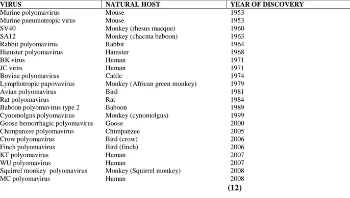
There are 22 existing known members of the Polyomaviridae, capable of infecting a range of species including monkeys, rodents and birds. The phylogenetic analysis of the polyomaviruses has revealed three genetically separate groups: (a) avian polyomaviruses, (b) mammalian polyomaviruses linked to MuPyV and (c) mammalian polyomaviruses linked to SV40. The natural host of SV40 is the Rhesus macaque (macaca mulatta), but several related species of monkey are also capable of being infected (12,13,14).
The spreading of SV40 in the monkey is thought to be by viral shedding in the urine, with host infection occurring by the oral, respiratory and subcutaneous routes. In healthy monkeys, SV40 appears to cause a low level persistent infection in the kidney, this demonstrates that SV40 is a nephrotropic virus (14). In immune compromised monkeys with Simian Immunodeficiency Virus (SIV), SV40 is associated with widespread infection, with the virus being detected in the brain, lung, kidney, lymph node, spleen and peripheral blood, suggesting SV40 may also have neurotropic and lymphotropic properties (15,16). Polyomaviruses are also present in the human host. BK virus (BKV) and JC virus (JCV) are exclusive human viruses and were both discovered in 1971 (17,18).
Recently, three new human polyomaviruses have been discovered (Table1, 12); KI virus (KIV), WU virus (WUV) and Merkel cell virus (MCV). The novel polyomaviruses KN and WUV have been established in respiratory fluids in individuals with respiratory infection and MCV is associated with Merkel Cell Carcinoma, a rare but aggressive human cancer of neuroendocrine origin (13,19,20, ). Phylogenetically, MCV is the only human polyomavirus that does not belong to the SV40 subgroup (21). This most recently discovered member of the Polyomaviridae has been shown to have the highest homology to the mouse polyomavirus subgroup and is most closely related to the lymphotropic polyomavirus (LPV, African green monkey polyomavirus), presumed to be of simian origin. While the polyomaviruses are thought to be highly species-specific and are believed to evolve in close association with their host, the close evolutionary relationship of MCV to LPV and SV40 to BKV and JCV calls this concept into question and may indicate the possibility that host switching can occur (21).
(12)
1.3: SV40 infection in humans
SV40 infection in the human host was seen as a rare event and only restricted to people living in contacts with the natural hosts-monkeys such as inhabitants of Indian villages located close to the jungles and workers attending to monkeys in zoos and animal facilities. Its association with the human host dates back to the 1950s and 1960s when SV40 contaminated vaccines occurred due to the ability of the virus to survive the formalin treatment used to inactivate the poliovirus (22). Human populations were exposed to SV40 by contaminated vaccines administered to hundreds of millions of people in United States, Europe Canada, Asia and Africa between the years of 1955-1963 (9, 23). Additionally, it is thought that a major European manufacturer distributed SV40 contaminated vaccines until as recently as 1978 (24).
Soon it was shown that children vaccinated with contaminated oral polio vaccine (Sabin vaccine or OPV) shed infectious SV40 in stools for at least 5 weeks after vaccination (25). Some children who received the same OPV, did not develop neutralizing antibodies even though they may have
VIRUS NATURAL HOST YEAR OF DISCOVERY
Murine polyomavirus Mouse 1953
Murine pneumotropic virus Mouse 1953
SV40 Monkey (rhesus macque) 1960
SA12 Monkey (chacma baboon) 1963
Rabbit polyomavirus Rabbit 1964
Hamster polyomavirus Hamster 1968
BK virus Human 1971
JC virus Human 1971
Bovine polyomavirus Cattle 1974
Lymphotropic papovavirus Monkey (African green monkey) 1979
Avian polyomavirus Bird 1981
Rat polyomavirus Rat 1984
Baboon polyomavirus type 2 Baboon 1989
Cynomolgus polyomavirus Monkey (cynomolgus) 1999 Goose hemorrhagic polyomavirus Goose 2000
Chimpanzee polyomavirus Chimpanzee 2005
Crow polyomavirus Bird (crow) 2006
Finch polyomavirus Bird (finch) 2006
KT polyomavirus Human 2007
WU polyomavirus Human 2007
Squirrel monkey polyomavirus Monkey (Squirrel monkey) 2008
MC polyomavirus Human 2008
received large doses of live SV40 compared with the potential inactivated SV40 in inactivated polio vaccine (Salk vaccine or IPV). Additionally, SV40 human contamination occurred in experimental infection with live respiratory syncytial virus to adult volunteers and a neutralizing antibody response in about two thirds of the volunteers was revealed. Inactivated vaccines against adenoviruses and hepatitis A virus contributed to human exposure to SV40, even though, the amount of SV40 infections was almost certainly lower than that administered with OPV or live respiratory syncytial virus (26, 27, 28).
The discovery of SV40 contaminated vaccine raised concerns and prompted research into the potential risk to human health. Soon after the identification of SV40 as a polio vaccine contaminant, several studies with hamsters started observing its generation of tumours in experimental animals. It was discovered that subcutaneous injection of rhesus monkey kidney cell extracts into newborn hamsters led to the formation of sarcomas at the site of inoculation (29). Afterwards, it was observed that intracranial injection of SV40 into hamsters induce ependymonas, a type of brain cancer (30). The Syrian golden hamster has been developed as an animal model for SV40-induced tumours with primary brain cancers, malignant mesotheliomas (31), bone tumours (32a) and systemic lymphomas (32b), developing in a manner depending on route of SV40 inoculation.
Furthermore, early serologic studies reported the presence of SV40 neutralizing antibodies at different titers, in the population that received IPV. Immune response appeared to correlate with the amount of SV40 present in the vaccine; 30-50% of individuals reached a significant antibody response against formalin-inactivated SV40 after three doses of the vaccine. Antibody titers persisted for a period of up to 3 years post inoculation. Additional serologic studies reported SV40 seropositivity in individuals with no history of immunization with contaminated IPV or other possible routes of SV40 infection. Studies such as Shah et al, detected antibodies to SV40 in adults and children born 1954 and after 1964 respectively, when IPV was free of SV40. This suggests that,
there is the possibility of human infection by SV40 irrespective of initial exposure to contaminated polio vaccine and that SV40 is being transmitted in the population to this day (1, 4, 33, 34).
To date, the route of transmission and prevalence of SV40 infection in human is still largely unknown. Serological studies have yet to provide comprehensive SV40 prevalence data. Nevertheless, recent studies with PCR and serological techniques indicate that SV0 infection occurs both in children and adults since SV40 DNA sequences was detected in normal and neoplatic tissues of people too young (1-30 years) or too old (60-80 years) to have been vaccinated with SV40-contaminated anti-polio vaccine. This finding may explain the lack of difference in cancer incidence between individuals vaccinated with SV40-contaminated and SV40-free anti-polio vaccines. Secondly, SV40 sequences and Tag were detected in blood and sperm specimens from normal individuals, oncological patients and in lymphoblastoid cells, which suggests that human PBMCs may represent a reservoir and vehicle of SV40 spreading in the tissues of the host and among individuals. Thirdly, detection of SV40 sequences in urine and stool samples from both children and adults indicates the possibility of haematic, sexual and or fecal routes of transmission, which are likely to be responsible for SV40 horizontal infection in humans. Furthermore, there has been detection of specific antibodies to SV40 capsid antigens in human sera, though, there was no comparative data on SV40 DNA prevalence in PBMC and antibodies presence to SV40 antigens in sera from the same patients (35).
Significantly, following laboratory experiments using hamsters, SV40 was shown to transform many human cell types in culture (36). In 1964, an interesting albeit unethical study by Jensen et a1., demonstrated that SV40-transformed human cells were able to produce subcutaneous tumours when injected into human volunteers (37). These SV40 transformed human cells grew as subcutaneous nodules for two weeks and then regressed, possibly because of an immune response. These early studies of SV40-mediated tumour induction in animal models indicated that SV40 was a potent cancer-causing virus and fuelled concerns about the exposure of the human population to
the SV40 in contaminated polio vaccines. SV40 has since been detected in various human tissues, both normal and malignant (11, 38, 39, 40).
Presently, the association between SV40 and human cancer remains very controversial. The discovery of the new human polyomavirus, MCV, integrated into merkel cell tumours, and the continuing studies on the cancer association of BKV and JCV, adds weight to the association of this family of viruses with human cancer.
Comparism of SV40, BKV and JCV
BKV and JCV are exclusive human pathogens and were both discovered in 1971 (17, 18). BKV and JCV infection is widespread in humans and usually occurs in childhood. Seropositivity for BKV reaches 90% in children aged five to nine, with JCV seropositivity at 50 to 60% by the age of ten (41). BKV infection usually occurs at an earlier age than JCV (42).
JCV and BKV share 69% sequence homology with SV40. The greatest homology is found in the early region coding for Tag and tags, whereas a lower homology is detected in the regulatory region. SV40 is phylogenetically closely related to JCV and BKV. They also evidence similarity with respect to size (5.2 Kb), genome organization and DNA sequence. The Tags of SV40, BKV and JCV strongly cross-react with the same antisera, while a less strong cross-reactivity is observed in most structural antigenic determinants of the viral proteins, named VP1, 2 and 3. The DNA sequences of SV40 share 70% homology with BKV, and 69% with JCV (41, 42).
BKV was first isolated from the urine of a renal transplant patient with ureteric stenosis (17). Although the virus is ubiquitous, it does not cause disease in the healthy host, but can produce pathological effects in immunocompromised individuals such as renal transplant recipients, in whom it can affect as many as five percent (43, 44). JCV was first isolated from the brain tissue of a patient with progressive multifocal leukoencephalopathy (PML) (18), a demyelinating disease of
the central nervous system caused by a lytic infection of oligodendrocytes (42). Similar to BKV, JCV is also associated with disease in the immunocompromised host, with PML usually only developing in individuals with a severely compromised immune system. PML was rare before the emergence of Human Immunodeficiency Virus (HIV) but this disease now affects about 5% of HIV-infected patients and is considered to be an Acquired Immunodeficiency Syndrome-defining disease (45).
1.4: SV40 Structure and Genome
SV40 is a non enveloped DNA tumour virus . The virion is about 45 nm, an icosahedral particle, with a density of 1.34–1.35 g/cm3. The viral genome is a circular, double-stranded DNA molecule of 5,243 bp in length in the reference strain SV40-776, with slight nucleotide variations in other strains, subdivided into three functional domains (34). On a genomic level SV40 is closely related to BKV and JCV, sharing approximately 70% homology (11). Although these three viruses are closely related, they can be distinguished at the DNA and protein levels and also serologically by neutralisation and haemagglutination inhibition assays (14). The genomic organisation of SV40, BKV and JCV is conserved with the genomes divided into three functional domains: the early region, the late region, and the regulatory region (46, 47, 48). The early genes are transcribed from one strand of the genome and the late genes are transcribed in the opposite direction from the complementary strand (42). The SV40 early viral coding regions (figure 1) comprises; the large T-antigen (Tag), small t-T-antigen (tag) and the 17kT (tiny T T-antigen). On the other hand, the late region encodes the structural proteins VP1, VP2 and VP3 and a small regulatory protein called agnoprotein, sometimes referred to as VPx (49). There is also, the pre-micro RNA (miRNA) in every mature virus (48).
1.4.1: SV40 Late Genome Region
The Capsid Proteins and Functions
The atomic resolution of the SV40 capsid is formed from the VP1(40kD), the major capsid protein forming the pentameric capsomers that make up the surface of the virus particle, the identical minor capsids VP2 (39kD) and VP3 (27kD) (14). The VP1 capsomers along with the VP2/3 complexes are then joined by the C terminal arms of VP1 to form the icosahedral capsid surrounding the minichromosome (Figure 1.3) and the four host-derived core histones [H2A, H2B, H3 and H4] (48, 50).
Figure 1.1: simian virus 40 (SV40) genome, showing the Tag, tag and tiny T antigen (17kT) in the early region. In the late region of the viral genome, the capsid proteins VPl, VP2 and VP3 are shown along with the regulatory protein, agnoprotein [agno]and a pre-microRNA (miRNA). The regulatory region (ori) contains sequences for the early and late promoter and the origin of replication(48).
SV40 Entrance into the Cell:
The cell infection starts by the binding of the SV40 virus to a receptor identified as the major histocompatibility complex (MHC) which is present on the plasma membrane of the host cell (51). SV40 binding to the host cell is co-directed by the capsid and VP2 (Figure 1.4). Subsequently, the bound virus traverses the membrane and enters a caveola which are large, 70-100 nm diameter flask-shaped invaginations on the plasma membrane (52), where after ~20mins, it is endocytosed and transported in caveolae-coated vesicles to the caveosome through an actin- and dynamin-dependent process. Each 60-70 nm transported vesicle contains a single SV40 virion (52, 53). SV40 particles tend to bud from the caveosome and traffic to the ER. From studies, the ER has been suggested to be the SV40 disassembling domain since by the aid of the ER-resident molecular chaperones, the genome and VP1 pentamers associated with VP2 and VP3 are liberated (51, 52, 54). Further dissociation of the VP1 pentamers tend to release the bound VP2 and VP3. The VP2 and VP3 are inserted into the ER through oligomarization to form a multimeric complex that aids in transporting the genome across the ER membrane. The VP2 and VP3 complex integrates in the contiguous nuclear and ER membrane to directly transport the genome into the nucleus. Intact SV40 has also been proposed to enter the nucleus through the nuclear pore complex (NPC).
However, the largest macromolecule the NPC has been shown to translocate measures 39 nm in diameter indicating that the NPC would exclude the entrance of the intact 50 nm SV40 capsid (55). Further integration of the VP2 and VP3 complex away from the nuclear boundary transports the genome into the cytoplasm (Figure 1.4), where one of the structural protein, ―VPX‖ utilizes its nuclear localization sequence and DNA-binding domains to traffic the genome into the nucleus (56).
However, uncoating of the viral capsid must occur before the genome can be translocated to the nucleoplasm for replication. Polyomavirus capsid undergoes endocytosis and is transported to the nucleus where the viral DNA is uncoated and transcription of the early region begins. The nuclear envelope poses a significant barrier to viral infection and the trafficking of viral components. The strict size limit for nuclear entry requires large viral capsids to undergo uncoating, unwinding or nucleic acid release prior to nuclear entry (55, 57, 58). Enveloped virions (influenza, retroviruses, calciviruses, flaviviruses and herpesviruses) are also subjected to these constraints, as their lipid envelopes are shed exposing the capsids, which must then release the replication competent genome.
Thus, capsids are exposed to cellular proteins that may be commanded by viruses to induce capsid uncoating or deformation to support nucleic acid release enabling genome delivery and replication. The nonenveloped dsDNA adenovirus (60-90 nm) employs cellular processes where, molecular chaperones and a virally encoded internal protease (p23) to weaken its capsid, allowing the genome to be released and enter the nucleus (59). The mechanism of uncoating is the most poorly understood stage in the life cycle of SV40 but the size of the SV40 capsid with its genome comprising of dsDNA associated with histones, indicates that the virus requires extensive disassembly before the genome is imported into the nucleus. This suggests that release of the SV40 genome would closely imitate adenovirus, requiring a cellular trigger for disassembly. Since SV40 traffics to the ER during cell entry, the ER likely contains the cellular trigger that initiates viral uncoating (Figure 1.4). The ER provides an oxidizing and calcium rich environment, optimized for the maturation and quality control of secretory proteins. It houses a variety of molecular chaperones, foldases and proteases, which play central roles in these fundamental cellular processes (60).
LT assists in the transition from viral DNA replication to the synthesis of the late transcripts ~30 hr post-infection. Two late viral transcripts are transcribed: a 16S RNA that is transported to the cytoplasm where it translates VP1; and a second 19S transcript, which encodes for VP2, VP3 and the agnoprotein. While VP1 is the central protein that forms the viral capsid, the roles and functions of VP2 and VP3 in the infection process are poorly defined. VP2 is myristylated on its amino terminal glycine residue (39, 60, 61). Deletion of the myristyl group in a mouse polyoma Py mutant strain resulted in a 20 fold decrease in virus infectivity due to an apparent defect at, or prior to virus uncoating (56, 61). In sharp contrast to these findings, deletion of the unique N-terminal portion of VP2, which contains the myristylated residue, has no effect on SV40 viability.
Since viral assembly occurs within the nucleus, newly synthesized viral structural proteins are directed for nuclear import by their intrinsic NLS. During infection, complexes of VP1, VP2 and
VP3 form in the cytosol and are transported into the nucleus for DNA binding and virion assembly. The low calcium concentration and reducing environment of the cytosol likely aids in the assembly of virions by inhibiting the generation of SV40 capsids prior to nuclear import. Once in the nucleus, VP2 and VP3 binding to the genome is proposed to facilitate SV40 assembly by forming a scaffold for VP1 capsomeres to assemble the capsid ( 62).
Viral lysis and Optimal Spreading
Little is known about the process involved in the dissemination of SV40 and nonenveloped viruses from the cell in general. SV40 infection is thought to result in cell lysis or death through a necrotic pathway due to the cellular trauma that results from the infection (63). However, electron microscopy studies have shown that SV40 virions saturate the apical surface of polarized cells ~48 hr post-infection, ~12-24 hr before cell permeabilization occurs (64).
Observation of SV40 within large smooth membrane vesicles suggest that SV40 utilizes a novel exocytic pathway that involves an unknown sorting signal to reach the cell surface. This would advantageously position the virus for the initial infection of adjacent cells prior to cell permeabilization and support the more efficient dissemination of virus for infection of distant cell populations upon cell permeabilization. This two-step process would enable the initial infections to occur without alerting the immune system, which becomes aware of the virus upon cell permeabilization. The plasma membrane of host cells is unquestionably compromised during infection. However, how this permeabilization is initiated is unclear (56, 60). Several enveloped and non enveloped viruses utilize proteins termed viroporins or virus porins to mediate membrane disruption during viral entry and release.
VP4 (Figure 1.5) is a recently described protein and its expression is thought to coincide with viral lysis. VP4 expression is thought to represent a mechanism by which SV40 regulates its life cycle to
enable optimal spreading of the virus (60). The nonenveloped poliovirus VP1 and VP4 proteins also aid in the delivery of viral RNA to the host. The poliovirus 2B protein has been proposed to initiate cell lysis. The 2B and myristylated VP4 proteins are small viral core components and VP1 contains an N-terminal α-helical domain that is also sequestered in the viral core. Upon receptor binding, the capsid undergoes a conformational change enabling VP1 and VP4 to insert into the host cell membrane and facilitate the transport of the viral RNA (65). A single point mutant in VP4 renders the channel ineffective, preventing the release of the viral RNA into the cytosol of the host (65).
Figure 1.4:
proposed mechanism by which SV40 regulates its life cycle A-C, formation ofpentamers for newly synthesized VP2 and VP3 by initially synthesized VP1, D-E, assembly of virion and five VP1 for VP2/3 so as to prevent them from integrating. Subsequently, VP4 is synthesized (F) and it oligomerises with VP3, possibly VP2 as well, then aims to the host cell (G) membrane so as to form a pore that initiates the lytic death of the host cell to release the progeny (Daniels et al., 2007).
1.4.2: SV40 Early Genome Region
The Large Tag
With the help of Tag and tag SV40 picks the lock on pivotal-check points in its life cycle control and achieves the transformation of host cells by expressing these two important proteins (Tag and tag) upon infection. This causes the cell to undergo an unrestrained proliferation circle that replicates and produces viral DNA and viral particles respectively.
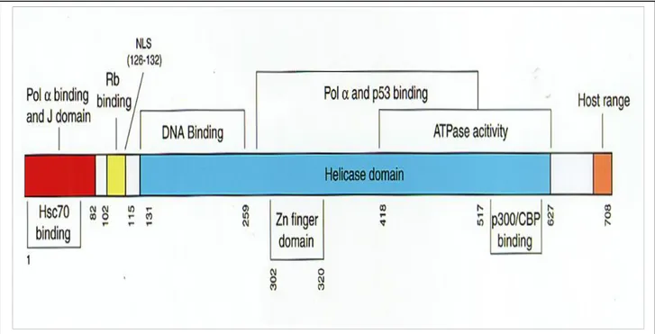
The large tumour antigen (Tag) of SV40 is a 708 amino acid protein that has many functions. It is the major transforming protein of the virus. T-ag is required for initiation of viral DNA synthesis and stimulates host cells to enter S phase. The oncogenic capacity of SV40 stems largely from the ability of this oncoprotein to bind to and inactivate the cellular tumour suppressor proteins and cell cycle regulatory proteins that include the retinoblastoma proteins such as pRB, p107 and p130/pRB2. The tumour suppressor proteins include p53 and the transcriptional co activators p300 and CBP (23, 66). Tag protein is found predominantly in the nuclei of SV40-infected and/or transformed cells (4).
Tag Transformation Domains
SV40 Tag is a multifunctional oncoprotein that possesses several defined functional domains that play a critical role in cell transformation and tumour induction (14). Figure 1.6 schematically illustrates different functional domains of SV40 large T antigen. The amino terminus of the Tag contains two distinct domains important in cell transformation. The far amino terminus of Tag includes the J domain involved in proper folding of protein complexes. This region shares 82 amino acid residues with small t antigen. The second region of the amino terminus of Tag mediates the binding to pRB and the pRB family members p107 and p130 (67). The J domains aslo coperate with LxCxE motif (residues 103-107) of the Tag to inactivate the functions of pRB family memebers and may also have additional transformation. The transforming ability of SV40 Tag is
sometimes abolished by mutations that disrupt their binding with either pRB or p53 (68). Furthermore, the transforming potentials of SV40 Tag is contributed by its possession of additional features such as, the ATPase and DNA helicase. These features provide specifically binding and unwinding functions to SV40 Tag at its origin of replication, thereby assisting in the viral DNA replication. The DNA binding domain (131-259) is mainly known for its function as a transcriptional transactivator. Tag, in addition to targeting cellular tumour suppressor proteins, also targets nuclear acetylases including CREB-binding protein (CBP), P/CAF and p300 (new family members of transcriptional co-activators).These regulatory proteins function as cofactors and play important roles in transcription and posttranslational modification of cellular tumour suppressor proteins (69, 70, 71).
Figure 1.5:
a schematic model representing the functional domains of SV40 Tag. Nearly, minimal regions of T-antigen that retain binding activity to polymerase -primase (Pol), tumour suppressor proteins Rb and p53, human heat shock protein 70 (hsc70) and coactivators p300 and CBP are illustrated. DNA binding domain, ATPase activity domain, nuclear localization signal (NLS) domain, helicase domain, host range domain, Zn finger domain, and J domain are also depicted.Tag interacts with these proteins through multiple regions (Eckner et al, 1996; ) and inactivates their important cellular functions. This is also thought to contribute to deregulation of cell cycle progression.
The SV40 Small T antigen (tag)
Small t antigen of SV40, is a 174 amino acid protein (48) produced by alternative splicing of early transcripts. It shows an all -helix structure with two zinc-binding sites in the unique domain (figure 1.7). Small ag J domain comprises three helices with structure similar to large T antigen crystal structure and NMR structure of poliomavirus DnaJ-like domain (72, 73).
Figure 1.6:
A, The domains of Small tag (St) displaying the 174 amino acid with unique regionsand the crystal structures similar to large T antigen and DnaJ-like domain. B, Small t antigen contains a J domain adjacent to a zinc binding domain that directs the association of small t antigen with the trimeric phosphatase pp2A. SV40 small tag interacts with the B subunit-binding region of the PP2A A subunit, resulting in the displacement of the B subunits from the PP2A complex by the small t (72).
B
A
Small tag forms complexes with the regulatory subunit of the protein phosphate 2A (PP2A) family as in (Figure 1.7B) of serine-threonine phospatases (48, 74). This association appears to inhibit the function of PP2A, which in turn leads to more phosphorylated and increased kinase activity of several cellular kinases including MAP kinase and its kinase ERK, Jun N-terminal kinase (JNK) and a key ion transporter, the Na/H anti-porter (75, 76, 77).
Tag assist solemnly on human cell transformation through its inactivation of the pRB and p53 pathways. Studying the tag mutants has provided important clues to the function of tag during cell transformation. Mutations in tag that prevent the interaction with and inhibition of PP2A demonstrate that this interaction is required for tag mediated transformation. On the contrary, a tag mutant that contains only the PP2A inactivation domain (amino acids 88-174) retains the ability to reduce transformation, which suggest that the alterations in PP2A activity is required for the transforming activity of tag (78).
Small tag in cell Proliferation and Transformation
Studies have shown that tag expression results in enhanced proliferation rates which suggests its involvement in the promotion and regulation of cell cycle progression. It supports the modification from G1 to S phase in cell cycle; tag expression or suppression of PP2A B56g enhances the ability of cells to proliferate in low-nutrient conditions (79, 80). Small tag also promotes the up-regulation of AP-1 transcriptional activity by regulating mitogen activated protein kinase (MAPK) through its over expression. Studies have observed the Raf, MEK1/2, ERK1/2 and KSR sites as possible spots MAPK regulation occurs. Additionally, expression of tag also gives rise to expression and elevation of certain proteins such as cyclin D1, B, thymidine kinase and dihydrofolate reductase linked with cell cycle progression (81). Small tag activate other transcriptional factors such as cAMP regulatory element binding protein (CREB), SP-1 and E2F transactivation linked to cell cycle progression. Small t antigen can also regulate transcription by trans activating the Ad E2A
promoter, which is transcribed by RNA polymerase II, and the Ad VA-I promoter, which is transcribed by RNA polymerase III (82).
Furthermore, tag has been observed to stabilize c-Myc that is directly dephosphorylated by PP2A in tag transformation assays by inhibiting the activity of PP2A (Figure 1.8). A stabilized c-Myc mutant is also capable of substituting for tag expression (83, 84). SV40 tag has been also implicated in anti-apoptotic pathways (Figure 1.8) and induction of changes in cytoskeleton. Studies have shown lines of evidence implicating tag in regulation of apoptosis. Transcriptional analysis of human cells expressing tag revealed up-regulation of anti-apoptotic targets of NF-kB (Figure1.8) such as ALDHI, SERPINB2 and surviving. NF-kB is activated by tag regulation through protein kinase and phosphatidylinositol 3-kinase (P13K) signaling (85). It is also involved in the regulation of the P13K/Akt pathway. Genetic substitution experiments have demonstrated that under tag expression either activated PI3K or a combination of the activated PI3K effectors Akt and Rac1 induce human cell transformation (Figure1.8). Equally, blocking PI3K function inhibits tag-mediated transformation (86).
Figure 1.7: cell transformation mediated by tag downstream expression leading to
inhibition of PP2A complexes that activates numerous signaling pathways and regulation of c-Myc, Akt and Anti-apoptotic NF-kB pathways (74).SV40 tag expression also aims at PP2A-dependent processes governing the cytoskeleton. Over expression of tag in epithelial cells induce dramatic F-actin rearrangements with increase Racl-induced membrane ruffling, Cdc42-initiated filopodia and loss of RhoA-dependent stress fibers (87). Studies on transcriptional profiling analyses have shown that a number of genes such as osteopontin, paxillin and gasoline involved in cellular adhesion and motility are up-regulated by the presence of tag. It also causes down regulation of several genes like Plakoglobin, claudin11, ICAM-1 and VCAM-ICAM-1 involved in cell-cell adhesion and junctional adhesion. This down regulation causes severe defects in formation and barrier properties of tight junction in cells with tag expression (88).
1.4.3: SV40 Regulatory region and Micro RNA
The viral genome of SV40 shows a regulatory region consisting of DNA replication promoter and enhancer elements. A non-coding region, it however functions to orchestrate viral transcription and replication. Its characteristic series of specific nucleotide motifs serve as binding or interaction sites for proteins involved in transcription or DNA replication (91).
The structure of the regulatory region of SV40 viruses can be distinguished in accordance with the number of enhancer elements observed. Hence, two types of regulatory region structure in the SV40 viruses based on the presence of enhancer elements present. Strains having two 72-base pair enhancer elements are referred to as having a 'non-archetypal' or complex regulatory region structure while regulatory regions lacking a duplicated enhancer element are considered 'archetypal' or simple structures (89).
In the case of the reference strain of SV40-776, variants containing one enhancer element are designated 776-1E (-1E virus) while viruses with a duplicated enhancer are called776-2E (-2E virus). These -2E viruses have been shown to replicate faster in tissue culture (89). It has been noted that while natural isolates of SV40 or virus isolated from human tumours generally do not contain enhancer duplicates, laboratory adapted monkey strains of SV40 usually contain two 72-base pair
enhancer elements (90). However, it is thought that a mixture of both archetypal and non archetypal regulatory regions may be present in monkey isolates and that laboratory tissue culture allows the selection of the faster growing variants (91).
Studies have shown that the regulatory region of SV40 may exert a significant influence on tumour incidence. In studies on SV40 tumour induction in Syrian golden hamsters (Mesocricetus auratus), the -lE virus was found to induce tumours in a larger proportion of animals than the -2E virus (92). While the faster speed of viral replication has typically been seen as an advantage for a virus in overcoming the ability of the immune system to control its growth, it has been proposed that slower replicating viruses may evoke a weaker immune response and therefore enhance their likelihood of persistence by circumventing immune surveillance (93). Based on this theory, it is thought that the faster replicating SV40 -2E virus is recognized and cleared more efficiently by the host immune response than the slower replicating -lE virus which would be more likely to evade the immune system and persist (90).
It has also been recently recognized that the viral regulatory region of SV40 may exert effects on vertical transmission in hamsters (94). In a study investigating SV40 transmission in the hamster model, it was found that hamsters inoculated with SV40 strains containing complex regulatory regions (-2E) transmitted virus more frequently than those infected with simple enhancer (-1E) viruses. Further studies are required to determine the relationship between the regulatory region and pathogenesis of SV40 infection, including the possibility that some SV40 strains or variants are more pathogenic than others.
Micro RNA
Since its discovery in the 1990s, relentless study efforts on micro RNA affirmd that SV40 encodes RNA that play important role in the viral life circle (48). Cellular and viral miRNAs are small regulatory RNAs that play critical role in gene regulation. (95).
miRNAs function by binding to complementary sequences in the 3' un-translated regions of messenger RNA (mRNAs), and then mediating mRNA degradation or repressing translation, ultimately leading to reduced protein expression. It has been known for some time that some viral genomes include non-coding RNA such as miRNAs, but their function is only starting to come to light. In fact, recent studies suggest that viral miRNAs may function to evade the host innate immune response, regulate gene expression and possibly contribute to viral-mediated tumourigenesis ( 96, 97). Micro RNAs are useful to the virus in that they are non-immunogenic, take up a small amount of genomic space and are powerful regulators of gene expression (98).
SV40 encodes a pre-miRNA, designated miR-S1, which is processed into two miRNAs that bind to the early RNAs, thereby directing their cleavage at a late stage of the viral cell cycle (98). The end result of the miRNA action in SV40 is a reduction in the expression of viral T antigens. It is known that some protein products of early SV40 genome, such as Tag, are very immunogenic and generate a strong cytotoxic lymphocyte (CTL) immune response. The function of the SV40 miRNA in down regulating expression of these immunogenic proteins suggests that the miRNA is involved in immune evasion strategies. Studies conducted using a mutant SV40 virus named 776-2E-SM, that lacks the viral miRNA, have shown that target cells infected with the mutant are more sensitive to CTL-mediated death than cells infected with a wild type SV40 (47).
In addition, release of the cytokine interferon-y was also diminished when CTLs encountered cells infected with the wild type SV40. Together these results suggest that SV40 miRNA may play a role in evading the adaptive immune response (47). Viral miRNAs in the herpes viruses Epstein Barr virus (EBV), human cytomegalovirus (HCMV) and Kaposi's sarcoma herpes virus (KSHV), have been shown to function in modulating the virus life cycle and human immune response, thereby underlining the importance of miRNAs in viral infections (99).
1.5: Life Cycle of SV40 in Host Cells
SV40 cellular infection begins by its binding to the major histocompatibility complex class 1 (MHC class 1) which is present on the plasma membrane of the host cell (Figure 1). The MHC class 1 molecule act as the specific cell surface receptor for SV40 (100, 101). The life cycle of SV40 in human cells is less well understood but the expression of MHC class I by nearly all nucleated cells thus explains the broad tropism of SV40 and its ability to infect and induce transformation in many types of cells and tissues (23). Not all cells expressing MHC class I are, however, susceptible to SV40 infection and differential ability in infecting polarized epithelial cells via the apical or basolateral surfaces have been reported, despite similar MHC expression on both membranes (42). This suggests the possibility of other factors contributing to SV40 tropism such as co-receptors factors for viral entry after binding or additional transcription factors that may need to be present for viral replication to occur (102).
In the natural monkey host, SV40 infection is known to result in a lytic infection. This viral production is controlled by the immune response and the virus thus persists in the kidney where it can be reactivated by immunosuppression (103). Further, after binding to the cell surface, the poliomavirus capsid is transported to the nucleus where uncoating and transcription of the viral DNA into early region of the genome occurs. The alternatively spliced transcription from the early region gives rise to two mRNAs that encode the large Tag and small tag. SV40 large Tag, a nuclear phosphoprotein of 94kD, is an essential factor for viral DNA replication. It binds to the viral origin of replication where it promotes unwinding of the DNA double helix and recruitment of cellular proteins that are required for DNA synthesis, including DNA polymerase-a and replication protein A (39).
SV40 depends on cellular enzymes and cofactors for DNA replication and these proteins are expressed in S phase. Large T antigen modulate cellular signaling path ways to induce cells to enter S phase and is thought to stimulate the cell cycle through its ability to bind to numerous cellular
proteins involved in crucial transduction path ways that control cell cycle progression and apoptosis (104). Small tag does not partake in lytic infection in culture rather assists the Large Tag in transformation of cells by SV40 and increases virus yield in permissive cells. In human cells, through binding to the cellular tumour suppressor proteins of the pRb family, Tag expression leads to the induction of S phase. pRb normally binds the transcription factor E2F to form a transcriptional repressor complex. However, on binding to SV40 Tag, a dissociation of E2F-pRb complexes occurs (106), releasing E2F to activate expression of growth stimulatory genes (Figure 1.9 A).
This pre-emptive signal for S phase induction and cell division and the resulting unbalanced DNA synthesis induces the cell to initiate a p53 response (tumour suppressor gene). p53 normally senses DNA damage and either pauses the cell cycle to repair the DNA or directs the cell to enter the apoptotic pathway, known as programmed cell death. However, Tag overcomes this obstacle by binding to p53 and inhibiting its function (Figure 1.9B). The cell is thus induced to proliferate and the p53-mediated checkpoint is abolished. This Tag-mediated subversion of cell cycle control allows cells with genetic damage to survive and enter S phase, leading to an accumulation of Tag-expressing cells with genomic mutations that may promote tumour growth as an accidental side effect of the viral replication strategy (105, 106, 107).
As SV40 virus replication proceeds, expression of the late genes begins. The gene products of the late region are the capsid proteins VP1, VP2, VP3, which assemble with the replicated viral DNA to form virions. In infection of the natural monkey host these virions are released upon cell lysis, resulting in death of the host cell (39). The life cycle of SV40 from infection to the production of new virions is thought to take 96 hours in permissive cells (Figure 1.5), resulting in about 300 infectious progeny virions per cell (60, 108).
Although polyomaviruses grow most efficiently in the cells of their natural host species they do not display absolute host specificity (75). Several outcomes are observed in cellular infection by a polyomavirus. In the natural host, cells are permissive to viral infection and are able to support viral replication which results in lytic infection and viral progeny (103). In general, these infections are non-oncogenic for their natural hosts (109). However, SV40 and other turnour viruses may become
Figure 1.8:
interaction of SV40 Tag with pRb and p53 Schematic representation of theinteraction of SV40 Tag with tumour suppressor proteins pRb and p53. (A) pRb normally binds transcription factor E2F, forming a transcriptional repressor complex in early GI phase of the cell cycle. Normally, when pRb is phosphorylated by cyclin-dependent kinases (cdk), E2F is released and activates growth-stirnulatory genes. Tag binding to pRb causes unscheduled dissociation of pRb-E2F complexes, releasing active E2F. (B) The function of p53 is to sense DNA damage and either cause the cell to pause in late GI phase for DNA repair or direct the cell to commit suicide through the apoptotic pathway if repair is not possible. p53 can transcriptionally induce p21, a cyclin-dependent kinase inhibitor, which blocks the activity of GI cyclin-cdk complexes arresting cell cycle progression in late GI phase. Binding of the SV40 Tag results in p53 being sequestered, abolishing its function and allowing cells with genetic damage to survive and enter S phase. As a result, Tag-expressing cells with genomic mutations accumulate that may promote tumourigenic growth (23).
more oncogenic when they cross species (2, 4, 110). SV40 DNA can become integrated into the chromosomal DNA of the cell especially upon infection of the non-permissive cells.
Different studies indicate that SV40 can replicate productively in human cells, including spongioblasts, fetal neural cells, new born kidney cells and some tumour cell lines, with poor growth outcome in human fibroblasts. Hence, the human cells are said to semi- permissive to SV40 infection (36, 103, 110). Some human cell types undergo visible cell lyses in response to SV40 infection, whereas other cells fail to exhibit cytopathic changes and produce low virus levels. Studies have shown that while SV40 infection of human fibroblasts leads to a productive outcome with viral replication, the virus establishes a persistent, non-lytic infection in mesothelial cells (3) and becomes transformed at high rate (1000 times higher than that of human fibroblast) by SV40 Tag alone, releasing infected virions due to persistent infection. Similar situation has also been observed in human lymphoblastoid B cell line where SV40 progeny is produced at a low level (38). The behavior of SV40 in mesothelial cell is very uncommon for a DNA tumour virus. Studies have evidenced that DNA tumour viruses either infect permissive cell lines with the production of an infectious viral progeny or infect and transform non-permissive cells without a productive viral cycle. The rate-limiting step to sustained transformation is thought to be the random integration of viral DNA into the cellular genome by non-homologous recombination. If this integration occurs such that the early coding sequences are intact allowing Tag to be expressed, the host cell and its subsequent descendants are transformed (4, 38, 48).
Possible Cell Transformation Phases from Primary Cell Growth.
Cell transformation could be described as the changes in types of proteins expressed and growth characteristics that take place in cells infected by some viruses, including tumour formation by retroviruses. Transformation could also be oncological or malignant (111,112). Investigating the process of the cell transformation induced by different agents in vitro can indisputably be a very powerful approach to elucidate this basic mechanism. Currently, this type of experiment has been carried out extensively with animal cell models, while only rarely has been observed with human cell types (113, 114).The difference in susceptibility and transformation between animal and human
Figure1.9:
possible Outcomes of SV40 infection in different Cells.Left—In SV40 infection of non-permissive mouse cells, no production of viral particles and malignant transformation can occur. Middle—Infection of permissive monkey cells with cell lysis that results in the production of progeny or many viral particles. Right—Infection of human mesothelial cells that leads to very limited production of viral particles with limited cell lysis and frequent malignant transformation (110).
cell models has been attributed to DNA repair capacity, response to oxidative stress, epigenetic factors such as DNA methylation(114).
In cell transformation, three stages/phases are observed (Figure 1.11): (a) production of cells with an extended life (EL. Cells) from primary diploid cultures that is characterized mainly of diploid primary cells in pre-crisis stage; (b) emergence of immortalized cells (I. Cells) from EL cells after passing through stage two crisis or post crisis stage; (c) development of tumourigenic cells (T. Cells) from EL or I cells in stressful cultural conditions (64, 115, ).
Furthermore, transformation and postponement of senescence of cells can be carried out in human cells such as lymphocytes (with short life span in culture) using different experimental approaches such as transforming by tumour viruses (Herpesvirus saimiri, infection with T-cell leukemia
virus-Figure 1.10:
schematic presentation of the three most frequently cited cell transformationStages/Phases with their characteristics. Phase 1 showing Extended life cells (EL) originated from the primary cells followed by Phase 2- the Immortalized cells (I) and in stressful cultural conditions tumourigenic cells –Phase 3 is developed from I and EL cells.
type1, transfection with Extein-Barrvirus, EBV), or with viral oncogens such as the large T antigen
(Tag) of different Polyomaviruses (116a, 116b). Previously, the transforming capacity of the Tag has been demonstrated in human lymphocytes in vitro (38, 117, 118).
1.6: The Immune Response to SV40
Usually, the viral replication cycle begins with the binding of the virus to the host cell through specific receptors (adsorption). For SV40, the host cell receptor is the MHC class 1 and it determines the tropism and specifity of SV40 virions. The effector in the cellular immune response to virus is the CD8+ cytotoxic T lymphocyte (CTL), which lyses virus-infected cells following recognition of virus encoded epitopes presented on the cell surface by MHC class 1. several factor like the proper generation of the virus epitopes, successful loading and stable binding of virus epitopes to MHC class 1 molecules, proper trafficking of the complex to the cell surface and the presence of T cell in the repertoire capable of recognizing the virus epitopes contribute to successful CD8+ T-cell recognition of virus epitopes (120). The MHC class 1 molecules comprises of a trans-membrane heavy chain that associate non-covalently with β2 microglobulin and a bound peptide representing the T cell recognized epitope. The epitope recognized by CD8+ T cells are typically peptides of 8-10 amino acids in length that can be derived from a variety of virus proteins. This length requirement is imposed by the structure of the peptide binding groove in which the closed ends of the groove accommodate the amino and carboxyl termini of the peptide.
Entry of virus proteins into the MHC class 1 antigen presentation pathway might occur through either direct infection of APCs or by ―cross-presentation‖ in which virus-infected cells or cellular debris are phagocytosed, and the antigens are shuttled into the MHC class 1 processing pathway. Recently, it has been observed that CD4+ T cells prime APCs via signaling through the CD40/ CD40 ligand receptor pair. These activated APCs are then capable of delivering co-stimulatory signals via B7-CD28 or similar engagements for the activation of naïve CD8+ T cells (119).
Studies addressing the requirement for CD4+ T cells help for successful priming of virus-specific CD8+ T-cells responses have suggested that some virus-specific responses are independent of CD4+ T-cell help, perhaps, due to alternate mechanisms for activation of APCs (119, 120).
SV40 Cellular Immune Response During Viral Lytic Cycle
Adaptive and innate immunity are firmly involved in defense against viral infections. However, nature of virus-host interaction determines the participation of the various arms of the immune response. Virus neutralization antibodies play a definitive role in interrupting viral spread while the T lymphocyte-mediated mechanism are involved in the elimination of virus-infected target cells. The CD8+ T lymphocyte (CTL) is responsible for this effect and it lyses virus infected cells by recognizing a viral epitope presented by the MHC class 1 antigens. In a typical immune response to viral infection, natural killer cells that recognize virus-infected cells are recruited followed by a burst of CTL after about 4-7 days. The CTL tends to fall to base line as the virus load declines and then persist as memory T cells which can be recalled upon re-exposure to the viral gene products. The mechanism of CTL effector is very essential for the elimination of virus- infected cells, specifically mainly on those viruses that cause persistent infections (121).
Furthermore, the CD4-mediated T-cells responses are involved in helping B cells produce antibodies and cytokines involved in the proliferation of CD8+ T cells and activation of APC. In human poliomaviruses, only SV40 has an available, reliable model which is the rehesus monkey and as its natural host. For BKV and JCV no animal models in which virus infection leads to its replication in vivo. This particular reason is affecting tremendously the study of T-cell mediated immunity during infection of natural hosts. Recently, a study have reported on the role of CD8+ T cells in interrupting the infectious cycle infection in SV40 in vitro. In this study, the murine CTL clone specific for H2-Kb and H2-Db were utilized along with restricted SV40 T antigen epitopes as probes for abrogating the SV40 infectious cycle in permissive monkey cells. To utilize the strategy,
a continuous line of monkey kidney cells, TC-7, was transfected with murine H2-Kb and H2-Db class 1 antigens, thus allowing the presentation of T-antigen epitopes to SV40 T-antigen-specific CTL clones generated by B6 mice. From this study, it was observed that, the interaction of T-antigen-specific CTL clones with SV40 infected TC7/H-2Db or TC7/H-2Kb cells for 5hrs reduced the SV40 yield by 70-90% as measured by the infectious center assay. This result suggests that: (a) the target of CTL is a non virion protein T antigen, which is synthesized before viral DNA replication; and (b) the CTL mediated events may also take place in the natural host undergoing SV40 infection (122, 123).
1.7: SV40 Association with Human Cancers
It has really been established that SV40 is a potent transforming virus, having observed its induction/generation of tumours in experimental animal models and various types of cells in vitro. More recent evidence from serological and molecular studies revealing its presence in humans raise the possibility that the virus could have transforming capabilities in human cells both in vivo and in vitro.
SV40 in Human Brain Tumours
Detection of SV40 DNA sequences in early studies was in rare cases due to low sensitivity used techniques. However, the studies supported previous findings of SV40 induction of brain tumours in hamsters (124). Recently, improved polymerase chain reaction methods (PCR) as amplification based methods has shown breakthrough in SV40 research, providing a sensitive method of DNA detection in human samples. PCR has been used to detect SV40 sequences in a variety of child hood brain tumours such as ependymoma and choroid plexus tumours, as well as thyroid, pituitary and parotid gland tumours. These human brain tumours correspond to the neoplasms that are induced by SV40 experimental inoculation in rodents or by generation of transgenic mice with the SV40 early region gene directed by its own early promoter-enhancer (39, 125).
There has been numerous studies with continuing reports of SV40 detection in brain tumours (90). Not only has the virus been detected but SV40 Tag has also been found complexed with tumour suppressors p53 and pRb in brain tumours, providing further evidence of a direct functional role for the virus in tumourigenesis (126). In addition to patient studies, an interesting report was published detailing the development of a meningioma in a laboratory scientist who had worked with an SV40-transformed cell line (127). This brain tumour was found to contain SV40 DNA sequences indistinguishable from the laboratory virus, suggesting that laboratory-acquired SV40 infection may have been responsible for turmour development. A meta-analysis of the association of SV40 with brain turmours revealed that samples from patients with brain tumours were almost four times more likely to have evidence of SV40 infection than those from controls (90). Furthermore, SV40 has been detected in multiple histological types of brain tumour, in both children and adults. Indeed, since brain tumours represent 21% of all cancers in children, it has been hypothesised that this high incidence of paediatric brain tumours may suggest acquisition of an infectious agent such as SV40 from the mother (90). However, other studies examining polyomaviruses in brain cancers using primers capable of detecting SV40 sequences have not reported co-infection (128).
SV40 in Malignant Mesothelioma
Malignant mesothelioma is an aggressive tumour of the lung pleura, pericardium or peritoneum, with mean patient survival of only one year from time of diagnosis (129). Mesothelioma has been firmly linked to asbestos exposure. However, since10-20% of occurrence is in people with no known exposure and only 5-10% of those exposed to high levels of asbestos develop this type of cancer, it is thought other factors may be involved in mesothelioma development (3). Asbestos and SV40 have been shown to be co-carcinogens, with SV40 infection lowering the amount of asbestos required for malignant mesothelioma to develop in hamsters. This raises the possibility that SV40 may act as a co-factor in other cancer types (130).