Summary. Poor histological differentiation is currently considered among the adverse histopathological factors associated with unfavourable clinical course of colorectal carcinoma (CRC). At present, the histological grade of CRC is assessed based on the percentage of glandular differentiation in the tumor according to the World Health Organization (WHO) criteria. However the prognostic value of the WHO grading system is limited by significant inter-observer variability in its assessment. In addition, the prognostic significance of WHO grading seems to depend on the microsatellite instability (MSI) status of the tumor. Finally, this grading does not apply to rarer histotypes of colorectal adenocarcinomas, such as the micropapillary, medullary, mucinous and signet ring cell variants.
Recently a novel grading system based on the counting of clusters of five or more cells lacking a glandular structure (poorly differentiated clusters) and set in the tumor stroma or at invasive edge has been proposed in CRC. There is evidence that grading based on poorly differentiated clusters (PDC) is more reproducible and has more robust prognostic significance compared to WHO grading in CRC.
In the present review we discuss the morphological features, criteria for the assessment, prognostic significance and correlation with biomolecular profiles of grading based on PDC counting in CRC.
Key words: Grading, Colorectal, PDC, Mucinous, Micropapillary
Introduction
Colorectal carcinoma (CRC) is one of the leading causes of cancer death in Western Europe and in the USA. Histologically, more than 90% of CRCs are adenocarcinomas and, among those, adenocarcinoma not otherwise specified (NOS) is the most common histological subtype (Hamilton et al., 2010). However, the current World Health Organization (WHO) Classification of Tumours of the Digestive System includes other histotypes such as medullary, mucinous, signet ring cell and micropapillary adenocarcinomas (Hamilton et al., 2010).
At present, the pathological Tumour Node Metastasis (pTNM) stage, established in accordance to the International Union Against Cancer (UICC-TNM) (Leslie et al., 2009) and to the American Joint Committee on Cancer (AJCC) (Edge et al., 2010), represents the most significant prognostic factor in CRC (Hamilton et al., 2010). For this reason, it has relevance in therapeutic decision-making in this tumor. Nevertheless, in spite of its strong prognostic value, pTNM stage only reflects the anatomic extent of the tumour in some cases, without any correlation with patient survival (Barresi et al., 2012). Hence, the search for additional prognostic factors which may predict the clinical course of CRC regardless of pTNM stage has been a main research focus.
Poor histological differentiation is currently
Review
Histological grading in colorectal
cancer: new insights and perspectives
Valeria Barresi1, Luca Reggiani Bonetti2, Antonio Leni1, Rosario Alberto Caruso1and Giovanni Tuccari2
1Department of Human Pathology, University of Messina, Messina and 2Department of Laboratory Integrated Activities, Anatomic Pathology and Legal Medicine, University of Modena and Reggio Emilia, Modena, Italy
Offprint requests to: Dr. Valeria Barresi, Dipartimento di Patologia
Umana, Policlinico G. Martino Pad D, Via Consolare Valeria, 98125 Messina, Italy. e-mail: [email protected]
considered to be a major adverse prognostic factor in CRC (Hamilton et al., 2010). Therefore histological grading is included in the histopathological report of CRC in routine practice. CRC was graded traditionally into well- (G1), moderately- (G2) and poorly (G3) differentiated on the basis of the percentage of gland formation. In detail, according to the WHO criteria (Hamilton et al., 2000), CRC was classified as G1 when showing more than 95% of gland formation, G2 when gland formation ranged between 50% and 95%, and G3 when gland formation was lower than 50%. However, due to the considerable inter-observer variability of this grading system (Chandler and Houlston, 2008) and to the similar behaviour of well- and moderately differentiated CRCs, the latest version of WHO Classification recommended the use of a two-tiered system, with G1 and G2 CRCs lumped together as low-grade and G3 considered as high-low-grade (Hamilton et al., 2010). Although the two-tiered system is more reproducible compared to the three-tiered one (Barresi et al., 2012), it still suffers from several limits. Firstly, although the WHO Classification criteria state that, in heterogeneous tumors, grading should be assessed considering the least differentiated component, the size of the neoplastic area to be considered is not specified. Hence the definition of poor differentiation still remains rather subjective by using this system. In addition, WHO histological grading could be applied only to the adenocarcinoma NOS, but not to other histotypes (Hamilton et al., 2010). Finally, the prognostic significance of WHO grade in CRC strongly depends on the status of microsatellite instability (MSI). Indeed, there is evidence that high-grade CRCs have poor prognosis only if they are microsatellite stable (MMS) (Rosty et al., 2014), while they behave as low grade tumors if they show high MSI (MSI-H) (Hamilton et al., 2010). Hence a grading system combining histological differentiation with MSI status was recently proposed (Rosty et al., 2014) and this combined grading system was shown to predict patients survival with higher accuracy compared to the current WHO grade (Rosty et al., 2014).
In the aim to standardize the criteria for the histological grading of CRC, a novel grading system based on the counting of clusters composed of ≥5 cancer cells and lacking a gland-like structure (poorly differentiated clusters) was recently proposed (Ueno et al., 2012). There is increasing evidence that grading based on the counting of poorly differentiated clusters (PDC) is more reproducible and has more robust prognostic significance compared to WHO grading (Ueno et al., 2012, 2014a,b, 2015; Barresi et al., 2012, 2014a,b; Kim et al., 2015).
The purpose of this review is to discuss the morphological features, criteria for the assessment, prognostic significance and correlation with biomolecular profile of grading based on PDC counting in CRC.
Morphological features of PDC
According to the original definition by Ueno and coll. (2012), PDC are composed of 5 or more cancer cells lacking a gland-like structure and set within the tumor stroma and/or at its invasive edge (Fig. 1a). Hence, compared to tumor budding (TB) foci -which are defined as isolated single cancer cells or clusters of fewer than five tumor cells at the invasive edge of the tumor (Ueno et al., 2002)- PDC consist of a higher number of cells and they may be found not only at the invasive front, but also within the tumour. The larger size of PDC makes their identification with conventional haematoxylin and eosin (H&E) stain easier compared to that of TB, with no need for adjuvant cytokeratin immunohistochemical staining (Ueno et al., 2012).
Transversally cut glandular crypts or fragmented neoplastic glands may mimic PDC (Fig. 1b-d) (Barresi et al., 2014a). However PDC have marked cytological atypia compared to neoplastic glands. Hence the comparison with the cytological aspect observed in the neoplastic glands nearby may help in the distinction between PDC and fragmented glands or transversally cut crypts.
On the other hand, intra-cytoplasmic mucin or vacuoles may be erroneously interpreted as glandular lumina and PDC showing these characteristics may be not recognized as such.
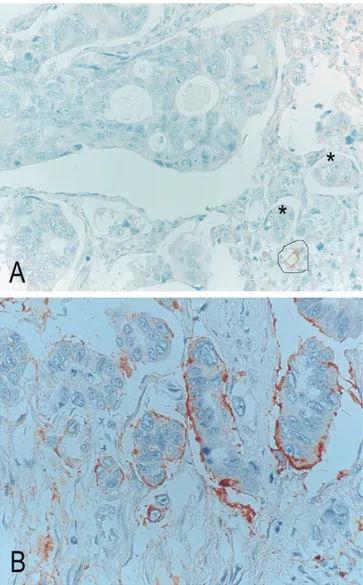
By looking at the images in the original paper by Ueno et al. (2012), a clear space is evident around PDC. The cleft surrounding PDC does not represent lymphatic vessel invasion; indeed immunohistochemistry against podoplanin demonstrates the absence of lymphatic endothelia (Fig. 2a) (Barresi et al., 2014c). However, a similar histological aspect is also found in the micropapillary variant of CRC, the hallmark of which is the presence of “small clusters of tumour cells within stromal spaces mimicking vascular channels” (Hamilton et al., 2010). We believe that PDC and micropapillary pattern may represent the same biological phenomenon in CRC (Barresi et al., 2014c). Indeed, simlarly to that observed in micropapillary CRC (Verdú et al., 2011), the clear space surrounding PDC is the result of reversed cell polarity of tumour cells, which display a secretory activity in the stroma-facing surface. Confirming this hypothesis, a reversed pattern of MUC1 expression is observed in PDC, similarly to micropapillary CRC (Barresi et al., 2014c) (Fig. 2b). For this reason we proposed that micropapillary could be considered as a CRC with high density of PDC (Barresi et al., 2014c). Besides, this peculiar histological aspect is related to tumor de-differentiation rather than to the presence of micropapillae. Indeed MUC1 is one of the proteins lost during epithelial-mesenchymal transition (EMT) (Guaita et al., 2002), a process by which neoplastic cells lose epithelial properties and acquire the mesenchymal cell potential to migrate through the extra-cellular matrix. Moreover, PDC have aberrant or lost expression of
e-cadherin (Barresi et al., 2014c), which is a protein essential for the establishment of cell-cell adhesion and which is down-regulated during EMT (Kalluri and Weinberg, 2009).
Assessment of PDC grading
According to the system originally proposed by Ueno and coll. (2012), CRC can be graded on the basis of PDC counting into three grades of malignancy. In detail, the whole tumor section, including its deepest part, is first scanned under a 4x objective lens with the aim to identify the areas with the highest number of PDC. Then, PDC are counted under the microscopic field of a x20 objective lens (i.e., a microscopic field
with a major axis of 1 mm) and the highest number in the whole section is recorded and considered for grading assessment. Cancers with <5, 5 to 9, and ≥10 clusters are classified as grade 1 (G1), grade 2 (G2) and grade 3 (G3), respectively (Fig. 3a-c).
Due to the higher standardization of the criteria used for grading, the PDC system has higher inter-observer reproducibility compared to WHO grading (Ueno et al., 2012; Barresi et al., 2014a,b).
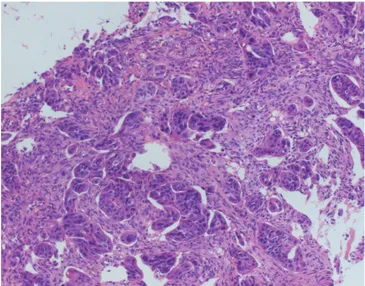
Histological grading of CRC is also commonly assessed in the preoperative endoscopic biopsies. Nevertheless, at present the clinical relevance of grading CRC biopsy is limited by low inter-observer reproducibility and poor concordance with the grading observed in the following surgical specimen (Fig. 4)
Fig. 1. a. PDC composed of ≥ 5 cancer cells with marked cytological atypia and no glandular formation in the stroma of CRC. A cleft-like space is evident around PDC. b-c. Transversally cut crypts (circles) may be confused with PDC. However they have less cytological atypia than PDC and display morphological characteristics similar to those of the nearby crypts. d. Fragmented neoplastic glands (circle) may also resemble PDC. Mild cytological atypia compared to PDC and comparison with the histological aspect of preserved neoplastic glands are helpful in the distinction. H&E stain. x200
(Burton et al., 2003). We showed that PDC grading system may also be applied to CRC biopsies (Barresi and Tuccari, 2013; Barresi et al., 2014a). Since CRC exhibits higher differentiation toward the surface, the probability of finding PDC is less in the biopsy compared to the surgical specimen (Barresi et al., 2014a). Hence, an adjustment of PDC grading scale is warranted for application in CRC biopsy. Although this needs verification in further studies, we showed that CRC biopsies with 0, 1-2 and >2 PDC under the microscopic field of a x20 objective lens may be graded as G1, G2 and G3, respectively, with good inter-observer agreement (Barresi et al., 2014a). According to our results, there is higher correspondence in grading assessed in the biopsy and surgical specimen by using the PDC system than the WHO one (Chandler and Houlston, 2008; Barresi et al., 2014a).
Major causes of disagreement in the assessment of PDC may be related to the difficulty in the distinction between PDC and transversally cut crypts or between PDC and fragmented neoplastic glands (Barresi et al., 2014a). However, the presence of a clear cleft around PDC but not around the crypts, and the marked cytological atypia observed in PDC but not in the crypts may be helpful in the differentiation. In addition, the comparison with the cytological aspect of the neoplastic glands in the specimen may be useful for the discrimination from fragmented glands. Finally, intra-tumoral inflammation may lead to the fragmentation of neoplastic glands and to the formation of clusters of neoplastic cells with no glandular lumen (Fig. 5). Nonetheless, since these clusters do not represent de-differentiation, they should not be considered to be PDC. Hence we suggest that areas showing inflammation should be excluded in the assessment of PDC grading in CRC (Barresi et al., 2014a).
PDC grading in the other histotypes of colorectal adenocarcinomas
According to the current WHO guidelines, grading does not apply to the micropapillary, medullary, mucinous and signet ring variants of CRC adeno-carcinoma (Hamilton et al., 2010). Indeed, these entities are said to carry their own prognosis (Hamilton et al., 2010). However, tumors of the same histotype may show different clinical outcome (Langner et al., 2012); for this reason, histopathological factors able to stratify patients for their individual recurrence risk may be useful also in the other histotypes of colorectal adenocarcinoma.
There is evidence that PDC grading may be assessed in micropapillary, medullary, mucinous and signet ring cell colorectal adenocarcinoma (Ueno et al., 2014a; Kim et al., 2015). As discussed above, micropapillary CRC might be considered as PDC G3 due to the high density of PDC as its main histological feature (Ueno et al., 2014a). On the other hand, medullary carcinoma, which is characterized by sheets of malignant cells with very rare PDC, could be mostly classified as PDC G1 (Ueno
et al., 2104a).
The histological grading of mucinous CRC is still an unsolved issue. In the previous edition of WHO Classification (Hamilton et al., 2000), mucinous adenocarcinoma was designated as poorly differentiated by convention (G3). Nonetheless, the current WHO guidelines state that the level of maturation of the epithelium determines differentiation in mucinous CRC, but MSI status should be also considered (Hamilton et al., 2010). Indeed mucinous CRCs with MSI-H behave as low-grade while those with low MSI (L-MSI) or microsatellite stability (MSS) behave as high grade (Hamilton et al., 2010). Interestingly, PDC grading can be easily assessed in this histotype by classifying as
Fig. 2. a. Absence of immunohistcohemical staining for podoplanin around PDC (stars) demonstrates that this histological aspect does not correspond to lymphatic invasion. Positive staining in a lymphatic vessel as internal positive control (circle). Podoplanin stain. b. Reversed pattern of MUC1 stain in PDC, with positive reaction at the cell membrane towards the stroma. MUC1 stain. x 200
PDC those clusters infiltrating the stroma with minimal extracellular mucin (Fig. 6a-c), but not those within a large mucin pool (ie, mucinous lake) (Ueno et al., 2014a; Kim et al., 2015). We recently demonstrated very good inter-observer agreement in the assessment of PDC grade in a cohort of 108 mucinous adenocarcinomas (unpublished data). Of note, as previously observed by Kim and coll. (2015), the percentage of PDC G3 cases was higher among mucinous carcinomas, compared to the adenocarcinomas NOS in our series (unpublished data).
Fig 4. High number of PDC in CRC endoscopic biopsy. H&E stain. x 100
Fig. 3. CRCs graded as G1 (a), G2 (b) and G3 (c) according to PDC grading. H&E stain. x 200
Fig. 5. Clusters of neoplastic cells within inflammatory infiltrate in CRC. Since these clusters may be formed by fragmentation due to inflammation, they should not be considered for PDC grading assessment. H&E stain. x 200
Finally, since signet ring cells may also aggregate to form clusters (Fig. 6d), there is some evidence that PDC grading might also be performed in signet ring cell CRC (Kim et al., 2015). Nonetheless, additional studies are needed to demonstrate the practical application of PDC grading in this variant.
Prognostic value of PDC grading
According to several studies, PDC grading is a strong, independent, prognostic factor in CRC (Barresi et al., 2012, 2014a,b; Ueno et al., 2012, 2014a,b; Kim et al., 2015). Indeed, significantly shorter disease free survival (DFS) and disease specific survival (DSS) were shown in patients with CRC of PDC grades 2 and 3 in any pTNM stages (Ueno et al., 2012, 2014a). Of note, the prognostic value of PDC grade for DFS and DSS is
superior compared to that of other histological parameters such as WHO grade, venous or lymphatic invasion, tumor depth and nodal status (Ueno et al., 2014a).
The prognostic significance of PDC grade is not restricted to colorectal adenocarcinoma NOS. For instance, micropapillary histotype, which is characterized by bad prognosis (Kim et al., 2006; Sakamoto et al., 2006; Xu et al., 2009; Verdú et al., 2011; Lino-Silva et al., 2012), can be considered as PDC grade 3 due to the high density of PDC. On the other hand, medullary carcinoma, which usually shows favorable prognosis (Sugao et al., 1997; Lanza et al., 1999), is commonly classified as PDC G1 due to the rare presence of PDC (Ueno et al., 2104a). Hence PDC grading would better describe the biological behavior of medullary carcinoma avoiding the need to differentiate
Fig. 6. Mucinous adenocarcinomas graded as G1 (a), G2 (b) and G3 (c) according to PDC grading. d. Clusters of neoplastic cells in signet ring cell CRC. H&E stain. x 200
this histotype from poorly differentiated CRC (Fiehn et al., 2015).
There is evidence that PDC grading may also stratify patients with mucinous adenocarcinoma for prognosis (Ueno et al., 2014a; Kim et al., 2015). Although mucinous histotype was associated with bad outcome in some studies (Mekenkamp et al., 2012; Verhulst et al., 2012), this histotype comprises CRCs with widely different clinical behavior according to others (Xie et al., 2009; Langner et al., 2012). In a study on 108 colorectal mucinous adenocarcinomas, we demonstrated that PDC grades II and III are significantly associated with shorter DFS and DSS compared to PDC grade I (unpublished data). Hence PDC grade may be included as a relevant histological prognostic parameter also in mucinous CRC.
Interestingly, high PDC grade is significantly associated with the presence of nodal metastases in all the studies performed up to now (Barresi et al., 2012, 2014a,b; Ueno et al., 2012, 2014a,b; Kim et al., 2015). Of note, it is a good predictor of nodal occult disease (micrometastases or isolated tumor cells) in pTNM stage I CRC (Barresi et al., 2012). In addition, the presence of PDC in pT1 CRC is a good predictor of nodal metastatic disease (Barresi et al., 2014b; Ueno et al., 2014b). This latter finding is noteworthy; indeed, due to the low incidence of nodal metastases (Beaton et al., 2013), this subset of CRCs may be submitted to conservative therapies such as submucosal dissection rather than to surgery (Nakadoi et al., 2012). However, the main limit of the conservative approach is the risk of understaging and under-treatment of pT1 N+ CRC, because of the impossibility of examining regional lymphnodes (Tytherleigh et al., 2008). Due to its significant correlation with nodal metastases, PDC grade may be included in the pool of the histopathological parameters predictive of nodal involvement and indicating the need for additional laparotomy in pT1 CRC (Barresi et al., 2014b). Given its higher reproducibility compared to other risk factors such as lymphatic invasion, TB, or WHO grade, it may be more easily used in routine practice with higher clinical relevance (Barresi et al., 2014b; Ueno et al., 2014b).
The strict prognostic significance of PDC grading presumably depends on the possibility that PDC reflect the EMT transition of tumor cells, i.e. cells with higher metastatic and invasive potential, as mentioned above.
PDC and biomolecular profile of CRC
MSI and mutations in KRAS and BRAF genes are the most common genetic alterations in CRC. MSI-H is more commonly found in mucinous and medullary histotypes and in CRC with pushing margin, an exophytic/polypoid growth pattern, lymphocytosis and signet ring cells (Young et al., 2001). The significant association between MSI-H and good clinical outcome in CRC limits the prognostic relevance of WHO grading (Rosty et al., 2014); indeed it was shown that WHO
high-grade CRC have bad clinical behavior only if microsatellite stable (MMS) (Rosty et al., 2014).
Kim and coll. (2015) found a significant association between MSI-H and PDC G3 in CRC. However, we did not observe any significant association between MSI status and PDC grade in pTNM stage II CRC (Barresi and Reggiani Bonetti, 2015) and in mucinous adenocarcinomas (unpublished data). What’s more, the prognostic significance of PDC grade was unaltered when tumors showing PDC G3 and MSI-H were considered to be PDC G1 in our cohorts. Hence our findings demonstrate that PDC grade may better reflect the biological aggressiveness of CRC compared to WHO grading and that it has prognostic significance regardless of the biomolecular profile of CRC.
With regards to the correlation between PDC grade and KRAS mutational status, we found a significantly higher rate of KRAS mutations in PDC G3 tumors by investigating KRAS mutations at codons 12, 13, 61, 117 and 146 in 175 surgically resected CRCs at different pTNM stage (Barressi et al., 2015). Similarly, the frequency of BRAF mutations was higher in PDC G3 CRCs compared to PDC G1 ones, although statistical significance was not reached (Barresi et al., 2015). To the best of our knowledge, no other data have been reported on the correlation between KRAS or BRAF mutations and PDC; however, Verdù and coll. (2011) previously showed that KRAS and BRAF mutational status does not differ between micropapillary -which may be considered to be PDC G3- and conventional CRC. Nonetheless, these authors investigated only KRAS mutations at codons 12 and 13 (Verdú et al., 2011). Interestingly, RAS oncogenic mutations were shown to induce de-differentiation of CRC cells (Haigis et al., 2008) and to drive EMT (Shao et al., 2014) by suppressing e-cadherin expression (Rachagani et al., 2011; Satow et al., 2014). Since PDC are supposed to reflect EMT due to their loss or aberrant (cytoplasmic) e-cadherin expression (Barresi et al., 2014c), it is tempting to speculate that the correlation between KRAS mutations and high PDC grade may depend on the role of mutated KRAS as a trigger of PDC formation. Besides, KRAS mutations were previously shown to be significantly associated also with TB (Prall and Ostwald, 2007), the phenotypic characteristics of which are similar to those of PDC (Barresi et al., 2014c). However, since high PDC counts are also present in CRCs without KRAS gene mutation, obviously other factors play a role in the development of PDC.
Conclusions
Although PDC have morphological similarity to TB, their larger size makes their identification easier in H&E stained sections of colorectal adenocarcinoma and their assessment more prompt and reproducible in routine practice. In addition, due to the higher standardization of the criteria used for its determination, PDC grading is more reproducible and prognostically informative
compared to the WHO grading system. A major advantage in the use of PDC grading compared to the WHO one is also its possible application in the other histotypes of colorectal adenocarcinoma, which are not currently graded according to the WHO guidelines. Of note, PDC grading maintains its prognostic relevance regardless of the MSI status of the tumor; hence it better mirrors the biological aggressiveness of CRC compared to the WHO grading. Given all these aspects, we believe that PDC grading may be used as a relevant additional prognostic factor in the routine histopathological evaluation of CRC.
References
Barresi V. and Tuccari G. (2013). Colorectal carcinoma grading quantified by counting poorly differentiated clusters: is it feasible on endoscopic biopsies? Am. J. Surg. Pathol. 37, 943-945.
Barresi V. and Reggiani Bonetti L. (2015). Correlation between microsatellite instability status and grading assessed by the counting of poorly differentiated clusters in colorectal cancer. Hum. Pathol. (in press). doi: 10.1016/j.humpath.2015.02.018.
Barresi V., Reggiani Bonetti L., Branca G., Di Gregorio C., Ponz de Leon M. and Tuccari G. (2012). Colorectal carcinoma grading by quantifying poorly differentiated cell clusters is more reproducible and provides more robust prognostic information than conventional grading. Virchows Arch. 461, 621-628.
Barresi V., Bonetti L.R., Ieni A., Branca G., Baron L. and Tuccari G. (2014a) Histologic grading based on counting poorly differentiated clusters in preoperative biopsy predicts nodal involvement and pTNM stage in colorectal cancer patients. Hum. Pathol. 45, 268-275. Barresi V., Branca G., Ieni A., Reggiani Bonetti L., Baron L., Mondello S. and Tuccari G. (2014b). Poorly differentiated clusters (PDCs) as a novel histological predictor of nodal metastases in pT1 colorectal cancer. Virchows Arch. 464, 655-662.
Barresi V., Branca G., Vitarelli E. and Tuccari G. (2014c). Micropapillary pattern and poorly differentiated clusters represent the same biological phenomenon in colorectal cancer: a proposal for a change in terminology. Am. J. Clin. Pathol. 142, 375-383.
Barresi V, Reggiani Bonetti L and Bettelli S. (2015). KRAS, NRAS, BRAF mutations and high counts of poorly differentiated clusters of neoplastic cells in colorectal cancer: observational analysis of 175 cases. Pathology. (in press).
Beaton C., Twine C.P., Williams G.L. and Radcliffe A.G. (2013). Systematic review and meta-analysis of histopathological factors influencing the risk of lymph node metastasis in early colorectal cancer. Colorectal Dis. 15, 788-797.
Burton S., Eddy B., Li W.Y., Reddy K., Aslam M., Owen E. and Weston J. (2003). Reliability of pre-operative biopsies in the histological grading of colorectal adenocarcinomas. Ann. R. Coll. Surg. Engl. 85, 23-25.
Chandler I. and Houlston R.S. (2008). Interobserver agreement in grading of colorectal cancers-findings from a nationwide web-based survey of histopathologists. Histopathology 52, 494-499.
Edge S.B., Byrd D.R., Compton C.C., Fritz A.G., Greene F.L. and Trotti A.E. (2010). AJCC Cancer Staging Manual. 7th ed. Springer. New York. pp 143-164.
Fiehn A.M., Grauslund M., Glenthøj A., Melchior L.C., Vainer B. and Willemoe G.L. (2015). Medullary carcinoma of the colon: can the
undifferentiated be differentiated? Virchows Arch. 466, 13-20. Guaita S., Puig I., Franci C., Garrido M., Dominguez D., Batlle E.,
Sancho E., Dedhar S., De Herreros A.G. and Baulida J. (2002). Snail induction of epithelial to mesenchymal transition in tumor cells is accompanied by MUC1 repression and ZEB1 expression. J. Biol. Chem. 277, 39209-39216.
Haigis K.M., Kendall K.R., Wang Y., Cheung A., Haigis M.C., Glickman J.N., Niwa-Kawakita M., Sweet-Cordero A., Sebolt-Leopold J., Shannon K.M., Settleman J., Giovannini M. and Jacks T. (2008). Differential effects of oncogenic K-Ras and N-Ras on proliferation, differentiation and tumor progression in the colon. Nat Genet. 40, 600-608.
Hamilton S.R., Volgelstein B., Kudo S., Riboli E,. Nakamura S., Hainaut P., Rubio C.A., Sobin L.H., Fogt F., Winawer S.J., Goldgar D.E. and Jass J.R. (2000). Carcinoma of the colon and rectum. In: Pathology and genetics of tumours of the digestive system. Hamilton S.R. and Aaltonen L.A. (eds).. IARC Press. Lyon. pp 104-119.
Hamilton S.R., Bosman F.T., Boffetta P., Ilyas M., Morreau H., Nakamura S.I., Quirke P., Riboli E. and Sobin L.H. (2010). Carcinoma of the colon and rectum. In: WHO classification of tumors of the digestive system. Bosman T., Carneiro F., Hruban R.H. and Theise N.D. (eds). IARC Press. Lyon. pp 134-146.
Kalluri R. and Weinberg R.A. (2009). The basics of epithelial-mesenchymal transition. J. Clin. Invest. 119, 1420-1428.
Kim M.J., Hong S.M., Jang S.J., Yu E., Kim J.S., Kim K.R., Gong G. and Ro J.Y. (2006). Invasive colorectal micropapillary carcinoma: an aggressive variant of adenocarcinoma. Hum. Pathol. 37, 809-815. Kim J.W., Shin M.K. and Kim B.C. (2015). Clinicopathologic impacts of
poorly differentiated cluster-based grading system in colorectal carcinoma. J. Korean Med. Sci. 30,16-23.
Langner C., Harbaum L., Pollheimer M.J., Kornprat P., Lindtner R.A., Schlemmer A., Vieth M. and Rehak P. (2012). Mucinous differentiation in colorectal cancer--indicator of poor prognosis? Histopathology 60,1060-1072.
Lanza G., Gafa R., Matteuzzi M. and Santini A. (1999). Medullary-type poorly differentiated adenocarcinoma of the large bowel: a distinct clinicopathologic entity characterized by microsatellite instability and improved survival. J. Clin. Oncol. 17, 2429-2438.
Leslie Sobin M.G. and Wittekind C. (2009). TNM classification of malignant tumors. Wiley Blackwell. Chichester. pp 100-105. Lino-Silva L.S., Salcedo-Hernández R.A. and Caro-Sánchez C.H.
(2012). Colonic micropapillary carcinoma, a recently recognized subtype associated with histological adverse factors: clinicopathological analysis of 15 cases. Colorectal Dis.14, e567-e572.
Mekenkamp L.J., Heesterbeek K.J., Koopman M., Tol J., Teerenstra S., Venderbosch S., Punt C.J. and Nagtegaal I.D. (2012). Mucinous adenocarcinomas: poor prognosis in metastatic colorectal cancer. Eur. J. Cancer 48, 501-509.
Nakadoi K., Tanaka S., Kanao H., Terasaki M., Takata S., Oka S., Yoshida S., Arihiro K. and Chayama K. (2012) Management of T1 colorectal carcinoma with special reference to criteria for curative endoscopic resection. J. Gastroenterol. Hepatol. 27, 1057-1062. Prall F. and Ostwald C (2007). High-degree tumor budding and
podia-formation in sporadic colorectal carcinomas with K-ras gene mutations. Hum. Pathol. 38, 1696-1702.
Rachagani S., Senapati S., Chakraborty S., Ponnusamy M.P., Kumar S., Smith L.M., Jain M. and Batra S.K. (2011). Activated KrasG¹²D is associated with invasion and metastasis of pancreatic cancer cells
through inhibition of E-cadherin. Br. J. Cancer 104, 1038-1048. Rosty C., Williamson E.J., Clendenning M., Walters R.J., Win A.K.,
Jenkins M.A., Hopper J.L., Winship I.M., Southey M.C., Giles G.G., English D.R. and Buchanan D.D. (2014). Should the grading of colorectal adenocarcinoma include microsatellite instability status? Hum. Pathol. 45, 2077-2084.
Sakamoto K., Watanabe M., De La Cruz C., Honda H., Ise H., Mitsui K., Namiki K., Mikami Y., Moriya T. and Sasano H. (2005). Primary invasive micropapillary carcinoma of the colon. Histopathology 47, 479-484.
Satow R., Hirano T., Batori R., Nakamura T., Murayama Y. and Fukami K. (2014). Phospholipase Cδ1 induces E-cadherin expression and suppresses malignancy in colorectal cancer cells. Proc. Natl. Acad. Sci. USA 111, 13505-13510.
Shao D.D., Xue W., Krall E.B., Bhutkar A., Piccioni F., Wang X., Schinzel A.C., Sood S., Rosenbluh J., Kim J.W., Zwang Y., Roberts T.M., Root D.E., Jacks T. and Hahn W.C. (2014). KRAS and YAP1 converge to regulate EMT and tumor survival. Cell 158, 171-184. Sugao Y., Yao T., Kubo C. and Tsuneyoshi M. (1997). Improved
prognosis of solid-type poorly differentiated colorectal adenocarcinoma: a clinicopathological and immunohistochemical study. Histopathology 31, 123-133.
Tytherleigh M.G., Warren B.F. and Mortensen N.J. (2008). Management of early rectal cancer. Br. J. Surg. 95, 409-423.
Ueno H., Murphy J., Jass J.R., Mochizuki H. and Talbot I.C. (2002). Tumour 'budding' as an index to estimate the potential of aggressiveness in rectal cancer. Histopathology 40, 127-132. Ueno H., Kajiwara Y., Shimazaki H., Shinto E., Hashiguchi Y., Nakanishi
K., Maekawa K., Katsurada Y., Nakamura T., Mochizuki H., Yamamoto J. and Hase K. (2012). New criteria for histologic grading of colorectal cancer. Am. J. Surg. Pathol. 36, 193-201.
Ueno H., Hase K., Hashiguchi Y., Shimazaki H., Tanaka M., Miyake O., Masaki T., Shimada Y., Kinugasa Y., Mori Y., Kishimoto M., Kameoka S., Sato Y., Matsuda K., Nakadoi K., Shinto E., Nakamura T. and Sugihara K. (2014a). Site-specific tumor grading system in colorectal cancer: multicenter pathologic review of the value of quantifying poorly differentiated clusters. Am. J. Surg. Pathol. 38,
197-204.
Ueno H., Hase K., Hashiguchi Y., Shimazaki H., Yoshii S., Kudo S.E., Tanaka M., Akagi Y., Suto T., Nagata S., Matsuda K., Komori K., Yoshimatsu K., Tomita Y., Yokoyama S., Shinto E., Nakamura T. and Sugihara K. (2014b). Novel risk factors for lymph node metastasis in early invasive colorectal cancer: a multi-institution pathology review. J. Gastroenterol. 49, 1314-1323.
Ueno H., Konishi T., Ishikawa Y., Shimazaki H., Ueno M., Aosasa S., Saiura A., Hase K. and Yamamoto J. (2015). Prognostic value of poorly differentiated clusters in the primary tumor in patients undergoing hepatectomy for colorectal liver metastasis. Surgery. 157, 899-908.
Verdú M., Román R., Calvo M., Rodón N., García B., González M., Vidal A and Puig X. (2011). Clinicopathological and molecular characterization of colorectal micropapillary carcinoma. Mod. Pathol. 24, 729-738.
Verhulst J., Ferdinande L., Demetter P. and Ceelen W. (2012). Mucinous subtype as prognostic factor in colorectal cancer: a systematic review and meta-analysis. J. Clin. Pathol. 65, 381-388. Xie L., Villeneuve P.J. and Shaw A. (2009). Survival of patients
diagnosed with either colorectal mucinous or non-mucinous adenocarcinoma: a population-based study in Canada. Int. J. Oncol. 34,1109-1115.
Xu F., Xu J., Lou Z., Di M., Wang F., Hu H. and Lai M. (2009). Micropapillary component in colorectal carcinoma is associated with lymph node metastases in T1 and T2 stages and decreased survival time in TNM stages I and II. Am. J. Surg. Pathol. 33, 1287-1292.
Young J., Simms L.A., Biden K.G., Wynter C., Whitehall V., Karamatic R., George J., Goldblatt J., Walpole I., Robin S.A., Borten M.M., Stitz R., Searle J., McKeone D., Fraser L., Purdie D.R., Podger K., Price R., Buttenshaw R., Walsh M.D., Barker M., Leggett B.A. and Jass J.R. (2001). Features of colorectal cancers with high-level microsatellite instability occurring in familial and spo-radic settings: parallel pathways of tumorigenesis. Am. J. Pathol. 159, 2107-2116. Accepted May 25, 2015