FINAL DEGREE PROJECT
Main area: IMMUNOLOGY
Secondary areas: PHARMACOLOGY
PHYSIOLOGY AND PATHOPHYSIOLOGY
MARÍA PAULA VARGAS DURÁN
Faculty of Pharmacy and Food Science
Universitat de Barcelona
June 2020
ANTIGEN-SPECIFIC CELL THERAPY:
Myelin Peptide - Loaded
Tolerogenic Dendritic Cells
for Multiple Sclerosis
Abbreviations
APC: Antigen Presentation Cell APL: Altered Peptide Ligand BBB: Blood-Brain Barrier
BDCA-1/3: Blood Dendritic Cell Antigen -1/3 CCL2: Chemokine C-C ligand-2
CLEC-2: C-Lecitin receptor-2 CNS: Central Nervous System CCR6: Chemokine Receptor 6 CSF: Cerebrospinal Fluid
DAMP: Damage-Associated Molecular Pattern DC: Dendritic Cell
DMT: Disease-Modifying Therapy
EAE: Experimental Autoimmune Encephalomyelitis EMA: European Medicines Agency
FDA: Food and Drug Administration Foxp3: Forkead box protein 3
GAEM: Grupo de Afectados de Esclerosis Múltiple GAL9: Galectin-9
GM-CSF: Granulocyte-Macrophage Colony-Stimulating Factor GMP: Good Manufacturing Practice
HLA DR: Human Leukocyte Antigen-antigen D Related HO-1: Heme Oxygenase-1
iDC: immature Dendritic Cell IDO: Indoleamine 2,3-Dioxygenase ILT-3/4: Ig-Like Transcripts-3/4 LC: Langerhans Cell
LPS: Lipopolysaccharide MBP: Myelin Basic Protein
mDC: myeloid Dendritic Cell
MMP-2/-9: Matrix Metalloproteinase-2/-9 MHC-II: Major Histocompatibility Class-II MRI: Magnetic Resonance Imaging
MS: Multiple Sclerosis
MoDC: Monocyte-derived Dendritic Cell MOG: Myelin Oligodendrocyte Glycoprotein NFAT: Nuclear Factor of Activated T-cell NLR: Nucleotide Like Receptor
NMSS: National Multiple Sclerosis Society 1,25(OH)2D3:1,25-dihydroxyvitamin D3
PAMP: Pathogen-Associated Molecular Pattern PBMC: Peripheral Blood Monocyte Cells pDC: plasmacytoid Dendritic Cell
PD-L1: Programmed Death-Ligand 1 PLP: Proteolipid Protein
PPMS: Primary Progressive Multiple Sclerosis RNS: Reactive Nitrogen Species
ROS: Reactive Oxygen Species
RRMS: Relapsing-Remitting Multiple Sclerosis SPMS: Secondary Progressive Multiple Sclerosis TCR: T-Cell Receptor
TLR: Toll-Like Receptor
Tol-DC: Tolerogenic Dendritic Cell
TRAIL: TNF-Related Apoptosis-Inducing Ligand VDR: Vitamin D Receptor
Index
1. Abstract ... - 1 -
2. Integration of three fields ... - 0 -
3. Introduction: Multiple Sclerosis ... - 1 -
3.1. Prevalence and Aetiology ... - 1 -
3.2. Diagnosis and Clinical Manifestations ... - 1 -
3.3. Three types of MS ... - 1 -
3.4. Treatment overview ... - 2 -
4. Objectives ... - 4 -
5. Methods and Material ... - 5 -
6. Results ... - 5 - 6.1. The Pathophysiology of MS ... - 5 - 6.1.1. Early Stages ... - 5 - 6.1.2. Advanced Stages ... - 6 - 6.1.3. Progressive Stages ... - 6 - 6.2. The Immunopathogenesis of MS ... - 7 -
6.2.1. The Role of Effector CD4+ T Cells, Microglia and Macrophages ... - 7 -
6.2.2. The Role of Effector CD8+ T Cells ... - 7 -
6.2.3. The Role of γδ Cells ... - 8 -
6.2.4. The Role of B Cells ... - 8 -
6.2.5. The Role of Astrocytes ... - 8 -
6.2.6. The Role of Regulatory and Natural Killer Cells ... - 9 -
6.3. DCs as a New Target for MS Treatment ... - 9 -
6.3.1. DCs Activation ... - 10 -
6.3.2. Human DCs Subpopulations ... - 11 -
6.3.3. Immunogenic DCs ... - 12 -
6.3.4. Tolerogenic DCs ... - 13 -
6.4. Experimental Autoimmune Encephalomyelitis (EAE): A Model of MS ... - 14 -
6.5. Antigen-specific therapy in MS ... - 15 -
6.6. Generation of Tol-DC in vitro ... - 15 -
6.6.1. How Does Estrogens Induce the Regulatory Phenotype of Tol-DCs? ... - 17 -
6.6.2. How Does Vitamin D3 Induce the Regulatory Phenotype of Tol-DCs? ... - 18 -
6.6.3. What Role Do Each of The Cytokines Play In The Generation Of Tol-DCs? ... - 18 -
6.7. Clinical trial with Tol-DCs ... - 20 -
6.7.1. How Was the Clinical Trial Conducted? ... - 20 -
6.7.2. Results and Conclusion of Clinical Trial ... - 21 -
6.8. Overview of Tolerance-Inducing Therapeutic Approaches for MS ... - 21 -
6.8.1. Peptide-Based Tolerance-Inducing Vaccination ... - 21 -
6.8.2. DNA Vaccination ... - 22 -
6.8.3. Cell-Based Tolerance-Inducing Vaccination ... - 23 -
7. Discussion ... - 25 -
8. Conclusion ... - 27 -
9. References ... - 28 - 10. Annex ... - 28 - - 33 - - 0 -
1. Abstract
Multiple sclerosis (MS) is a multifactorial disease characterized by immune dysregulation, neurodegeneration, and failure of the central nervous system (CNS) repair mechanisms. Identifying specific myelin-derived peptides as important targets of the autoreactive immune response has opened the possibility to develop antigen-specific therapeutic approaches. Dendritic cells (DCs) are the most potent professional antigen-presenting cells (APCs) of the immune system capable of inducing or suppressing the T cell response, their effect depends on different factors such as the degree of maturity, signals obtained from the local microenvironment, communication with other immune cells or the DCs subtype. Therefore, DCs have proved to be one of the most promising tools in immunotherapy in order to modify the immune response and restructure immune tolerance. There are some established protocols in vitro for generating monocyte-derived tolerogenic-DCs (tol-DCs) that exhibit numerous immunosuppressive mechanisms. However, the therapeutic potential of DCs has not yet been fully exploited clinically. In this report, I describe the immunopathogenesis of MS, different subsets of DCs, the central role of DCs in the initiation of antigen-specific tolerance, and protocols for generating tol-DCs. In addition, I will discuss the characterization of tol-DCs for clinical application, as well as recent clinical trials based on tol-DCs to treat MS.
Terapia Celular Antígeno-Específica: Células Dendríticas Tolerogénicas Cargadas con Péptidos de Mielina para la Esclerosis Múltiple
La esclerosis múltiple (EM) es una enfermedad multifactorial caracterizada por la desregulación inmune, la neurodegeneración y la alteración de los mecanismos de reparación del sistema nervioso central (SNC). Después de identificar los péptidos derivados de mielina específicos como importantes dianas en la respuesta autoinmune, se abrió la posibilidad de desarrollar aproximaciones terapéuticas antígeno-específicas. Las células dendríticas (CDs) son las células presentadoras de antígeno más potentes del sistema inmune capaces de inducir o suprimir la respuesta de células T, su efecto depende de varios factores como el grado de madurez, las señales obtenidas del microambiente local, la comunicación con otras células inmunes o el subtipo de CD al que pertenecen. Por eso, las CDs han resultado ser una de las herramientas más prometedoras en la inmunoterapia con el objetivo de modificar la respuesta inmune y reestructurar la tolerancia inmune. Hay varios protocolos in vitro establecidos para generar CDs tolerogénicas (tol-CDs) derivadas de monocitos y estables que exhiben numerosos mecanismos inmunosupresores. Sin embargo, aún no se ha explotado por completo el potencial terapéutico de las CDs en clínica. En este proyecto, describiré la inmunopatogenia de la EM, los diferentes subtipos de CDs, el papel central de las CDs en el inicio de tolerancia específica de antígeno y los protocolos de generación de tol-CDs. Además, discutiré la caracterización de las tol-CDs para la aplicación clínica, así como los recientes ensayos clínicos basados en tol-CDs para tratar la EM.
2. Integration of three fields
This project has been developed to integrate three different fields related to the Pharmacy degree: Immunology, as the main discipline and Physiology and Pathophysiology, and Pharmacology and Therapeutics, as secondary scopes.
The principal field is Immunology due to this work is focused on investigating how a group of immune system cells of a patient who suffers from MS can become a therapeutic strategy to modify the course of disease. To do so, an exploration of the DCs function and classification has been made, making a clear distinction between immunogenic DCs and tol-DCs. Moreover, this subject has been critical to understanding the immune dysregulation that develops in MS.
Physiology and Pathophysiology has been integrated into the development of this project at the beginning of this work when an overview of the most relevant characteristics of MS, in terms of diagnosis and clinical manifestations, is made. This scope has also been useful for explaining the pathophysiological process leading to MS.
Finally, Pharmacology and Therapeutics has been present when a brief description has been made of the pharmacological therapies currently applied in individuals with MS. Furthermore, at the end of the work, not only is the antigen-specific therapy discussed from the immunological point of view but also pharmacological.
3. Introduction: Multiple Sclerosis
MS is an autoimmune disease in the CNS that is characterized by loss of motor and sensory functions, that results from immune-mediated inflammation, demyelination and subsequent axonal and neuronal degeneration. (1)
3.1. Prevalence and Aetiology
MS is one of the most frequent neurological diseases with 2.5 million affected people worldwide and the second cause of disability in people between 20 and 40 years old (2,3). It has three times more incidence in woman than in men, and in northern locations (3,4). Besides, MS is considered to be a multifactorial disease, involving genetic susceptibility and environmental factors such as viral pathogens, smoking, chemicals, diet and vitamin D levels (5).
3.2. Diagnosis and Clinical Manifestations
The clinical diagnosis is developed through medical history, neurological examination and various tests that allow identifying demyelinating lesions such as Magnetic Resonance Imaging (MRI) and cerebrospinal fluid (CSF) analysis. According to the criteria, to diagnose MS, the neurologist must find evidence of damage in at least two separate areas of the CNS, that the damage occurred at different points in time and rule out other neurological diseases. (6,7)
The range of clinical manifestations of MS is variable and the evolutionary course is highly unpredictable due to various pathogenic mechanisms. The most frequent symptoms of MS correspond to deficiencies: movement, sensitivity, vision, coordination and balance, pain and fatigue, cognitive and psycho-emotional functions, among others. The symptoms that MS patients report early are fatigue, double or blurred vision, weakness, loss of strength, difficulty walking, numbness or tingling. Finally, due to the diversity in the presentation of symptoms, as well as their frequency and intensity, it is considered that there is no typical MS. (6,8)
3.3. Three Types of MS
Three basic MS disease courses (also called types or phenotypes) have been defined (6):
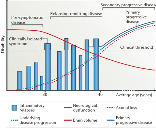
§ Relapsing-remitting MS (RRMS): it is characterized by an initial episode of neurological dysfunction (clinically isolated syndrome). It is also distinguished by attacks (relapses or exacerbations) due to the occurrence of symptoms of neurologic dysfunction for more than 24 hours. Focal CNS inflammation correlate with relapses and demyelination are appreciable as white matter lesions, using MRI. These attacks are followed by periods of partial or complete recovery (remissions) (See Figure 1: black line). During remissions, all symptoms may disappear, or some
symptoms may continue and become permanent. However, during periods of remission, there is no apparent disease progression. It affects 85% of people at the time of diagnosis (1,2,6,9).
§ Secondary progressive MS (SPMS): approximately 50% of those affected by RRMS may refer to SPMS about 15 years after diagnosis. SPMS is characterized by a progressive worsening of neurologic function (accumulation of disability) over time (6,9). Progressive neurological declines is caused by CNS atrophy; that is, increased axonal loss and decreased brain volume (See Figure 1: red lines) (2). § Primary progressive MS (PPMS): characterized by worsening neurologic
function from the onset of symptoms and absence of early relapses or remissions (See Figure 1: blue line). Approximately affects 15% of people at the time of diagnosis (6,9).
3.4. Treatment Overview
Treatments used in MS have different objectives (6,8,10): P Modify the natural course of the disease.
P Treat relapses, exacerbations or attacks. P Alleviate the presence of symptoms.
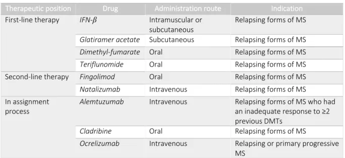
In this review we summarize the most relevant disease-modifying therapies (DMT) approved by the Food and Drug Administration (FDA):
1. The immunomodulators (Interferon-β (IFN-β), Glatiramer acetate, Dimethyl fumarate and Teriflunomide) whose mechanism of action is to downregulate cytokines and chemokines production in the inflammatory cascade, thereby inhibiting T cells activation and migration through the blood-brain barrier (BBB) and their arrival in the CNS (5). This group represents the first-line therapy (4). 2. The general immunosuppressant (Mitoxantrone) that interfere with DNA repair
causing nucleotide crosslinking and DNA strand breaks. It inhibits lymphocytes and monocytes migration, B cells function, and secretion of Tumor Necrosis Factor-α (TNF-α), Interleukin-2 (IL-2) and Interferon-γ (IFN-γ). (11)
3. Immune - selective intervention (11):
a. Blockade (Natalizumab): humanized monoclonal antibody against the α4 subunit of VLA-4 integrin. This antibody blocks the binding between VLA-4 and VCAM-1 and fibronectin, preventing B cells and T cells migration into the CNS.
b. Sequestering (Fingolimod): its target is the sphingosine-1-phosphate receptor. It works sequestering lymphocytes in lymph nodes by inhibiting lymphocyte egress. Furthermore, it inhibits the entry of DCs into secondary lymphoid organs.
Both previous drugs represent the second-line therapy (4).
c. Depleting (Alemtuzumab): humanized monoclonal antibody against a cluster of differentiation (CD)52 on T and B cells. This antibody leads to depletion of T and B cells expressing the CD52 marker, while regulatory T (Treg) cells are maintained due to their low expression of CD52.
d. Decreased signaling (Daclizumab): humanized antibody against CD25, IL-2 receptor. It prevents the signaling of IL-2 and increases CD56+ NK cell activity
(11). The European Medicines Agency (EMA) has withdrawn it from the market after detecting cases of severe inflammatory brain disorders, including encephalitis and meningoencephalitis (12).
e. Decreased presentation (Ocrelizumab): humanized antibody against CD20+
B cells. Thus, it depletes CD20+ B cells and reduces pathogenic B cell antigen
presentation.
f. Apoptosis (Cladribine): its target is adenosine deaminase. It involves immune cells depletion by induction of lymphocyte apoptosis. It also reduces CD4+ T
IFN-β: Interferon-beta; MS: Multiple Sclerosis; DMTs: Disease-Modifying Therapies
Many of these treatments involve general immunosuppression, which entails numerous adverse side effects (such as multifocal progressive leukoencephalopathy), they are partially effective, and especially in the initial stages (9,11). For this reason, there is a need to investigate safer treatments capable of stopping or delaying the progression of disability caused by MS.
In this review, we focus on a specific treatment with human tol-DCs loaded with myelin peptides as a highly selective and disease-specific therapeutic approach.
4. Objectives
The main objective of this project is to carry out a comprehensive bibliographic research on one of the most promising immune-based therapies: tol-DCs loaded with myelin-derived peptides as a therapeutic tool for MS. So far, DCs have been investigated in several clinical trials to promote an immune response against infectious diseases or cancer. However, very few clinical studies have exploited their specific immunosuppressive potential.
For this reason, the main goal of this work is to know the immunosuppressive mechanism by which tol-DCs may represent a novel therapeutic strategy for MS. The secondary objectives, closely linked to the main one, are the following:
P Study the immunopathological mechanisms that lead to MS.
P Investigate which are the different protocols established to generate tol-DCs in vitro applied in clinical trials.
P Analyse the entire procedure from peripheral blood monocyte cells (PBMCs) are obtained, through the isolation and treatment of monocytes, until myelin-peptides loaded tol-DCs are administered to MS patients.
Therapeutic position Drug Administration route Indication
First-line therapy IFN-β Intramuscular or
subcutaneous Relapsing forms of MS Glatiramer acetate Subcutaneous Relapsing forms of MS Dimethyl-fumarate Oral Relapsing forms of MS Teriflunomide Oral Relapsing forms of MS Second-line therapy Fingolimod Oral Relapsing forms of MS Natalizumab Intravenous Relapsing forms of MS In assignment
process Alemtuzumab Intravenous Relapsing forms of MS who had an inadequate response to ≥2 previous DMTs
Cladribine Oral Relapsing forms of MS
Ocrelizumab Intravenous Relapsing or primary progressive MS
P Know if the clinical trials conducted so far have proved safe, well-tolerated and clinical effectiveness by MS patients.
P Make an overview of immunotherapies with a similar strategic approach followed by the tol-DCs that are currently underway.
5. Methods and Material
This project belongs to the bibliographic research category. The compilation of information to conduct this project has involved databases such as PubMed, Scopus or Google Scholar to search for bibliographic articles, clinical trials and reviews in recognized journals like Frontiers in Immunology. The selected material was filtered based on the number of citations in which those articles have been mentioned, publication date and journal impact factor, giving preference to those that have been reported more recently and a high number of citations. The keywords for precise search were: (“Multiple Sclerosis” [AND] “Tolerogenic Dendritic Cells”) and (“Multiple Sclerosis” [AND] “EAE” [OR] “Experimental Animal Encephalomyelitis”).
Besides consulting the previous sources, information has also been obtained from web pages related to the field of inquiry. It is the case of the website “MS online” corresponding to the Grupo de Afectados de Esclerosis Múltiple (GAEM) Foundation, National Multiple Sclerosis Society (NMSS), and Food and Drug Administration (FDA). Finally, Medline Plus has served as a medical encyclopaedia that has resolved doubts about technical medical terms.
6. Results
6.1. The Pathophysiology of MS
The pathology of MS is characterized by the presence of plaques or lesions, demyelinated areas of white and grey matter of the brain and spinal cord, that demonstrate a loss of myelin sheaths and oligodendrocytes. While axons and neurons are preserved in early disease, gradual neuroaxonal loss occurs in advance disease, correlating with the disability degree of individuals with MS. (2,13)
6.1.1. Early Stages
The inflammation process occurs at all stages of MS, but it is less prominent in the remissions phases than in the relapses phases. In the early stages, the brain and spinal cord show little damage in areas outside of focal lesions (also called normal-appearing white matter). However, general brain atrophy has been contemplated. These early lesions are characterized by infiltrated peripheral immune cells such as macrophages, T CD8+ cells, T CD4+ cells, B cells and plasma cells and leakage of the BBB. (2,14)
6.1.2. Advanced Stages
As the disease progresses, microglial and astrocyte activation, myelin loss, axonal injury, and inflammatory T-cell and B-cell are evident. As a result, even more pronounced atrophy of grey and white matter (13). About the inflammatory infiltration, the proportion of B cells and plasma cells increases, but the composition of T cells does not differ (15). Note that, throughout the course of the disease, microglia and macrophages remain in a chronic state of activation (16).
6.1.3. Progressive Stages
CNS inflammation is characterized by a low proportion of invasive peripheral cells in the lesions. Studies have shown that SPMS can present tertiary lymphoid structures, that is, inflammatory aggregates in the meninges which could help tissue injury and cortical demyelination. (17)
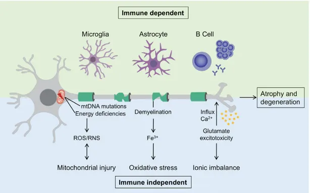
Two mechanisms can be distinguished by which neurodegeneration and axonal atrophy are caused. The first mechanism is immune-dependent and is characterized by the performance of the immune cells resident in the CNS, also involving microglia, astrocytes, and potentially B cells from ectopic germinal centers. The second mechanism is immune-independent and diverse events take place: mitochondrial injury due to the mitochondrial DNA mutation and the reduction of the respiratory chain activity, resulting in neuronal energy deficit, owing to the enhanced production of reactive oxygen species (ROS) and reactive nitrogen species (RNS); oxidative stress enhanced by iron (Fe3+) release from active demyelination areas; ionic imbalance due
to glutamate excitotoxicity causing a massive influx of calcium into neurons. (11)
6.2. The Immunopathogenesis of MS
Immune dysregulation affects both innate and adaptative immunity (5).
6.2.1. The Role of Effector CD4+ T Cells, Microglia and Macrophages
The immunological cascade begins when an antigen binds to peripheral DC receptor. This DC processes the antigen and communicated it to the naïve CD4+ T cell
into the lymph node. The adaptative immune response is activated. (5)
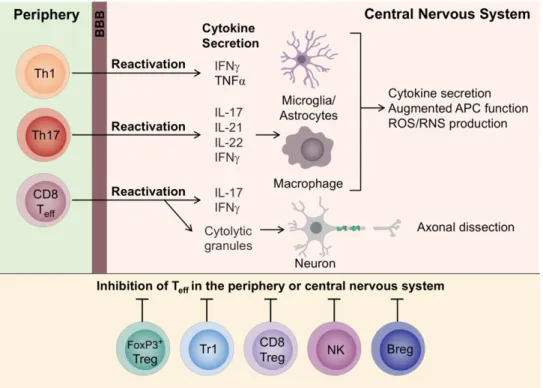
DCs have an activated phenotype through expression of the cell surface marker CD83. Once activated, DCs induce differentiation of T helper 1 (Th1) and T helper 17 (Th17) cells. The differentiation occurs based on the cytokines that Antigen Presentation Cells (APCs) secrete; in the presence of IL-12, naïve CD4+ T cells differentiate into Th1 cells,
secretors of IFN-γ and TNF-α. Whereas in the presence of Interleukin-23 (IL-23), naïve CD4+ T cells differentiate into Th17 cells, secretors of IL-17, IL-21, IL-22 and IFN- γ.
Studies reveal individuals with MS experience increased Th17 cells in peripheral blood during acute relapse. (5,11)
Different mechanisms of pathogenicity of Th17 cells are involved in MS. On the one hand, they produce matrix metalloproteinase (MMP-2 and MMP-9) and ROS that contribute to the increase of BBB permeability (18,19). Furthermore, these cells are involved in the formation of ectopic lymphoid follicles. Finally, Th17 cells produce granulocyte-macrophage colony-stimulating factor (GM-CSF). GM-CSF promotes the continuity of inflammation by mobilizing bone marrow-derived monocytes and CD103+
DC, and by upregulating major histocompatibility complex class II (MHC II) and cytokines in APCs (5,20).
Pro-inflammatory cells migrate, cross the BBB to the CNS, these are reactivated locally in response to CNS autoantigens presented by APCs, leading to an inflammatory response. Pro-inflammatory cytokines activate macrophages (infiltrated cells into CNS in response to injury) and microglia (resident CNS cells derived from myeloid progenitors) which, in turn, produce other pro-inflammatory mediators such as TNF-α, ROS and RNS, ultimately responsible for demyelination and axonal loss. So, microglia and macrophages. play a crucial role in the process of inflammatory attack on myelin antigens. (5,21) It has been observed in animal models that Th1 and Th17 cells produce different diseases in consonance with immunophenotype. A predominant Th1 cells response involves infiltration mainly into the white matter of meningeal and parenchymal mononuclear cells. In contrast, a predominant Th17 cells response facilitate the formation of ectopic lymphoid follicles in the meninges (22). Also, it has been demonstrated that a predominant Th1 cells response localizes the pathogenic cells infiltration mainly in the spinal cord, whereas the predominant Th17 cell response, in the brain (23).
6.2.2. The Role of Effector CD8+ T Cells
CD8+ T cells are also involved in the pathophysiology of MS. It has been found
BBB through α-4 subunit of VLA-4 integrin binding. CD8+ T cells secrete
pro-inflammatory mediators such as IL-17, IFN-γ and can also release cytolytic granules causing axonal damage. Moreover, the characteristic relapses in some stages of MS could be related to the loss of terminally differentiated of autoregulatory CD8+ T cells. (5,11)
6.2.3. The Role of γδ Cells
The γδ cells are a subpopulation of T cells. During the early stages of the disease, these cells can be found in large amount in the CSF. δγ cells have been characterized by being CD161+ CCR6 + and secreting IL-17. Activation of these cells can boost responses
effector T cells under the effect of IL-23 and deprived of the activity of Treg cells. This process further promotes autoimmunity. (5)
6.2.4. The Role of B Cells
B cells are involved in MS immunopathogenesis in different ways. First, these cells are capable of activating effector CD4+ T cells by binding between MHC class II of
B cell and T cell receptor (TCR) of T cells. T cells contribute in the proliferation and differentiation of B cells. Second, regulatory B cells (B reg) can inhibit effector T cells by secreting IL-10, IL-35, transforming growth factor-β (TGF-β), and programmed death-ligand 1 (PD-L1). In MS patient, the regulatory function of B cells is defective and instead, they have a deposit of autoreactive B cells. Third, B cells can differentiate into plasma cells to produce autoantibodies, both in the periphery and in the CNS. These autoantibodies are directed against myelin antigens and other components of CNS. Fourth, B cells can pass through the BBB and collaborate in ectopic germinal centers formation. Ectopic lymph nodes in the CNS are compartmentalized and operate independently to lymph nodes located on the periphery. Last but not least, B cells secrete pro-inflammatory cytokines such as TNF-α, GM-CSF and IL-6 which activate microglia and astrocytes. (11)
Within CSF, memory B cells can differentiate into plasmablasts causing oligoclonal bands (24). The presence of oligoclonal bands is correlated with a progressive clinical course of the disease. As discussed in the introduction, one of the methods for diagnosing MS is by observing oligoclonal bands in CSF analysis. However, CSF analysis alone cannot confirm or exclude a diagnosis of MS because these abnormalities can also be found in other neurological diseases (5,6).
6.2.5. The Role of Astrocytes
Astrocytes are responsible for controlling the infiltration of peripheral pro-inflammatory leukocytes through the BBB and regulating oligodendrocytes, microglia and cells of the adaptive immune system. A study has reported that elevated clusters of astrocytes enzyme B4GALT6 have been found in lesions of brain promoting inflammation and neurodegeneration during chronic CNS inflammation. (5)
6.2.6. The Role of Regulatory and Natural Killer Cells
In the immunopathology of MS also fits involving regulatory cells capable of modulating immune activation and thus, control the development of autoimmunity. Several subsets of regulatory CD4+ T cells have been identified, but among the most
prominent Treg are those that express the transcriptor factor Forkhead box protein 3 (FoxP3) and inhibitory immune checkpoint surface molecules that facilitate the suppression of T cells proliferation. Another main regulatory CD4+ T cells is the Tr1
regulatory CD4+ T cell able to inhibit cell proliferation via the secretion of IL-10. Natural
Killer cells (NK cells), through their ability to kill activated and pathogenic CD4+ T cell,
act as immune suppressors with regulatory properties. (11)
6.3. DCs as a New Target for MS Treatment
Dendritic cells are the most potent antigen-presenting cells that exist and they are characterized by regulating both immune and tolerogenic responses depending on the context in which the interaction between the antigen and DCs occurs. (25)
The rationale for working with DC in antigen-specific cell therapy approach is its functional plasticity, its property of generating highly specific immune responses and the possibility of obtaining DC in vitro under suitable conditions for clinical applications (25). In addition, the results obtained in experimental animals models suggest tol-DCs treatment could represent a therapeutic tool for autoimmune diseases such as MS (26).
6.3.1. DCs Activation
Immature DCs (iDCs) are found in peripheral tissues where they patrol and absorb large amounts of membrane-bound or soluble antigens by micropinocytosis and phagocytosis. Along these lines, iDCs acquire pathogen-associated molecular patterns (PAMPs) and damage-associated molecular patterns (DAMPs) in situ through a set of surface and intracellular receptors, such as cell surface-C-type lectins, surface and intracellular Toll-like receptor (TLRs), and intracellular helicases. (27,28)
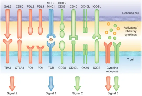
Once they interact with a pathogen, iDCs undergo phenotypic and functional transformations, through a process called maturation (25). Maturation implicates that antigens-loaded iDCs migrate into the draining lymph nodes via afferent lymphatics where they lose the ability to phagocytose antigens and acquire stimulatory properties to present antigens to T cells. Antigen presentation and T cells differentiation requires increased expression of surface MHC class II molecules to be recognized by T-cell receptor (TCR) on T lymphocytes and increased expression of costimulatory molecules associated with the ability to promote or suppress T cells through different signaling axes: CD80/CD86-CD28, CD40-CD40L, OX40L-OX40, ICOSL-ICOS and galectin (GAL)9-TIM3, CD80-CTLA4, PDL1-PD1, PDL2-PD1, respectively (see Figure 4); as well as enhanced production of pro-/anti-inflammatory cytokines and chemokines. This process leads to the development of an immune response T cell subsets (4,27,28).
Figure 4. Different signals involved in the induction of T-cell mediated immunity or tolerance by DCs.
Signal (1) Antigen presentation. Modulation of T cell activity occurs by the presentation on MHC I and MHC II molecules. Signals (2) and (3) Co-stimulatory molecules [belonging to the B7 and TNF protein families] and soluble cytokines can prime T cell response (green arrows and receptors). Contrarily, CTLA4: cytotoxic T lymphocyte antigen 4; PD1: programmed cell death protein 1; PD-L1: programmed cell death-1 ligand and TIM-3: T cell immunoglobulin and mucin-domain containing-3 and soluble factors such as IL-10 can suppress T cell activation (red arrows and receptors). (28)
6.3.2. Human DCs Subpopulations
DCs comprise a heterogeneous population of bone-marrow-derived cells that are founded in spleen, blood, skin and non-lymphoid tissues. Five main DCs subtypes have been distinguished (4,28,29):
§ Myeloid DC (mDC): mDCs are resident in lymphatic tissue and are also present in the blood, peripheral tissues and lymph nodes, where they constantly uptake blood or tissue antigens. Regarding their role in autoimmune diseases, they are implicated in their progression by increased pro-inflammatory cytokines production and T cell activation.
o Type 1 conventional DC (cDC1): correspond to the CD141+/blood DC antigen
(BDCA)-3.
- Main surface markers: CD11clow, HLA-DR+, DEC205+, XCR1+.
- cDC1 are characterized by their efficient antigen cross-presentation and CD8+
and Th1 cells priming. Due to the excellent processing and cross-presentation of exogenous antigens on MHC class I molecules to activate CD8+ T cells
and prime Th1 response, cDC1s gain a unique potential to induce immunity against intracellular pathogens and malignant cells.
o Type 2 conventional DC (cDC2): correspond to the CD1c+ /BDCA-1.
- Main surface markers: CD11c+, C11b+, HLA-DR+, CD172a+.
- cDC2s are defined by several functions such as CD4+ T cells priming, Th17
activation, Th1 and Th2 response induction and Treg cells activation. They present peptides on MHC class II molecules to CD4+ T cells.
§ Plasmacytoid DC (pDC): pDCs are a small subset of DCs which concentrate mainly in lymph tissues and blood and they access the lymph nodes through the blood circulation. About their participation in autoimmune diseases, they are implicated in their progression by increasing IFN-α production and decreasing ability to prime Treg cells.
- Main surface markers: CD11c-, CD123+, CD303+, CD304+, chemokine
receptor (CCR)2+, CXC-Chemokine receptor (CXCR)3+, HLA-DRlow.
- Their main function is antiviral immunity, upon foreign nucleic acid, they start to produce IFN type I. They are also characterized by their poor ability to prime naïve T cells. pDC present peptides only after cell activation. However, at steady state pDCs can impulse T cell anergy and promote Treg cells development, thus they can also induce tolerogenic immune responses. § Langerhans cell (LC): resident in the epidermis. Its role in autoimmune diseases is
not yet well-defined.
- Main surface markers: Langerin, Epcam, BDCA1+, CD1a+, CD11chigh.
- LCs participate in immunological surveillance. LCs are responsible for maintaining the tolerance towards commensals skin agents while retaining the selective ability to respond to certain pathogens.
§ Monocyte-derived DC (MoDC): MoDCs are mainly differentiated from monocyte in peripheral tissues during inflammation.
- Main surface markers: CD11c+, CD11b+, HLA-DR+, CD1c+, CD206+,
CD209+, CD1a+, CD172a+, CCR2+.
- MoDCs induce context-dependent differentiation of CD4+ T cells toward a Th1, Th2 or Th17 phenotype.
To summarize, from all DCs subsets above mentioned, the cells have been described to generate both immunological and suppressive function are cDC2, pDCs, LCs and MoDCs.
6.3.3. Immunogenic DCs
iDCs act as sentinels that detect pathogens by membrane-bound TLR or through nucleotide-binding oligomerization domain-like receptors (NLR) within cytosol, for that reason they populate almost all surfaces of the body. They use proteolytic machinery (endolysosomal and proteasomal) through which they partially degrade the antigens to peptides. After encountering exogenous antigens and appropriate signals, iDCs differentiate into mature DCs, which are characterized by upregulation of MHC class I and II molecules and costimulatory molecules and responsiveness to inflammatory chemokines. Mature antigen-loaded DCs drive differentiation and activation of T cells, by providing immunomodulatory signals through cell-cell contact and pro-inflammatory cytokines, into effector T cells with specific functions and cytokines profiles. Although several cells of the immune system such as B cells or macrophages also have the property of presenting antigens, DCs differ because they have the unique ability to activate naïve CD4+ and CD8+ T cells and induce antigen-specific immune responses. (27,28)
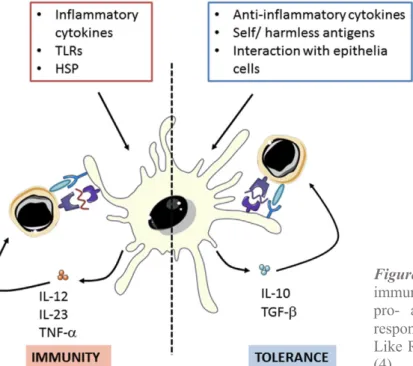
Figure 5. Dendritic cells (DCs) can polarize
immune response through stimulate both pro- and anti-inflammatory activities in response to different signals. TLR: Toll-Like Receptors, HSP: Heat Shock Proteins (4).
The full activation of naïve T cells requires a three-step signaling process (27,30): 1. The binding of the TCR to its cognate antigen presented in MHC molecules by
DCs.
2. The binding of CD28 with co-stimulatory molecules like members of the B7 protein family CD80 and CD86.
3. The interaction of pro-inflammatory cytokines secreted by DCs with the respective cytokine receptors.
6.3.4. Tolerogenic DCs
iDCs can present self-antigens to T cells in the thymus to maintain immunological tolerance through clonal T cell deletion, induction of T cell anergy, inhibition memory and effector T cell responses, and generation or the differentiation of CD4+ CD25+Foxp3+
Treg cells. For this reason, they are essential in maintaining central and peripheral tolerance. (27,28)
Regarding the phenotypic characteristics of tol-DCs, they show an immature or semi-mature phenotype distinguished by the low amount of surface MHC and co-stimulatory molecules such as CD80 and CD86 (31). The activation of anergy-associated genes under the control of nuclear factor of activated T-cell (NFAT) and induction of T cell anergy are the consequence of antigen presentation to T cells in the absence of sufficient CD80/CD86 stimulation of CD28 molecules on T cells. Furthermore, induction of Treg cell differentiation takes place as a result of low or no signal through the CD28 receptor (28).
6.3.4.1. Mechanisms of Tolerance Induction
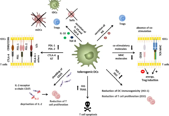
Different immunosuppressive mechanisms have been proposed by which tol-DCs contribute to the induction and maintenance of immune tolerance are described below (4,27):
P The low expression of MHC molecules and co-stimulatory molecules on the surface of DCs makes it impossible to stimulate cells effectively, resulting in T cells anergy, with regulatory capacity.
P Secretion of the anti-inflammatory cytokine IL-10, TGF-β or retinoic acid is involved in Treg cells and Tr1 induction and inhibition of effector T cells function.
P Expression of IL-1 related receptor plays an important role in maintaining low levels of co-stimulatory molecules and regulating the Treg cells expansion.
P Expression of immune- modulatory/-inhibitory molecules like PD-L1/2, CTLA-4 and Ig-like transcripts-3/4 (ILT-3/4) or expression of death receptors like TNF-Related Apoptosis-Inducing Ligand (TRAIL) or FAS represent ways to inhibit efficient responses of T cells.
P Deprivation of nutritional factors through the expression of indoleamine 2,3-dioxygenase (IDO) and heme oxygenase-1 (HO-1), with an inefficient antigen presentation, leads to a reduction of T cells proliferation and Treg cells induction, respectively.
P Following a similar mechanism to the previous one, soluble CD25 shedding involves IL-2 deprivation and the consequent reduction of T cells proliferation.
It has been demonstrated that from peripheral CD4+CD25- Foxp3- T cells exposed to
antigen in the presence of TGF-β and IL-10, without IL-6 or IL1-β, at steady state, peripheral Treg cells increase, which promotes the upregulation of Foxp3. (32)
Recent research has shown that upregulation of C-lectin receptor-2 (CLEC-2) is associated with Treg induction. Furthermore, platelet growth factor impulses the secretion of IL-10 by DCs, which leads to the consequent induction of Treg cells. (33)
6.4. Experimental Autoimmune Encephalomyelitis (EAE): A Model of MS
The use of animals has been very useful to understand the pathophysiology of MS. One of the most commonly used animal models to represent MS in rats and mice is the experimental autoimmune encephalomyelitis (EAE). The EAE model has allowed to discover the most effective treatments and/or beneficial effects they have on them. However, in some studied treatments beneficial effects have been observed in EAE that have not been generalized in MS, in some cases even leading to a worsening of disease indices. Nonetheless, most of the immunological processes first determined in EAE are directly adaptable to MS. (4,34)
The EAE animal model has been able to demonstrate the participation of CD11b+ and
CD11c+ DCs in both Treg and Tr1 cells generation (35). Therefore, one of the key
mechanisms to induce antigenic tolerance is the stimulation of DCs with a regulatory profile (4).
Other researchers have made comparisons regarding the use of oral immunosuppressive drugs (e.g., Vitamin D3) during days before and days after induction of EAE in animals. The results obtained were similar in both: an increase in CD4+Foxp3+ Treg cells, an
increase in IL-10 levels in lymph nodes of treated animals and a significant improvement in clinical severity. These suggest that the main target of the tolerogenic effect of vitamin D3 are DCs. Other projects indicate that in DCs absence during the priming process of autoreactive T cells leads to a deficiency of cell generation that, which ultimately results in a violent attack against the CNS. (26,35,36)
6.5. Antigen-specific therapy in MS
The exploration of immune-tolerogenic therapies to treat autoimmune disease such as MS is justified by the need to find more selective therapies designed to restructure tolerance itself without causing general immunosuppression. Currently, common treatment options are life-long systematic immune suppression, which can cause serious side effects such as opportunistic infections, secondary autoimmunity and increased risk of malignant tumors. (4,25,27)
Antigen-specific immunotherapy has several advantages. These include the ability to restructure, enhance, or induce peripheral immune tolerance for pathogenic adaptive response of T and B cells without reducing overall immune surveillance against microbes or tumors (37). Furthermore, it is a highly selective approach, which should lead to high safety and efficacy in immunotherapies (38).
Focusing on the MS, the specific target antigen is undiscovered yet, however, it is known that the proteins in the myelin sheath, such as myelin peptides of myelin basic protein (MBP), myelin oligodendrocyte glycoprotein (MOG) and proteolipid protein (PLP) are important targets in the autoreactive immune response. Several myelin immunodominant peptides from MS patients have been identified ex vivo, to develop antigen-specific tolerance for MS. These peptides have been tested in clinical trials using autologous leukocytes chemically coupled with these peptides. (38,39)
6.6. Generation of Tol-DC in vitro
In recent years, many protocols to generate stable human monocyte-derived tol-DCs in vitro have been established. Many of those are based on monocytes (CD14+) as
precursor cells of DCs due to the high number of cells obtained in peripheral blood. (25) DCs can be produced in vitro from monocyte cultured with IL-4 and GM-CSF, which promote differentiation and growth. DCs can become tol-DCs when their regulatory phenotype is induced with anti-inflammatory cytokines like IL-10 or TGF-β, immunosuppressive drugs like vitamin D3, estriol, corticosteroids (dexamethasone) or rapamycin (see Figure 7 (40)). Then, to boost peptide-specific properties to tol-DCs,
they are cultured with a defined maturation cytokine cocktail like prostaglandin E2, IL-1β, TNF-α, IL-6 or lipopolysaccharide (LPS) activation (4,25,27,38). Lastly, to achieve efficient clinical responsiveness following tol-DCs administration, tol-DCs require pulsing with relevant antigens e.g., myelin peptides in MS (28). The tol-DCs generation process must comply with good manufacturing practice (GMP) conditions (38).
According to some studies, tol-DCs generated with dexamethasone and other stimuli have a stable phenotype, intermediate expression of co-stimulatory molecules CD80 and CD86, low expression of CD40, secrete high amounts of IL-10 and TGF-β (anti-inflammatory cytokines) and low amounts of IL-12, IL-23 and TNF-α (pro-inflammatory cytokines). (4)
In a clinical trial conducted in patients with Crohn’s disease, it was observed that intraperitoneal administration of dexamethasone/vitamin D3-treated tol-DCs had low expression of CD1a and CD14, and high expression of CD209, which has been associated with an increase in Treg cells, so a clinical improvement in some patients (38,41). Regarding to tol-DCs migration in vivo, researches conducted so far have not been able to determinate tol-DCs can reach the inflamed CNS. However, recent data suggest that tol-DCs shuttle across the BBB could be facilitated by the introduction of
de novo CCR5 expression using mRNA electroporation into tol-DCs (28,38).
It is important to highlight that a crucial part in the generation of tol-DCs is that they must have particular characteristics after culture (42):
P Low expression of co-stimulatory molecules.
P High stability after maturation stimuli and its production of IL-10. P Low activation of T cells.
Nonetheless, the generation of tol-DCs in vitro has different disadvantages, such as patient-specific, high cost and laborious. To overcome these limitations, other approaches have been conducted to establish tol-DCs in vivo. One possible approach, among several others, is exploit coadministration of free autoantigens or autoantigens encapsulated with nanoparticles, microparticles or liposomes, carrying tolerogenic factors that are released specifically to DCs. (28)
Xie et al. (26). has provided the appropriate information to make below sections (6.6.1., 6.6.2. and 6.6.3.).
6.6.1. How Does Estrogens Induce the Regulatory Phenotype of Tol-DCs?
Estrogens (e.g., estriol) play a beneficial role in the pathogenesis of MS since the incidence of relapses is lower in pregnant patients with MS. Estrogen-treated mature DCs had inhibited pro-inflammatory cytokine secretion in vitro and presented a low ability to present antigens. Reduction in both Th1 and Th2 cytokine production and inhibition of T cell responses occur when estrogen-activated DCs induce IDO expression. In MS patients, estrogen upregulated IDO expression on DCs and inhibited Th1 and Th2 cytokine secretion. However, estrogen is not commonly used clinically due to its side effects, although there is evidence estrogen can relieve the MS symptoms and reduce relapses incidence.
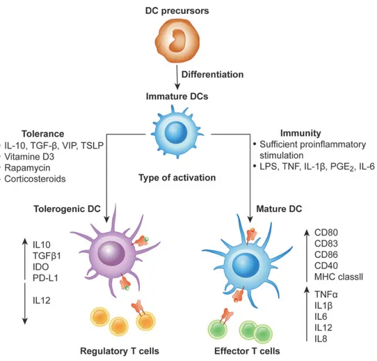
Figure 8. Differentiation of monocyte-derived immunologic and tolerogenic DCs. DCs differentiate
from monocytes into iDCs in the presence of IL-4 and GM-CSF. In the presence of maturation signals (pro-inflammatory cytokines), DCs are activated and acquire a stimulatory phenotype, which they induce effector/cytotoxic T responses. Differently, in the absence of maturation factors, the incubation of iDCs with anti-inflammatory mediators or genetic modification of DCs can lead to the generation of tol-DCs, which promote anergy, apoptosis or activation of Treg cells. VIP: Vasoactive Intestinal Peptide; TSLP: Thymic Stromal Lymphoietin. (28)
6.6.2. How Does Vitamin D3 Induce the Regulatory Phenotype of Tol-DCs?
As discussed in the introduction, sun exposure deficiency increases the risk of MS, that is, vitamin D deficiency is a risk factor for MS (5). Vitamin D levels in MS patients are lower than in healthy individuals. Similarly, vitamin D levels in MS patients who are in relapse are lower than those during remission states.
Vitamin D plays an immunomodulatory role when it interacts with vitamin D receptor (VDR), which is expressed in lymphocytes. 1,25-dihydroxyvitamin D3 (1,25(OH)2D3) is
the active form of vitamin D in vivo. In vitro, 1,25(OH)2D3 inhibits the proliferation of
MBP-specific T cells and increases Treg cells from MS patients. In addition, in vitro, 1,25(OH)2D3 can partially block monocyte differentiation, directed by GM-CSF and
IL-4, in DCs. When PBMCs, in the presence of GM-CSF and IL-4 in vitro, differentiated into DCs and were treated with 1,25(OH)2D3, their ability to mature APCs was inhibited.
Likewise, when monocyte-derived DCs from RRMS patients were treated with 1,25(OH)2D3, DCs were characterized by an immature phenotype, inhibited IL-12p40
secretion, and increased chemokine C-C ligand-2 (CCL2) secretion.
Tol-DCs induced by1,25(OH)2D3 and stimulated with myelin peptides led to stable
antigen-specific hyporesponsiveness in myelin-reactive T cells from patients with RRMS. Furthermore, a significant increase in IDO expression was shown with vitamin D-induced tol-DCs. All of these studies revealed that vitamin D-induced tol-DCs have potential immunotherapy value.
6.6.3. What Role Do Each of the Cytokines Play in the Generation of Tol-DCs in vitro?
Protocol generation of tol-DCs in vitro requires the representation of a set of cytokines. Each of them with a specific function.
First, monocytes isolated from PBMCs pool are culture with cytokines to induce differentiation to DCs with immature phenotype:
§ GM-CSF: GM-CSF is a cytokine required for Th17 to become pathogenic T cells (differentiation) and promote DCs maturation. A study has demonstrated that autoreactive helper T cells with normal IL-17 or IFN-γ expression, but specifically lacking GM-CSF in mice, failed to induce EAE. Whereas those autoreactive helper T cells without IL-17 or IFN-γ expression, but that secreted GM-CSF was sufficient to induce EAE.
§ IL-4: IL-4 is a cytokine with potent anti-inflammatory properties. IL-4 therapy has been achieved protection from EAE and MS, and a more severe form of clinical disease has developed in IL-4 deficient mice. (43)
Second, iDCs pass into tol-DCs by adding cytokines, in culture, that impose the regulatory phenotype (anti-inflammatory cytokines):
§ IL-10: IL-10 can inhibit immune responses and induce immune tolerance as it can inhibit Th1 cells and induce Tr1 cells. These cells are essential in peripheral immune tolerance. IL-10 suppresses the autoimmune response by inhibiting self-antigen-specific T cell and induces Treg cells, thus playing a beneficial role in MS. In vitro, monocyte-derived DCs obtained from MS individuals and treated with IL-10 induced the production of IL-4 and IL-10 by autologous lymphocytes, whereas cDCs derived from patients with MS and exposed to IL-10 became resistant to LPS maturation. Besides, IL-10-treated DCs expressed a low level of IL-12 and were unable to stimulate T cell proliferation both in vitro and in vitro. § TGF-β: TGF-β is an anti-inflammatory cytokine. DCs treated with TGF-β were
characterized by immature or tolerogenic phenotype, with less ability to stimulate T cells. Therefore, TGF-β-treated DCs have been shown to be potentially therapeutic in EAE rats.
Third, tol-DCs are cultivated in the presence of a define cytokine cocktail in order to boost their tolerogenic properties (4,26):
§ TNF-α: two types of TNF-α are distinguished: membrane-bound TNF-α (mTNF-α) and soluble form of TNF-α. TNF receptor-1 (TNFR1) is widely expressed and binds to both types of TNF, while TNF receptor-2 (TNFR2) is expressed on lymphocytes and binds to soluble mTNF-α. TNF-α is a pro-inflammatory cytokine, so mice treated with antibodies against TNF-α were resistant to EAE. Those mice deficient in TNFR1 or TNFR1/TNFR2 are also resistant to EAE, while severe EAE symptoms were exhibited in TNFR2 deficient mice. These findings suggest that TNFR2-linked mTNF-α signal has a protective role. This protection role could justify the reason that patients treated with anti-TNF-α agent aggravated the disease in a clinical trial.
§ IL-1β: studies reveal that a significant reduction in disease severity during EAE was observed in those IL-1β receptor knock-out mice. IL-1β receptor knock-out mice leads to a significant decrease in VCAM-1 expression and, consequently, a decrease in leukocytes infiltration in the spinal cord in rodents exposed to EAE. Elevated levels of IL-1β were found in brain lesions of MS subjects.
§ IL-6: IL-6 is a pro-inflammatory cytokine and, along with TNF-β, promotes Th17 differentiation. IL-6-knock-out mice are completely resistant to EAE induction. Furthermore, mice that have the IL-6 receptor blocked, through inhibition of Th17 differentiation, prevent the EAE induction. Researchers have found DCs are the key cells required to produce IL-6 and boost EAE. Finally, IL-6 was found in the brain of MS individuals.
6.7. Clinical trial with Tol-DCs
Recently, Zubizarreta and her colleagues developed a phase Ib clinical trial in which they included eight patients with MS to test increasing concentrations of autologous tol-DCs loaded with various peptides of myelin proteins. The main objective of the trial was to assess safety and tolerability, while the secondary objective was to observe possible clinical deviations (relapses and disability), MRI and immune responses. (9,38)
6.7.1. How Was the Clinical Trial Conducted?
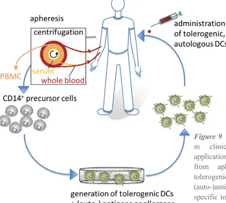
Autologous monocyte-derived DCs were obtained from the patients by leukapheresis (procedure to separate and collect monocytes from the blood (44)). Monocytes were cultured in the presence of IL-4 and GM-CSF to accomplish the differentiation process, anti-inflammatory cytokines and dexamethasone to induce tolerogenic phenotype. Then, a mixture of cytokines was added including IL-1β, IL-6, TNF-α and prostaglandin E2 as well as immunogenic peptides in order to increase the tolerogenic properties of peptide-specific DCs. DCs were stimulated with myelin peptides from MBP, PLP and MOG. Finally, tol-DCs were progressively administered by IV injection in increasing doses (38). Since the cells are from the patients himself (autologous), they do not suffer rejection and can perform their function after being injected (9).
Figure 9. Tolerogenic dendritic cells (DCs)
in clinical application. For clinical
applications, CD14+ monocytes are isolated
from apheresis products to generate tolerogenic DCs, which are loaded with (auto-)antigens. Subsequently, antigen-specific tolerogenic DCs are reinjected into the patients to affect the inflammatory immune response of autoimmune disease. (27)
6.7.2. Results and Conclusion of the Clinical Trial
They obtained the following results (38):
- Safety: tol-DCs therapy was well tolerated, with no serious side effects or therapy-related adverse reactions.
- Efficacy and clinical monitoring: patients remained clinically stable in terms of relapse, disability and various measurements with MRI.
-
Immunological responses: a significant increase in the production of IL-10levels was observed in peptide-stimulated PBMC, as well as an upward trend in the frequency of a subtype of Treg cells, Tr1 (CD4+ IL-10+) and a significant
decrease in memory CD8+ and NK cells.
Evidence was obtained that tol-DCs therapy induces peptide-specific tolerance by promoting T cells adaptive regulatory function. Therefore, as individuals with MS are considered to have decrease Tr1 cells function, increasing Tr1 cells function by tol-DCs could contribute to restructuring peripheral immune tolerance in patients with autoimmune disease. As the trial was not developed to evaluate efficacy at the clinical level, no conclusions should be drawn in terms of efficacy, but future trials with the power to show clinical efficacy (phase II and/or phase III clinical trials, including control (placebo) group) will be able to demonstrate whether the increase in Tr1 cells response can actually modify MS course. With these results, they conclude that IV administration of myelin peptide-loaded DCs is safe and feasible without remarkable side effects and showing a promising immunological and clinical results. (4,38)
6.8. Overview of Tolerance-Inducing Therapeutic Approaches for MS
After the discovery of the myelin proteins (MBP, MOG and PLP) as a critical target of the autoreactive immune response in parallel ways to the generation of tol-DCs approach as a therapeutic tool, several investigations were launched with the same purpose: inducing antigen-specific tolerance in MS. (4,45)
Three main branches are identified within the tolerance-inducing therapeutic approaches that entered the clinical phase in MS:
6.8.1. Peptide-Based Tolerance-Inducing Vaccination
1. Transdermal administration of MOG, MBP, PLP peptides: induction of immunological tolerance was demonstrated by activation of LCs and subsequent induction of IL-10 secreted by T cell. Immunological effects resulted in a significant reduction in annualized relapse rate and lesion activity on MRI (measurements of disease). (46,47)
2. Subcutaneous administration of altered peptide ligand (APL): clinical trial consisted of incorporating amino-acids substitutions at TCR contact positions. Exacerbations of the disease were demonstrated following treatment, which is why the trial was halted. (48)
3. Intradermal administration of apitopes (AXT-MS-1467): a technique that consists of designing synthetic soluble peptides that mimic the naturally processed epitopes, also called apitopes. These apitopes induce antigen-specific expansion of Treg cells, capable of “switching off” pathogenic T cells responsible for myelin damage in the CNS. Two clinical trials have been carried out with the ATX-MS-1467 apitope (a mix of four short peptides from MBP) in which no serious treatment-related adverse effects. A reduction in brain lesions observed by MRI was also revealed. (49,50)
4. Subcutaneous administration of mannosylated liposomes containing MBP peptides: another alternative is to reach MBP peptides to APCs in vivo for natural processing, presentation in a tolerogenic environment, and involving antigen-specific T cells. This process is allowed by determining specific markers expressed on the surface of APCs, such as CD-206 (mannose receptor). In this context, encapsulation of selected immunodominant MBP peptides into mannosylated liposomes improved the uptake of the peptide, through the CD-206 receptor, by DCs. This resulted in immune tolerance to myelin-derived antigen. In clinical trials, researchers observed a significant reduction in serum levels of CCL2, CCL4, IL-7 and IL-2. (51,52)
5. Both intradermal and intramuscular administration of TCR peptide vaccination: this approach consists of administering short amino-acid sequences from the TCR-derived of pathogenic T-cell clones, to induce immunoregulation mediated by T cell directed to T-cells expressing those TCRs. Intradermal injection of synthetic TCR peptides resulted in clinical improvement, along with beneficial immunological effects, such as increased TCR peptide-specific T cells and reduction of MBP-specific T cells. Intramuscular injection of TCR peptide also produced immunoregulatory effects. (53)
6.8.2. DNA Vaccination
1. Intramuscular administration of MBP-encoding DNA: BHT-3009 is a genetically engineered DNA vaccine that encodes the full-length human MBP. The purpose is to restore tolerance to MBP. In the clinical trials done so far, a reduction in the brain lesions observed on MRI and a reduction in the proliferation of IFN-γ-producing T cells accompanied by a reduction myelin-specific autoantibody titers in CSF have been observed. (54)
6.8.3. Cell-Based Tolerance-Inducing Vaccination
1. Subcutaneous administration of T cells:
a. Irradiated autologous T cells: vaccination consists of autologous MBP-specific T cells isolated from peripheral blood that are inactivated by irradiation. Upon administration of irradiated, autologous MBP- specific T-cell clones, an immune response is generated to eliminate other pathogenic autoreactive T-cells in the patient circulation without affecting the rest of the immune system. Reduced relapse rate, stabilization in disease progression, and lesion activity on MRI are observed in the trials conducted. (55)
b. Mixture of attenuated myelin-reactive T cells: vaccination consists of attenuated myelin reactive T cells selected with multiple peptides derived from MBP, PLP and MOG. Promising results were seen in a small clinical trial involving RRMS and SPMS patients. (56)
2. Intravenous administration of autologous PBMC chemically coupled with a mixture of myelin-derived peptide: after selecting a set of seven immunodominant myelin-derived peptides, a trial was conducted in which MS patients received a single infusion of autologous PBMC pulsed with these seven peptides and chemically fixed. The authors concluded that the antigen-coupled cells had well-tolerance and a favorable safety profile. (39)
3. Intradermal/Intranodal/Intravenous administration of tol-DC pulsed with myelin-derived peptides: to date, IV administration of tol-DCs cells is known to be safe and feasible. As commented in the previous section of the work,
Zubizarreta et al. observed an increase in IL-10 production and frequency in
Vaccination strategy
Developmental progress
Administration
route Clinical outcomes Mode of action References
Peptide-based tolerance-inducing vaccination
MOG, MBP, PLP
peptides Phase I/II Transdermal Reduction in ARR; reduction in MRI measurement of disease Activation of LCs; generation of IL-10 secreting cells (46,47) Altered peptide
ligand Halted Subcutaneous 62% increase in number of active lesions; two pts demonstrated disease exacerbations associated with vaccination strategy (48) Apitopes (AXT-MS-1467)
Phase IIa Intradermal Safe; 79% decrease in lesions at MRI Expansion of Treg (49,50) Mannosylated liposomes containing MBP peptides
Phase I Subcutaneous Safe Decrease of CCL2, CCL4, IL-7 and IL-2 at study completion (51,52) TCR peptide vaccination Phase I Both intradermal and intramuscular
Safe Generation of IL-1 secreting TCR peptide-specific T cells; reduction of MBP-specific T cells (53) DNA vaccination MBP-encoding
DNA vaccine Phase II Intramuscular Safe and well-tolerated; reduction in number of active lesions; decrease in clinical relapse rate
Reduction of IFN-γ-producing myelin-reactive T cells; decrease of myelin-specific autoantibody titers in the CSF. (54)
Cell-based tolerance-inducing vaccination
Irradiated autologous T cells
Phase I Subcutaneous Safe and feasible;40% reduction in relapse rate; stabilization of disease progression and lesion activity on MRI
Generation of cytotoxic T-cell response against myelin-reactive cells; depletion of myelin-reactive T cells (55) Mixture of attenuated myelin-reactive T cells
Phase IIb Subcutaneous Clinical endpoints not met (details not published) (56) Autologous PBMC chemically coupled with a mixture of myelin-derived peptides
Phase I Intravenous Safe and feasible; stabilization of clinical and MRI parameters of disease activity at study completion Decrease in myelin-specific T-cell reactivity in pts receiving the highest dose of cells (>1x10⁹) (39) Tol-DC pulsed with myelin-derived peptides Phase I Intradermal, Intranodal and Intravenous
Safe and feasible. IV administration increase in IL-10 production and frequency of Tr1 cells.
(38)
7. Discussion
Due to the high prevalence of MS, this disease has been the central focus of several investigations in different institutions. One of the objectives that has involved the most effort has been finding the ideal strategy to silence precisely those immune responses that are impaired in MS.
Currently, only more than ten commercialized drugs are available, and they are partially effective and especially in the initial inflammatory phases. The concern is even greater in progressive forms of MS, as to date, there is only one approved drug for PPMS (Ocrelizumab). Furthermore, most of the treatments used in MS involve general immunosuppression, which leads to numerous side effects (infections and even malignant tumors). For these reasons, the search for therapies based on antigen-specific immune tolerance aims to control inflammation and delay progression or improve the degree of disability, as well as being well-tolerated without altering all immune surveillance. Among the diversity of immunological therapeutic tools, DCs have demonstrated their great potential due to their functional plasticity and the possibility of their generation in
vitro. So far, DCs have been investigated in various clinical trials to enhance the immune
response for cancer or infectious diseases. However, the immunosuppressive potential of DCs has been little exploited to treat human autoimmune diseases, organ transplant rejection or even allergies.
Multiple protocols of tol-DCs generation in vitro have been established based on PBMCs. In general, these protocols indicate monocytes must be cultured with differentiation and growth cytokines to obtain iDCs, subsequently with regulatory cytokines and immunosuppressive drugs such as dexamethasone or vitamin D3 to obtain tol-DCs, and for boosting their tolerogenic properties, pro-inflammatory cytokines must be added to the culture. Finally, these tol-DCs can be loaded with myelin peptides so that specific tolerance to these peptides is created and, therefore, does not lead to an autoimmune reaction. Although the generation of tol-DCs for clinical application has many positive aspects, it should be mentioned that has certain disadvantages, such as that it is very laborious, patient-specific or high cost.
Focusing on the phase Ib clinical trial coordinated by Dr. Benítez and Dr. Villoslada at Hospital Clínic, it was decided to generate tol-DCs in vitro using dexamethasone to induce the regulatory phenotype, since in a previous study which followed the same therapeutic strategy involved patients with Crohn’s disease, was the immunosuppressor agent that provided the best results regarding the increase of Treg cells. Although the target autoantigens of the myelin sheath are undetected, the target proteins of the myelin with which tol-DCs can be loaded and generate a local immunosuppression situation are known. The Hospital Clínic research group selected peptides identified in previous studies that were recognized by T lymphocytes from patients. However, Dr. Benítez points out that it is important to identify even more specific targets on which the T lymphocytes focus (See Annex).