DEPARTMENT OF BIOENGINNERING
DOCTORAL PROGRAM IN BIOENGINEERING
A new laser self-mixing interferometer system for
the assessment of the chest-wall mechanics
Doctoral Dissertation of:
Ilaria Milesi
Tutor: Prof. Antonio Pedotti
Advisor: Prof. Raffaele Dellacà
Supervisor: Prof. Maria Gabriella Signorini, Ph. D.
Grazie a Raffaele per i suoi preziosi insegnamenti, la sua onestà e la capacità di vedere molto più lontano di me!
Grazie al prof. Pedotti per avermi consentito di portate avanti questo progetto di tesi. Grazie a tutti i miei colleghi/amici con i quali ho condiviso tre anni intensi di fatica e soddisfazione, in particolare grazie a Ema, Leo, Pasquo, Chiara, Miriam, Ale!
Grazie a Peter Frykholm e al suo gruppo per la generosità ed entusiasmo dimostrati! Grazie a tutti gli amici che mi sono stati vicini, anche nei momenti di crisi. Come avrei fatto senza i caffè con Stefano, Francesca e Bob!
E infine, ma non in ordine di importanza, grazie alla mia famiglia che mi ha sostenuto nelle mie scelte e ha creduto in me.
SUMMARY
EXTENDED ABSTRACT ... i
INTRODUCTION ... xi
1. ASSESSMENT OF CHESTWALL KINEMATICS AND MECHANICS ... 1
1.1. RESPIRATORY SYSTEM ... 1
1.1.1. ANATOMY ... 1
1.1.2. VENTILATION MECHANICS ... 3
1.1.3. MECHANICAL MODELING OF THE RESPIRATORY SYSTEM ... 5
A. ONE COMPARTMENT MODEL ... 5
B. ADVANCED MODELING ... 6
C. MECHANICAL ELEMENTS ... 7
1.2. ASSESSMENT OF RESPIRATORY MECHANICS IN CLINICS ... 12
1.2.1. LUNG VOLUME DEFINITIONS ... 13
1.2.2. LUNG VOLUME MEASUREMENTS ... 13
A. SPIROMETRY ... 14
B. PLETHYSMOGRAPHY ... 14
A. FLOW AND PRESSURE PLETHYSMOGRAPHY ... 15
B. RESPIRATORY INDUCTIVE PLETHYSMOGRAPHY ... 15
1.2.3. PRESSURE DEFINITIONS ... 17
1.3. PATHOLOGICAL CONDITIONS ... 18
1.3.1. ASSESSMENT OF PATHOLOGICAL CONDITIONS ... 18
A. FEV1, FVC FEV1/FVC ... 18
B. FLOW- VOLUME LOOP AND PEF ... 19
C. VOLUME –PRESSURE CURVE... 20
1.3.2. ARDS ... 21
A. CAUSES ... 22
B. DIAGNOSIS ... 23
1.4. ALTERNATIVE TECHNIQUE TO MEASURE MECHANICAL PROPERTIES... 32
1.4.1. OSCILLATORY MECHANICS AND FOT ... 32
D. COMPLEX IMPEDANCE... 32
E. REMARKS ABOUT STIMULUS ... 33
F. INPUT AND TRANSFER IMPEDANCE... 34
1.4.2. OPTOELCTRONIC PLETHYSMOGRAPHY ... 46
1.5. MEASUREMENT OF LOCAL PROPERTIES ... 48
1.5.1. REGIONAL IMPEDANCE ... 49
G. MEASUREMENTS OF RIBCAGE AND ABDOMEN COMPARTMENTS ... 49
H. IMPEDANCE MAP BY OEP AND FOT ... 51
1.6. REMARKS... 55
1.7. REFERENCES ... 57
2. CONTACTLESS TECHNOLOGIES FOR DISPLACEMENT MEASUREMENTS ... 64
2.1. MICHELSON CONFIGURATION ... 64
2.1.1. TWO BEAMS INTERFEROMETER ... 66
2.1.2. HETERODYNE LASER SYSTEM... 67
2.2. PERFORMANCE AND MEASUREMENTS LIMITS ... 67
2.2.1. DETECTION LIMITS ... 67
2.2.2. SPECKLES ... 68
2.3. SELF MIXING INTERFEROMETRY ... 69
2.3.1. WORKING PRINCIPLES ... 69
A. THREE-MIRRORS CAVITY MODEL ... 69
B. THEORY FOR SELF-MIXING IN LASER DIODE ... 70
2.3.2. REMARKS ON SELF-MIXING INTERFEROMETRY ... 73
2.3.3. OPERATIONS ON DIFFUSIVE TARGETS ... 73
2.4. LASER SELF MIXING INTERFEROMETER FOR MEASUREMENTS ON HUMANS 77
2.4.1. ALGORITHM FOR LOW INJECTION REGIME ... 78
A. ESTIMATION OF THE REQUIRED SAMPLE FREQUENCY ... 78
B. ALGORITHM CONCEPTS ... 79
2.4.2. SYSTEM VALIDATION IN VITRO ... 84
A. SET UP ... 84
B. RESULTS AND DISCUSSION ... 85
2.4.3. SYSTEM VALIDATION IN VIVO ... 87
A. SET UP ... 87
B. DATA PROCESSING ... 88
C. IN VIVO RESULTS AND DISCUSSION ... 89
2.4.4. CONCLUSION ... 93
2.5. PROXIMITY SENSORS ... 94
2.5.1. LASER SENSORS ... 95
2.5.2. INFRARED PROXIMITY SENSORS ... 97
A. REFLECTED IR STRENGTH ... 97
B. TRIANGULATION SENSORS ... 98
2.5.3. PROXIMITY SENSORS FOR BIOMEDICAL MEASUREMENTS ... 99
A. ALGORITHM TO DETECT ANGLES AND TO LOCALIZE MEASURED POINTS ON THE SKIN ... 99
B. REMARKS ABOUT SPATIAL RESOLUTION REQUIRED ... 101
C. SELECTION OF THE APPROPRIATE DISTANZIOMETER AND REMARKS ... 103
D. FRONT END BOARD ... 103
E. CALIBRATION PROCEDURE ... 106
F. VALIDATION OF THE ALGORITHM FOR ANGLE ESTIMATION ... 108
2.6. REFERENCES ... 111 3. VALIDATION OF A NEW SCANNING SYSTEM BASED ON LASER INTERFEROMETRY
3.1.1. FOT UNIT ... 118
3.1.2. OPTICAL SCANNING UNIT ... 119
3.1.3. ELECTRONIC UNIT ... 119
A. POLYGRAPH BOARD HARDWARE ... 122
B. POLYGRAPH BOARD FIRMWARE ... 125
3.1.4. SOFTWARE ... 128
3.2. MEASUREMENTS ON ANESTHETIZED PATIENTS ... 130
3.2.1. ATELECTASIS IN ANAESTESIA ... 130
A. ATELECTASIS DEFINITION ... 130
B. ETHIOLOGY ... 131
C. ATELECTASIS PREVENTION ... 133
3.2.2. PILOT STUDY: AN ANIMAL ARDS MODEL ... 135
A. BACKGROUND... 136
B. METHODS ... 136
C. DATA ANALYSIS ... 138
D. RESULTS AND DISCUSSIONS ... 139
3.2.3. MEASUREMNENT ON HUMANS ... 142
A. PROTOCOL ... 142
B. DATA ANALYSIS ... 145
C. RESULTS AND DISCUSSIONS ... 145
D. DISTAZIOMETER VALIDATION FOR IN VIVO MEASUREMENTS ... 155
3.3. REFERENCE ... 163
EXTENDED ABSTRACT
The respiratory system is a complex system which accomplishes gas exchange by means of the synergy among functions and structures which have specifically developed at this aim. Processes involved in gas exchange are: 1) ventilation, that is air transport inside and outside the lungs; 2) oxygen and carbon dioxide diffusion among alveoli and blood; 3) oxygen and carbon dioxide delivering for the blood to the cell and finally, 4) ventilation regulation. The respiratory system functionalities are not only reliant on its components, that are a complex branching system of tubing (airways), a huge exchange region (the alveoli) where gases can diffuse from and into the blood, but its behaviour is deeply influenced also by the respiratory muscles and structures that form a boundary of it, such as the chest-wall and the diaphragm. Impairment at only one of these structure or functions can seriously affect not just the respiratory system but it can yield to a general worsening in the life condition of the subjects due to laboured breathing or asthma attacks, fatigue and difficulty with mobility, heightened sensitivity to ordinary substances and chemicals, and compromised immunity to infection.
In particular ventilation capability, which represents the first step in breathing activity, relies on the mechanical properties of the whole respiratory system: the resistance of the airways, the compliance and elastic recoil of the lung parenchyma, stiffness in chest wall and abdomen, all these components contribute in different and complex ways in the relationship between the pressure generated by active components inside the respiratory chain and the volume of gas introduced.
Diseases affecting the respiratory system, such as ARDS (acute respiratory distress syndrome), restrictive diseases and lung oedema, produce alterations and inhomogeneities in the lung structure and in the chest wall which cause changings in the mechanical properties.
In more details, ARDS is defined as a syndrome of inflammation and increasing permeability that is associated with a constellation of clinical, radiological and physiological abnormalities that cannot be explained by, but may coexist with, left atrial or pulmonary capillary hypertension. The physiological consequences are severe hypoxaemia due to right-to-left shunting of blood and decreased pulmonary compliance due to filling and closure of alveoli. As the airspaces fill with fluid, the gas exchange and mechanical properties of the lung deteriorate.
Restrictive lung diseases are characterized by reduced lung volume, either because of an alteration in lung parenchyma or because of a disease of the pleura, chest wall, or
ii
neuromuscular apparatus. The many disorders that cause reduction or restriction of lung volumes may be divided into two groups based on anatomical structures. The first is intrinsic lung diseases or diseases of the lung parenchyma. The second is extrinsic disorders or extra-parenchymal diseases, which involve the chest wall, pleura, and respiratory muscles, the components of the respiratory pump. Diseases of these structures result in lung restriction, impaired ventilatory function, and respiratory failure (eg, non-muscular diseases of the chest wall, neuromuscular disorders).
Also pulmonary oedema can cause alterations in lung mechanics that directly contribute to clinical morbidity and mortality rates. Both the location of the oedema fluid (interstitital versus alveolar pulmonary oedema) and the aetiology of the pulmonary oedema contribute to the severity and type of abnormalities of lung mechanics observed. The alterations in lung mechanics associated with the adult respiratory distress syndrome may involve the direct effects of released mediators, alterations in pulmonary surfactant, and altered airway reactivity, as well as the direct effects of the oedema fluid.
Up to the present day, the diagnosis and the management of these kinds of pathologies have been carried out mainly by chest radiography, which reveals diffuse opacity according to the stage of the pathology and CT. More specifically ARDS can be detected also by means of measurement of lung endothelial and alveolar epithelial barrier function and BAL (Bronchoalveolar lavage), while restrictive pathology can be detected also by means of some laboratory tests aimed to measure the concentration of well-known markers such as elevated creatine kinase level. Anyway all of them present some intrinsic limitations, indeed they are invasive or required long time to be carried out and thus they can’t be used to monitor in a continuative way the pathology evolution.
On the contrary several published data suggest that global but also local chest wall mechanical properties are parameters that might be useful for the evaluation of structural elasticity and inertia on the overall behaviour of the respiratory system and for estimating the possible impact of both restrictive and obstructive diseases on respiration, thus it could be useful in the assessment of the pathology providing a handy tool for clinicians during the diagnosis but also the therapy of the disease. Although the rich informative content of the chest wall mechanical properties, up to now this parameter has been almost disregarded because of the intrinsic difficulties in its measurement. Therefore a new approach for the detection and the monitoring of these diseases can be based on the measurements of the local and global chest-wall mechanics and the extraction of parameters which are sensitive to pathophysiological alteration. The more promising and inspiring approach has been reported by Dellacà et
al [2002] which suggest the union of FOT (Forced Oscillation Technique) and OEP (Optoelectronic Plethysmography) to estimate the impact of low amplitude pressure stimuli given at the mouth at the level of vibration induced at the chest-wall.
Briefly FOT method is based on the measurements of the response of the respiratory system to small pressure stimuli generated externally and applied during normal breathing. There are two different approaches in studying oscillatory mechanics of the respiratory system: one could study the response of the system by analysing the flow that the pressure stimulus has been able to induce from the mouth or analyse how the stimulus has travelled through the system by measuring the displacement of the external surface of the thorax. The former, widely used in oscillatory mechanics, allows the estimation of the input impedance by combining the measurement of pressure and flow at the airway opening, while the latter allows the estimation of the transfer impedance by combining airway opening and chest wall vibrations.
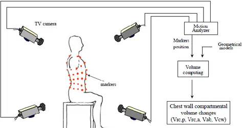
OEP is a system for motion capture specifically developed to estimate breathing volumes. It is able to measures the three-dimensional coordinates of several passive markers applied to body landmarks on the chest-wall by using several TV cameras which frame the subject by different angles. From these data a dedicated algorithm computes the three-dimensional coordinates of the markers by perspective projection described by the collinearity equations (stereo-photogrammetric methods). Then by means of triangulation on the markers detected or by using the Gauss theorem it is possible to calculate the volume enclosed into the markers surface.
In the past years, most of the published studies were focused on the estimation of the input impedance obtained from the response of the respiratory systems to a pressure stimulus given at the mouth providing information about the average properties of the respiratory system and in particular about the mechanical properties of the airways, on the contrary little is known about the transfer impedance. In the above mentioned study, global transfer impedance and its distribution on the chest wall has been estimated at different frequencies on healthy subjects showing how impedance is not equally distributed on the thoraco-abdominal wall but the ribcage is the predominant component at those frequencies. This study opens interesting insight into the comprehension of chest wall mechanics but it is limited by the complexity in the methodological approach- use of OEP system with 69 markers and cameras which should be carefully calibrated- and costs connected. The already uncovered point in this approach is the measurement of chest wall vibration in a reliable and resolute way by means of low costs and not invasive devices.
The objective of this work is to develop and validate a new method that allow the estimation of the local mechanical properties of the chest-wall based on FOT and
iv
optical devices in a non-invasive, low cost and compact way. This new approach will relay on the possibility to have reliable and resolute measurements of the displacement of the chest-wall in order to allow the estimation of impedance map: its knowledge will allow to develop new methodological and technological tools useful for the bed-side assessment of patients affected by pathologies which alter the chest-wall mechanics such as ARDS and restrictive diseases.
To achieve these results new technology, at least for the respiratory system, are needed in particular an optimum candidate is laser self-mixing interferometry which allows the measurement of very little relative displacement (theoretical resolution less than one micrometre).
The work will be presented in four sections whose contents are resumed below. 1 ASSESSMENT OF CHESTWALL KINEMATICS AND MECHANICS:
In this section, the background and the aim of the work are presented. Specifically it is organized in three parts.
In the first one a brief introduction about the respiratory mechanics will be presented together with its assessment by means of the most common techniques used by clinicians. Then a brief look at different pathologies and their classification into obstructive and restrictive diseases is reported focusing on the alterations induced on the mechanics of breathing and in particular in the chest wall mechanics. The main studied pathologies will be ARDS and fibrosis which can be taken as an example of alteration in respiratory mechanics.
The second part deals with non-conventional techniques and technologies which have been proposed in the last years for the measurements of the respiratory mechanics in a non invasive way.
In more details, this section starts with the basics of oscillatory mechanics and the basics of FOT (Forced Oscillation Technique). The FOT represents the current state-of-the art in state-of-the assessment of lung function and has reached a high level of sophistication. Here the measurement set up are presented and the main parameters and interpretative models are described and commented
The second technologies described here is OEP which allows the estimation of chest-wall volume variation and its compartmentalization into at least two parts: the rib cage and the abdomen.
The third part of this chapter describes a recent approach that by combining OEP and FOT allows the estimation of the oscillatory mechanics of the chest wall, in this way it is possible to study how a pressure stimulus travels inside the chest wall according to the different structure it finds in its pathway and to build maps which describe how impedance is distributed over chest wall surface. Although this methods give some
interesting hints into local properties of the respiratory system, its limits, which are mainly due to methodological issues and to incapacity to reliable measure movements less than 0.5 mm, are underlined. Anyway a strong connection between the measurement of local mechanical properties of the respiratory system and the punctual measurements of point of the chest wall is evidenced. So the measurements of the chest wall mechanics requires the use of new technologies-methodologies which should answer to some fundamental requirements such as spatial and time resolution and reliability, but also at some additional features which make it user friendly such as no need of contact with the skin or no need for calibration. A candidate which has all the wanted property, could be laser self mixing interferometer associated with optical distantiometer.
2 NEW TECHNIQUE TO MEASURE DISPLACEMENT
This chapter will be split into two parts according to the technologies and its validation.
The first part deals with interferometry, which is a well-established technology used to measure relative displacement of target with a very high spatial and time resolution. The main interferometer configurations will be covered starting from the Michelson prototype and coming to the modern self-mixing one. The last one is the most compact and easiest to use, since it doesn’t require the use of any mirror because interference effects take place directly inside the resonant cavity laser and, above all, it allows also the measurement of diffusive target for little measurements. On the contrary, at the state of the art, big displacement (less than one cm) are not carried out because the signal quality is dramatically reduced by the presence of speckles, which are darkness zones due to interference in the light diffuse by rough surface. The very important feature of being suitable also for diffusive target suggests the possibility to use laser self mixing interferometers for measurement directly on the skin of patients and on the chest-wall in order to measure both the little vibrations due to pressure stimuli and the wide movement due to breathing. The introduction of this technology allows fixing the main problem for chest-wall mechanics estimation, that is the measurement of its displacement. The only limitation is the presence of speckles which doesn’t allow using traditional algorithm for processing interferometer signal. In the following part of this section, the introduction of a new algorithm and methodological approach for measure chest-wall displacement, will be descripted. The skin behaves as a grey surface diffuser, inducing low back-reflection to the laser diode. In order to analyse the interferometric signal, we propose a new method for estimating the movement direction also in the low-injection regime ,since traditional algorithms, based on fringe counts and extraction of their slope, are not reliable in this
vi
application The developed algorithm was based on two assumptions: 1) the frequency of the fringes is linked to the relative velocity of the target, which must be continuous and 2) each time the target changes its movement direction, the velocity equals zero. To validate the algorithm in vivo e in vitro measurements have been carried out. The set-up, protocol, algorithm and results are discussed for the validation on a test object.
Interferometer was carefully aligned with a piston of a linear servo motor and its signal was recorder during the motion of the motor which is controlled to produce a sinusoidal displacement.
Piston velocity, estimated by interferometric signal as described above, was then integrated to obtain displacement. Motor displacement amplitude and the interferometric one were compared; in particular minima and maxima displacement values were detected and used to compute linear regression and to perform the Bland et Altman analysis. A good linearity, r2=0.99, and the absence of any biased errors demonstrates the algorithm skills to reconstruct displacement from interferometric signals.
In the following part validation of the algorithm when the interfeometer is used directly on skin is reported.
A sinusoidal pressure signal was generated and was applied to the subject through a connecting tube and a mouthpiece. The pressure at the airway opening was measured by a piezo-resistive pressure transducer and the thoraco-abdominal wall velocities in correspondence of the Lewis Angle and approximately 1 cm below the umbilicus were detected using self-mixing interferometer. The interferometer was fixed on a mechanical frame above the subject at a distance of about 60 cm from the subject lying in supine position. Contemporary the scene is surveyed by SMART cameras which are sensitive to interferometer spot, in this way it has been possible to compare displacement measured by interferometer with the one measured by SMART CAMERA The results obtained were compared. The interferometer displacement shows good agreement with the SMART. In conclusion our data demonstrate that laser interferometry could be used to measure accurately displacements of the chest wall making this technique suitable for all the applications involving the measurement of wide chest wall movement as well as for the detection of very small displacement as those occurring when a pressure wave reaches the chest surface.
Estimation of the phase delay between the pressure signal and the chest-wall vibrations has also be performed using the same set up
Eight normal healthy subjects in supine position during quiet breathing while submitted to a sinusoidal pressure forcing at the mouth with components at 5, 11 and
19 Hz. Displacements of 9 chest wall points along three imaginary lines (nipples lines, left and right, and midline) were measured by laser self mixing interferometers. The phase displacement among these points was estimated by correlation technique between the pressure at the mouth and the velocity of the points measured by the interferometer. The results show spatial inhomogeneities strongly dependent on position and frequency. The results are in good agreement with previous data measured by OEP. In conclusion laser interferometry may be an attractive technique for the assessment of local CW motion.
The basic set up proposed here allows to validate the algorithm and to estimate the phase shift of some points on skin but it can’t be proposed to acquire more signal and to generate an impedance maps because of the long time required to move manually the interferometer and because the lack of knowledge about the distances between the points measured.
OPTICAL DISTANTIOMETER FOR MEASUREMENT ON SKIN
Interferometers allow measuring relative displacement but they can’t provide information about the absolute position of the points and the direction of the interferometer respects to the skin, for this reason a second optical technology has been sided to interferometry to solve these issues, it is optical distantiometry.
Basic principle behind optical triangulation distantiomer are explained: for distances of a few inches with high accuracy requirements, laser triangulation sensors measure the location of the spot within the field of view of the detecting element and are so named because the sensor enclosure, the emitted laser and the reflected laser light form a triangle. The input-output characteristic of these devices is not linear, so a careful calibration procedure is needed.
The section goes on with a description of the calibration procedure which consists in pointing the distantiometer towards a target which is moved of a known quantity, then the best fitting polynomial waveform is estimated and the calibration coefficients are extracted. A validation protocol has also been carried out; it consists in measuring the distances of a target with distantiometers and contemporary with SMART camera. A model made by three markers placed on the distantiometer and three markers placed on the target, which is a plane surface, has been implemented The distances so measured show a good agreement with the one estimated by OEP with an error less than 0.5 mm.
The last part of this section deals with the description of a method which allows estimating the angle between the optical beam of the distantiometer and a plane, which can be used to correct for not orthogonally of the interferometer. Basically, if the distantiometer is pointing towards a plane and it rotates of a given angle, by
viii
measuring the distances between it and the plane before and after the rotation, it is possible to estimate the incidence angle by means of easy trigonometrically equations. An analysis of the sensitivity of this method according to error in measurement estimation is performed. The in vitro validation performed mainly consists in pointing the distantiometer toward a plane with a given inclination and in rotating it by means of a stepper motor, the measured distances are used to perform the estimation of the angle. The results are good although they are strongly dependent on the polynomial degrees of the waveform selected in the calibration procedure.
3 ASSESSMENT OF THE MECHANICAL PROPERTIES OF THE CHESTWALL DURING INDUCTION OF ANAESTHESIA
Once the feasibility of laser interferometer measurements directly on skin has been demonstrated and validated, a new system which allows the measurements of the local mechanical properties of the respiratory system has been proposed. The most innovative aspects are the introduction of new technologies for this context such as interferometry and the synergy between already used technique such as FOT with new technologies such as interferometry and distantiometry.
The realized system has been carefully design to fulfill the design requirements of: 1) high spatial resolution, 2) localizing point on the chest-wall, 3) non-invasiveness, 4) easy methodological approach, 5) short measurement time, 6) contactless, 7) modularity which allows a fast set-up also in clinical environment.
Once a system for scanning the chest-wall and measuring impedance maps is available it is possible to employ it in studying different pathologies or not physiological conditions which are supposed to induce alteration in the chest-wall mechanics. In particular a protocol for the measurement of mechanical impedance of patients during anesthesia induction has been design and implemented.
This chapter is organized into two sections: the first one deals with the description of the scanning system, while the second one is focused on the protocol.
The realized system has been carefully designed to fulfill the requirements in particular it has been organized in three conceptual blocks, each one with a well-defined function:
The FOT unit provides the pressure stimulus and the measurement of the pressure and flow at the open airways. It can be organized according to the different condition in which the measurement should be performed, i.e. during mechanical ventilation or spontaneous breathing. The FOT approach satisfies the requirement of a non-invasiveness and non-cooperation required to the patients;
The optical scanning unit provides the measurement of velocity and absolute distance of several points (up to five) of the chest-wall by means of interferometers and
distantiometers which can be moved to scan the whole chest-wall. Interferometer spatial resolution allows the estimation of the small vibration produced by the pressure stimulus and grants for non-invasive and contactless features
The electronic unit allows: to record all the data, to drive the pressure stimulus generator and finally to move the optical scanning unit.
Then the measurement protocol is described. ARDS, thoracic surgery, and anesthesia are associated with atelectasis, disturbed ventilation-perfusion relationship, and hypoxemia. It has been shown that in these patients the mechanical behavior of the respiratory system is altered, with a reduction in functional residual capacity and compliance. Mechanical ventilation is essential in the treatment of all these patients and, in particular, positive end-expiratory pressure (PEEP) is used to reverse atelectasis and improve lung function. Optimal open lung PEEP may be defined by a mechanical point of view which can be evaluated by measuring input impedance, as proved in previous works, but also taking advantages of the variations in the chest-wall mechanics.
All patients were studied in the supine position while ventilated in pressure support (PS) mode. Just before surgery measurements of oscillatory mechanics were performed at different stages measuring input impedance at 5-11-19 Hz by FOT, local transfer function at 5-11-19 Hz, FRC (Functional Residual Capacity)
The patients were connected to the mechanical ventilator and the laser scanning system has been adopted to measure chest wall movements.
At the end of the chapter results and discussions are reported. Since this group of patients is healthy from the respiratory system point of view, derecruitment is not awaited, while symmetry in the left and right part can be explained as a substantial homogeneity of the respiratory system.
4 CONCLUSION AND DISCUSSIONS
The research activity presented in this work was aimed at proposing s new system and methodology to study local chest wall mechanics by means of oscillatory mechanic which can find an application for the study and the clinical management of patients affected by diseases which induce alterations in the chest wall mechanics. For this purpose new experimental setups and protocols have been designed and used to measure oscillatory mechanics.
Our data demonstrate that laser interferometry could be used to measure accurately displacements of the chest wall as small as 400nm making this technique suitable for all the applications involving the measurement of wide chest wall movement as well as for the detection of very small displacement as those occurring when a pressure wave reach the chest surface.
x
Phase delay between pressure at the mouth and the chest-wall surface can be estimated by matching FOT and optical devices. Phase delay changes according to the different compartments, and in relation to the chest-wall status.
In conclusion, oscillatory lung mechanics can be considered as a powerful and quantitative tool to measure lung mechanics. This information could be used as a complement of the standard techniques in order to diagnose, treat and monitor in a more specific and effective way pathologies and non-physiological conditions characterized by alteration in chest-wall mechanics
INTRODUCTION
The respiratory system is a complex system which accomplishes gas exchange by means of the synergy among functions and structures which have specifically developed at this aim. Processes involved in gas exchange are: 1) ventilation, that is air transport inside and outside the lungs; 2) oxygen and carbon dioxide diffusion among alveoli and blood; 3) oxygen and carbon dioxide delivering for the blood to the cell and finally, 4) ventilation regulation. The respiratory system functionalities are not only reliant on its components, that are a complex branching system of tubing (airways), a huge exchange region (the alveoli) where gases can diffuse from and into the blood, but its behavior is deeply influenced also by the respiratory muscles and structures that form a boundary of it, such as the chest-wall and the diaphragm. Impairment at only one of these structure or functions can seriously affect not just the respiratory system but it can yield to a general worsening in the life condition of the subjects due to labored breathing or asthma attacks, fatigue and difficulty with mobility, heightened sensitivity to ordinary substances and chemicals, and compromised immunity to infection.
In particular ventilation capability, which represents the first step in breathing activity, relies on the mechanical properties of the whole respiratory system: the resistance of the airways, the compliance and elastic recoil of the lung parenchyma, stiffness in chest wall and abdomen, all these components contribute in different and complex ways in the relationship between the pressure generated by active components inside the respiratory chain and the volume of gas introduced.
Diseases affecting the respiratory system, such as ARDS (acute respiratory distress syndrome), restrictive diseases and lung edema, produce alterations and inhomogeneities in the lung structure and in the chest wall which cause changings in the mechanical properties.
In more details, ARDS is defined as a syndrome of inflammation and increasing permeability that is associated with a constellation of clinical, radiological and physiological abnormalities that cannot be explained by, but may coexist with, left atrial or pulmonary capillary hypertension. The physiological consequences are severe hypoxaemia due to right-to-left shunting of blood and decreased pulmonary compliance due to filling and closure of alveoli. As the airspaces fill with fluid, the gas exchange and mechanical properties of the lung deteriorate.
Restrictive lung diseases are characterized by reduced lung volume, either because of an alteration in lung parenchyma or because of a disease of the pleura, chest wall, or
xii
neuromuscular apparatus. The many disorders that cause reduction or restriction of lung volumes may be divided into two groups based on anatomical structures. The first is intrinsic lung diseases or diseases of the lung parenchyma. The second is extrinsic disorders or extra-parenchymal diseases, which involve the chest wall, pleura, and respiratory muscles, the components of the respiratory pump. Diseases of these structures result in lung restriction, impaired ventilatory function, and respiratory failure (e.g., non-muscular diseases of the chest wall, neuromuscular disorders). Also pulmonary edema can cause alterations in lung mechanics that directly contribute to clinical morbidity and mortality rates. Both the location of the edema fluid (interstitial versus alveolar pulmonary edema) and the etiology of the pulmonary edema contribute to the severity and type of abnormalities of lung mechanics observed. The alterations in lung mechanics associated with the adult respiratory distress syndrome may involve the direct effects of released mediators, alterations in pulmonary surfactant, and altered airway reactivity, as well as the direct effects of the edema fluid.
Up to the present day, the diagnosis and the management of these kinds of pathologies have been carried out mainly by chest radiography, which reveals diffuse opacity according to the stage of the pathology and CT. More specifically ARDS can be detected also by means of measurement of lung endothelial and alveolar epithelial barrier function and BAL (Bronchoalveolar lavage); while restrictive pathology can be detected also by means of some laboratory tests aimed to measure the concentration of well-known markers such as elevated creatine kinase level. Anyway all of them present some intrinsic limitations, indeed they are invasive or required long time to be carried out and thus they can’t be used to monitor in a continuative way the pathology evolution.
On the contrary several published data suggest that global but also local chest wall mechanical properties are parameters that might be useful for the evaluation of structural elasticity and inertia on the overall behavior of the respiratory system and for estimating the possible impact of both restrictive and obstructive diseases on respiration, thus it could be useful in the assessment of the pathology providing a handy tool for clinicians during the diagnosis but also the therapy of the disease. Although the rich informative content of the chest wall mechanical properties, up to now this parameter has been almost disregarded because of the intrinsic difficulties in its measurement. Therefore a new approach for the detection and the monitoring of these diseases can be based on the measurements of the local and global chest-wall mechanics and the extraction of parameters which are sensitive to pathophysiological alteration. The more promising and inspiring approach has been reported by Dellacà et
al [2002] which suggest the union of FOT (Forced Oscillation Technique) and OEP (Optoelectronic Plethysmography) to estimate the impact of low amplitude pressure stimuli given at the mouth at the level of vibration induced at the chest-wall.
Briefly FOT method is based on the measurements of the response of the respiratory system to small pressure stimuli generated externally and applied during normal breathing. There are two different approaches in studying oscillatory mechanics of the respiratory system: one could study the response of the system by analyzing the flow that the pressure stimulus has been able to induce from the mouth or analyse how the stimulus has travelled through the system by measuring the displacement of the external surface of the thorax. The former, widely used in oscillatory mechanics, allows the estimation of the input impedance by combining the measurement of pressure and flow at the airway opening, while the latter allows the estimation of the transfer impedance by combining airway opening and chest wall vibrations.
OEP is a system for motion capture specifically developed to estimate breathing volumes. It is able to measures the three-dimensional coordinates of several passive markers applied to body landmarks on the chest-wall by using several TV cameras which frame the subject by different angles. From these data a dedicated algorithm computes the three-dimensional coordinates of the markers by perspective projection described by the collinearity equations (stereo-photogrammetric methods). Then by means of triangulation on the markers detected or by using the Gauss theorem it is possible to calculate the volume enclosed into the markers surface.
In the past years, most of the published studies were focused on the estimation of the input impedance obtained from the response of the respiratory systems to a pressure stimulus given at the mouth providing information about the average properties of the respiratory system and in particular about the mechanical properties of the airways, on the contrary little is known about the transfer impedance. In the above mentioned study, global transfer impedance and its distribution on the chest wall has been estimated at different frequencies on healthy subjects showing how impedance is not equally distributed on the thoraco-abdominal wall but the ribcage is the predominant component at those frequencies. This study opens interesting insight into the comprehension of chest wall mechanics but it is limited by the complexity in the methodological approach- use of OEP system with 69 markers and cameras which should be carefully calibrated- and costs connected. The already uncovered point in this approach is the measurement of chest wall vibration in a reliable and resolute way by means of low costs and not invasive devices.
The objective of this work is to develop and validate a new method that allow the estimation of the local mechanical properties of the chest-wall based on FOT and
xiv
optical devices in a non-invasive, low cost and compact way. This new approach will relay on the possibility to have reliable and resolute measurements of the displacement of the chest-wall in order to allow the estimation of impedance map: its knowledge will allow to develop new methodological and technological tools useful for the bed-side assessment of patients affected by pathologies which alter the chest-wall mechanics such as ARDS and restrictive diseases.
To achieve these results new technology, at least for the respiratory system, are needed in particular an optimum candidate is laser self-mixing interferometry which allows the measurement of very little relative displacement (theoretical resolution less than one micrometer).
1
1. ASSESSMENT OF CHESTWALL KINEMATICS AND
MECHANICS
The role of respiration is to bring oxygen to tissues and to carry out carbon dioxide, which is reached through four steps: 1) pulmonary ventilation, in order to transport air from the atmosphere to alveoli, 2) diffusion of oxygen and carbon dioxide through alveolar-capillary membrane, 3) transport of oxygen and carbon dioxide from and to cells in blood and liquids, 4) control of ventilation and of other features of respiration.
These vital functions are performed not only by means of its components such as the airways, the lung parenchyma and the pleura, but they rely also on the behavior of the respiratory muscles and on the mechanical properties of the chest-wall, the box which encloses the whole system.
1.1. RESPIRATORY SYSTEM
1.1.1. ANATOMY
The respiratory tract (or system) begins at the nose and ends in the most distal alveolus as reported in Figure 1.1. Thus, the nasal cavity, the posterior pharynx, the glottis and vocal cords, the trachea and all the divisions of the tracheobronchial tree are included in the respiratory system. The upper airways consist of all structures from the nose to the vocal cords, including sinuses and the larynx, whereas the lower airways consist of the trachea, bronchi and alveoli. The major function of the upper airways is to "condition" inspired air that means to heat it and to humidify it. The nose also acts as a filter, entraps, and clears particles greater than 10 μm in size.
The major structures of the larynx include the epiglottis, arytenoids, and vocal cords. The epiglottis and arytenoids cover the vocal cords during swallowing. Thus, under normal circumstances, they inhibit aspiration into the lower respiratory tract.
The tissue of the lung is called parenchyma, it is very elastic and it can easily distend. It contains elastin and collagen fibers. The collagen fibers normally are folded and they distend during the lung expansion; the elastin fibers stretch when the lung expands. Both fibers, however, exert a force to regain their initial condition. In every healthy human even at the end of the deepest expiration, when the lung contains the minimum amount of air, the fibers of the parenchyma remain always stretched to a certain extent, exerting elastic recoil.
2
The pleurae are serous, thin, smooth and transparent membranes, covered with mesothelium and forming closed sacks placed between lungs and the internal of thoracic cavity.
.
Figure 1.1: Segments: 1, Apical; 2, posterior; 3, anterior; 4, lateral (superior); 5, medial (inferior); 6, inferior; 7, medial basal; 8, anterior basal; 9, lateral basal; 10, posterior basal. Bronchopulmonary segments, anterior view.
Every pleura consists of two sheets of membranous tissue: pulmonary pleura, wrapping the lung, and parietal pleura which wrap the inside of the thorax. The space in between these two membranes (intrapleural cavity) is filled by a very thin layer of fluid, the intrapleural
fluid, having excellent lubricating properties. The excess of this fluid is continuously
removed through the lymphatic system, in order to maintain a slight suction in the pleural cavity, that is, the pressure of the pleural fluid is sub-atmospheric. The negative intrapleural pressure makes the lung volume to follow closely volume changes of the thoracic cage but, at the same time, thanks to the pleural space and to the pleural fluid, the lung and the thoracic cage are allowed to slide each other. Figure 1.2 shows the relative position of the lungs and the thoracic cage in the resting position, at the end of a quiet expiration, and at the end of the inspiration. We can notice that the lungs slide into the comer of the thoracic cavity, to fill the room left empty when the diaphragm contracts.
3 Figure 1.2: Both the lungs and the internal wall of the thoracic cavity are covered by a membranous sheet of tissue called pleura. a. The two pleura are folded in such a way to create two distinct intrapleural cavities (their dimensions are here exaggerated). b. At inspiration, when the diaphragm contracts, the lungs slide along the thoracic cavity to fill the freed space
1.1.2. VENTILATION MECHANICS
The ventilation of the lungs is accomplished alternating phases in which air is flowing into the lungs (inspiration) and phases in which air exits the lungs (expiration). The mechanics of these air movements are strictly related to the peculiar anatomy of the chest.
Inspiration is determined by the contraction of the inspiratory muscles, which are basically the diaphragm and the external intercostal muscles. The diaphragm represents the lower closure of the chest; when relaxed it is dome shaped while when it contracts it moves downward and it flattens.
In this way the bowels are pressed down and the volume of the thoracic cavity increases. During quiet respiration the diaphragm alone is responsible for the 75% of volume expansion of the thoracic cavity.
External intercostal muscles are located in between the ribs, elongated forward and downward, as shown in Figure 1.3. The same figure shows the forces acting on the ribs when they contract, and the moments of these forces around the pivot points at the spinal cord. Accordingly to the third principle of dynamics, each muscle exerts the same force on both ribs it is attached to. The force it exerts on the lower rib, however, has a greater arm than the force acting on the upper rib, due to the physiological leaning of the muscles. The imbalance of the two moments takes the rib to rotate upward. Since ribs normally slant downward, when rotating, they push the sternum in front. This increases the antero-posterior diameter of the thoracic cage and therefore the volume of the thoracic cavity
4
enlarges. The expansion of the thoracic cavity produces negative pressure in the intrapleural space, which induces the lungs to expand. The alveoli, the alveolar ducts and the bronchioles in their turn enlarge, creating a negative pressure (with respect to atmospheric) inside them, which recalls air from the outside.
Figure 1.3: When the external intercostal muscles contract, the ribs are pulled upward and forward, and they rotate on an axis joining the tubercle and head of rib. As a result, both the lateral and anteroposterior diameters of the thorax increase. The internal intercostals have opposite action.
Expiration in quiet breathing occurs passively, without the recruitment of any muscle. When the diaphragm and external intercostal muscles stop contracting, the weight of the thoracic cage and its own elasticity bring it to its resting position. At the same time the compressed bowels push back the relaxed diaphragm as well and the volume of the thoracic cavity resumes its smaller dimensions.
If the volume of the lungs decreases, the pressure inside them rises, becoming greater than the atmospheric pressure. This forces the air out, through the airways. When expiration needs to be faster, as during exercise or coughing, the expiratory muscles are recruited to provide greater ventilation. Expiratory muscles are mainly the abdominal muscles and the internal intercostal muscles. When contracting the abdominal muscles squeeze the viscera, which in turn push up the diaphragm. Internal intercostal muscles work similarly to external intercostal muscles (inspiratory muscles); they are also located in between the ribs but they are turned symmetrically, that is the expiratory muscles lean backward and downward. When these muscles contract, the ribs are pulled down, so that the sternum and the rib cage regain fast their resting position. Expiratory muscles, besides, are able to force the thoracic cavity to a smaller volume than at the end of a passive expiration. Nevertheless, whatever effort is placed in forcing the expiration, it is not possible to empty completely the lungs, which physiologically will always contain some amount of air.
5
1.1.3. MECHANICAL MODELING OF THE RESPIRATORY SYSTEM
A. ONE COMPARTMENT MODEL
From the anatomical and physiological description given above it is possible to deduce simplified models of the respiratory system, which resume the features that are essential in determining the efficiency of the ventilation. A classical model is represented in Figure 1.4. According to this model the whole respiratory system is subdivided in subsystems and each of them is characterized by one lump parameter. Similar models provide a simplified description of the actual interpretation of the respiration mechanics and also the measurements that are performed for diagnostic purposes are coherent with the same clinical interpretation.
Figure 1.4: A model of the ventilation mechanics. The airways oppose resistance to the air flow; the lungs and the thoracic cage are schematized as elastic bags.
The influence of the airways on the ventilation mechanics is classically described by the resistance they oppose to the air flow. The overall resistance of the airways is determined mainly by the flow conditions in the trachea and in the large bronchi, while the small bronchi and the bronchioles give a small contribution. The upper airways are responsible for the 80-90% of the overall resistance, while the remaining 10-20% is due to the flow condition in the smaller airways. This is explained by two reasons: firstly all the air flow passes through the upper airways where, consequently, air velocity is high and the flow is turbulent. Turbulent flow gives rise to a greater pressure drop; hence it corresponds to a greater resistance than laminar flow. Secondarily, although the terminal airways are very small and for this reason singularly they oppose high resistance to the air flow, the lungs account millions of terminal bronchioles and, considered all together, they add up to a large cross section, which minimally stops the air flow. The effect is similar when in an electrical circuit many large resistors are connected in parallel.
6
B. ADVANCED MODELING
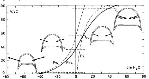
In our model the lungs and the chest wall are represented in the form of elastic bags one contained into the other. When a system can be schematically represented in this form, its static behavior is completely described by the relationship connecting the volume inside the elastic bag to the pressure difference between the inside and the outside of the bag. This relationship is determined by the mechanical features of the bag's wall. The curves drawn in Figure 1.5 provide a graphical representation of the volume/pressure relationship for the lungs when they are taken out of the chest wall (PL) and for the thoracic cage without the lungs inside (PW).
Figure 1.5: Static curves of the lungs (PL), of the chest wall (PW) and of the lungs-plus-chest wall
(Prs). The slope of these curves is related to the compliance of the systems.
The first curve (PL) is determined by the characteristics of the parenchyma. The curve of the
chest wall alone (PW) derives from the passive behavior of the tissues that construct the
chest and all organs, which somehow interfere with lungs expansion (the bowels for instance are compressed by the diaphragm during inspiration). The third curve represents the whole system lungs-plus-chest wall (Prs); it is the sum of the other two curves.
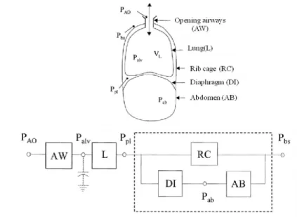
In order to describe chest wall mechanics during spontaneous breathing in normal subjects, in the past several models have been proposed. The simplest model of chest wall motion was developed by Konno and Mead (1) , who partitioned it into the rib cage and the abdomen. Successively, it was demonstrated that the rib cage cannot be considered as a single compartment and therefore, different two-compartment rib cage models were proposed, in which the part apposed to the lung (pulmonary rib cage) was distinguished from the part apposed to the diaphragm (abdominal rib cage). In Figure 1.6, we reported a detailed mechanical model of respiratory muscle actions on the chest wall, divided into pulmonary rib cage, abdominal rib cage and abdominal wall.
7 Figure 1.6: Simplified mechanical model of the chest wall incorporating a two-compartment rib cage, i.e. pulmonary (RCp) and abdominal (RCa) rib cage, and abdominal wall (ABw). The respiratory muscles are grouped into: upper rib cage muscles (RCM); lower rib cage muscles (RCMa) divided into insertional (RCMa,ins) and non-insertional (RCMa,nins) on the abdomen; costal and crural diaphragm (COS and CRU, respectively); abdominal muscles (ABM) divided into insertional (ABM,ins) and non-insertional (ABM,nins) on the lower rib cage. Pao, Ppl and Pab represent the airways opening, pleural and intra-abdominal pressures, respectively.
Upper rib cage muscles act on pulmonary rib cage and lower rib cage muscles act on abdominal rib cage. Lower rib cage muscles are divided into insertional and non-insertional on the abdominal wall. The diaphragm is split into its costal and crural parts. The abdominal muscles are divided into insertional and non-insertional on the rib cage. The spring represents the mechanical linkage between pulmonary rib cage and abdominal rib cage and describes the restoring force developed by distortions between the two compartments away from the relaxation configuration.
In conclusion, the partitioning of the pressures differences across different parts of the chest wall and the possibility to measure the volume displacements of the two-compartment rib cage and abdomen have proved major advances in our knowledge of the act of breathing in normal conditions. This is important to better understand the behavior of the chest wall also in different "pathological" conditions.
C. MECHANICAL ELEMENTS
Evaluation of mechanical respiratory system properties consists in studying system cinematic and its dynamic. The first deals with balances configuration that means the positions and the forces acting on the system, while the second deals with the relationship between system movements and the forces which cause them.
8
The measurements on mechanical system are force and positions, since the respiratory system has a tridimensional geometry, these can be translated into volume, flow (volume variation) and pressure.
Another fundamental aspect is the estimation of passive elements mechanical properties (resistance, elastance and inertance) as reported in Table 1.1 and the estimation of active components action (pressure, work, power) such as the respiratory muscles.
Usually to make easier the comprehension of respiratory system mechanics, an analogue model is used, in particular the respiratory system can be considered as a three dimensional analogue of a mass-spring-damper system.
The respiratory system is usually modeled as an assemblage of just three simple types of primitives passive elements, that are elastance, inertance and resistance; each of them is associated to a different way of handling energy.
In particular the elastance is connected with storage by potential energy, inertance with storage by means of kinetic energy, resistance with dissipation by means of friction.
Table 1.1: Analogue measure for respiratory system, mechanical and electrical models
Other used components are ideal flow and pressure generators.It is a convention to characterize each element in terms of drop pressure at its terminals and flow passing through it. The relationship between pressure and flow is not necessary linear (Figure 1.7 and 1.1) but to avoid complex non linear characteristics, it is possible to set volume history, lung volume and volumetric flow rate and to consider just little movements around the equilibrium point.
̇ 1.1
Where ̇ is a function of the volumetric flow rate and P is gas pressure. Rohrer’s equation 1.2 for flow resistance is an example of a well-known constitutive relation, where k is a constant and the relation is not linear.
Respiratory system Mechanical model Electrical model
State: Volume: V Position: x Charge: q
Flow: dt t dV V () Velocity v: dt t dx x () Current: i=dq )dt(t Flow variation:
dt
t
V
d
V
(
)
Acceleration: 2 2(
(
))
)
(
dt
t
x
d
dt
t
x
d
x
a
Current variation:dt
t
di
i
(
)
Pressure: P Force: F Voltage: V
Properties: Elastance E=P/V Stiffness: K=F/x Capacity: C=q/v
Resistance: R= P/
V
Friction: B= F/v Resistance: R= v/i9
̇ ̇| ̇| 1.2
The linearized relationship of pressure difference to volume flow is called an ideal elemental equation.
Figure 1.7: Rohrer equation taken as an example of non linear pressure-flow relations linearized for small departure about any given bias flow rate.
According to the analogue circuital model, passive component have positive power consumption.
Elastance (compliance)
An ideal fluid elastance (E) has pressure differences in direct proportion to volume, and it can be also thought as the reciprocal of compliance.
Here the equation 1.3
̇
1.3
Under static conditions in completely relaxed patients, airway pressure is equal to the elastic recoil pressure of the respiratory system (2). Thus, compliance is measured as the change in lung volume per unit change in applied static pressure (elastic recoil pressure). The units are liters per cmH2O or milliliters per cmH2O. Compliance is the mathematical inverse of elastance, the amount of pressure required to change the volume of the lung by a given amount (3). Although both terms are useful, the clinical practice and refers to compliance more frequently than elastance will be followed.
The lung and chest wall are said to be aligned in series, since pressure applied to the airway is first transmitted to the lung. From the lung, a reduced amount of applied pressure is transferred to the chest wall. Because of this series relationship, the pressure to distend the respiratory system is the sum of the pressures required to distend the lung and the chest wall (1.4, 1.5). Thus the elastic of the respiratory system (Ers) is the sum of the lung elastance (EL) and chest wall elastance (ECW).
flow
pr
es
su
10
1.4
1.5 The lung and the chest wall display different volume. The resulting pressure-volume relationship of the respiratory system is sigmoidal in shape, and compliance is greatest in the midvolume range, where breathing normally occurs. At the completely relaxed static equilibrium volume of the respiratory system, elastic recoil of the lung and the chest wall exactly balance each other. Also at this point, compliance of the lung and chest wall is approximately equal in normal subjects. In this midvolume range, the elastic work of breathing and fluctuations in transpulmonary pressure will be minimized (4).
Resistance
An ideal fluid resistance exhibits a pressure drop proportional to flow rate according to the elemental relation reported in the following equation (1.6).
̇ 1.6
For steady fully developed laminar flow at low Reynolds number (parabolic velocity profile), in a long straight pipe of length l and cross sectional A, containing fluid viscosity the resistance can be expressed as in the following equation (1.7):
1.7
This is the Poiseuille formula for flow resistance.
With fully turbulent flow, the pressure difference is proportional to the square of the gas flow multiplied by another constant (K2), which is related to the density of the gas and is independent of its viscosity (1.8):
| ̇|̇ 1.8
In the case of the respiratory system, resistance is rarely linear, and the relationship between pressure and flow is usually expressed by Rohrer's equation (1.9):
̇ | ̇|̇ 1.9
The customary units of respiratory resistance are cmH2O/(liter/s). The mathematical inverse of resistance is conductance, but this term is seldom used. The resistance of the respiratory system can itself be subdivided into elements that are in series and hence additive (5). These elements include pulmonary resistance (RL) and resistance of the chest wall (RCW). Pulmonary resistance is itself further subdivided into airway resistance (Raw) and a lung tissue component. Each of these components is determined by measuring the net pressure required to produce flow of the component .The relationship between driving pressure and airflow depends on whether flow is laminar, turbulent, or a mixture of both (5)
11
Inertance
An ideal fluid inertance links pressure to first derivate flow as shown in the following equation (1.10):
̇
1.10 Where the inertance is the value reported in 1.11:
1.11
Fluid inertance increases proportionally with tube length (l) and gas density (), but decreases as the reciprocal of the cross section (A).
At frequencies normally encountered during spontaneous and mechanical ventilation, the effects of inertance are usually insignificant and are customarily ignored (6). The pressure generated by inertance is in the opposite direction to that generated by the elastance of the respiratory system. Hence, inertial forces tend to slightly offset the impedance to flow provided by the stiffness of the respiratory system.
The sum of the elastic and inertial forces is referred to as the reactance of the respiratory system.(7)
Active elements
There are active elements capable of delivering energy to the system over an extended period that account for sustained respiratory system motions; they may be respiratory system muscles or external loudspeaker or pumps.
Two useful idealized sources are the volume flow (current source) and the pressure source (voltage source); the first can provide a fixed flow independently from the drop pressure at its terminals, while the second can maintain a fixed pressure despite of the flow through it.
The equation of motion
A linear respiratory system model is formulated connecting the passive elements listed above and imposing mass conservation and Newton law.
The mass conservation principle is the equivalent of Kirchhoff’s current law in electrical circuits; the second is the principle of compatibility. It applies to pressure drop in the system and guarantees that the net pressure drop taken in a specific direction around any closed path in the system circuit, must be zero, which is the analogous of Kirchhoff’s voltage law.
12
Figure 1.8: An Electrical analogue of the respiratory system. The analogy is between flow and current, volume and charge, pressure and voltage.
The simplest model of respiratory system is represented in Figure 1.8; it is just a RLC series. Applying the continuity and the compatibility principles to the model, the following equation (1.12) can be found:
1.12
Substituting the elemental relations into Eq. 1.8 and rearranging the term, then one obtains 1.12 and 1.13:
̇ ̈ 1.13
This is a very important equation known as the motion equation.
1.2. ASSESSMENT OF RESPIRATORY MECHANICS IN
CLINICS
As described in the previous paragraph, the mechanical properties of the respiratory system may be express by means of few lumped parameters whose measurement has a diagnostic value in identifying the presence of a disease or in assessing what kind of disease is eventually present but, at the same time, it is time consuming and not easy to perform. Therefore up to now in clinical practice the objectives of measurement procedures are not exactly the airways resistance or the compliance of the lungs or the thoracic cage, but different parameters related to the same conceptual representation of the respiratory mechanics. In particular, the state variables are flow, volume and pressure, here a review of the most significant terms is reported.
13
1.2.1. LUNG VOLUME DEFINITIONS
The measurement of the lung volumes are normally obtained by means of a spirometer. In Figure 1.9 is shown a typical spirometric track where is depicted the excursion of lung volume during normal breathing and during a maximal inspiration and expiration. The lung volumes depicted are:
1. VT is the tidal volume representing the air volume normally breathed; 2. TLC is the total lung capacity, the lung volume at maximal expansion; 3. RV is the residual volume, the gas volume after a maximal expiration; 4. IRV is the inspiratory residual volume;
5. ERV is the expiratory residual volume;
6. FRC is the functional residual capacity, it is the lung gas volume after a normal expiration;
7. IC is the inspiratory capacity, the maximum gas volume breathed in starting at FRC; 8. VC is the vital capacity, the maximum gas volume breathed in starting at RV.
Figure 1.9: Spirometric tracks showing the principle lung volume measurements.
In order to measure breathing volumes, in clinical environment are used two different approaches based on a direct measurement of the volume or on the measurement of the flow, whose integration provides volume.
1.2.2. LUNG VOLUME MEASUREMENTS
The instrumentation commonly operated to measure volumes in testing the pulmonary function can be subdivided into two major categories: volume sensing devices, commonly known as spirometers, and flow sensing devices, called respiratory flowmeters or
14
pneumotachometers. Since the air flow is defined as the volume of air inspired or expired
per unit of time, the overall volume moved results from integration of the air flow or, vice versa, knowing the moved volume at every instant of time, we can derive the instantaneous flow just differentiating the volume. For this reason both kinds of devices can theoretically be used to yield to the same results. The measurements of lung volumes and respiratory flow are certainly non-invasive. The measurement devices remain outside of the body and they exploit the fact that the patient naturally moves air in and out of its lungs. This doesn't mean that the measurements can be considered absolutely free of any risk for the patient and for the physician, anyway the main hazards are connected with the risk of cross contamination between patients employing successively the same device or they can be associated to specific situations as pneumothorax, unstable cardiovascular status, in recent surgery.
A. SPIROMETRY
It consists in collecting gas which passes through the opening airways and computing the volume taken in the lung.
The most common spirometer is the bell one, Figure 1.10 which collects air into a sliding bell. By knowing the bell geometry and its movement, it is possible to estimate volume.
B. PLETHYSMOGRAPHY
Plethysmography is a general term used to describe all volume measurements, in particular lung volume estimation for respiratory system.
Figure 1.10: A spirometer consists of a floating drum put over a chamber, with the drum counterbalanced by a weight. The drum filled with air or oxygen is connected with the mouth of the patient by a tube. If the patient exhales into the spirometer, the drum rises, by inhaling in the drum falls. On a moving sheet of paper an appropriate recording is made
Kymograph drum Counter balance weight Floating drum Oxigen chamber water