A
cta
DV
A
cta
DV
A
dvances in dermatology and venereology
A
ctaD
ermato-V
enereologicadoi: 10.2340/00015555-2609 This is an open access article under the CC BY-NC license. www.medicaljournals.se/acta
The aim of this retrospective study was to determine the type and prevalence of vascular patterns in the ulcerated and non-ulcerated portions of histologi-cally proven basal cell carcinomas (BCCs) and corre-late them with other dermoscopic and clinical featu-res, including the clinically supposed diagnosis. Three authors retrospectively collected 156 clinical and 156 dermoscopic digital images of ulcerated BCCs (histo-logically confirmed); each image was blindly evalua-ted by 2 other authors, who did not know the histo-logical diagnosis. Seventeen lesions were completely ulcerated, while 139 lesions presented ulcerated and non-ulcerated portions. Correct clinical diagnosis was associated with the type of lesion, in particular 90.6% of partially ulcerated lesions were correctly diagno-sed with clinical-dermoscopic examination, compared with 11.8% of totally ulcerated lesions (χ2 = 64.00, p = 0.000). Presence of arborizing pattern in the
ulce-rated portion was associated with a correct diagno-sis (Fisher’s exact test, p = 0.015). Correct diagnodiagno-sis was also associated with absence of dotted pattern in the non-ulcerated area (χ2 = 16.18, p = 0.000); the
ab-sence of hairpin (χ2 = 6.08, p = 0.000) and glomerular
patterns were associated with correct diagnosis in the ulcerated areas (χ2 = 18.64, p = 0.000). In case of
com-pletely ulcerated BCC the clinician lacks the means to correctly identify the correct nature of the lesion, and is driven towards an incorrect diagnostic conclusion.
Key words: dermoscopy; basal cell carcinoma; melanoma;
vas-cular pattern; ulcerated basal cell carcinoma.
Accepted Jan 12, 2017; Epub ahead of print Jan 17, 2017 Acta Derm Venereol 2017; 97: 612–616.
Corr: Angela Filoni, Section of Dermatology, Department of Biomedical
Science and Human Oncology, University of Bari, Piazza Giulio Cesare, IT-70124 Bari, Italy. E-mail: [email protected]
B
asal cell carcinoma (BCC) is defined as a slow-growing skin malignancy, predominantly affecting middle-aged and fair-skinned individuals. Clinically BCC can manifest differently, with nodular, superficial, morpheic, and pigmented variants (1).Dermoscopy is an in vivo technique that has proven useful to differentiate BCC from other cutaneous ma-lignancies, such as squamous cell carcinoma (SCC) and melanoma (2–5).
Characteristic dermoscopic features of BCC include large blue-grey ovoid nests, leaf-like areas, multiple
blue-grey globules and spoke-wheel areas (6, 7). In addi-tion, specific vascular patterns may aid the dermoscopic diagnosis of BCC, especially when the above-mentioned traditional criteria are lacking (8–11). Arborizing vessels, in particular, are known to be a characteristic and com-mon feature of BCC, with a positive predictive value of 94.1% (9–12). Moreover, short fine telangiectasias are commonly observed in superficial BCCs (13, 14). How-ever, several additional morphological types of vessels have been reported (11–15).
The aim of our study was to determine the type and prevalence of vascular patterns in the ulcerated and non-ulcerated portions of histologically proven BCCs and correlate them with other dermoscopic and clinical features, including the clinically supposed diagnosis.
MATERIALS AND METHODS
Three of the authors (DB, NA, GG) retrospectively collected 156 clinical and 156 dermoscopic images of histologically confirmed ulcerated BCCs, developed by 153 patients treated at the Derma-tology Unit of University of Bari Policlinico hospital between January 2011 and December 2013. Anagraphical data (such as age and sex) and lesion-related data (such as presence of pigmentation, lesion anatomical site and diameter) were recorded.
Dermoscopic images had been taken via digital dermoscopy (VideoCAP™, DS Medica, Milano, Italy) and polarized handheld
dermoscopy devices (DermoGenius® II, DermoScan GmbH,
Regensburg, Germany and DermLite DL3N, 3 Gen LLC, San Juan Capistrano, CA, USA) mounted on a digital camera (10× magnification). Clinical images had been taken via videoCAPTM or digital camera. Skin preparation prior to dermoscopy included hyperkeratotic skin removal by wet gauze and application of pet-rolatum oil in case of immersion dermoscopy. Each digital image was blindly evaluated by 2 of the authors (AF, MV), who did not know the histological diagnosis, for the presence of the following types of vessels, diversified for the ulcerated and non-ulcerated portions of the lesions: arborizing, dotted, linear-irregular, comma, hairpin, crown, glomerular. In the presence of 3 or more types of vessels, the vascular pattern was defined as polymorph. Diffuse reddish coloration (milky-red globules/areas and erythema) was also considered. Moreover, the prevalent vascular pattern for each lesion was noted. Other dermoscopic features were assessed: large blue-grey ovoid nests, leaf-like areas, multiple blue-grey globules and spoke-wheel areas (classic BCC patterns); brown-to-black dots/globules, pseudopods, blue/white veil, pigment network and peppering (classic melanoma patterns). Pigmented BCCs were defined by the presence of dermoscopic pigmentation on more than 25% of the lesion’s surface.
On the basis of the above characteristics a clinical-dermoscopic diagnosis was advanced by the 2 blinded authors.
Vascular Patterns in Cutaneous Ulcerated Basal Cell Carcinoma:
A Retrospective Blinded Study Including Dermoscopy
Nicola ARPAIA1, Angela FILONI1, Domenico BONAMONTE1, Giuseppe GIUDICE2, Margherita FANELLI3 and Michelangelo
VESTITA2
1Section of Dermatology, Department of Biomedical Science and Human Oncology, 2Section of Plastic and Reconstructive Surgery, Department
A
cta
DV
A
cta
DV
A
dvances in dermatology and venereology
A
ctaD
ermato-V
enereologica Statistical analysisTo compare the prevalence of each vascular pattern between ulcerated and non-ulcerated portion of the lesions the McNemar test was performed, excluding from the analysis the 17 lesions that were completely ulcerated. Associations between the vascular pat-terns and other dermoscopic and clinical features were evaluated
by χ2 test. To compare the prevalence of single patterns, as well
as dermoscopic and clinical features between correctly diagnosed
lesions and not, χ2 test was also employed. A logistic regression
was performed to identify, by a multivariate analysis, which pat-tern or feature led to a correct diagnosis.
RESULTS
A total of 156 clinical and 156 dermoscopic images of ulcerated BCCs, which had developed on 153 patients, were assessed. Characteristics of lesions and patients are shown in Table I. Seventeen lesions were completely ulcerated, while 139 lesions presented ulcerated and non-ulcerated portions.
Partially ulcerated lesions
The prevalence of the discrete vascular patterns in the 139 lesions with both ulcerated and non-ulcerated areas
is shown in Table II and Fig. 1. A statistically significant difference in the prevalence of the arborizing, dotted, linear-irregular, comma, hairpin, and polymorph patterns between the ulcerated and non-ulcerated areas was noted.
In the ulcerated portion the comma pattern was as-sociated with the presence of pigmentation (χ2 =11.137,
p = 0.001) and with the presence of large blue-grey ovoid
nests (χ2 = 4.91, p = 0.027). In the non-ulcerated
por-tion, diffuse reddish coloration was associated with the presence of leaf-like areas (χ2 = 8.29, p = 0.004); and the
presence of blue/white veil was associated with linear-irregular pattern (χ2 = 4.41, p = 0.036) and with diffuse
reddish coloration (χ2 = 8.47, p = 0.004). The absence of
blue/white veil was associated with arborizing pattern (χ2 = 10.6, p = 0.001). An inverse association was noted
between the presence of hairpin pattern and the presence of at least one of the classic BCC patterns; in fact 36.4% of lesions with hairpin pattern had at least one of the clas-sic BCC patterns vs. 67.6% of lesions without hairpin pattern presenting at least one of these typical patterns (χ2 = 4.4, p = 0.036).
As expected, the classic dermoscopic criteria of BCC were highly associated with a correct clinical diagnosis in the 139 partially ulcerated lesions: ovoid nest (χ2 = 4.59,
p = 0.032), blue-gray globules (χ2 = 4.95, p = 0.026),
leaf-like (χ2 = 7.83, p = 0.005) and arborizing vascular pattern
(χ2 = 9.1, p = 0.003). Ninety-four percent of lesions with at
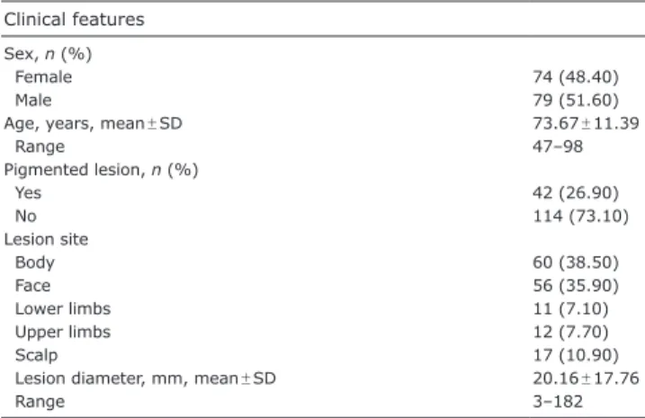
Table I. Characteristics of all the 156 considered lesions, developed on 153 patients, assessed retrospectively
Clinical features Sex, n (%)
Female 74 (48.40)
Male 79 (51.60)
Age, years, mean ± SD 73.67 ± 11.39
Range 47–98 Pigmented lesion, n (%) Yes 42 (26.90) No 114 (73.10) Lesion site Body 60 (38.50) Face 56 (35.90) Lower limbs 11 (7.10) Upper limbs 12 (7.70) Scalp 17 (10.90)
Lesion diameter, mm, mean ± SD 20.16 ± 17.76
Range 3–182
SD: standard deviation.
Table II. Pattern prevalence comparison between ulcerated and non-ulcerated portion in the 139 partially ulcerated lesions
Vascular pattern Ulcerated portion % Non-ulcerated portion % p-value Arborizing 16.5 83.5 0.000 Dotted 82.7 8.6 0.000 Linear-irregular 55.4 25.9 0.000 Comma 16.5 1.4 0.000
Diffuse reddish coloration 0 12.9 –
Hairpin 5.8 0.7 0.039
Glomerular 4.3 1.4 0.219
Crown 0.7 0 –
Polymorph 28.1 2.2 0.000
Significant values are given in bold.
40 50 60 70 80 90 100 % ulceratedportion
notulceratedportion
p=0.001* p=0.001* p=0.001* 0 10 20 30
Arborizing Dotted Linear
irregular Comma Erythema Hairpin Glomerular Crown Milky red areas Polymorph
p=0.001*
p=0.039*
p=0.219*
p=0.001*
A
cta
DV
A
cta
DV
A
dvances in dermatology and venereology
A
ctaD
ermato-V
enereologicaleast one of these classic criteria of BCC were clinically diagnosed correctly, compared with 61.1% of lesions without these criteria (χ2 = 24.59, p = 0.000).
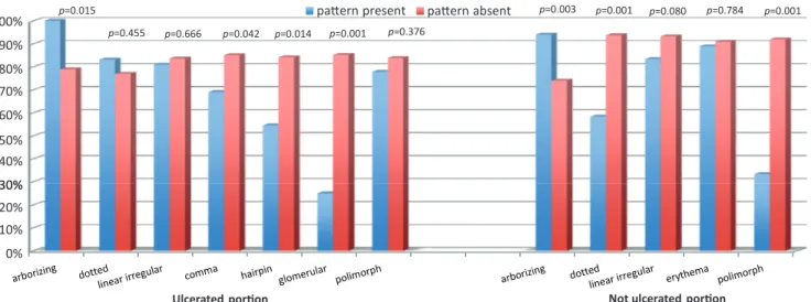
Presence of arborizing pattern in the ulcerated portion was associated with a correct diagnosis (Fisher’s exact test, p = 0.015). One-hundred percent of lesions with arborizing pattern were correctly diagnosed vs. 78.9% of lesions without that pattern. A similar association was evident for the non-ulcerated portion (Fisher’s exact test,
p = 0.008) (Fig. 2).
Correct diagnosis was also associated with absence of the dotted pattern in the non-ulcerated area (χ2 = 16.18,
p = 0.000); and absence of hairpin (χ2 = 6.08, p = 0.000)
and glomerular patterns were associated with correct diagnosis in the ulcerated areas (χ2 = 18.64, p = 0.000).
Logistic regression analysis, performed on the 139 lesions with ulcerated and non-ulcerated portions, also revealed that the vascular features influencing the correct diagnosis most were: presence of arborizing pattern in the ulcerated portion; absence of dotted pattern in the non-ulcerated areas, and absence of hairpin and glomerular patterns in the ulcerated areas, which are those commonly associated with melanoma and/or SCC.
Lesion size was not associated with any of the factors studied, while there was a significant association between the presence of spoke-wheels and the upper limb site (χ2 = 12.53, p = 0.014). No differences in the lesions’
cha-racteristics were observed between males and females.
Totally ulcerated lesion
Concerning the 17 totally ulcerated lesions, the dotted and linear-irregular dermoscopic patterns were the most frequent (88.2% and 70.6%, respectively). The comma and polymorph patterns occurred in ~35% of these le-sions. Other patterns were rare (hairpin and glomerular) or non existing.
Fig. 3 describes the association between the clinical
diagnosis and the prevalence of vascular patterns in the
17 totally ulcerated lesions. Ten out of 17 lesions were blindly diagnosed as malignant melanoma; 5 out of 17 as SCC; and 2 out of 17 as BCCs. In the group mis-diagnosed as melanoma, 9 out of 10 lesions presented the dotted pattern, 4 the linear-irregular pattern, and 3 the polymorph pattern.
All lesions
Considering all 156 lesions, the correct clinical diagnosis was associated with the type of lesion (totally or partially ulcerated); in particular, 90.6% of partially ulcerated le-sions were correctly diagnosed with clinical-dermoscopic examination, compared with 11.8% of totally ulcerated lesions correctly diagnosed (χ2 = 64.00, p = 0.000).
DISCUSSION
This study confirms a significant correlation between the correct clinical diagnosis and known classic dermoscopic characteristics of BCC, such as arborizing vessels, large
30% 40% 50% 60% 70% 80% 90%
100% p=0.015 pattern present pattern absent
p=0.042 p=0.014 p=0.001 p=0.455 p=0.666 p=0.003 p=0.001 p=0.080 p=0.784 p=0.376 p=0.001 0% 10% 20% 30%
Ulcerated portion Not ulcerated portion
Fig. 2. Percentage of correct clinical-dermoscopic diagnosis in relation to presence/absence of different patterns in the 139 partially ulcerated lesions.
Fig. 3. Clinical-dermoscopic diagnosis and type of patterns blindly observed in the 17 totally ulcerated basal cell carcinoma lesions.
4 5 6 7 8 9 dotted linear irregular comma hairpin glomerular polymorph 0 1 2 3
A
cta
DV
A
cta
DV
A
dvances in dermatology and venereology
A
ctaD
ermato-V
enereologicablue-grey ovoid nests, leaf-like areas, multiple blue-grey globules, and spoke-wheel areas (2–12).
More interestingly, in the 139 lesions presenting both an ulcerated and non-ulcerated area, significant dif-ferences in the prevalence of certain vascular patterns were noted between the 2 areas. In particular, the dotted, linear-irregular, hairpin, comma and polymorph patterns were highly represented in the ulcerated areas, whereas the arborizing pattern was prevalent, as expected, in the non-ulcerated areas (Table II, Fig. 1).
Considering exclusively the 17 BCCs that were com-pletely ulcerated, these lesions lacked the classic der-moscopic criteria of BCC (leaf-like, spoke-wheels, ovoid nest, blue-grey globules, arborizing vascular pattern), while showing a distinct prevalence of dotted, linear-irregular and polymorph vascular patterns (Figs 3 and 4). Of the lesions presenting both an ulcerated and a non-ulcerated portion, 90.6% were diagnosed correctly through clinical and dermoscopic examination (Fig. 4); conversely, only 11.8% of completely ulcerated lesions were diagnosed correctly (Fig. 5).
The results of this study raise concern about a high percentage of incorrect diagnosis in those lesions (his-tologically proven as BCCs) that were, both clinically and dermoscopically, blindly interpreted as different malignant neoplasms, melanoma in particular. In fact, in case of completely ulcerated BCCs lacking specific dermoscopic criteria, and presenting vascular patterns traditionally associated with melanoma and/or SCC (Fig. 5) (16, 17), the clinician simply lacks the means to correctly identify a BCC, and is driven towards an incorrect diagnosis of melanoma or SCC. This results in overdiagnosis of SCC/melanoma and in more urgent and radical surgical removal; this, of course, does not pose
any particular concern to the patient management (except perhaps increased patient psychological stress and un-necessary increased burden of urgent surgical removals), as the lesion will be ultimately excised. However, from a clinical and scientific aspect, this has long remained a grey area. To the best of our knowledge there are no reported data in the literature describing dermoscopic vascular patterns in ulcerated cutaneous BCC. Of note, our results pertain only to ulcerated histologically pro-ven BCCs; we cannot assume that they apply to other ulcerated lesions of a different nature.
Study limitation
The limitation of this study is its retrospective nature; further controlled studies are warranted to confirm our preliminary results.
The authors declare no conflicts of interest.
REFERENCES
1. Roewert-Huber J, Lange-Asschenfeldt B, Stockfleth E, Kerl H. Epidemiology and etiology of basal cell carcinoma. Br J Dermatol 2007; 157: 47–51.
2. Altamura D, Menzies SW, Argenziano G, Zalaudek I, Soyer HP, Sera F, et al. Dermatoscopy of basal cell carcinoma: morphologic variability of global and local features and ac-curacy of diagnosis. J Am Acad Dermatol 2010; 62: 67–75. 3. Lallas A, Apalla Z, Argenziano G, Longo C, Moscarella E, Specchio F, et al. The dermatoscopic universe of basal cell carcinoma. Dermatol Pract Concept 2014; 4: 11–24. 4. Popadić M. Dermoscopic features in different morphologic
types of basal cell carcinoma. Dermatol Surg 2014; 40: 725–732.
5. Seidenari S, Bellucci C, Bassoli S, Arginelli F, Magnoni C, Ponti G.High magnification digital dermoscopy of basal cell carcinoma: a single-centre study on 400 cases. Acta Derm Venereol 2014; 94: 677–682.
6. Popadić M. Statistical evaluation of dermoscopic features in basal cell carcinomas. Dermatol Surg 2014; 40: 718–724. 7. Puig S, Cecilia N, Malvehy J. Dermoscopic criteria and
ba-sal cell carcinoma. G Ital Dermatol Venereol 2012; 147: 135–140.
8. Argenziano G, Zalaudek I, Corona R, Sera F, Cicale L, Petrillo
Fig. 4. Handheld dermoscopy of a partially ulcerated basal cell carcinoma of the trunk in a 62-year-old man. Notice the arboriform
pattern on the right (non-ulcerated portion) and the polymorph pattern (dotted, linear irregular, comma) on the left (ulcerated portion) (magnification x20).
Fig. 5. Digital dermoscopy image of a completely ulcerated basal cell carcinoma of the left forearm in an 80-year-old woman. Prevalent
A
cta
DV
A
cta
DV
A
dvances in dermatology and venereology
A
ctaD
ermato-V
enereologicaG, et al. Vascular structures in skin tumors: a dermoscopy study. Arch Dermatol 2004; 140: 1485–1489.
9. Kreusch JF. Vascular patterns in skin tumors. Clin Dermatol 2002; 20: 248–254.
10. Micantonio T, Gulia A, Altobelli E, Di Cesare A, Fidanza R, Riitano A, et al. Vascular patterns in basal cell carcinoma. J Eur Acad Dermatol Venereol 2011; 25: 358–361.
11. Zalaudek I, Kreusch J, Giacomel J, Ferrara G, Catricalà C, Argenziano G. How to diagnose nonpigmented skin tumors: a review of vascular structures seen with dermoscopy: part II. Nonmelanocytic skin tumors. J Am Acad Dermatol 2010; 63: 377–386.
12. Menzies SW, Westerhoff K, Rabinovitz H, Kopf AW, McCarthy WH, Katz B. Surface microscopy of pigmented basal cell carcinoma. Arch Dermatol 2000; 136: 1012–1016. 13. Giacomel J, Zalaudek I. Dermoscopy of superficial basal cell
carcinoma. Dermatol Surg 2005; 31: 1710–1713.
14. Scalvenzi M, Lembo S, Francia MG, Balato A. Dermoscopic patterns of superficial basal cell carcinoma. Int J Dermatol 2008; 47: 1015–1018.
15. Sakakibara A, Kamijima M, Shibata S, Yasue S, Kono M, Tomita Y. Dermoscopic evaluation of vascular structures of various skin tumors in Japanese patients. J Dermatol 2010; 37: 316–322.
16. Warszawik-Hendzel O, Olszewska M, Maj M, Rakowska A, Czuwara J, Rudnicka L. Non-invasive diagnostic techniques in the diagnosis of squamous cell carcinoma. J Dermatol Case Rep 2015; 9: 89–97.
17. Chappuis P, Duru G, Marchal O, Girier P, Dalle S, Thomas L. Dermoscopy: a useful tool for general practitioners in melanoma screening: a nationwide survey. Br J Dermatol 2016; 175: 744–750.