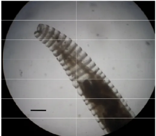
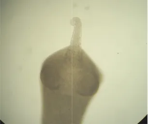
Fig.1.Male of Uncinaria stenocephala Bar = 100 µm
Fig.3.Male of Molineus legerae Bar = 50 µm
Fig.5.Angiostrongylus vasorum an infected fox (arrows)
Uncinaria stenocephala (bursa). Bar = 100 µm
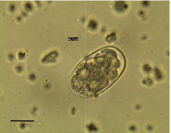
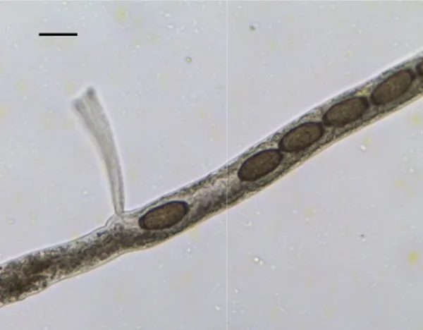
Fig. 2. Ancylostomatidae egg Bar = 100 µm
Molineus legerae (bursa). Bar = 50 µm
Fig. 4. Caudal end of
legerae. Bar = 20 µm
Angiostrongylus vasorum in the heart of an infected fox (arrows)
Fig. 6.Male of Angiostrongylus vasorum (bursa). Bar = 50 µm
Ancylostomatidae egg. Bar = 100 µm
. Caudal end of a female of Molineus Bar = 20 µm
Angiostrongylus vasorum ). Bar = 50 µm
Fig.7.First stage larva of vasorum. Bar = 50 µm
Fig.9. First stage larva of Bar = 50 µm
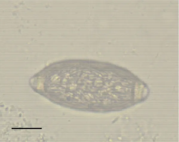
Fig.11. Egg of Toxa Bar = 25 µm
First stage larva of Angiostrongylus . Bar = 50 µm
Fig. 8. Cephalic part of (adult). Bar = 20
First stage larva of Crenosoma vulpis. Bar = 50 µm
Fig.10. Egg of Toxocara canis Bar = 25 µm
ascaris leonina. Bar = 25 µm
Fig.12. Apical part of
part of Crenosoma vulpis Bar = 200 µm
Toxocara canis. Bar = 25 µm
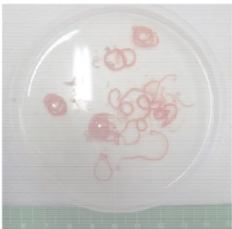
Fig.13.Adult of Pterygodermatites affini
Fig.15. Parasitic nodules due to Spirocerca lupi infection (stomach of a fox)
Fig.17. Microfilaria of Acanthocheilonema reconditum from a positive dog (Knott test).
Bar = 50 µm
Pterygodermatites affinis Fig. 14. Adults of
Parasitic nodules due to Spirocerca infection (stomach of a fox)
Fig. 16. Eggs of Bar = 20 µm.
Acanthocheilonema from a positive dog (Knott test).
0 µm
Fig.18. Microfilariae of
reconditum and Dirofilaria repens (below). Bar = 50 µm
Adults of Spirocerca lupi
Eggs of Spirocerca lupi. 20 µm.
e of Acanthocheilonema and Dirofilaria repens (below).
Fig.19. Aonchotheca putorii Bar = 30 µm.
Fig.21 Adult of Eucoleus aerophilus trachea. Bar = 500 µm.
Fig.23. Female of Eucoleus boehmi in the uterus. Bar = 30 µm.
Aonchotheca putorii eggs in utero. 30 µm.
Fig.20 Egg of Aonchotheca putorii Bar = 20 µm.
Eucoleus aerophilus in the . Bar = 500 µm.
Fig. 22 Egg of Eucoleus aerophilus examination). Bar
Eucoleus boehmi with eggs . Bar = 30 µm.
Fig.24. Egg of Eucoleus boehmi flotation). Bar = 30 µm.
Aonchotheca putorii. Bar = 20 µm.
Eucoleus aerophilus (lung ). Bar = 20 µm.
Eucoleus boehmi (coprological Bar = 30 µm.
Fig. 25. Pearsonema plica eggs. Bar = 50 µm.
Fig. 27.Male of Trichuris vulpis
Fig. 29.Egg of Calodium hepaticum
liver parenchima.
Pearsonema plica female, vulva and eggs. Bar = 50 µm.
Fig. 26. Eggs of Pearsonema plica uterus. Bar =
Trichuris vulpis (spicule). Fig. 28.Egg of
Bar = 30 µm
Calodium hepaticum in the Bar = 20 µm.
Fig. 30. Egg of Hymenolepis diminuta.
Bar = 30 µm
Pearsonema plica in the uterus. Bar = 30 µm
of Trichuris vulpis. Bar = 30 µm
Hymenolepis diminuta. Bar = 30 µm
Fig. 31. Scolex of Taenia polyacantha
Fig. 33. Egg capsule of Dipylidium caninum (coproscopy of a dog).
Fig. 35. Scolex of Mesocestoides
Taenia polyacantha Fig. 32. Scolex of
Dipylidium caninum (coproscopy of a dog).
Fig. 34. Proglottids of a Dilepididae.
Mesocestoides spp. Fig. 36. Proglottids of
Scolex of a Dilepididae.
Proglottids of a Dilepididae.