UNIVERSITÀ DEGLI STUDI DI ROMA
"TOR VERGATA"
FACOLTA' DI MEDICINA E CHIRURGIA
DOTTORATO DI RICERCA IN
MICROBIOLOGIA MEDICA ED IMMUNOLOGIA
XX CICLO
"Transcriptional regulation by thymosin alpha-1
in peripheral blood monuclear cell from
HIV positive patients"
Nome e Cognome
ANTONELLA MINUTOLO
Docente Guida: Dott.ssa Beatrice Macchi
Coordinatore: Prof. Enrico Garaci
INTRODUCTION
THYMOSIN α 1: history and biology
The isolation from the thymus gland of the thymosins, a family of biologically active molecules with hormone-like properties, was first described in 1966 by AL Goldstein and A White. Since that time, significant progress has been made in understanding the role of the thymosins in immunity and in the nature of the growth factors, cytokines and chemokines that they modulate. The thymosins include a family of biochemically and functionally distinct polypeptides with clinically important physiological properties. In the early 1970s, preclinical studies establishing the immunorestorative effects of a partially purified thymosin preparation termed thymosin fraction 5 (TF5) provided the scientific foundation for the first clinical trials with TF5 in 1974. TF5 was effective in stimulating the immune system in children with DiGeorge syndrome and other thymic dysplasias. These trials led to further interest in the active components in TF5 and to the chemical characterization of the biologically active thymosins. Several of these molecules are showing significant clinical promise in cancer, infectious diseases and wound healing (Goldstein et al 2004).
TF5 consists of a family of at least 40 mostly small acidic polypeptides, with molecular weights ranging from 1000 to 15,000 Dalton. Several of these polypeptides contribute individually to the biological activity of the parent compound: polypeptide beta 1, thymosin alpha 1 (Tα1), prothymosin alpha 1, parathymosin and thymosin beta 4 have been characterized chemically and biologically (Low et al., 1979).
Thymosin α1 is a 28 amino acid biologically active proteincleaved from positions 2–29 of a precursor protein known as prothymosin. This is a nuclear protein present in all mammalian cells with unknown biological activity and is believed to be strongly associated with cell
proliferation, transcription, processing and export of m-RNA from the nucleus (Tao et al., 1999).
Thymosin α has been widely studied both in vivo and in vitro and is currently known as lymphokine or thymic hormone since it is produced by lymphocytes and thymic epithelium. Like many cytokines, Thymosin α is a an immune modulating pleiotropic peptide.
The activities associated with Tα1 include:
1) The influence in the maturation of T cells through induction of differentiation markers;
2) Induction of various cytokines secretion, including MIF, INF-γ, IL-2 and its receptor (CD25) on natural killer cells (NK), T lymphocytes and blood monucleate cells (Svedersky et al., 1982; Sztein et al., 1986; Oates et al., 1989);
3) It was shown that Tα1 stimulate maturation of stem cell CD34+ in CD4+ CD3+ cells in an experimental model in vitro co-culture with thymic epithelium, inducing the synthesis of IL-7 (Knutsen et al. 1999), indicating the role of Tα1 in the maturation and differentiation of hematopoietic cells.
Therapeutic application of Tα1
Tα1has shown a variety of effects on cells and pathways of theimmune system, its central role in modulating dendritic cell(DC) function has only recently been appreciated. As DCs have the ability to sense infection and tissue stress and to translate collectively this information into an appropriate immune response,an action on DCs would predict a central role for Tα1 in inducing different forms of immunity and tolerance. Ithas been shown the capacity of Tα1 of activating infected dendritic cells through Toll-like receptor signalling, thus influencingthe inflammation balance, and increasing the expression oftumour, viral, and
major histocompatibility complex (MHC) I antigens (Giuliani et al. 2000; Romani et al. 2007).
In vitro studies suggest that Tα1 may act via pathways commonlyused by various cytokines. This raises the possibility that Tα1 and cytokines may have synergistic activity through potentiation of cytokine activity by Tα1. The immunopharmacology of Tα1 predicts an important clinical role for Tα1 in the restoration of cellular immune activity when used in combination with cytokines in vivo (Naylor et al 2007). In this study patients who experience immune suppression due to the presenceof tumour, irradiation, and/or chemotherapy or aging of the hostwould most benefit from this treatment combination. Thymosin alpha1 is also able to potentiate the action of cytokines reducing the hematological toxicity of cytotoxic drug therapy (cyclophosphamide-, 5-fluorouracil-, dacarbazine- or ifosfamide-based regimens) and this demonstrate the mechanism of action of Tα1 and its role as an immune system enhancer (Billich, 2002).
Tα1 plays an important role as an in vivo antitumoral in experimental models of animals and humans, alone or in combination with other commonly used anticancer.
It has been emphasized that combination therapy of Tα1 and low doses of interferon (IFN) or interleukin 2 (IL-2) carries a positive effect in the reconstitution of the immune response inhibiting tumour growth and / or cytostatic drugs (Favalli et al., 1989; Mastino et al., 1991; Mastino et al., 1992; Pica et al., 1998).
In combination therapies, Tα1 markedly enhances the effects of anticancer chemotherapy reducing the toxic effects of these drugs in a Lewis lungcarcinoma animal model (Garaci et
al., 2000; Garaci et al., 2003), using a combination of thymosin alpha1 (Tα1) and interferon (IFN) after cyclophosphamide. The efficacy of the combination therapywith Tα1 and either IFN or IL-2 in addition to chemotherapy has confirmed in preclinical models cancer such as
et al. 1994) where Tα1 has shown antitumor effects. In the last decade many studies have also highlighted the importance of using biological response modifiers (BRM) in the treatment of different types of immunological dysregulation, related to age, cancer or infectious diseases (Garaci et al. 1997, Garaci et al. 2003).
Immunotherapy using Tα1 has been found effective in infectious disease control (Rasi et al, 1996; Garaci et al. 1994). The combinationof Tα1 and IFN-α was also used in patients with chronic hepatitis B and C including IFN-non responders and patients infected with precore mutants or genotype 1b (Rasi et al, 1996). Further studies demonstratedadditional biological activities clarifying the mechanism ofaction of Tα1, partially explaining the synergism with IFN. Basedon these information, two clinical trials are ongoing: a largephase II study on advanced melanoma patients treated with Tα1 at differentdoses after dacarbazine and a phase III study, on IFN-resistant hepatitis C virus (HCV) patients treated with a triple (IFN, ribavirin, and Tα1)combination (Rasi et al., 2003, Garaci E. et al 2007; Camerini et al 2007). By July 2001, Tα1 was in phase III trials in the USA in combination with PEGylated interferon-alpha and later the same month it was approved in the Philippines as a monotherapy or in combination with interferon, in the treatment of HCV infection.
Tα1 has been launched in Argentina, China, Peru, the Philippines and Singapore for the treatment of chronic HBV infection.
The effects of combination therapy with Tα1 and natural human lymphoblastoid interferon-alpha were also investigated in human immunodeficiency virus infection. The effects of combination therapy with Tα1, interferon-alpha and zidovudine in patients with CD4+ lymphocytes ranging from 200 to 500/mm3 was analyzed in a randomized non-blinded study and found that the treatment was well tolerated after 12 months of therapy and was associated with a substantial increase in the number and function of CD4+ T cells. In vitro this combination treatment resulted in synergistic stimulation of the cytotoxic activity against
natural killer-sensitive target cells of lymphocytes collected from human immunodeficiency virus-infected donors and did not interfere with the antiviral activity of zidovudine. A similar effect was not observed in human immunodeficiency virus patients treated with zidovudine alone or associated with single agents (Garaci et al., 1994).
It was also identified that prothymosin alpha (ProTα)in HVS transformed CD8+ T cells wasa potent inhibitor of HIV-1 replication following proviral integration. These data suggest that the anti-HIV-1activity of ProTα is mediated through the modulation of a numberof genes that have been reported to suppress HIV-1 replication including the dysregulation of transcription factors and theinduction of PKR and Rantes mRNA (Mosoian et al., 2007). There is also a pilot study of the safety and efficacy of thymosin alpha 1 in augmenting immune reconstitution in HIV-infected patients with low CD4+ cells counts taking highly active antiretroviral therapy (Chadwick et al., 2003).
The Tα1 has been used as adjuvant during vaccinations. An example of clinical efficacy has been shown in vaccination with influenza vaccine in the elderly (Ershler et al., 2007).
In addition, there are cases in the literature of vaccination in animal models supported by Tα1 and prothymosin α 1, which has confirmed the ability of peptides to function as thymic adjuvant in immune response (Shmelev et al., 1994; Shiau et al., 2001).
Tα1 is thought to activate and increase T cells at the site of immunization and in the draining lymph nodes, these observations have gained interest in the clinical study of thymic peptides, especially in view of the possibility of considerable strengthening of innate immune response (Naylor and Hadden, 2003).
A previous study using microarray system to analyse the expression of 8300 genes in peripheral blood mononuclear cells (PBMCs) from healthy human donors after 48 hours of exposure to the Tα1, have shown that Tα1 induces an elevated transcriptional response in PBMCs (Garaci et al., 2003). These results could explain the mechanisms through which Tα1
increases the immune response and exerts its action, especially when used in combination therapy, for the treatment of cancer or infectious diseases.
The use of Tα1, as immunotherapeutic treatment in combination with conventional drugs, has shown that it was an important lymphokine for immune restoration without substantial toxic effects for the patient and earning an in-depth study of the mechanisms that govern their activities.
Human immunodeficiency virus type 1 (HIV-1)
The first cases of the acquired immune deficiency syndrome (AIDS) were reported in 1981 but it is now clear that cases of the disease have been occurring unrecognized for at least 4 years before its identification. The disease is characterized by an increased susceptibility to infections caused by opportunistic pathogens or by the occurrence of an aggressive form of Kaposi's sarcoma or B-cell lymphoma, accompanied by a profound decrease in the number of CD4+ T cells. As it seemed to be spread by contact with body fluids, it was early suspected to be caused by a new virus. By 1983 the agent now known to be responsible for AIDS, called the human immunodeficiency virus (HIV), was isolated and identified (Barre-Sinoussi, 1996). The initial infection with HIV generally occurs after transfer of body fluids from an infected person to an uninfected one. The virus is carried in infected CD4+ T cells, dendritic cells, and macrophages, and as a free virus in blood, semen, vaginal fluid and milk. Most patients who are infected with HIV will eventually develop AIDS, after a period of apparent quiescence of the disease known as clinical latency or the asymptomatic period (Figure A).
Figure A The course of HIV infection
This period is not silent, however, there is persistent replication of the virus and gradual decline in the function and numbers of CD4+ T cells. At this point, which can occur anywhere between 2 and 15 years or more after the primary infection, the period of clinical latency ends and opportunistic infections begin to appear. However, it has become increasingly clear that the course of the disease can vary widely. Thus, although most people infected with HIV go on to develop AIDS and ultimately to die of opportunistic infection or cancer, this is not true for all individuals (Baltimore, 1995; McCune, 1995).
Biology of HIV
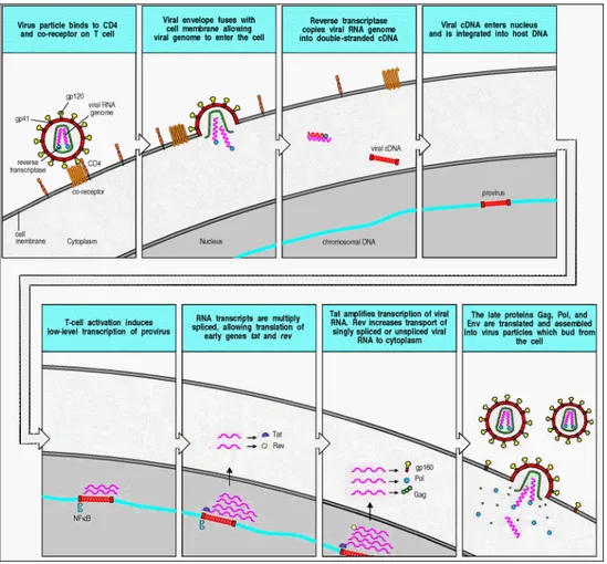
One of the proteins that enters the cell with the viral genome is the viral reverse transcriptase, which transcribes the viral RNA into a complementary DNA (cDNA) copy. The viral cDNA is then integrated into the host cell genome using viral integrase, which also enters the cell with the viral RNA. The infectious cycle up to the integration of the provirus is shown in
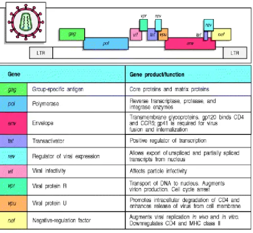
provirus, as shown in the next section. However, HIV can, like other retroviruses, establish a latent infection in which the provirus remains quiescent. This seems to occur in memory CD4+ T cells and in dormant macrophages, and these cells are thought to be an important reservoir of infection. The entire HIV genome consists of nine genes flanked by long terminal repeat sequences (LTRs), which are required for the integration of the provirus into the host cell DNA and contain binding sites for gene regulatory proteins that control the expression of the viral genes (Figure C). Like other retroviruses, HIV has three major genes gag, pol, and
env. The gag gene encodes the structural proteins of the viral core, pol encodes the enzymes
involved in viral replication and integration, and env encodes the viral envelope glycoproteins. The gag and pol mRNAs are translated to give polyproteins chains that are cleaved by the viral protease (also encoded by pol) into individual functional proteins. The product of the env gene, gp160, has to be cleaved by a host cell protease into gp120 and gp41, which are then assembled as trimers into the viral envelope. HIV has six other, smaller, genes encoding proteins that affect viral replication and infectivity in various ways (Kinoshita, 1997, Cullen 1998). The production of infectious virus particles from an integrated HIV provirus is stimulated by a cellular transcription factor that is present in all activated T cells. Activation of CD4+ T cells induces the transcription factor NFκB, which binds to promoters not only in the cellular DNA but also in the viral LTR, thereby initiating the transcription of viral RNA by the cellular RNA polymerase. This transcript is spliced in various ways to produce mRNAs for the viral proteins. The Gag and Gag-Pol proteins are translated from unspliced mRNA; Vif, Vpr, Vpu, and Env are translated from singly spliced viral mRNA; Tat, Rev, and Nef are translated from multiply spliced mRNA. At least two of the viral genes,
tat and rev, encode proteins, Tat and Rev respectively, that promote viral replication in
activated T cells. Tat is a potent transcriptional regulator, which functions as an elongation factor that enables the transcription of viral RNA by the RNA polymerase II complex. Tat encloses two binding sites, contained in one domain, named the transactivation domain.
Figure C The Genome of HIV
The first of these allows Tat to bind to a host cellular protein, cyclin T1. This binding reaction promotes the interaction of the Tat protein through the second binding site in its transactivation domain to an RNA sequence in the LTR of the virus known as the transcriptional activation region (TAR). The consequence of this interaction greatly enhances the rate of viral genome transcription, by causing the removal of negative elongation factors that block the transcriptional activity of RNA polymerase II. The expression of cyclin T1 is greatly increased in activated compared with quiescent T lymphocytes. This, in conjunction with the increased expression of NFκB in activated T cells, may explain the ability of HIV to lie dormant in resting T cells and replicate in activated T cells (Cullen, 2000; Cullen, 1998; Emerman and Malim, 1998; Fujinaga et al., 1999; Kinoshita et al., 1997; Subbramanian and Cohen, 1994; Trono, 1995).
HIV infection: CD4+ T cells, dendritic cells, and macrophages
The ability of HIV to enter particular types of cell, known as the cellular tropism of the virus, is determined by the expression of specific receptors for the virus on the surface of those cells. HIV enters cells by means of a complex of two noncovalently associated viral glycoproteins, gp120 and gp41, in the viral envelope. The gp120 portion of the glycoprotein complex binds with high affinity to the cell-surface molecule CD4. This glycoprotein thereby draws the virus to CD4 T cells and to dendritic cells and macrophages, which also express some CD4. Before fusion and entry of the virus, gp120 must also bind to a co-receptor in the membrane of the host cell. Several different molecules may serve as a co-receptor for HIV entry, but in each case they have been identified as chemokine receptors.
Chemokine and HIV-1: interactions and clinical implications.
Chemokines are small peptides that are potent activators and chemoattractants for leukocyte subpopulations and some non-hematopoietic cells. These chemotactic cytokines belong to the chemokine superfamily, which can be divided into 4 groups or families (CXC, CX3C, CC, and C) based on a cysteine motif (Table 1). Their actions are mediated by a family of 7-transmembrane G-protein-coupled receptors (Onuffer and Horuk, 2002). Chemokines are known to function as regulatory molecules in leukocyte maturation, trafficing, homing of lymphocytes and in the development of lymphoid tissues. Besides these functions in the immune system, they also play a critical role in many pathophysiological processes such as allergic responses, infectious and autoimmune diseases, angiogenesis, inflammation, tumor growth and hematopoietic development (Onuffer and Horuk, 2002). Alteration and inappropriate expression of chemokine receptors is thought to be implicated in multiple
addition, certain chemokines and their receptors are involved in HIV pathogenesis. The discovery of chemokine receptors as co-receptors for HIV-1 has opened the door for a number of novel anti-viral approaches. Two chemokine receptors, known as CCR5, which is predominantly expressed on dendritic cells, macrophages, and CD4+ T cells, and CXCR4, expressed on activated T cells, are the major co-receptors for HIV. After binding of gp120 to the receptor and co-receptor, the gp41 causes fusion of the viral envelope and the plasma membrane of the cell, allowing the viral genome and associated viral proteins to enter the cytoplasm. There are different variants of HIV, and the cell types that they infect are determined to a large degree by the chemokine receptor they bind as co-receptor. The variants of HIV that are associated with primary infections use CCR5, which binds the CC chemokines RANTES, MIP-1α, and MIP-1β, as a co-receptor, and require only a low level of CD4 on the cells they infect. These variants of HIV infect dendritic cells, macrophages, and T cells in vivo. However, they are often described simply as ‘macrophage-tropic' because they infect macrophage but not T-cell lines in vitro and the cell tropism of different HIV variants was originally defined by their ability to grow in different cell lines. In contrast, ‘lymphocyte-tropic' variants of HIV infect only CD4+ T cells in vivo and use CXCR4, which binds the CXC chemokine stromal-derived factor-1 (SDF-1), as a co-receptor. The lymphocyte-tropic variants of HIV can grow in vitro in T-cell lines, and require high levels of CD4 on the cells that they infect. Infection of CD4+ T cells via CCR5 occurs early in the course of infection and continues to occur, with activated CD4+ T cells accounting for the major production of HIV throughout infection. Late in infection, in approximately 50% of the cases, the viral phenotype switches to a T-lymphocyte-tropic type that utilizes CXCR4 co-receptors, and this is followed by a rapid decline in CD4+ T-cell count and progression to AIDS (Chan and Kim, 1998) and can be blocked by therapeutic antagonists of the CCR5 receptor.
Cytokines and HIV-1: interactions and clinical implications.
Cytokines play an important role in controlling the homoeostasis of the immune system (Table 2). Infection with HIV results in dysregulation of the cytokine profile in vivo and in
vitro. During the course of HIV-1 infection, secretion of T-helper type 1 (Th1) cytokines,
such as interleukin (IL-2), and antiviral interferon (IFN)-gamma, is generally decreased, whereas production of T helper type 2 (Th2) cytokines, such as IL-4, IL-10, proinflammatory cytokines (IL-1, IL-6, IL-8) and tumour necrosis factor (TNF)-alpha, is increased. Such abnormal cytokine production contributes to the pathogenesis of the disease by impairing cell-mediated immunity. A number of cytokines have been shown to modulate in vitro HIV-1 infection and replication in both CD4+ T lymphocytes and cells of macrophage lineage. HIV-inductive cytokines include: TNF-alpha, TNF-beta, IL-1 and IL-6, which stimulate HIV-1 replication in T cells and monocyte-derived macrophages (MDM), IL-2, IL-7 and IL-15, which upregulate HIV-1 in T cells, and macrophage-colony stimulating factor, which stimulates HIV-1 in MDM. HIV-suppressive cytokines include: IFN-alpha, IFN-beta and IL-16, which inhibit HIV-1 replication in T cells and MDM, and IL-10 and IL-13, which inhibit HIV-1 in MDM. Bifunctional cytokines such as IFN-gamma, IL-4 and granulocyte-macrophage colony-stimulating factor have been shown to have both inhibitory and stimulatory effects on HIV-1. The beta-chemokines, macrophage-inflammatory protein (MIP)-1alpha, MIP-1beta and RANTES are important inhibitors of macrophage-tropic strains of HIV-1, whereas the alpha-chemokine stromal-derived factor-1 suppresses infection of T-tropic strains of HIV-1 (Kedzierska and Crowe, 2001).
Highly active antiretroviral therapy (HAART)
Considerable progress has been made in the development of substances that interfere with the various stages of the replication cycle of HIV. Current anti-HIV therapeutic strategies were developed by the early 1990s, and they consist of at least three different low molecular weight inhibitors of the HIV enzymes: reverse transcriptase and the HIV protease. Used properly, these compounds are highly active against HIV. The first group of drugs encompasses both nucleoside and nucleotide analogues and the non-nucleoside reverse transcriptase inhibitors. The second group includes protease inhibitors of HIV-1 blocking the maturation of the viral precursor proteins, preventing the generation of viral proteins copies.
Used alone, these medicines determine the onset of mutants that can resist their action and it is for this reason that in therapy nucleoside inhibitors are associated with those of protease, such anti-HIV drug combination regimes have been generally referred to as HAART (for highly active antiretroviral therapy) .
HAART patients show a significant increase in the absolute number of CD4+ lymphocytes and CD8+, a reduction in the expression of molecules and activation of a transient improvement of immune response. The HAART has allowed us to make the transformation of AIDS into a chronic disease since it allows you to sets aside the plasma viremia for a long time, although it has some side effects such as toxicity and development of resistance (Richmann , 2006). There are, at present, 22 compounds which have been formally approved by the US Food and Drug Administration for the treatment of HIV infection (AIDS). According to their point of intervention with the HIV replicative cycle, these compounds can be classified in 5 categories: (1) NRTIs (nucleoside reverse transcriptase inhibitors): azidothymidine, didanosine, zalcitabine, stavudine, lamivudine, abacavir and emtricitabine; (2) NtRTIs (nucleotide reverse transcriptase inhibitors): tenofovir, administered as its oral prodrug form TDF (tenofovir disoproxil fumarate); (3) NNRTIs (non-nucleoside reverse transcriptase inhibitors): nevirapine, delavirdine and efavirenz; (4) PIs (HIV protease
inhibitors): saquinavir, ritonavir, indinavir, nelfinavir, amprenavir, lopinavir, atazanavir, fosamprenavir, tipranavir and darunavir; and (5) FIs (fusion inhibitors): enfuvirtide. Starting from the drugs which are currently available for the treatment of AIDS, numerous combinations could be envisaged. Drug combinations are, in principle, aimed at obtaining synergism between the compounds, reasonably expected if they act by different mechanisms, while reducing the likelihood for drug resistance development (De Clercq, 2007).
New therapeutic approaches
Currently, a number of compounds that act on new viral targets are being investigated including integrase inhibitors, which were found to be extremely toxic. There are ongoing clinical studies on antagonists of HIV coreceptors, while at an early stage of experimentation are compounds that interact with cellular factors that bind the LTR, and molecules which can act as regulators and inhibitors of cell transcriptional factor NF-kB and Tat viral protein (De Clercq, 2002). Information was already available on HIV fusion and the earlier event of HIV attachment, the last and first stages of entry, respectively (Gallo, 2002). Identifying and elucidating the mechanisms of action of naturally occurring HIV suppressive factors, led to the discoveries of RANTES, MIP-1α, and MIP-1β as HIV inhibitors. In addition, after the discoveries of the chemokine coreceptor usage in HIV, the design of agonists and antagonists of them represent a new avenue of therapy. Their identification help to lead to the discoveries of the HIV coreceptors (CCR5 and CXCR4), a major development in HIV pathophysiology. Other HIV suppressor factors exist and their identification and elucidation of their molecular mechanisms for inhibiting HIV will help to open other new avenues of therapy (Gallo, 2002).
Tα1 and HIV therapy
The possibility to combine antiretroviral chemotherapy with immunotherapy has been suggested years ago. The introduction of the highly active antiretroviral therapy (HAART), able to reduce the viral load in most of the patients, has remarkably reduced the mortality associated to HIV infection. The most relevant immunological consequence of HAART in responders patients is an appreciable quantitative and functional, al least in part, recovery of the CD4+ T cell compartment. Treatment of HIV-infected patients with HAART leads to immune reconstitution as shown by increases in CD4+ lymphocyte counts, decreased risk of opportunistic infections and improved survival (Palella et al., 1998; Valdez et al., 2001). However, a variable percentage of individuals exhibits a discordant response to HAART. Particularly, some patients show an unsatisfactory increase of CD4+ T cells, despite a successful virological response (immunological failure), with risk of serious clinical events. (Saravolatz et al., 1996; Floridia et al., 2002). These patients may benefit from the adjunctive use of immunomodulatory compounds that enhance immune reconstitution. In a previous clinical trials in HIV-infected patients, thymosin α1 was shown to induce increase in CD4+ cell counts, but only when given in combination with either interleukin (IL)-2 or interferon (IFN)-α (Garaci, 1994). These studies were conducted in patients taking zidovudine monotherapy and the efficacy of thymosin α1 in patients with full viral suppression on combination antiretroviral therapy is unknown. A pilot study (Chadwich et al., 2003) was conducted to determine the safety and efficacy of thymosin α1 injections in conjunction with combination antiretroviral therapy in patients with advanced HIV infection. It was demonstrated that thymosin α1 was very well tolerated in this group of patients, which is in agreement with other clinical trials of thymosin α1 in HIV-infected patients and in patients with hepatitis B and C infection (Lau et al., 2002). The good tolerability contrasts with immunomodulatory cytokines such as IFN-α or IL-2 that have been shown to have substantial toxicity in patients with HIV infection as well as other groups. No benefit of thymosin α1 in
augmenting CD4+ cell count response to HAART was demonstrated. It is conceivable that a higher dose of thymosin α1 might have a greater effect on CD4+ cells, although the dose used for the study was relatively high in comparison to previous studies in patients HIV infection and currently recommended dosage for use in the treatment of hepatitis virus infections. It is also possible that an effect might have become apparent after a longer duration of therapy. In one previous trial, thymosin alpha 1 (given in conjunction with IFN-α) had its greatest effect after 12 months of treatment (Garaci, 1994) in a group of patients with advanced immunodeficiency and it is possible that a greater CD4 response might be obtained in patients with higher initial CD4 counts. HAART has been shown to allow reconstitution of both subsets of T cells (Douek et al., 1998) and there is recent evidence that one mechanism for this effect is through production of de novo naive T lymphocytes in the thymus. Immunophenotyping studies were performed to assess the effect of thymosin 1α in stimulating expansion of naive and memory T cells (Chadwich et al., 2003). Some studies have suggested that impaired T cell restoration in patients with low CD4+ cell counts when starting HAART may result from impaired thymic function (Teixeira et al., 2002). The lack of any specific effect of thymosin 1α in this study, in terms of increases in numbers of naive or memory CD4+ or CD8+ T cells, did not support a role of thymosin in selectively stimulating these particular lymphocyte subtypes. The most interesting effect of thymosin was the significant increase in signal joint (sj) T cell receptor excision circles (TRECs) levels seen after 12 weeks of treatment. Increase in the de novo production of naive T cells by thymopoiesis may be an important component of immune reconstitution in HIV-infected patients taking HAART ,and sjTREC levels have been shown to predict survival in HIV-infected patients as well as the total CD4 count response to HAART after several years (Hatzakis et al., 2002; Pido et al.,2002; Mussini et al., 2002). Further studies are needed to investigate which genes can be regulated by Tα1 in HIV infected individuals in order to
obtain new information on transcriptional activity of Tα1 as adjuvant immunotherapy to HAART.
AIM OF THE STUDY AND EXPERIMENTAL DESIGN
Thymosin alpha-1 (Tα1)has shown a variety of effects on cells and pathways of theimmune system. Tα1 has been shown to exert an immunomodulatory activity towards both T cell and NK maturation. Moreover, Tα1 has been shown to act on effector functions of mature lymphocytes, such as cytokine production and cytotoxic activity. However, molecular mechanisms underlying the potent activity of Tα1 have not completely elucidated. Several clinical studies on patients with infectious or other diseases, have already demonstrated the high tolerability and the absolute safety of Tα1 (Garaci et al., 2000). In addition, Tα1 has the ability to activate infected dendritic cells through Toll-like receptor signalling, thus influencing the inflammation balance, and increasing the expression of tumour, viral, and major histocompatibility complex (MHC) I antigens (Giuliani et al., 2000; Romani et al., 2007). However, information regarding a possible use of Tα1 in patients with HIV infection is very limited. There is only one recent study, showing Tα1 utilization in combination with HAART, with encouraging results on T-cell compartment maturation (Chadwick et al., 2003).A previous study using a microarray to analyse the expression of 8300 genes in PBMCs from healthy human donors after 48 hours of exposure to the Tα1, has shown that Tα1 induced an elevated transcriptional response in lymphoid cells (Garaci al., 2003), thus giving a novel impetus in understanding the pleiotropic effects exerted by Tα1 and the molecular mechanisms involved in its action. The principal aim of the experiments performed in the framework of this Ph.D. thesis was to investigate which genes belonging to immune response pathways could be regulated by Tα1 in vitro in PBMCs from HIV infected individuals. This, to obtain new information on the effect of Tα1 on the
response in PBMCs from HIV+ patients and a possible helpfulness of the use Tα1 in combination with antiretroviral therapy for the control of HIV infection. To this purpose, a cohort of HIV+ individuals was enrolled in an open cross-sectional study by the “AIDS Center”, S. Giovanni Hospital, Rome. In vitro PBMCs from HIV+ individuals and from healthy donors were isolated. Fresh cells were used for flow cytometry analysis of surface molecule expression (CD4, CD8). Cells were then incubated with Tα1 (50µg/ml) for 48h to evaluate gene expression profile using the "Human Autoimmune & Inflammatory Response" microarray systems (SuperArray). Based on the results of microarray analysis, the expression of some selected genes, presumably modulated by Tα1, was quantified using quantitative Real Time PCR.
MATERIALS AND METHODS
Patients and samples
Eleven HIV-1-infected patients with different response to HAART, were enrolled in a open cross-sectional study by Department of Infectious and Tropical Disease, University of Rome “La Sapienza,”orthe “AIDS Center”, S. Giovanni Hospital, Rome. The characteristics of the patients are listed in the results (Table 4).
The study received the ethics approval by the institutions participating in the study and informed consent was obtained from the patients.
PBMCs from HIV+ individuals and from healthy HIV- donors were isolated from heparinized blood by centrifugation gradient according to standard methods. PBMC isolated were stimulated by Tα1 (50µg/ml, SciClone Pharmaceuticals International, USA) for 24 h in RPMI 1640 (Life Technologies) supplemented with 10% FCS (Life Technologies), 2 mM glutamine, 50 IU/mL penicillin, and 50 IU/mL streptomycin (Hyclone), and then readded for others 24h. Cells were then harvested, washed twice in PBS and used for flow cytometry analysis of cellular proteins and RNA preparation to evaluate gene profile expression.
Viral Load
HIV-1 RNA Viral Load (VL) levels were determined using a quantitative ultrasensitive reverse transcription-polymerase chain reaction assay (limit of detection was 50 HIV RNA copies/mL).
Immunostaining and flow cytometry analysis of cellular proteins
For the extracellular protein staining, approximately 5 x 10⁵ cells were resuspended in 50 µl of PBS with PE conjugated anti-human CD8 and PerCP conjugated anti-human CD4 (BD Bioscences) or irrelevant control antibody and incubated at 4°C for 30 min. After treatment, cells were washed twice in PBS and stored at 4°C. Flow cytometry analysis was performed by a FACScan flow cytometer and Cell QuestTM software.
Extraction of RNA
RNA isolation was performed using Trizol (Life Technologies, Grand Island, NY), according to the manufacturer’s instructions. The quantity and the quality of all RNA preparations were assessed by gel electrophoresis and OD260/OD280 ratios. RNA was used for microarray analysis as well as for quantitative reverse transcription-polymerase chain reaction (Real-time PCR).
Mycroarray
For these experiments it was used a Oligo GEArray Human Autoimmune & Infiammatory response microarray systems (SuperArray, Bethesda, MD). This Oligo GEArray profiles the expression of 440 genes involved in the autoimmunity and inflammation immune responses. It represents expression of cytokines, chemokines, and their receptors, as well as genes related to cytokine metabolism, production and cytokine-cytokine receptor interaction. Genes involved in the acute phase, inflammatory and humoral immune responses are also represented. Controls are represented by four potentially normalized genes: glyceraldehyde-3-phosphate dehydrogenase (GAPDH), peptidylprolyl isomerase A (PPIA), glucoronidase beta
(GUSB), and β-actin. Starting from total RNA (3,5 µg) it was utilized a proprietary and patent-pending linear RNA amplification and labelling procedure to synthesize labelled antisense RNA (aRNA) also known as labeled cRNA target. Total RNA was amplified, and cRNA probes weregenerated by biotin-16-dUTP (Roche Diagnostics, GmbH) incorporation using the AmpoLabeling-LPR Kit (SuperArray, Frederick, MD), according to the manufacturer'sinstructions.
The array membranes (SuperArray) were prehybridizedin GEAhyb (SuperArray) solution for 2 h at 60°C. Labeled cRNA probes were hybridized overnight at 60°C with continuous agitation. After two wash hybridized cDNA probes were detected by chemiluminescence. Images of the membranes were recorded on x-ray films (Amersham Bioscences, UK ltd) and weredigitally recorded on a personal densitometer scanner. Data were further processed with GEArray Analyzer software (http://www.superarray.com).All signal intensities were PUC18 plasmid (negative control) DNA background subtracted, and normalized to the housekeeping genes.
Relative gene expression levels were calculated as the ratio between the mean values of gene expression in PBMCs from HIV infected in patients or healthy donors treated with Ta1 and the mean values obtained from untreated control .
Changes in gene expression was expressed as a fold increase/decrease. The cut-off to express the fold induction or inhibition of gene expression in comparison with controls was 1.5 or 0.6 fold-changes respectively. Genes which suited both above criteria were considered to be induced or suppressed. Scatter plots were made from normalized signals.
Real-time PCR
Real-time PCR was performed on a ABI Prism 7300 instrument (PE Applied Biosystems, Foster City, CA, USA) with gene-specific primers and the SybrGreen protocol to confirm the gene expression changes observed in the microarray analysis. All primers employed were cDNA specific and were purchased from Primm (MI, Italy). Names and primers for all genes quoted in the text are listed in Table 3. An amount of 0.75 micrograms of total RNA from each sample was reverse-transcribed in a total volume of 20 µl with high-capacity cDNA Reverse Transcription Kits (Applied Biosystems), according to the manufacturer’s instructions.
Amplification of specific PCR products was detected using the SYBR Green PCR Real Master Mix (SuperArray). The RQ-PCR was performed in duplicated in a total reaction volume of 25 µL containing 11,25 µL SYBR Green PCR Master Mix, 150 nM forward and reverse primers, 10,25 µL dH2O, and 750 ng cDNA as a template. Samples were heated for 10 min at 95ºC and were subjected to 45 cycles of PCR amplification, each cycle consisting of 15s at 95ºC and 60s at 60ºC. Within each experiment, no template control (NTC) and housekeeping genes β-actin (ACTB), used as reference, were run in parallel to verify any contamination and to determine amplification efficiency. Each run was completed with a melting curve analysis to confirm the specificity of amplification and lack of unspecific products and primer dimers. Quantification was performed using the Ct comparative method, relative gene mRNA levels were calculated as follows: 2 - [ ∆Ct (Tα1) - ∆Ct (CTR) ] = 2 - ∆∆Ct , where ∆Ct (sample) = [Ct( gene target)−Ct(ACTB)] represents the difference, in threshold cycle number, between genes and ACTB.
Gene bank number name SEQUENCE 5'→3' NM_002988 CCL18 (PARC) FORWARD REVERSE CCAAGCCCCAGCTCACTCT TGCAAGGCCCTTCATGATG NM_002982 CCL2 (MCP-1) FORWARD REVERSE CAAGCAGAAGTGGGTTCAGGAT TCTTCGGAGTTTGGGTTTGC NM_002990 CCL22 (MDC) FORWARD REVERSE TGCCCTGGGTGAAGATGATT AAGGCCACGGTCATCAGAGT NM_002983 CCL3 (MIP1alpha) FORWARD REVERSE CCATGGCTCTCTGCAACCA GCGGTCGGCGTGTCA NM_021006 CCL3L1 (LD78 beta) FORWARD REVERSE GCTGACACTCGAGCCCACAT AGCAGTGGAGACCTGCATGAT NM_001001435 CCL4L1 FORWARD REVERSE CACCGCCTGCTGCTTTTC TAATCTACCACAAAGTTGCGAGGAA NM_002985 CCL5 (Rantes) FORWARD REVERSE AGCCTCTCCCACAGGTACCA GGCAGTAGCAATGAGGATGACA NM_005211 CSF1R FORWARD REVERSE CCTGCTGACTGTTGAGACCTTAGA CACTCCCCACGCTGTTGTG NM_000758 CSF2 FORWARD REVERSE TTCTGCTTGTCATCCCCTTTG CTCATCTGGCCGGTCTCACT NM_000760 CSF3R FORWARD REVERSE AGGCGATCTGCATACTTTAAGGA AACAAGCACAAAAGGCCATTG NM_022059 CXCL16 FORWARD REVERSE GGAGTCTCGCTCTGTCATCCA GTTGCAGTGAGCCGAGATCA NM_002341 LTB FORWARD REVERSE CACCCCGATATGGTGGACTT CCATCACGGCCCCAAAG NM_002415 MIF FORWARD REVERSE GCGGGTCTCCTGGTCCTT GCACGTTGGTGTTTACGATGA NM_002468 MYD88 FORWARD REVERSE GAGCTGGCGGGCATCAC TCGAAACGCTCAGGCATATG NM_003264 TLR2 FORWARD REVERSE GCGGGAAGGATTTTGGGTAA TGGTCTTAAATATGGGAACCTAGGA Table 3
Statistical Analysis
Statistical Analysis were done using SPSS (version 12.0, SPSS). For statistical comparisons of the laboratory data from HIV+ patients and HIV- individuals, the independent samples t-test analysis was performed; for statistical comparisons of the laboratory data within each patients group, the paired samples t-test analysis was performed; for non-parametric bivariate correlation, Spearman’s rho correlation coefficient was calculated. For statistical and graphical reasons, an arbitrary value of 50 HIV RNA copies/mL was assigned to samples under the limit of HIV-RNA detection and the numbers of HIV copies/mL were transformed as decimal logarithms.
RESULTS
Patients characteristics: virological and immunological status.
Eleven HIV-1-infected patients with different response to HAART, were enrolled in a open cross-sectional study on the effect of thymosin alpha 1 in vitro, at the Department of Infectious and Tropical Disease, University of Rome “La Sapienza,” and at the “AIDS Center”, S. Giovanni Hospital, Rome. For comparison, a group of HIV, HBV, HCV negative healthy donors were also enrolled in the study. The characteristics of the patients are shown in Table 4. The table list encompasses the age of the patients (average 41), the sex (3 female and 8 male), the HIV-1 RNA viral load (VL) levels (8 patients < 50 copies/ml; 1 patient < 5000 copies/ml; 1patient = 49915 copies/ml and 1 patient = 135000 copies/ml); the average CD4+ cell number was 587 (cells/mm3) and the average CD8+ cell number was 1163 (cells/mm3). As a percentage value, the mean CD4+ cells were 23,82% and CD8+ cells were 46,27%. All patients, except one, were under HAART during this study. The results, reported in Figure 1, show a statistically significant (p<0,001) inverse ratio of CD4+ and CD8+ peripheral blood mononuclear cells in HIV+ patients, while the healthy donors showed a normal phenotype. In fact, the percentages of positive cells, as detected by flow cytometry, in fresh PBMCs were: CD4+ = 41,94 ± 3,5 % and CD8+ = 38,52 ± 2,87 %, for healthy donors, and CD4+ = 23,81 ± 2,52 % and CD8+ = 46,27 ± 1,83 %, for HIV+ patients, respectively (mean ± standard error). As shown in Figure 2, in this group of patients VL levels, expressed as Log10 of the RNA copies number per mL, were significantly inversely correlated with levels of CD4+ cells per mm3 (Spearman’s rho= -723; p=0,018). Taking into consideration that most of the patients were under therapy and that the viral load was <50 copies/mL, results demonstrate a general, both virological and immunological, positive response to the therapy.
Patient Age Sex HIV-RNA (copies/ml) CD4 (cells/mm3) CD8 (cells/mm3) CD4 % CD8 % THERAPY 1 34 M 50 831 1220 32 47 LPV/r+3TC+DDI 2 44 F 50 624 1170 24 45 TDF+TMC114+R 3 52 M 50 513 1642 15 48 ATV200+DDI+3TC 4 46 M 50 571 1088 21 40 3TC+DDI+EFV 5 43 M 1300 511 1022 24 48 ATV150+ABC+3TC 6 43 M 50 829 800 41 38 3TC+AZT+LPV/r 7 44 F 49915 536 839 28 44 no therapy 8 26 M 50 687 1026 24 45 TPV+TDF+3TC 9 49 M 135000 106 791 9 46 LPV/r+3TC+DDI (no aderente) 10 33 M 50 674 2089 20 62 ATV/r+ddi+3TC 11 36 F 50 580 1108 24 46 TPV+TDF+3TC Table 4 Characteristics of patients.
Evaluation of the effects of thymosin alpha 1 on the transcriptional activity of PBMCs from HIV+ and HIV- individuals, by microarray assay
In order to provide new information on the effects of thymosin alpha 1(Tα1) in vitro on the transcriptional activity of PBMCs from HIV+ patients and to compare these effects on those exerted on PBMCs of healthy donors, the “Human Autoimmune and Inflammatory response” microarray system (SuperArray) was used. PBMCs from HIV+ or HIV- healthy donors were stimulated with Tα1 (50 µg/ml) for 48 h and the mRNA from different samples was used to perform the microarray analysis of 440 gene expression. The results were expressed as fold change with respect to corresponding untreated control samples, after subtraction of background and normalization to the housekeeping expression from either counterpart. The cytokine, chemokine and related receptor genes correspond to about 41% of the total genes present in the microarray. Other genes involved in cytokine and cytokine-receptor interaction represent another 15% of the total genes. The genes involved in humoral immune and inflammatory responses correspond to 12% and 32% of the total genes present in the microarray, respectively (Figure 3, panel a). The summary of percentage values of the Tα1-regulated genes in PBMCs from HIV+ patients according to their functional gene grouping is reported in Figure 3, panel b. Cytokine, chemokine and their receptor genes represented 24 % of the total genes modulated by Tα1, while other genes involved in cytokine-cytokine receptor interaction represented another 20%. The remaining genes modulated by Tα1 were grouped in the humoral immune response sub-group for 18% , and in the inflammatory response subgroup for 38 %.
Conversely, Figure 3, panel c shows the distribution of the Tα1-modulated genes in healthy donors. The percentage values of modulated genes were: 29% for cytokine, chemokine and receptor gene subgroup, 14% for the other genes involved in cytokine-cytokine receptor interaction, 16% for the humoral immune response subgroup and 41% for the inflammatory
response subgroup. These results show that the effects on the transcriptional response by thymosin alpha 1 were effective on genes belonging to the same functional groups in PBMCs from HIV+ as well as in HIV- donors.
The number of positively and negatively Tα1-modulated genes in PBMCs from HIV+ and in healthy donors is shown in Figure 4. Ninety-four genes were regulated in HIV + patients, of which 64% were up regulated and 36% were down regulated. In HIV- donors the total number of genes regulated were 74, of which 62% were positively modulated and 38% were negatively modulated. These results show that the number of genes modulated by Tα1 was relatively high in HIV+ as well as in HIV- donors, and that the distributions of positively and negatively modulated genes were comparable in the two groups.
The distribution of the 94 mRNAs whose transcription was significantly different in the presence of Tα1 in PBMCs from HIV infected patients, compared to untreated control samples, and selected on the basis for the family group, are listed in Tables 5-9. On the other hand, Tables 10-12 show the gene subgroup distribution of the 74 mRNAs modulated by Tα1 in PBMCs from healthy donors. Each table contains the gene names, a brief description of the function, the gene bank access number of the mRNA sequence, and the fold change. Particularly, the fold change represents the ratio between the mean values of gene expression in PBMCs from HIV+ patients or healthy donors treated with Tα1 and the mean values obtained from corresponding untreated control samples.
The effects of Tα1 treatment on the transcriptional response of PBMCs from HIV+ patient and HIV– donors is described in Figure 5-9. The relative gene expression levels is expressed as fold change.
The expression of several genes from cytokine, chemokine and their receptors were modulated (17 positively modulated and 13 negatively modulated). In HIV+ patients, genes as MCP-1(CCL2), MIP-1alpha (CCL3), LD78 beta (CCL3L1), CCL4L11, Rantes (CCL5),
PARC (CCL18), MDC (CCL22), interleukin 8 (IL8), CCR1, CCR7, CCRL2, IL4R, IL6R, IL10RA and IL10RB were positively modulated by Tα1, while eotaxin (CCL11), CXCL1, CMTM1, CCR2, CXCR6, IL5RA IL2RG, IFNA1-2 and LEFTY1- 2 were negatively modulated by the same treatment in vitro (Figure 5a). Several of these genes are also involved in the humoral and inflammatory response (Figure 7a and Figure 8), together with other genes such as interferon regulatory factor 7 (IRF7), myeloid differentiation primary response gene (MYD88), Allograft inflammatory factor 1 (AIF1), Arachidonate 5-lipoxygenase (ALOX5), Leukotriene A4 hydrolase (LTA4H), Toll receptor 2 (TLR2) and Toll receptor 8 (TLR8). Genes involved in cytokine-cytokine receptor interaction were also modulated (16 positively modulated and 9 negatively modulated); among the others, Colony stimulating factor 1 receptor (CSF1R), Colony stimulating factor 2 (CSF2), Colony stimulating factor 3 receptor (CSF3R), Lymphotoxin beta (LTB), Macrophage migration inhibitory factor (MIF) and CD74 were up regulated, while CD28 and chemokine receptor 1 (XCR1) were down regulated (Figure 6a). Within the inflammatory response group a high number of genes were regulated in PBMCs from HIV+ patients (34 positively modulated and 13 negatively modulated) (Figure 8). Tα1 induced an elevated transcriptional response in HIV+ as well as in HIV- donors, but only few mRNAs were expressed in both groups. In healthy donors, several different cytokines, chemokines and their receptors were modulated by Tα1 (15 positively modulated and 13 negatively modulated). Among the others, CCL7, CCL17, CCL19, MCP-1, CXCL2 and CXCL10 (Figure 5b). A number of these genes are also involved in the humoral immune response (Figure 7b) together with other genes such as CD86, IL7R and CD22. Genes involved in cytokine-cytokine receptor interaction were also regulated (8 positively modulated and 6 negatively modulated); among the others, lymphotoxin alpha (LTA), CD86 and IL5 were down regulated (Figure 6b). In the inflammatory response pathway, 21 genes were positively modulated and 17 were down
the transcriptional response of a high number of genes involved in the immune response. Interestingly, the transcriptional response to Tα1 treatment is elevated in HIV+ patients as well as in healthy donors, but the degree of modulation is different between the two groups, suggesting a differential effect owing to the basal condition of cellular activation of the PBMCs from HIV+ patients and healthy donors.
Gene expression confirmation by Real time PCR analysis
In order to confirm the results obtained by the microarray analysis, 15 genes which resulted as positively modulated by Tα1 in PBMCs from HIV+ patients were selected and their expression was quantified by Real-time-PCR analysis. These genes include the following: Eight chemokines (MCP-1 (CCL2), MIP 1alpha (CCL3), Rantes (CCL5), PARC (CCL18), MDC (CCL22), LD78 beta (CCL3L1), CCL4L1 and a small inducible cytokine B6 (CXCL16) and 7 genes involved in the inflammatory response and in cytokine-cytokine receptors interaction, such as Macrophage migration inhibitory factor (MIF), Colony stimulating factor 3 receptor (CSF3R), Colony stimulating factor 2 (CSF2), Colony stimulating factor 1 receptor (CSF1R), Lymphotoxin beta (LTB), Myeloid differentiation primary response gene (MYD88) and Toll receptor 2 (TLR2). Quantification was performed using the Ct comparative method and expressed as fold change Log2 (2-∆∆Ct). For statistical comparisons of the data within each group of individuals, the paired samples t-test analysis was performed. Figure 10a shows the results of the Real time PCR analysis for the quantitative evaluation of the expression of genes for chemokines in PBMCs from HIV+ patients after in vitro treatment with thymosin alpha 1. The results of the microarray assay for the gene expression of 4 chemokines were completely confirmed by statistical analysis of Real time PCR results: Rantes (mean fold change= 1,55; p=0,022), PARC (mean fold change= 0,67; p= 0,014), MDC (mean fold change= 0,91; p= 0,009) and CXCL16 (mean fold
change= 2,1; p= 0,041). The positive modulation of CCL4L1 by microarray was confirmed (fold change = 0,88), but the difference compared to the untreated control samples was not statistically significant (p>0.050). Conversely, expression of MIP-1 alpha transcripts was found unchanged (fold change = 0,05) and transcripts for two genes were negatively modulated by Tα1 by Real time analysis. These results didn’t confirm those obtained with the microarray assay. However, the values were not statistically significant (MCP-1, fold change = -0,54, and LD78 beta, fold change= -1,04 , p>0.050).
The expression of the same genes was analyzed in PBMCs from healthy donors treated with Tα1, using Real time PCR (Figure 10b). Three genes that were found to be positively modulated by the microarray assay were not confirmed by Real time PCR: MCP-1 (fold change = -1,14), Rantes (fold change= -0,43) and CCL4L1 (fold change = -1,8), but the fold change was not statistically significant (p>0.050). On the other hand, CXCL16 down modulation by microarray analysis, was confirmed by Real time PCR (fold change = -2,3), even if the difference with the untreated control was not statistically significant (p>0.050). Regarding to other analysed genes that showed no significant modulation by microarray, some of them were down modulated by Real time PCR: however, the difference with the untreated control samples was not statistically significant (p>0.050). These genes were: MIP1alpha (fold change = 0,85), PARC (fold change = 0,39), LD78 beta (fold change = -0,85) and MDC (fold change = -2,25). Figure 11a shows the Real time PCR results for gene expression of genes involved in the inflammatory response and in the cytokine-cytokine receptors interaction. In PBMCs from HIV+ patients treated with thymosin alpha 1, CSF2 and TLR2 expression was not regulated (respectively fold change = 0,058 and 0,06). The others selected genes that were positively modulated by microarray and were significantly up regulated also by Real time analysis were the following: CSF1R (fold change = 1,47; p= 0,039), CSF3R (fold change= 2,84; p= 0,005), LTB (fold change = 2,5; p=0,003), MYD88
healthy donors treated with Tα1, the same genes were analysed by Real time PCR (Figure 11
b). All genes were found to be not significantly modulated by Tα1 (p>0.050) except for the TLR2 (p=0,002). The Real time PCR analysis for CSF3R expression (fold change= -1,88) confirmed the result of the microarray assay. Conversely, the Real time PCR analysis for CSF1R gene expression (fold change = -1,14) did not confirm the result of the microarray. Two genes analysed that were not significantly modulated by microarray, were found to be up regulated by Real time PCR; they are: CSF2 (fold change = 0,28) and MYD88 (fold change = 0,15). Three other genes were found to be negatively modulated by Real time PCR, i.e. LTB (fold change = -1,89), MIF (fold change = -2,19). and TLR2 (fold change = -0,46; p=0,002), although they were not modulated by microarray assay. The above reported results indicate that Tα1 specifically modulated the expression of genes belonging to the chemokine family or involved in the inflammatory response and in cytokine-cytokine receptors interaction, in PBMCs from HIV+ patients.
Figure 12a, reports the values of some gene expression, normalized for the housekeeping
gene and expressed as Log 2 (2-∆Ct), in HIV+ and HIV- individuals. For statistical comparisons of the laboratory data from HIV+ patients and HIV- individuals, the independent samples t-test analysis was performed. Levels of expression of these genes in the untreated samples were not significantly different between the HIV+ and the HIV- groups. After treatment with Tα1, CXCL16, CCL4L1, CSF3R, LTB and PARC genes were positively modulated in PBMCs from HIV+ patients, with a significant difference in comparison with the gene expression in the PBMCs from healthy donors. CXCL16 (untreated samples: HIV+= -2,03 , healthy donors= -2,04; Tα1 treated samples: HIV+= 0,06 , healthy donors= -4,33 , p= 0,021), CCL4L1 (untreated samples: HIV+= -0,48 , healthy donors= -1,96; Tα1 treated samples: HIV+= 0,4, healthy donors= -3,75 , p< 0,001), CSF3R (untreated samples: HIV+= -2,1 , healthy donors= -1,22; Tα1 treated samples: HIV+= 0,74 , healthy donors= -3,10 , p= 0,021),
LTB (untreated samples: HIV+= -2,01 , healthy donors= -1,3; Tα1 treated samples: HIV+= 0,47, healthy donors= -3,19 , p=0,019) and PARC (untreated samples: HIV+= 0,8 , healthy donors= 1,47; Tα1 treated samples: HIV+= 1,47, healthy donors= 0,27 , p=0,017). As a show in Figure 12b the expression of MIF (untreated samples: HIV+ = 6,96 , healthy donors= -0,53 , p= 0,016; Tα1 treated samples: HIV+ = -3,9 , healthy donors= -2,73) and MDC (untreated samples: HIV+= -0,22, healthy donors= 2,21 , p= 0,023; Tα1 treated samples: HIV+= -0,69, healthy donors= -0,03) was significantly different in untreated PBMCs from healthy donors compared to untreated PBMCs from HIV+ patients. Interestingly, treatment with Tα1 minimized the difference in expression of this gene between HIV+ and HIV- individuals. Figure 12b reports also the analysis of the expression of TLR2. The expression of this gene was not modulated by Tα1 in PBMCs from HIV+, while it was negatively modulated in healthy donors (untreated samples: HIV+= -0,61 , healthy donors= -0,52; Tα1 treated samples: HIV+= -0,55 , healthy donors=0,98 p=0,003) with a significant difference after treatment between the two groups. These results indicate a differential transcriptional regulation induced by thymosin alpha-1 in PBMCs from HIV+ patients and healthy donors.
DISCUSSION AND CONCLUSIONS
The importance of the thymus in regenerating a functional immune system has been clearly shown after cancer chemotherapy, bone marrow transplantation and combination antiretroviral therapy in HIV infection. In fact, the production of new T cells by the thymus is necessary for the reconstitution of a functional immune system (reviewed in Fang et al., 2008). It is known that the thymic peptide Tα1 has the ability to promote the maturation of CD4+/CD8+ thymocytes in CD4+ and CD8+ T cells and to stimulate the cell maturation in bone marrow by IL-7 production. Thus, using Tα1 in combination with antiretroviral drugs and other cytokines or growth factors to improve the immune reconstitution induce by antiviral therapy, seems an attractive strategy for the treatment of HIV infection.
In this Ph.D. thesis the effects of Tα1 in vitro on the transcriptional activity of PBMCs from HIV+ patients in comparison with a group of healthy donors, have been studied. The technical approach to perform this study started with the use of a microarray assay. In fact, this technique provides a powerful methodology for studying biologic systems on genomic scale. A previous study using a microarray system to analyse the expression of 8300 genes in PBMCs from healthy human donors after 48 hours of exposure to the Tα1 in vitro, showed that Tα1 induced an elevated and multiple transcriptional response in lymphoid cells (Garaci
al., 2003). Those findings provided new information to explain the mechanisms through
which Tα1 increases the immune response and exerts its action, especially when used in combination therapy, for the treatment of cancer or infectious diseases. Aim of the present Ph.D. thesis was to extend that observation by investigating which genes belonging to immune response pathways could be regulated by Tα1 in PBMCs from HIV infected individuals. This was considered to be useful not only to get new basic information on molecular mechanisms underlying impairment of immune cell functions during HIV
infection and the immune regulatory activity of Ta1, but also to obtain new information to further understand the potential helpfulness of using Tα1 in combination with antiretroviral therapy. To this aim, a well characterized cohort of HIV+ individuals were enrolled in an open cross-sectional study. In this group of patients, the levels of viral load inversely correlated with the number of CD4+ cells per mm3 of blood and taking into consideration that most of the patients were under therapy and that the viral load was <50 copies/mL, it is possible to assume that these patients are representative for the general positive response of the patients to the most updated antiretroviral regimens during HIV infection.
To provide new information on the regulation of the transcriptional activity by Tα1 on PBMCs from HIV+ patients, a targetted microarray systems for the analysis of the expression of selected 440 genes was used. The targetted gene list included cytokine, chemokine and related receptors, other genes involved in cytokine and cytokine-receptor interaction, genes involved in the humoral immune response and in the inflammatory response. The results obtained by the microarray analysis confirmed the ability of Tα1 to regulate, in lymphoid cells, the transcriptional response of a wide number of genes involved in immune regulation and response. Actually, the number of genes modulated by Tα1 was relatively very high in HIV+ as well as in HIV- donors, with a distribution of positively and negatively modulated genes that was comparable in the two groups. Interestingly, a general aspect of the results is that, in spite of a comparable, elevated number of identical genes for which the transcriptional response was modulated both in HIV+ patients and in healthy donors, the sort of the modulation was different between the two groups. This finding suggests that: i) Tα1 exerts its regulatory action on a quite wide, but still specifically selected, number of target genes, ii) being difficult at the moment to imagine how Tα1 could simultaneously regulate the transcription of such an elevated number of genes, it seems plausible that its activity could be exerted indirectly, acting as a transcription factor regulator, rather than directly acting as a
transcription factor by itself and iii) that the differential response was probably due to the dissimilar basal state of cellular activation of the PBMCs from HIV+ patients and healthy donors. Moreover, the phenotype analysis showed a clear inverse ratio of CD4+ and CD8+ cells in HIV+ patients, while the healthy donors showed a normal phenotype. Hence, the different transcriptional response by Tα1 in the two groups could be due to the different distribution of the target cellular subpopulations . In fact, some differential effects of Tα1 on the transcriptional response of PBMCs from HIV+ patients could be attributable to the capability of Tα1 to exert a preferential regulatory activity on the expression of specific genes on CD8+ target cells, rather on CD4+ target cells. Further studies are necessary to address this issue. Nevertheless, regarding to this point it is worthy to remind that a major component of anti-HIV immune response is the innate immunity and, particularly, the CD8(+) cell antiviral non-cytotoxic response (CNAR), mediated by a novel CD8(+) cell antiviral factor (CAF) (Levy J.A, 2003). It has not been established if the CNAR response is mediated by a single potent factor, unknown at the moment, or if it is the result of a mix of antiviral factor produced by CD8+ T cells. In this contest the role of the production of anti-HIV chemokines has been hypothesized. The stimulation of the production of chemokines by Tα1 in PBMCs from HIV+ patients indicates for a dual role of this peptide in the simultaneously immune modulator and antiviral action. The phenotype of HIV+ PBMCs showed an higher percentage of CD8+ cell. Thus, the different modulation of chemokines in the patients and healthy donors, might be due to the preferential cell target of Tα1.
Interestingly, among the others, several genes of cytokines, chemokines and related receptors were found to be modulated by Tα1 in PBMCs from HIV+ patients. Chemokines are small peptides whose actions are mediated by a family of 7-transmembrane G-protein-coupled receptors (Onuffer and Horuk, 2002). These peptides are known to function as regulatory molecules in leukocyte maturation, trafficking, homing of lymphocytes and in the
development of lymphoid tissues, as well as mediators of the inflammatory response of monocytes/macrophage cellular subset. Besides these functions in the immune system, they also play a critical role in many pathophysiological processes such as allergic responses, infectious and autoimmune diseases, angiogenesis, inflammation, tumour growth and hematopoietic development (Onuffer and Horuk, 2002). In addition, certain chemokines and their receptors are also involved in HIV pathogenesis. Identifying and elucidating the mechanisms of action of naturally occurring HIV suppressive factors, led to the discoveries of RANTES, MIP-1α, and MIP-1β as HIV inhibitors. The discovery of chemokine receptors as co-receptors for HIV-1 has opened the door for a number of novel anti-viral approaches. Two chemokine receptors, known as CCR5, which is predominantly expressed on dendritic cells, macrophages, and CD4 T cells, and CXCR4, expressed on activated T cells, are the major co-receptors for HIV. The ‘macrophage-tropic' variants of HIV that are associated with primary infections use CCR5, which binds RANTES, MIP-1α, and MIP-1β, as a co-receptor, and require only a low level of CD4 on the cells they infect. These variants of HIV infect dendritic cells, macrophages, and T cells in vivo. The ‘lymphocyte-tropic' variants of HIV infect only CD4 T cells in vivo and use CXCR4, which binds the stromal-derived factor-1 (SDF-1), as a co-receptor. Infection of CD4 T cells via CCR5 occurs early in the course of infection and continues to occur, with activated CD4 T cells accounting for the major production of HIV throughout infection. Late in infection, in approximately 50% of the cases, the viral phenotype switches to a T-lymphocyte-tropic type that utilizes CXCR4 co-receptors, and this is followed by a rapid decline in CD4 T-cell count and progression to AIDS (Chan et al., 1998). Thus, the fact that Tα1 showed to exert an strong effect on the modulation of numerous genes encoding for chemokines in PBMCs from HIV+ patients, as well as from HIV- individuals, could open new perspectives for future studies. For these reason, in particular, MCP-1 (CCL2), MIP 1alpha (CCL3), Rantes (CCL5) , PARC (CCL18), MDC
were selected as genes whose expression was quantitatively evaluated using Real Time PCR to confirm the microarray analysis. Actually, expression of Rantes, PARC, MDC and CXCL16 showed a significant increase after Tα1 treatment in PBMCs from HIV+ patients. The positive modulation of CCL4L1 by microarray was confirmed, although the difference with the untreated control was not statistically significant. Differently, the positively modulation of MIP 1alpha, LD78beta and MCP-1 was not confirm by Real Time analysis. Other than the potent HIV inhibitor Rantes, which expression has been found to be induced by Tα1 in PBMC from HIV+ patients, others chemokines have been demonstrated to be endowed with an antiviral activity toward HIV infection. LD78 beta (CCL3L1), a natural isoform of MIP 1alpha, is a potent inhibitor of HIV through the interaction of CCR5 (Nibbs et al, 1999; Aquaro et al, 2001) and CCL4L1 showed the ability to inhibit HIV infection but different authors are discordant in the effectiveness of this chemokine (Howard et al, 2005; Shao et al., 2007). Interestingly, Tα1 induced also the expression of MDC, CXCL16 and PARC. MDC has been described in 1997 has an inhibitor of HIV (Pal et al., 1997). More recently, it has been reported that the production of HIV-suppressive chemokines MDC and MIP 1alpha is associated with better outcome in children receiving antiretroviral regimens in settings where drug-resistant mutations are prevalent (Lambert et al., 2007). CXCL16 is a transmembrane chemokine ligand of the HIV-coreceptor Bonzo (also known as STRL33 and TYMSTR) (Matloubian et al., 2000). CXCL16 may function in promoting interactions between DCs and CD8 T cells and in guiding T cell movements in the splenic red pulp. CXCL16 was also found in the thymic medulla and in some nonlymphoid tissues, indicating roles in thymocyte development and effector T cell trafficking. CCL18/PARC primarily targets lymphocytes and immature DC, although its agonistic receptor remains unknown so far, and is mainly expressed by a broad range of monocytes/macrophages and DC. CCL18 is constitutively present at high levels in human plasma and likely contributes to the
physiological homing of lymphocytes and DC and to the generation of primary immune responses (Schutyser et al., 2005).
Among other new therapeutic approaches, there are ongoing clinical studies on antagonists of HIV coreceptors (De Clercq, 2002). Moreover, the possibility that other biological HIV suppressor factors exist and that their identification and elucidation of their molecular mechanisms for inhibiting HIV will be an interesting strategy for future therapies, has been hypothesized by years (Gallo, 2002). Thus, results reported in this Ph.D on the regulatory effects of Ta1 on chemokine transcription could furnish a rational basis for a reasonable use of Tα1 in HIV infection.
Other seven genes involved in the inflammatory response and in cytokine-cytokine receptors interaction, i.e. Macrophage migration inhibitory factor (MIF), Colony stimulating factor 3 receptor (CSF3R), Colony stimulating factor 2 (CSF2), Colony stimulating factor 1 receptor (CSF1R), Lymphotoxin beta (LTB), Myeloid differentiation primary response gene (MYD88) and Toll receptor 2 (TLR2) were selected to confirm microarray analysis by Real Time PCR. In PBMCs from HIV+ patients treated with Tα1, CSF2 and TLR2 were not confirmed for positive modulation. The others selected genes were positively modulated in microarray and were significantly up regulated also by Real time analysis.
CSFR1 is the receptor of the M-CSF. Has been reported that the HIV protein Nef negatively regulates cell surface expression of CSFR1, thereby inhibits M-CSF functions in monocytes/macrophages (Suzu et al, 2005; Hiyoshi et al., 2008). Nef has been shown to have a positive role in viral replication and pathogenesis, Nef interacts with host cell signal transduction proteins to provide for long term survival of infected T cells and for destruction of non-infected T cells. Nef also advances the endocytosis and degradation of cell surface proteins, including CD4 and MHC proteins (Peterlin and Trono, 2003). This action possibly impairs cytotoxic T cell function, thereby helping the virus to evade the host immune