Chapter 2
Microfabrication Techniques in Scaffolds
Realization
2.1 Principles of Microfabrications
The design and fabrication of biodegradable scaffolds are keystones to progresses of tissue engineering and organ regeneration. Similarly, the widespread applications of microfabrication strategies has proven to be beneficial both in elucidating complex biological processes and improving cell function though a many ways. Although many techniques and approaches have been developed to study the cell behavior, expanding micro-scale functionalities to tissue engineering scaffolds could prove beneficial in controlling cell function in vivo. Incorporating micro-scale systems and strategies for tissue and organ regeneration into scaffolds requires the ability to develop advanced microfabrication techniques tailored specifically for biomaterials. This chapter is dedicated to describing current strategies for microfabrication of biomaterials in order to realize a scaffold with micro- and nano-scale features. In general, these processes and approaches implement changes to traditional micro-scale fabrication techniques, such as replica molding, soft lithography, and electrospinning, thereby expanding processing capabilities to include either natural or synthetic biomaterials. More advanced techniques, such as solid free-form fabrication and the production of in situ cell-seeded scaffolds, are also reviewed. The next generation of scaffold fabrication will benefit from adapting novel generalized materials-processing strategies to expand functionality and match corresponding advances in novel biomaterial development. The design and engineering of suitable biodegradable scaffolds are central to the field of tissue engineering and organ regeneration. Traditional advancements in scaffold fabrication have focused on developing new types of biomaterial systems with more desirable characteristics, such as reduced toxicity or immune response, increased strength, and elastomeric properties. Parallel fabrication strategies have also been improved and developed to accommodate novel biopolymeric systems and to some extent have also been modified to
improve and control a similarly defined parameter space, including for example, biocompatibility, pore size, porosity, and pore connectivity [1].
Integrating drug delivery techniques to administer appropriate growth factors or growth factor-encoding plasmids can lead to improved cell and tissue function and in some cases can pomote improved tissue function and vascularization of the construct [2]. This general approach has been shown to be useful in the application of designing systems to support the growth of small volumes of simple tissues of primary cell type, such as epidermis, cartilage, and the bladder. Complex or highly vascularized organs and tissues such as the muscle, liver and kidney require the integration of many cell types, where the functionality of organ is highly dependent on spatially defined microenvironments and subsequent heterotypic cell-cell interactions. While there have been substantial advancements in producing vascularized scaffolds, the inability to produce large volumes of organs is still problematic. Controlling the mechanical, chemical an spatial cellular microenvironments within a scaffold is essential to designing tissue engineering systems.
The integration of micro- and nano-scale technologies with biology and bioengineering has led to significant advancements in the field of tissue engineering. Probing cells and biological systems with tools that operate at the micron- and submicron-length scales have led to the elucidation of some of the fundamental parameters of the cellular microenvironment that influence cell processes and phenotype. Studying and controlling cell-matrix interactions is also of extreme importance. While the chemistry and biology of cell-matrix interactions have been studied extensively, the topography of this interface also plays an important role in regulating cell function. The extracellular matrix is known to contain nanometer-scale features, which provide cues that influence essential cell functions such as proliferation, migration, and spreading. Numerous synthetic systems with a variety of submicron-scale feature sizes and geometries have been used to study the behavior of cells in response to substrates rich in nanometer-scale topographical cues [3]. Cells have also been known to respond to randomly oriented topography such as nano-scale roughness in addition to well defined substrates with submicron-scale fabricated features. Topographic features on the order of 1 micron or smaller can influence a number of cell functions, including cell attachment, morphology, and directed migration, which are important cellular processes to control in fabricating cell-scaffold constructs. Cell alignment, for example, has been shown to play an important role in developing stronger tissues in the cases
of smooth muscle cells, skeletal muscle, and fibroblasts. Topography has also been shown to influence the gene profile, including the up-regulation of fibronectin mRNA levels in fibroblasts (4). An understanding of the interactions between cells and chemical, topographical, and spatial microenvironments is an important aspect for the rational design of tissue engineering systems. The corpus of work performed in the field of microfabrication for tissue engineering has focused primarily on studying cellular interactions in two dimensions. Translating the systems and techniques developed to control cell function in two dimensions must be expanded and applied to demonstrate similar control in three-dimensional scaffolds in order to utilize tissue engineering as a viable therapeutic option. The current paradigm of biomimicry in the fabrication of tissue engineering scaffolds requires the ability to control the cellular environment on a micron and submicron level. A wide range of top-down and bottom-up processes have been developed to meet the corresponding increase in demand of micro- and nanometer-scale precision in developing tissue and organ regeneration systems.
2.2 Traditional Micro-Scale Techniques applied to
Scaffold Fabrication
Photolithographic-based processes originally employed in microfabrication of integrated circuits, used in combination with techniques such as replica molding of polymers and patterning of biomolecules, function as the primary method for studying the effect of microenvironmental parameters on cell function. Manipulating cell geometries at micro scale can lead to precise control over cell functions such as differentiation, migration, proliferation, and cell fate. In addition to spatially dependent signals in homogeneous cell populations, heterotypic cell-cell interactions have been proven critical in controlling cell function.
Because nearly all functional tissues are heterotypic, defining the spatial organization of multiple cell types precisely has been shown to lead to improved tissue function. As the complexity of the organ increases, so does the importance of defining appropriate cell-cell interactions precisely. The following section is dedicated to describing the application of traditional microfabrication techniques to the fabrication of tissue-engineering scaffolds.
Photolithographic-Dependent Processes
Photolithography for use in silicon micromachining and soft lithography is a keystone for developing microfabricated systems for tissue engineering with precise spatial control of structures. The inherent two-dimensional nature of photolithographic-based systems led to the development of microdevices that interface directly with biomolecules and cells, which has proven useful in the study of cell-matrix and heterotypic cell-cell interactions. Microfabrication of inorganic materials such as silicon and quartz for etching and replica molding of poly(di-methyl-siloxane) (PDMS) has been used extensively for biomedical applications, including biosensors and microfluidic networks [4].
While they are biocompatible, microscale systems constructed using inorganic materials found in traditional microfabrication techniques are inherently limited to in vitro applications such as diagnostic systems. Microfabricated silicon or replica-molded PDMS molds produced with soft lithography can also be interfaced directly with natural or synthetic polymers to create microsystems for biomedical applications.
Biodegradable polymers can be cast onto microfabricated molds to produce structures on substrates with feature resolutions as small as 20 μm. Fabricating tissue engineering scaffolds with therapeutic potential requires the expansion of two-dimensional microfabrication techniques to enable the production of three dimensional systems. Lamination techniques are suitable for the integration of multiple micromolded biopolymer layers into a complex three-dimensional structure. Soft-lithography techniques have been used to fabricate molds with PDMS for use in the solvent casting of concentrated PLGA solutions or embossing of solid PLGA to produce microfabricated layers [5]. Multiple thin layers of PLGA (100 μm thickness) can be laminated together to form substantially thick structures. Bonding of micromolded PLGA has been accomplished by applying pressure and elevated temperatures to produced scaffolds in a variety of hierarchical geometries. The processing window to bond layers and maintain microfluidic structures is heavily dependent on temperature and time of bonding. The combination of heat and pressure results in a fusion of PLGA molecules at the points of contacts, leading to a nearly indistinguishable interface.
One typical application of microfabricated biopolymers is development of biodegradable microfluidic devices [6]. Polymeric microfluidic systems provide a platform technology for the
development of microfluidic scaffolds, which are advantageous for traditional porous scaffolds, due to the potential for rapid perfusion and improved control over the microenvironment. These systems have been seeded with a variety of cell types, which maintain their long-term function and viability via perfusion. Furthermore, after fabrication and in vitro seeding, these devices have the potential for host implantation. One could also envision the expansion of biodegradable polymeric microfabrication to produce implantable diagnostic systems, such as temporary, resorbable biosensors. Microfluidic devices have also been fabricated from calcium alginate hydrogels, another versatile material used extensively in drug delivery and tissue-engineering applications [7]. In this reported method, calcium alginate gels are patterned using an adapted soft-lithography technique, which are able to produce microchannels with cross-sectional areas between 100 x 200 microns and 25 x 25 microns. The channels are sealed via chemical cross-linking by first chelating calcium from each of the faces to remove cross-links, laminating the hydrogels slabs together, and then inducing chemical link-ages via the addition of calcium chloride. Flowing solutions of both small molecules and high-molecular-weight compounds result in a variety of possibilities of transient concentration profiles throughout the hydrogel network. Parameters such as flow geometry, volumetric flow rate, concentration of solute, molecular weight of solute, and cross-linking density of hydrogel can be adjusted to control the spatial and temporal coordinates of concentration. The utility of such a system lies in the ability to potentially perfuse the ambient hydrogel network, which would presumably be housing seeded cells, to enhance nutrient supply and waste removal. This application would be especially useful for highly metabolically active cells, such as hepatocytes. Microfluidic systems have also been fabricated via photo cross linking of acrylated PCL and PLA hybrid polymers. These systems have been fabricated using an adapted soft-lithography technique as well, in which a PDMS mold is used to form microchannels during the UV-initiated photopolymerization process. The mold is removed immediately after cross-linking, and the microfabricated materials remains. Human umbilical endothelial cells (HUVECs), fibroblasts, and HepG2, a human hepatocarcinoma cell line, have been cultured on microstructures fabricated in this manner.
Change of traditional photolithographic techniques can be used to expand the set of potential fabrication methods to produce more complicated systems. The creation of three dimensional structures is a desirable thrust for use as masters in the fabrication of advanced microscale systems, such as microfluidic systems with complex layouts or biomimetic structures. The formation of three dimensional structures via traditional two dimensional photolithographic
techniques requires numerous masks, multiple photolithographic steps, and precise alignment between each cycle of photolithography and etching. These complications lead to increased cost and a reduction in speed. A convenient method for rapidly producing three dimensional structures using a modified electroplating technique can circumvent many of the previously defined issues [8].
First, a patterned substrate consisting of an array of conductive islands with a precise, predefined lateral arrangement is patterned on an insulating substrate through a single lithographic metal patterning step, such as gold liftoff. A wire lead is connected to a single conductive island and corresponding counter-electrode, respectively, and placed in a plating solution of a conductive material such as nickel or poly(pyrrole), an electrically conducting biocompatible polymer. At the start of electrodeposition, material is plated only on the island initially in contact with the lead. As electroplating continues, the conductive material grows laterally, spanning the insulating regions, and vertically, increasing the feature height, at rates that are governed by plating material and processing parameters. When conductive material bridges an insulating region and comes in contact with a nearby conductive island, material is then plated on both patterns. Due to the lag in electroplating imparted by the insulated region, the structures plated earlier in the first structure will be taller and wider than features plated on subsequent conductive regions. This process of bridging insulating gaps is repeated as electroplating continues, where resulting differences in feature height and width of subsequent features is governed by the lateral spacing of the conducting islands and intrinsic anisotropy of plating velocities of the electrodeposited material. In addition to being helpful for creating a wide variety of MEMS components and devices, this technique can be used to fabricate masters for the production of microfluidic vascular scaffolds with channels that posses aspect ratios around one.
This technique may serve to overcome the issue of connecting macroscopic fluid-handling systems with microscale fluidics by eliminating the presence of high-aspect-ratio-microchannels at the inlet and outlet, which create difficulty in establishing patency. Creating a microfluidic master with similar flow characteristics using a traditional process consisting of a single photolithographic step would lead to undesirable feature geometries with extremely high aspect ratios that can exceed 1 : 500.
Electrospinning
Photolithography is a time-consuming and expensive method to produce a well-defined microfabricated features with accurate spatial characteristics. Alternative methods to photolithographic-based processes allow the fabrication of biomaterial surfaces with nanoscale topography, which can improve cell functions such as adhesion, migration and proliferation. Elecrospinning is a convenient method for producing arrays of loose fibrous networks containing rich nanometer-scale texture with individual fiber diameters on the order of hundreds of nanometers.
The electrospinning process draws a continuous narrow stream of material from a reservoir of polymer melt or solution to a collecting plate, where the material accumulates, producing the fibrous mat. This is accomplished by inducing charge build up on the surface of the solution through the application of strong voltages. When voltage is sufficiently strong, electrostatic potential overcomes energy associated with surface tension of bulk material at the orifice, and solution is accelerated toward grounded collector. As the polymer solution is propelled to the collector, the solvent evaporates, resulting in a continuous stream of ultrathin fibers. Electrospinning has been used to fabricate fibrous scaffolds using a variety of natural materials, such as silk fibroin, collagen, polypeptides, and synthetic polymers such as PLGA, PCL, poly(vinyl alcohol) (PVA) and poly(ethylene oxide) (PEO) [9].
Ceramics and composite fiber networks have also been produces using electrospinning techniques. The diameters of individual fibers can range from approximately 30 nm to 1 micron, and they are varied by controlling the properties of solution, such as polymer concentration, viscosity, and conductivity, while also carefully defining electrospinning parameters, such as applied voltage. Nanofibrous scaffolds produced by elecrospinning typically result in a thin three dimensional film of nonwoven mesh consisting of randomly oriented fibers. These mesh constructs can then be laminated together by heating the scaffold to temperatures slightly above the melting point of the polymer or by the solvent dissolution and melding. These processes produce fiber bonds at the points of contacts between the fibers and results in across-linked network. Although random-fiber mats are useful in producing surfaces with rich texture and topography, improving the utility for electrospun materials in tissue engineering is dependent on the developing processes to better control the large-scale fiber alignment.
Nanofibrous systems with aligned fibers have also been synthesized by using a rotating drum as a collector [10] or, in the case of PLGA, by annealing fibrous network after applying mechanical forces [11]. Fabricating topographically rich scaffolds with aligned nanofibers can exploit the contact guidance response in cells and lead to engineered cell functions, such as directed motility.
2.3 Three-Dimensional Scaffold Assembly
The motivation for scaffold design must be focused on ability to create scaffolds with predefined and precisely controlled unit cell geometries to enable optimization of pore structure that can accommodate constraining parameters such as mechanical properties and nutrient transport. Controlling pore structure on a micron scale requires the recruitment of computational topology-design tools.
The general field of rapid prototyping forms three-dimensional objects from computer-generated solid or surface models. Programs such as computer-aided design (CAD) coupled with the ability to fabricate arbitrary and complex three-dimensional structures through use of rapid prototyping and solid free-form fabrication (SFF) techniques have allowed production of designer scaffolds with predefined microarchitecture. One of the major limitations in microfabricated scaffolds has been inherent limitation of photolithography to two-dimensional micropatterning.
Although some photolithographic processes can be stepped to create three-dimensional structures, SFF techniques, trough implementation of controlled deposition using computer programs, do not require the use of multiple photomasks or an alignment step between layers. Systems that can freely incorporate controlled microfabrication processes in z-direction have complete control over the microscale geometries and the relevant resulting macroscopic properties. A number of fabrication in three dimensions have been devised to fabricate many classes of materials, including polymers, composites, and ceramics. Of particular interest is controlling the mechanical moduli found in either soft tissues (0.5-350 MPa) or hard tissues (10-1500 MPa).
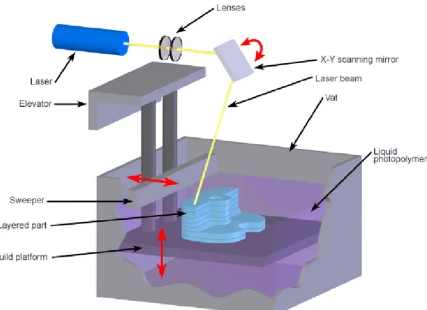
Stereolithography, Selective Laser Sintering and Three-Dimensional Printing
Stereolithography (SLA) (fig 1) and selective laser sintering (SLS) (fig 2) are SFF techniques that use a laser-based curing device to photopolymerize liquid monomers or to sinter powered materials, respectively. Both processes begin by design a three-dimensional structure using CAD software, which is then converted into geometrically patterned two-dimensional slices of approximately 100 microns in thickness. A computer-controlled servo mechanism transmits information to the scanning laser and guides it within the x-y plane, selectively bonding the material to form the structure. The build platform is then stepped down the same distance as the slice thickness and a new slice is scanned. SLA selectively polymerizes material for a vat of photopolymeric solution in a layer-by-layer process. SLA has been used to fabricate scaffolds from poly(propylene fumarate) and diethyl fumarate mixtures for hard tissues, including the repair of bone defects [12]. PEG-DMA scaffolds incorporated with PLGA microparticles have also been fabricated for use in the tissue engineering of soft tissues. One previous limitation of SLA-based processes has been inability to fabricate structures, such as scaffolds, that were large enough for clinical applications.
SLS, an SFF method that operationally is similar to SLA, selectively bonds material from a powder bed using a CO₂ laser that is directed on a powder bed of material. The laser is scanned across the surface and sinters polymers or composites in a preselected pattern for each layer. Low compaction forces during fabrication in SLS lead to highly porous structures. SLS has been employed in fabrication of scaffolds using calcium phosphate, PLA, PCL, and poly(ether ether ketone).
Figure 2: Selective Laser Sintering (SLS)
The spatial resolution and minimum feature size of SLA and SLS are fixed by laser spot size in x-y plane and by step size in z-dimension. Laser spot sizes around 250 μm are commonly attainable, while spot sizes as low as 70 μm have been achieved for specialy systems. Feature resolutions in z-axis are theoretically limited by precision of mechanical stepping system that governs slice thickness and is typically no smaller than 100 μm. In general, reducing feature resolution and slice thickness in SLA and SLS processes results in a dramatic decrease in production speed, which
prompts need to solve the engineering optimization problem to balance the benefits of these two fabrication characteristics. Variations of SLA processes could overcome the limitations due to slice thickness. Cross-linking of biomaterials via multiphoton excitation could also provide an efficient method of three-dimensional scaffold microfabrication. Multiphoton excitation cross-linking using UV has been applied to forming three-dimensional matrices of a wide range of polymers and bulk protein formulations, such as collagen, laminin, fibronectin, polyacrylamide, bovine serum albumin, alkaline phosphatase, and various blends of these [13]. The distribution of intensity irradiation in z-direction theoretically could reduce minimum feature sizes along the axis to approximately 20 μm or smaller. Remarkably, protein matrices fabricated through this method have been shown to remain active in this polymer matrix environment [13]. Further pursuit of this technology could lead to rapid and efficient fabrication of three-dimensional structures using a wide variety of biomaterials while simultaneously maintaining delicate conditions that can preserve biological activity.
Three-dimensional printing (3DP) (fig 3) is similar in practice to that of SLA and SLS, in that a three-dimensional object is fabricated on two-three-dimensional slice at a time using a stepping system. One exception is that 3DP utilizes inkjet printer technology to control deposition of a chemical binder and to selectively fuse material in a powder bed in order to create the object. Drug delivery and tissue-engineering devices have been fabricated from PEO, PCL, and PLGA using 3DP. However, any biological material could be fabricated in principle using 3DP given selection of an appropriate chemical binder. The resolution of objects created using 3DP varies with the complexity of the object. Lines 200 and 500 microns in width have been produced using 3DP of polymer solutions and pure solvent, respectively. Feature resolutions of approximately 1 millimeter are more likely for complex geometries. One significant limitation in the application of 3DP for scaffolds is the addition of the chemical binder. Even if composition of chemical binder itself is found to be nontoxic and biocompatible, introduction of cytotoxic organic solvents such as chloroform and methylene chloride is undesirable. Post-fabrication efforts to remove residual solvent, such as vacuum drying, are not completely effective; therefore issue of cytotoxicity in 3DP-fabricated scaffolds remains.
Figure 3: 3D Printing (3DP)
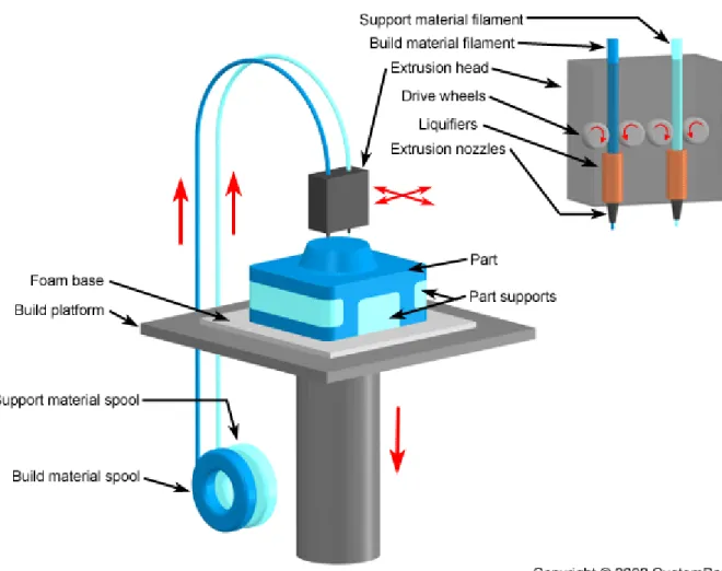
Fused Deposition Modeling
Fused-deposition modeling (FDM) (fig 4) is another method of SFF that has been employed in fabrication of three-dimensional scaffolds. FDM uses a layer-by-layer deposition technique, in which molten polymers or ceramics are extruded through a nozzle with a small orifice, which merges with material on previous layer. The pattern for each layer is controlled by mechanical manipulation of x-y position of nozzle and can be different or arbitrary for each deposited layer. This technique has been applied to production of three-dimensional scaffolds using polymers (PCL, high-density polyethylene) and composites (PCL/Hydroxyapatite). A modification of FDM, termed fused deposition of ceramics, has also been developed for fabrication of scaffolds from 48% to
77%. PCL scaffolds fabricated via FDM have a compressive stiffness ranging from 4 to 77 MPa, which spans mechanical stiffness for both soft and hard tissues [14].
One of primary advantages of FDM and related processes is high degree of precision that can be achieved in the x-y plane. However, control in the z-direction is limited and fixed by the diameter of the material extruded through the nozzle. Additional limitations, including pore anisotropy and the geometry of pore connectivity, are substantially limited due to the continuous deposition process. The extrusion process limits the types of materials that can be processed and therefore has limited the wide applicability of FDM to scaffold fabrication. FDM is typically limited to synthetic thermoplastic polymers, thereby eliminating many natural biomaterials and thermoset synthetic polymers.
Microsyringe Deposition
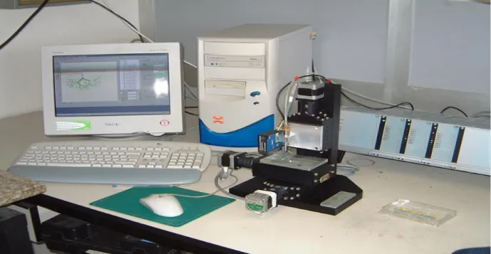
Microsyringe deposition is similar in practice to FDM. A polymer solution is patterned onto a precision-controlled surface using a continuous stream of material via layer-by-layer deposition in order to create three-dimensional structures [5]. Pressure-assisted microsyringe-based deposition (PAM) (fig 5) uses compressed air to eject a polymer solution in volatile solvent through a narrow capillary needle, which has a diameter of between 10 and 20 μm. Control of placement of solution in the x-y plane is controlled by a micropositioning system, which can achieve lateral precisions of 0.1 microns, while physical dimensions of structures can range from 5 to 600 microns, depending on various processing parameters.
PAM has been used to fabricate three-dimensional microfabricated scaffolds with to any polymer, synthetic or natural, that is soluble in a volatile solvent. PAM presents several distinct advantages over traditional SFF methods, including dramatically improved spatial resolution and feature dimensions. Traditional SFF processes are known to have minimum resolutions around hundreds of microns or larger and have principal limitations in minimum feature size. PAM also offers a convenient method for fabrication of multiphase scaffolds with micron-scale precision placement of multiple polymers. Low temperatures also allow potential integration systems for controlled release of proteins and other biomolecules to create favorable microenvironments for tissue regeneration.
2.4 Microfabrication of Cell-Seeded Scaffolds
From the perspective of material properties, mass transport, mechanics, electrical conductivity, surface chemistry, and topology all mediate cell behavior at nano-, micro-, and macroscopic scales. These properties are often correlated with scaffold chemical compositions and structures in an independent way. For example, while a higher porosity may result in a beneficial increase in the mass transport capabilities of a scaffold, it may also compromise the scaffold’s mechanical strength. Achieving optimal scaffold functional performance for a particular tissue-engineering application, therefore, requires balancing different factors. An understanding of fundamental scaffold properties and functions must form design of any higher-order hierarchical scaffold design involving temporal and/or spatial controls. Temporally, scaffold properties at onset of in vitro seeding (time zero) or during acute phase of host response following direct implantation in vivo will provide an initial set of environmental conditions for cells and tissues. These initial scaffold properties and associated functions will then evolve over time in response to passive and active interactions with the culture environment and cells, respectively. Temporal features, enabled mainly by scaffold degradation and release of bioactive factors, are obligatory for presenting time-appropriate signals corresponding with each particular stage of tissue development. Spatially, tissues and organs are 3D-structures having a characteristic organization consisting of patterns of different types of cells and ECM constituents. Anisotropic biochemical and structural properties are designed in scaffolds to induce cell an ECM orientations and to guide multiscale organizations from molecular and cellular level up to the gross tissue or organ level.
Understanding cell-matrix as well as heterotypic cell-cell interactions, both in vitro and in vivo, is a critical component in designing tissue engineering systems. Coordinating the placement of cells on scaffolds is a promising method of capitalizing on specific cell-cell interactions that may lead to favorable conditions for tissue formation. However, three-dimensional scaffolds that are postseeded with cells cannot typically retain spatial segregation or control specific cell-cell interactions across multiple cell types. While a photolithography-based cell-patterning techniques are a powerful method for controlling such interactions in a two-dimensional microenvironment, these methods translate to three-dimensional systems with much difficulty. Advances in microfabrication of biomaterials and SFF have led to potential to control the placement of polymers and cells with micron-scale precision. Combining aspects of these existing technologies
with a layer-by-layer approach is a promising way to fabricate scaffolds that are preloaded with cells that are spatially controlled in arbitrary geometries. Typical scaffold fabrication techniques often employ harsh conditions, such as cytotoxic solvents or elevated temperatures. Hence, developing novel scaffold fabrication processes that are capable of maintaining viable cells is essential. This limitation also serves to reduce drastically the number of biomaterials that are suitable for such processes. Despite these challenges, a number of possibilities have been demonstrated using this approach to scaffold fabrication.
Scaffolds Future Direction
Engineering the spatial and temporal microenvironments on a precise level is critical for inducing desired cellular response, which is especially important in field of tissue engineering and organ regeneration. Consequently, designing microarchitecture of scaffolds used for said applications is an important first step in inducing favorable cell-matrix and cell-cell interactions. The need for microscale control has led to development of a number of fabrication routes that are able to control spatial location of the scaffold features. One viable model that has produced the current state of art and may continue to direct progress in field of advanced scaffold manufacturing is the adaption of traditional material-processing techniques. Replica molding, electrodeposition, and solid-free-form fabrication all represent examples of general materials-processing techniques that have been applied to develop advanced processing platforms for scaffold fabrication. This trend will likely continue as future technologies begin to mature, such as self-assembly and three-dimensional photolithography. The adaption of nascent materials-processing technologies in realization of scaffold systems, where appropriate, could lead to advances in organ regeneration, with the potential for clinical therapy.
Often, driving force for scaffold design and fabrication is desire to create a biomimetic system. This manifests itself as designing materials and processes that try to mimic tissue properties, surface topography, or chemical cues. The notion of the pursuit of biomimicry as effective design criteria for organ regeneration may or may not prove to be most efficient means. However, biomimetic strategies for scaffold fabrication provide challenges that drive the overall improvement of tissue engineering systems and technology. Advancing engineering systems at present will therefore mature the technology in anticipation of application of knowledge to be
gained by future advancements in unraveling the complexity of biological systems, such as further elucidation of proteome and mapping of cell signaling networks. Hence, full potential of organ and tissue regeneration therapy in the future may only be realized on combining advanced scaffold fabrication techniques with design criteria outlined by information on fundamental biological processes derived from integrated system biology.
2.5 Scope of the Work
The recent discovery of CNT’s has created new trends in nano‐engineering and bionano‐ engineering. In particular, Carbon Nanotubes (CNTs) offer a natural platform for obtaining composite microfabricated scaffolds thanks to their excellent mechanical and electronic properties, coupled with a good biocompatibility.
The term “bio” is related to the biocompatibility properties that CNT’s showed in biological systems, making possible the introduction of several advantages to in‐vitro and in‐vivo studies. That’s why I started this project realizing two- and three-dimensional composite PLLA/CNTs scaffolds with the PAM system and then performing their mechanical characterization and a cell behavior analysis for bone tissue engineering applications.
In the specific, I want to design and create 3‐D microfabricated composite scaffolds for tissue engineering, simulating μct (micro computed tomography) of bone tissue, and testing them in‐vitro at first (with human fetal stem cells) , and then in‐vivo for bone surgeries to give bone‐cells growth directions.
In particular I want to demonstrate trough mechanical characterization at first, (performing stress-strain tests, cycle-compressive tests and nano-indentation tests) and trough a cellular behavior analysis then (performing cell viability assays), that higher stiffness of PLLA/CNTs composite scaffolds better stimulate and induce osteoblast proliferation than pure PLLA scaffolds and 2D-PLLA flat films.
The ultimate goal is the realization of an innovative 3D scaffold for bone implants using human fetal stem cells (hfob 1.19) and making them differentiate into osteoblastic cells.
Finally, I want to show if and how scaffold topology (in terms of 3D structure) affects cell density, behavior, proliferation and differentiation.
References
1) Murphy WL, Dennis RG, Kileny JL, Mooney DJ (2002): Salt fusion: an approach to improve pore interconnectivity within tissue engineering scaffolds. Tissue Eng. 8: 43-52
2) Lee KY, Peters MC, Anderson KW, Mooney DJ (2000): Controlled growth factor release from synthetic extracellular matrices. Nature. 408: 998-1000
3) Fleming RG, Murphy CJ, Abrams GA, Goodman SL, Nealey PF: Effects of synthetic micro- and nanostructured surfaces on cell behavior. Biomaterials. 20: 573-588
4) Borenstein JT, Terai H, King KR, Weinberg EJ, Kaazempur-Morfrad MR, Vacanti JP: Microfabrication technology for vascularized tissue engineering. Biomed. Microdevices. 4: 167-175
5) Vozzi G, Flaim C, Ahluwalia A, Bhatia S: Fabrication of PLGA scaffolds using soft lithography and microsyringe deposition. Biomaterials. 24: 2533-2540
6) Bettinger CJ, Winberg EJ, Kulig KM, Vacanti JP, Wang Y, Borenstein JT, Langer R: Three-dimensional microfluidic tissue engineering scaffolds using a flexible biodegradable polymer. Adv. Mater. 18: 165-169
7) Cabodi M, Choi NW, Gleghorn JP, Lee CSD, Bonassar LJ, Stroock AD: A microfluidic biomaterial. Chem. Soc. 127: 13778-13789
8) Lavan DA, George PM, Langer R: Simple, three-dimensional microfabrication of electrodeposited structures. Chem. 42: 1262-1265
9) Li D, Wang Y, Xia Y: Electrospinning of polymeric and ceramic nanofibers as uniaxially aligned arrays. Nano Lett. 3: 1167-1171
10) Xu CY, Inai R, Kotaki M, Ramakrishna S: Aligned biodegradable nanofibrous structure:a potential scaffold for blood vessel engineering. Biomaterials. 25: 877-886
11) Zhong XH, Ran SF, Fang DF, Hsiao BS, Chu B: Control of structure, morphology, and property in electrospun poly(glycolide-co-lactide) nonwoven membranes via postdraw treatments. Polymer. 44: 4959
12) Cooke MN, Fisher JP, Dean D, Rimnac C, Mikos AG: Use of stereolithography to manufacture critical-sized 3D biodegradable scaffolds for bone ingrowth. J.Biomed.Mater.Res. 64B: 65-69
13) Basu S, Campagnola PJ: Properties of crosslinked protein matrices for tissue engineering applications synthesized by multiphoton excitation. J.Biomed.Mater.Res. A71A: 359-368