INTRODUCTION
1 Mycobacterium genus
1.1 General CharacteristicsBacteria of the genus Mycobacterium are acid-fast, aerobic, non-motile and non-spore-producing rods, with a slow generation time. The rods are 2-4m in length and 0.2-0.5m in width. They belong to the phylum Actinobacteria which are characterised by a high GC content DNA (61-71 %) and high lipid content in the cell wall. Few species within the genus are pathogenic in human, they include Mycobacterium tuberculosis (Mtb) and the regional subtypes M. africanum and M. canettii, which are the major causative agents of human tuberculosis (TB), M. microti and M. bovis which are mostly pathogenic in a wide range of domesticated and wild animals, and can be transmitted to humans, and M. leprae, the causative agents of leprosy.
Other species are considered nonpathogenic or opportunistic pathogens and cause disease when host-defences are compromised. Representative species belonging to this group areM. avium, M. kansasii and M. scrofulaceum (Horsburgh 1991; Nightingale 1992). Apart from few pathogen species within the genus which are host-dependent and preferentially reside in the intracellular environment of mononuclear phagocytes, most of them, such as M. smegmatis, M. vaccae, M. gordonae, replicate without restraint in their natural ecosystems and rarely cause diseases in higher vertebrates.
1.2 Cell wall
The cell envelope of Mycobacterium species is rather complex and differs substantially from the cell wall structures of both gram-negative and gram-positive bacteria (Karakousis et al., 2004). The most striking feature is the extraordinarily high lipid content of the mycobacterial cell envelope, which constitutes up to 40% of their dry weight (Brennan and
Nikaido 1995). Slow cell division, resistance to detergents and certain antimicrobial agents, persistence in the environment and an acid/alcohol fast nature are due to the characteristic hydrophobic cell wall.
The lipid rich cell wall does not take up stain readily, but resists decolourisation when destained with an alcohol wash. One acid-fast staining method for mycobacteria is the Ziehl-Neelsen stain. This staining technique comprises of a primary stain, a decolouriser and a counterstain. The primary stain, which is typically concentrated carbol fuchsin is made by dissolving the dye basic fuchsin in phenol. Once stained by this method, intense decolourization (strong acid or acid/alcohol) does not release the primary stain from the waxy cell wall and the mycobacteria retain the red colour. The acid-fast bacilli are then visualized by counterstaining with methylene blue solution. Mycobacteria appear red in a contrasting background (Brennan and Nikaido 1995; Danilchanka 2008) (Fig.1).
In addition to conferring unique tinctorial properties, which are exploited for diagnostic purposes, the cellular envelope of Mycobacterium is essential for the interacting of the host with Mycobacterium species (Daffè and Draper 1998). This cell wall forms an extremely hydrophobic layer that confers to mycobacteria the properties of resistance to solutes including many antibiotic and therapeutic agents. In addition, some of these lipids also display strong biological activities and therefore may be considered as essential virulence factors (Cox et al., 1999; Camacho et al., 2001).
Fig.1 Mtb visualization using Ziehl-Neelsen stain.
The cell envelope of Mycobacterium consists of three layers: the cytoplasmic membrane, the outer layer or mycomembrane and the capsule (Fig.2). The outer layer or mycomembrane is the most remarkable of these structures and consists mainly of one large complex, consisting of three different covalently linked structures: peptidoglycan, arabinogalactan and mycolic acids.
The peptidoglycan of mycobacteria consists of β-(1,4) linked N-acetylglucosamine (NAG), N-acetylmuramic acid (NAM) residues, and a tetra-peptide chain. The tetra-peptide chain usually includes L/D-alanine, D-isoglutamine and meso-diaminopimelic (mDAP), and can be cross-linked to form a three dimensional mesh-like layer (Madigan and Martinko, 2005). In Mtb the cross-link occurs between two mDAP residues or between mDAP and D-alanine (Brennan, 2003; Lavollay et al., 2008).
Fig.2 Schematic representation of mycobacterial cell wall. The cell wall mainly
contains a large cell-wall core that contains three different covalently linked structures (peptidoglycan, arabinogalactan and mycolic acids). The covalent linkage of mycolic acids provides an extremely hydrophobic layer known as the mycomembrane. The outer part of the mycomembrane is composed of various free lipids including cord factor or dimycolyl trehalose, phenolic glycolipids, phosphatidylinositol mannosides and sulpholipids that are intercalated with the mycolic acids. Most of these lipids are specific for mycobacteria. The capsule mainly contains polysaccharides (glucan and arabinomannan). (From Brennan and Crick, 2007).
Situated immediately outside the peptidoglycan, the arabinogalactan layer is a branched polymer of galactose and arabinose. The galactose containing non-reducing end of arabinogalactan is covalently bound to the NAG residue of peptidoglycan by a unique linker, a disaccharide bridge α-L-Rhap-(1→3)-D-GlcNAc-(1→P) (McNeil et al., 1990). Arabinogalactan biosynthesis is essential for the growth and survival of mycobacteria. Arabinogalactan has a linear galactan chain of about 30 β-D-galactofuranose (β-D-Galf) residues which is further elaborated by arabinan chains (Daffe et al., 1990; Besra et al., 1995; Alderwick et al., 2007). In addition, almost two-thirds of the arabinan chains are esterified with mycolic acids at the non-reducing termini of the arabinan chain (Daffe et al., 1990).
Mycolic acids are extremely large hydroxylated branched-chained fatty acids (C30-C90) and their covalent linkage results in a hydrophobic layer of extremely low fluidity. The mycolic acids are most likely orientated perpendicular to the cytoplasmic membrane (Minnikin 1991). The entire complex, encompassing peptidoglycan and arabinogalactan esterified with mycolic acids, is also known as the mycolyl-arabinogalactan-peptidoglycan complex.
In addition, free lipids, most of which are specific for mycobacteria, are intercalated with the mycolic acids. These lipids include phenolic
glycolipids, phthiocerol dimycocerosates, cord
factor/dimycolyltrehalose, sulfolipids and phosphatidylinositol mannosides (PIM).
Lipoarabinomannan (LAM), together with its precursors, lipomannan (LM) and PIMs are major lipoglycans found in the mycobacterial cell wall. They are important not only for the structure of the cell wall but also for modulating the host response during infection (Guerardel et al., 2003; Briken et al., 2004).
Some mycobacterial species contain capsular-like outer layers, which consist mainly of polysaccharides, proteins, and small amounts of lipids (Daffè and Draper 1998). Electron microscopy led to the term “electron
transparent zone” (Rastogi et al., 1991). Unlike other bacterial capsules, the mycobacterial capsule does not seem to offer protection against phagocytosis, and its function has not been completely determined up to now. It may protect the bacteria against enzymatic digestion after phagocytosis. Furthermore, capsular polysaccharides possibly trigger the interaction between bacteria and host cells during the first steps of infection, and it has been assumed that they contribute to intracellular persistence and replication. Taken together, capsular polysaccharides are believed to be potent virulence factors (Frehel et al., 1991). Three polysaccharides of the capsule of Mtb have been isolated and partly characterized, i.e. a glycogen-like glucan with an estimated molecular mass of 100 kDa, a mannan, and an arabinomannan (Daffe and Draper 1998).
The existence of an outer membrane bilayer, analogous to the outer membrane of gram-negative bacteria, has long been postulated in mycobacteria. Based on the chemical structures of the main cell envelope constituents, several models of the hypothetical outer membrane bilayer of mycobacteria, were developed (McNeil and Brennan 1991; Liu et al., 1995; Puech et al., 2001; Minnikin et al., 2002). According to these models, the innermost leaflet consists mainly of the mycolic acids, which are, at least in part, covalently linked to the cell wall arabinogalactan. The outermost leaflet is proposed to be composed of various glycolipids, including trehalose monomycolate and trehalose dimycolate; of phospholipids; and of species-specific lipids such as glycopeptidolipids, phthiocerol dimycocerosate, and sulfolipids (Liu et al., 1995; Daffè, 2005). Recently, Zuber et al. have provided a direct visualization of the mycobacterial outer membrane, demonstrating that mycolates are essential constituents of this structure (Zuber et al., 2008).
1.3 Pathogenesis of TB
The principal route of entry of the tubercle bacillus into the body is via the respiratory tract through the inhalation of respiratory droplet nuclei, which are small enough in size (1 to 2m or less) to allow passage into the lower respiratory tract (Riley et al., 1995). Droplets of a larger size are efficiently excluded from the lower respiratory tract by the physical barriers of the nasopharynx and upper respiratory tract. These droplets can persist in the atmosphere for several hours and, because the infectious dose is in the range of one to ten bacilli, this makes transmission an extremely efficient process (Russel et al., 2009). The bacteria that reach the lung are phagocytosed by alveolar macrophages. At this stage, the destruction of mycobacteria depends on the intrinsic microbicidal capacity of host phagocytes and virulence factors of the ingested mycobacteria. Mycobacteria which escape the initial intracellular destruction will multiply, and this will lead to disruption of the macrophage. When this happens, blood monocytes and other inflammatory cells are attracted to the lung. These monocytes will differentiate into macrophages which again readily ingest but do not destroy the mycobacteria (van Crevel et al., 2002).
These monocytes form the cellular matrix of the early granuloma, which is the primary characteristic of this disease. In its early stage, the granuloma has a core of infected macrophages enclosed by foamy macrophages and other mononuclear phagocytes, surrounded by lymphocytes. This tissue response contains the infection and determines the end of the period of rapid replication of Mtb (Russel et al., 2009) (Fig.3).
Fig.3 Process of the development of TB infection. Following inhalation by the
host, Mtb is taken up by alveolar macrophages and DCs. Migration of the infected cells to the lymph nodes stimulates naive T cell populations, including CD4+ and CD8+ T cells. Antigenic T cells induce granuloma formation, in which the Mtb infection is contained. The granuloma can contain the tubercle bacilli for many years; however infection in the immunocompromised host can lead to caseation of the granulomas and this may results in active transmission of the bacilli to a new host. (From Russell et al., 2009).
Tumour necrosis factor (TNF) and inflammatory chemokines produced by the infected macrophages result in recruitment of neutrophils, macrophages, dendritic cells (DCs), natural killer (NK) cells, CD4+ and CD8+ T cells each of which in turn make its own cytokines and chemokines that increase the cellular chemotaxis and remodelling of lung tissue in the infected regions. The inflammatory response is replaced and controlled by a particular cellular immune response which is associated with production of interferon- (IFN-). In fact, at this stage, the recruited cells are employed as building blocks of tubercles (Flynn and Chan, 2005; Ulrichs and Kaufmann 2006; Russel, 2007). As the granuloma matures, it develops an extensive fibrous capsule that encases the macrophage core and excludes the majority of lymphocytes from the center of the structure. Concomitant with this transition is a considerable decrease in the number of blood vessels penetrating the granuloma. At this stage there is a noticeable increase in the number of foamy macrophages in the fibrous capsule (Russel, 2009).
In the late stage, the granulomas lose their vascular appearance and become necrotic. Cytokine production and lymphocyte proliferation mainly occur in these structures which are composed of B-cells, antigen presenting cells (APCs), CD4+ and CD8+ T cells (Jo, 2008). In addition, recent histological studies of human granulomas supported the role of the follicle-like structures and have revealed that considerable number of the bacteria or bacterial products are associated with the macrophages found both bordering the central necrotic regions and outside the fibrotic margin where lymphocytes proliferate (Marmiesse et al., 2004). Up till this stage, the granuloma functions as a container of the bacteria and prevents its dissemination which leads to latent infection.
Different factors such as senescence, malnutrition, alcohol consumption and HIV/AIDS suppress the immune system, providing proper conditions for reactivation of the pre-existing infection. Following such a change in immune surveillance, caseation occurs and the bacilli
are released into airways, leading to active transmission, which is termed “reactivation of TB”.
1.4 Epidemiology and control measures
TB is an ancient disease that currently presents an immense global health challenge. It is estimated that one third of the world‘s population (approximately 2 billion people) are infected with the tubercle bacilli. The World Health Organisation (WHO) estimate that globally in 2011 there were 8.7 million cases of active TB leading to 1.4 million deaths (WHO 2012). However, infection by the organism does not necessarily lead to disease and only 5-10% of these individuals will progress to active disease each year (WHO 2012). The remaining 90% of infected individuals will initially be asymptomatic and experience latent infection, from which reactivation may occur. The geographic incidence of infection and therefore disease burden varies greatly. Most of the estimated number of cases in 2012 occurred in Asia (59%) and Africa (26%). The 22 high-burden countries (in terms of absolute numbers of cases) account for 82% of all estimated cases worldwide. China, India and Russia alone account for an estimated 60% of TB cases worldwide (WHO 2012).
Living in or having visited a high incidence area are key risk factors, but host factors also play a role in the risk of patients progressing to active disease. The immunosuppressed individuals, especially those infected with HIV, are particularly susceptible to the disease. Individuals who have poor nutrition and general bad health, such as the homeless and those who misuse alcohol and drugs are also at increased risk. Other immunocompromised patients, such as patients with chronic renal disease, neoplastic disorders and those receiving immunosuppressive therapy are also susceptible. The vaccination status of the individual may also play a role.
The treatment of TB differs from that of the other infectious diseases due to the long treatment time needed to cure the patient (Calver et al.,
2010). A characteristic difficulty of TB is the persistence of the pathogenic mycobacteria, regardless of prolonged antibiotic treatment. The micro-environment containing dormant bacteria could change over a period of time causing the bacteria to recommence growth, at which stage they are vulnerable to the standard drugs (Parrish et al., 1998; Dick 2001). Because some of the subpopulation of Mtb may not be eliminated effectively with standard antibiotics, prolonged periods (i.e. > 6 months) of the treatments are required (Barry et al., 2009).
Antibiotics against TB can be classified into two lines of combination treatments, of which application of the second line is dictated by the development of drug resistance to the first. The decision to commence with a treatment regime for TB should not be taken lightly, due to the severe side effects that can occur. For example first line drugs can cause drug induced hepatitis, nausea, deafness and progressive loss of vision. The first line of drugs includes isoniazid, rifampicin, ethambutol, pyrazinamide and streptomycin used for the treatment of drug sensitive TB (Somoskovy et al., 2001). Second line drugs, such as ethionamide, fluoroquinolones and macrolides, are used for the treatment of multi drug resistant TB (MDR). However these drugs have more toxic side effects. They are difficult to obtain in developing countries or are less effective than first line drugs. Extensive drug resistant (XDR) TB brought to the concept of “third line” drugs, such as rifabutin, that may be used in such extreme cases as a last resort.
M. bovis bacillus Calmette–Guérin (BCG) is the only available TB vaccine. It is prepared from a strain of the attenuated bovine TB bacillus, M. bovis, that has lost its virulence in humans by being specially subcultured in an artificial medium for 13 years. BCG was first administered orally in 1921, and since then many clinical trials in different parts of the world have evaluated its efficacy in preventing TB. These trials demonstrated that BCG confers consistent protection against TB meningitis and disseminated TB in children, and leprosy (Merle et al., 2010) in areas of the world where that disease is endemic
(Rodrigues et al., 1993; Sterne et al., 1998; Trunz et al., 2006). However, protective efficacy of BCG vaccine highly varies (from 0 to 80%) against pulmonary disease, which accounts for the major burden of global TB mortality and morbidity throughout the world (Fine, 1995; Brewer, 2000). Explanations for this inconsistency include differences in trial methodology, host population genetics, use of different BCG vaccine strains (Behr and Small, 1997), and heterogeneous immunity to a variety of environmental mycobacteria that may interfere with the protection provided by BCG (Brandt et al., 2002).
As a result, there is an urgent global public health need to develop newer efficacious and safer TB vaccines that may have an impact in both preventing TB infection and, in previously infected populations, halting the progression to TB disease.
1.5 Mtb secretion systems
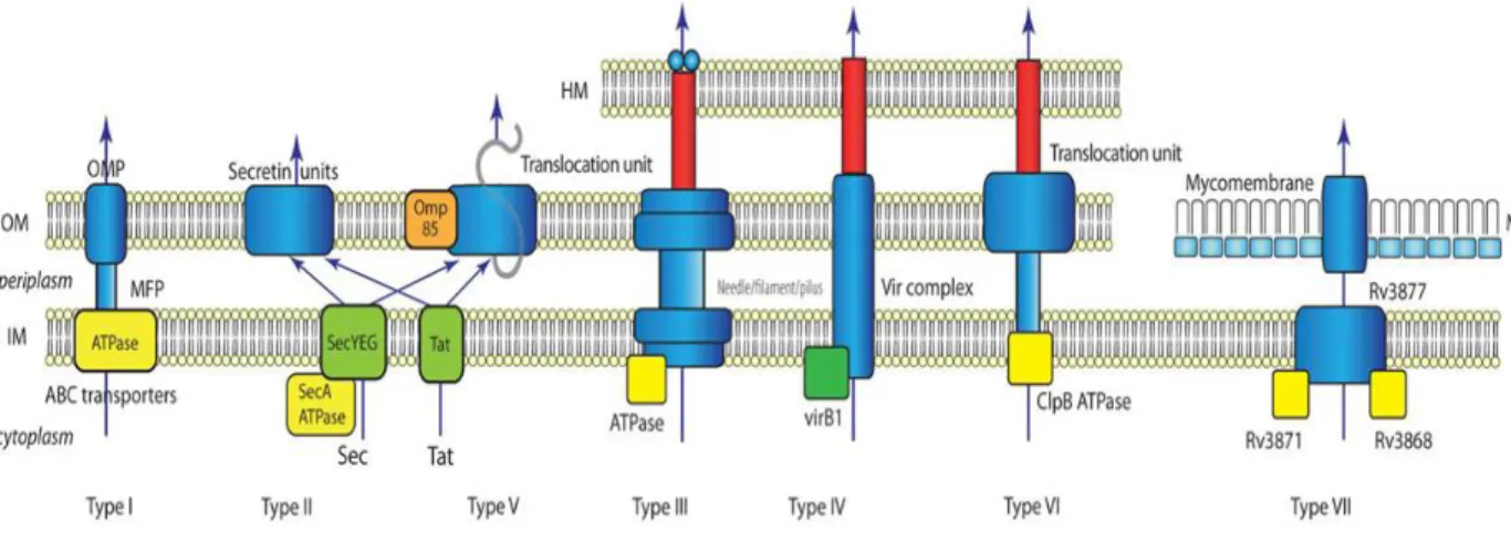
Bacterial pathogenicity depends on the ability to secrete virulence factors, which can be displayed on the bacterial cell surface, secreted into the extracellular milieu or injected directly into a host cell (Finlay and Falcow, 1997). The mechanisms of protein secretion have been most extensively investigated in Gram-negative bacteria, which resulted in the identification of different specialized secretion systems, designated type I–VI (Fig.4).
Fig. 4 Summary of known bacterial secretion systems. In this simplified view
the basics of each secretion system are sketched. Type III, Type IV and Type VI secretion system penetrate the host membrane (target cell membrane) with needle-like structures. The T VII secretion system of mycobacteria is separated from the other systems of Gram-negative bacteria. HM: Host membrane; OM: outer membrane; IM: inner membrane; MM: mycomembrane; OMP: outer membrane protein; MFP: membrane fusion protein. ATPases and chaperones are shown in yellow. (Tseng et al., 2009).
Protein secretion in Gram-negative bacteria is particularly complex because these bacteria are surrounded by two membranes that secreted proteins must pass through to enter the extracellular environment or host cell. In contrast to Gram-negative bacteria, Gram-positive bacteria are regarded as simpler in structure as they lack a second membrane; secretory proteins of Gram-positive bacteria therefore only need to traverse the cytoplasmic membrane and the peptidoglycan layer to enter the extracellular environment (van Wely et al., 2001). However, recent studies have provided evidence that there is an alternative protein-secretion system in Gram-positive bacteria (Pym et al., 2003; Stanley et al., 2003; Hsu et al., 2003; Guinn et al., 2004). Perhaps not surprisingly, this specialized secretion system was identified in Mtb, a bacterium with an extremely complex cell envelope.
The outcome of mycobacterial infection is dependent on both effector molecules secreted by the bacteria and host factors. Therefore, the secreted mycobacterial proteins and, accordingly, the respective systems responsible for their export are critical for virulence. Secreted proteins of Mtb play an important role in virulence and in pathogenesis of the disease, by altering host resistance or by modifying the
environment of the infected host cells. For instance, the inability of the live vaccine strain BCG to cause disease is, partially, due to the absence of the secreted proteins the 6 kDa Early Secreted Antigenic Target (ESAT-6) and the 10kDa culture filtrate protein (CFP-10) (Pym et al., 2002; Lewis et al., 2003; Pym et al., 2003; Hsu et al., 2003). Bioinformatic analysis and secretome studies of the extracellular proteome of Mtb cultures showed that this bacterium secretes a large number of different proteins, albeit in relatively small amounts (Jungblut et al., 1999; Rosenkrands et al., 2000).
Three secretion pathways mediate the protein transport across the complex mycobacterial cell envelope (Bitter et al., 2009):
- the SecA1-operated pathway, responsible for secretion of proteins containing typical N-terminal signal sequences, or the alternative SecA2-operated pathway involved in secretion of proteins without an obvious signal sequence;
- the twin arginine translocation (TAT) system, devoted to secretion of a subset of proteins containing an atypical N-terminal signal sequence, characterised by a couple of arginine residues;
- the recently described type VII secretion system specialised for secretion of small, highly immunogenic proteins lacking a classical N-terminal signal sequence, and characterized by the aminoacidic motif Trp-Xaa-Gly and a size of approximately 100aa (WXG100-family) (Pallen, 2002).
1.5.1 Type VII secretion systems
The type VII secretion system (ESX) represents a novel secretion pathway recently identified in mycobacteria. The ESX systems are named for the first known secreted substrate of any ESX pathway, EsxA (ESAT-6) of Mtb. The hallmark of the ESX systems is that they secrete small proteins which lack Sec or Tat signal peptides and share the characteristic WXG100 motifs. The Mtb genome harbours five gene clusters coding for type VII secretion systems, named ESX-1 - ESX-5
(Cole et al., 1998; Tekaia et al., 1999) (Fig.5). Phylogenetic analyses and comparative genomics have revealed that the five different ESX systems in the genus Mycobacterium probably evolved by gene duplication, in the order ESX-4, ESX-1, ESX-3, ESX-2 and most recently, ESX-5 (Gey Van Pittius et al., 2001).
Fig.5 Genetic organization of the ESX loci in Mtb H37Rv with the proposed
nomenclature. The abbreviation ecc stands for esx conserved component (Bitter et al., 2009).
ESX-1 is the most extensively characterised type VII secretion system, due to its crucial implication in virulence of pathogenic mycobacteria through secretion of effector molecules such as ESAT-6, its protein partner EsxB (CFP-10), as well as EspA and other associated proteins (Pym et al., 2003; Fortune et al., 2005; MacGurn et al., 2005; McLaughlin et al., 2007; Frigui et al., 2008; Raghavan et al., 2008; Gordon et al., 2009; Simeone et al., 2009; Carlsson et al., 2009; Garces et al., 2010; Sani et al., 2010; Bottai et al., 2011).
While the biological function of ESX-2 and ESX-4 systems remains entirely unknown, the ESX-3 was shown to be particularly important, as its corresponding proteins are involved in iron uptake via the siderophore mycobactin and are essential for in vitro growth of pathogenic mycobacteria (Serafini et al., 2009; Siegrist et al., 2009). Although the role of the ESX-1 and ESX-3 systems in Mtb has been elucidated, predictions for the function of the ESX-5 system came from data obtained in M. marinum. Nevertheless, recently, a growing number of studies are aimed to investigate its role in Mtb.
1.5.2 ESX-5 secretion system
The ESX-5 system represents the most recently evolved ESX system, whose orthologues are conserved in the genomes of various slow growing mycobacteria, including the human pathogens M. leprae and M. ulcerans, or the fish pathogen M. marinum, but not in the genomes of fast growing saprophytic species such as M. smegmatis (Gey Van Pittius et al., 2001). Similarly to other ESX clusters, the ESX-5 locus consists of a pair of esx genes, coding for EsxM and EsxN proteins, immunodominant antigens, which induce strong CD4+ T-cell responses both in humans and in different animal models (Alderson et al., 2000; Jones et al., 2010). Immediately upstream of esxM/esxN the ESX-5 locus harbours the ppe25-pe19 gene cluster, coding for members of mycobacteria-specific protein families, named after the conserved N-terminal proline-glutamic acid (PE) or proline-proline-glutamic acid (PPE) motifs (Cole et al., 1998; Tekaia et al., 1999). The ppe-pe-esx genes are flanked by blocks of ecc (esx conserved components) genes that encode membrane proteins and ATP-binding proteins, predicted to be components of an ATP-powered secretion machine involved in the export of the corresponding Esx proteins (Brodin et al., 2004; Bitter et al., 2009). First insights into the role of ESX-5 were gained in the last few years by using M. marinum as model (Abdallah et al., 2006; 2009; Daleke et al., 2011; Weerdenburg et al., 2012). Analysis of secretomes of
two M. marinum ESX-5 transposon mutants revealed that a functional ESX-5 system is required for secretion/ transport of various PPE and PE proteins, such as the heterogeneously expressed PPE41 protein from Mtb (Abdallah et al., 2006) or PPE and PE proteins belonging to the PPE_MPTR and PE_PGRS subgroups (Abdallah et al., 2009). In M. marinum, the ESX-5 system is implicated in modulating innate immune responses of macrophages and in facilitating M. marinum dissemination from infected macrophages (Abdallah et al., 2007; Weerdenburg et al., 2012).
In contrast to M. marinum, the biological function of the ESX-5 system in Mtb, as well as the impact of this system in virulence and host-pathogen interaction remains largely unknown. Very recently, the crucial role of ESX-5 in virulence and the ESX-5-dependent secretion of EsxN and PPE41 were described for Mtb H37Rv and it has been shown that disruption/repression of single core components of the ESX-5 secretion machinery strongly impacts the Mtb in vitro growth properties, suggesting the importance of an intact and functional ESX-5 for Mtb viability (Bottai et al., 2012, Di Luca et al., 2012). In addition, it has been demonstrated the strong CD4+ T cell immunogenicity of ESX-5-associated PE/PPE proteins, demonstrating, thus, the critical impact of the Mtb ESX-5 secretion system in antimycobacterial immunity and pathogenic potential in immunocompetent mice (Sayes et al., 2012).
2 Host innate immune response to the pathogens
2.1 Pathogen recognition by innate immune systemInnate immunity relies on a limited number of receptors and secreted proteins that are encoded in the germline and that recognize features common to many pathogens (Janeway and Medzithov 2002). In contrast, adaptive immunity uses a process of somatic cell gene rearrangement to generate an enormous repertoire of antigen receptors that are capable of fine distinctions between closely related molecules (Bonilla and Oettgen, 2010; Chaplin 2010). Nonetheless, the innate immune system discriminates very effectively between host cells and pathogens, providing initial defenses and also contributing to the induction of adaptive immune responses.
Innate immune recognition (also known as pattern recognition) is based on the detection of molecular structures that are unique to microorganisms. Pattern recognition is unusual since each host receptor (Pathogen Recognition Receptor, PRR) has a broad specificity and can potentially bind to a large number of molecules that have a common structural motif or pattern. The targets of PRRs are sometimes referred to as pathogen-associated molecular patterns (PAMPs), although they are present on both pathogenic and non-pathogenic microorganisms (Medzhitov, 2007; Rasmussen et al., 2009). PAMPs are well suited to innate immune recognition for three main reasons. First, they are invariant among microorganisms of a given class. Second, they are products of pathways that are unique to microorganisms, allowing discrimination between self and non-self molecules. Third, they have essential roles in microbial physiology, limiting the ability of the microorganisms to evade innate immune recognition through adaptive evolution of these molecules. The detection of these structures by the innate immune system can signal the presence of microorganisms (Medzithov, 2007).
The innate immune system serves to monitor for more than just the presence of microbes; it also contains PRRs that recognize danger signals produced by cells in response to pathogenic conditions. These danger signals or danger associated molecular patterns (DAMPs) are released in conditions of cellular damage or stress or formed by pathogen-mediated modification of host proteins and can be recognized by the PRRs of the innate immune system. Thus the innate immune system can identify pathology occurring independent of infectious causes. In fact the recognition of DAMPs is known to be a crucial factor in sterile inflammatory responses, in which the innate immune system responds to tissue damage mediated by non-microbial insults such as ischemia or trauma (Kono and Rock 2008; Pedra et al., 2009).
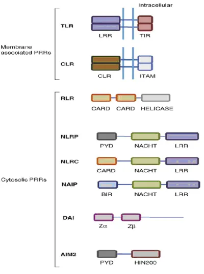
There are several functionally distinct classes of PRRs which include Toll-like receptors (TLRs), retinoic acid-inducible gene I-like receptors (RLRs), and nucleotide-binding oligomerization domain-like receptors (NLRs) (Fig.6).
Fig. 6 Schematic representation of the basic structure of PRRs. The basic
structural domains of different PRRs are compared in diagrammatic form. LRR=leucine rich repeats; TIR=Toll/interleukin-1 receptor interacting domain; CLR=C type lectin receptor domain; ITAM=Immunoreceptor tyrosine-based activation motif; CARD=caspase recruitment and activation domain; Helicase=helicase domain; PYD=pyrin domain; NACHT=nucleotide binding and oligomerisation domain, BIR=Baculovirus inhibitor of apoptosis repeat; HIN200=Hin 200 domain. Z=Z-DNA binding domain. (Bryant and Fitzgerald, 2009).
The best characterized class is TLRs. To date, 12 members of the TLR family have been identified in mammals (Akira and Takeda, 2004). TLRs are type I integral membrane glycoproteins characterized by the
extracellular domains containing varying numbers of leucine-rich-repeat (LRR) motifs and a cytoplasmic signaling domain homologous to that of the interleukin 1 receptor (IL-1R), termed the Toll/IL-1R homology (TIR) domain (Bowie and O’Neill, 2000; van Duin et al., 2006). Based on their primary sequences, TLRs can be further divided into several subfamilies, each of which recognizes related PAMPs: the subfamily of TLR1, TLR2, and TLR6 recognizes lipids, whereas the highly related TLR7, TLR8, and TLR9 recognize nucleic acids. TLRs are expressed on various immune cells, including macrophages, DCs, B cells, specific types of T cells, and even on non-immune cells such as fibroblasts and epithelial cells. Expression of TLRs is not static but rather is modulated rapidly in response to pathogens, a variety of cytokines, and environmental stresses. Furthermore, while certain TLRs (TLRs 1, 2, 4, 5, and 6) are expressed on the cell surface, others (TLRs 3, 7, 8, and 9) are found almost exclusively in intracellular compartments such as endosomes, and their ligands, mainly nucleic acids, require internalization to the endosome before signaling is possible (Akira et al., 2006).
NLRs are a family of intracellular sensors that have key roles in innate immunity and inflammation. In humans the NLR family is composed of 22 members (Meylan et al., 2006).
NLRs are defined by an architecture that contains a LRR domain that is variable in the repeats composition and number, a middle nucleotide binding and oligomerization domain and a variable N-terminal protein-protein interaction domain, that can be either a caspase recruitment and activation domain (CARD), a pyrin domain (PYD), or a baculovirus inhibitor of apoptosis repeat domain (BIR). The LRR domain has been implicated in ligand sensing and autoregulation of the NLRs; however the ability of the NLRs to bind directly to their ligands has not yet been demonstrated. The PYD, CARD or BIR domains facilitate downstream signaling through protein-protein interactions (Inohara et al., 2005; Fritz et al. 2006; Werts et al., 2006; Proell et al., 2008).
The more recent discovery of NLRs as cytosolic PRRs suggests that microbes evading extracellular surveillance encounter a second line of recognition in the host cytosol (Lamkanfi and Dixit, 2009).
Parallel to TLR3 and 7/8, two cytosolic PRRs named retinoic acid-inducible gene 1 (RIG-I) and melanoma differentiation associated gene 5 (MDA5) detect intracellular RNA species, associated with virus infection, and initiate activation of downstream signaling and induction of cytokines (Yoneyama et al., 2004; Yoneyama et al., 2005).
RIG-I and MDA5 are homologous IFN- inducible proteins containing two amino-terminal CARDs, a carboxy-terminal DExD/H-Box RNA helicase domain and a C-terminal regulatory domain (RD). The helicase domain and the regulatory domain interact with specific RNA species and the CARDs are responsible for downstream signaling. Recently, it has been shown that RIG-I and MDA5 bind short ssRNA and long ssRNA, respectively. In addition, RIG-I has been found to detect 5’ triphosphate RNA. Besides RIG-I and MDA5, the family of RLRs also includes a third member, LGP2 (Laboratory of Genetics and Physiology 2), which lacks the CARD domains and may act as a negative regulator molecule, possibly by forming heterodimeric complexes with RIG-I and MDA5, although the precise mechanism by which it works is still poorly understood.
2.2 Macrophages and DCs
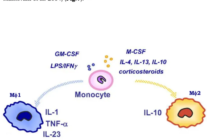
Macrophages play a critical role in host defense and the maintenance of homeostasis. They are derived from circulating blood monocytes, and display a remarkable plasticity since they can change their physiology in response to environmental cues. These changes can give rise to different populations of cells with distinct functions (e.g. host defence, wound healing and immune regulation) (Gordon, 2003; Mantovani et al., 2005; Benoit et al., 2008). For this reason, the mononuclear phagocyte system can be defined as a dynamic continuum of functional states. In an effort to emulate the T helper 1 (Th1) and T helper 2 (Th2)
cell literature, the extremes of this continuum have been termed type 1 (M1) and type 2 (M2) macrophages (Goerdt and Orfanos, 1999: Mantovani et al. 2004) (Fig.7).
Fig. 7 Macrophages polarize and acquire different functional properties in
response to environment-derived signals. Macrophage exposure to GM-CSF, IFN- or LPS drives M1 polarization, with potentiated cytotoxic and anti-tumoral properties, whereas M2, induced by exposure to M-CSF, 4 or IL-13, are in general more prone to immunoregulatory activities (Modified from Sica et al., 2005).
M1 secrete a battery of pro-inflammatory cytokines, which help to orchestrate and amplify Th1 immune response, and they can efficiently kill intracellular pathogens. M2 promote the Th2 response and contribute to the production of the extracellular matrix, therefore their primary function seems to be related to wound healing.
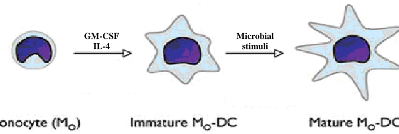
DCs are bone marrow immune cells which populate lymphoid and non-lymphoid tissues of the body (Lipscomb and Masten, 2002). They are
referred to as «professional antigen presenting cells» and possess the unique capability of activating naïve T-cells. These cells are capable of integrating innate immune responses to initiate adaptive immunity in the host. Many features of DCs are divided in time and place and translated into two developmental stages, namely immature and mature DCs (Bell et al., 1999; Mellman and Steinman 2001) (Fig.8). Immature DCs (iDCs) are present in virtually all tissues where they encounter both self and non-self antigens. Triggered by a multitude of signals, including selected pathogens and pro-inflammatory mediators, iDCs mature, after which they display an extraordinary immune stimulatory capacity (Wenink et al, 2006).
Fig. 8 Monocytes can convert to DCs as a result of incubation with
Granulocyte Macrophage-Colony-Stimulating Factor (GM-CSF) and IL-4. Further activation of DCs might require direct contact with pathogens or other mediators in the microenvironment (Modified from Naik, 2008).
2.3 IL-1 family cytokines
Orchestration of an appropriate immune response against these microbial threats is accomplished in part through the production of potent pro-inflammatory cytokines that initiate biological effects associated with inflammation (Dinarello, 1996; Lamkanfi and Dixit, 2009). The most prominent pro-inflammatory cytokines belong to the
GM-CSF IL-4
Microbial stimuli
IL-1-like family, which comprises 11 members: IL-1, IL-1, IL-1 receptor antagonist (IL-1Ra), IL-18, IL-33 and IL-1F5 – IL-1F10 (Busfield et al., 2000; Kumar et al., 2000; Sims et al., 2001).
IL-1, which was the first interleukin to be described, is known for the ability to cause a wide variety of biological effects associated with infection, inflammation and autoimmune processes (Dinarello et al., 1986). There are two forms of IL-1: IL-1 and IL-1, which signal through the same receptor complex and have identical biological activities in solution. But, despite their identical activities, IL-1 and IL-1 differ in several ways. First, IL-1 is secreted and circulates systemically, whereas IL-1 is generally associated with the plasma membrane of the producing cell and so acts locally. Second, IL-1 is mainly produced by innate immune cells such as monocyte/macrophages and DCs, whereas IL-1 expression is more widespread, for example, it is highly expressed by keratinocytes and endothelial cells. Third, the two genes are differentially regulated during development and in response to environmental cues, which results in different functional contributions from these cytokines during immune responses.
IL-1 is a pivotal pro-inflammatory cytokine which has pleiotropic effects. It was initially described as an ‘endogenous pyrogen’ because of its ability to induce fever in animal models. IL-1 was also called ‘lymphocyte-activating factor’, based on its ability to stimulate T-cell proliferation (McKean et al., 1985; Dinarello, 2004; Martinon et al., 2009). More recently, IL-1 was observed to enhance the differentiation of CD4+ T cells into Th2 or T helper 17 cells, the latter being a newly described subset of T cells involved in immune responses to fungi as well as in autoimmunity (Acosta-Rodriguez et al., 2007; Kool et al., 2008). A number of studies also suggest a direct role of IL-1 in the control of pathogenic infections, of fungal, bacterial and viral origin. Moreover, it was found that excessive production of this cytokine
provokes local and systemic manifestations of inflammation (Hise et al., 2009; Joly et al., 2009; Allen et al., 2009; Ichinohe et al., 2009).
Although IL-18 lacks the pyrogenic activity of IL-1, it is mainly known as a factor that induces IFN- in activated T cells and NK cells, thereby contributing to Th1 cell polarization. IL-18 is also involved in the induction of Fas ligand, as well as several secondary pro-inflammatory cytokines, chemokines, cell adhesion molecules, and nitric oxide synthesis (Takeda et al., 1998; Horwood et al., 1998; Tamura et al., 2002; Maxwell et al., 2006).
The more recently described member of the IL-1 family is Interleukin-33. IL-33 is thought to be involved in Arthritis, Alzheimer disease and allergy, and is expressed primarily by epithelial cells. Although relatively new, thus far, the cytokine has been associated with Th2 mediated immune reactions by stimulating eosinophils, basophils and mastcells (Haraldsen et al., 2009).
Because of their potency and extensive functions, the synthesis and release of IL1β and IL-18 need to be tightly controlled. First, accumulation of intracellular stores of pro-IL-1 and pro-IL-18 occurs via transcriptional regulation. This is followed by cleavage of pro-IL-1 and pro-IL-18 and release of the mature cytokines. These events require at least two distinct stimuli. An initial microbial stimulus propagated through innate PRRs causes accumulation of intracellular stores of pro-IL-1 and IL-18. The second event leading to the cleavage of pro-forms into biologically active cytokines involves multiprotein complexes, commonly referred to as “inflammasomes’.
2.4 The inflammasome
Inflammasomes are multiprotein platforms that control the activation of the cystein aspartate protease caspase-1 and the cleavage of pro-IL-1 and pro-IL-18 enabling the release of the active mature forms of these cytokines. Several members of the NLR family can assemble multimolecolar complexes in response to various activators, leading to
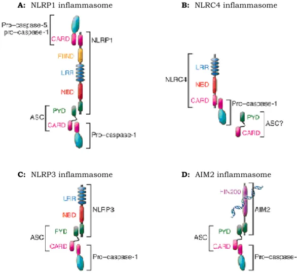
the activation of inflammatory caspases. Four inflammasome complexes have been partially characterized to date (Fig.9). The first to be identified was the NOD Like Receptor Protein 1 (NLRP1) inflammasome (also known as NALP1 inflammasome), comprising NLRP1, caspase-1, caspase-5 and the adaptor proteins apoptosis-associated speck-like protein containing a CARD (ASC) and CARD inhibitor of NF- B-activating ligands (CARDINAL) (Martinon et al., 2002; Faustin et al., 2007). It has been shown that NLRP1 inflammasome play a role in anthrax immunity (Boyden and Dietrich 2006; Squires et al., 2007; Wickliffe et al., 2008; Reig et al., 2008). The NLR family CARD domain-containing protein 4 (NLRC4) (also called IL-1-converting enzyme Protease Activating Factor, IPAF) inflammasome consists of NLRC4, ASC and caspase-1 (Bryant and Fitzgerald, 2009). NLRC4 recognizes bacterial flagellin from Salmonella and Pseudomonas species and causes activation of caspase-1 (Ren et al., 2006; Molofsky et al., 2006; Miao et al., 2006). Whether ASC is required for signaling by the NLRC4 inflammasome is somewhat controversial. Since NLRC4 itself contains a CARD domain, it could recruit caspase-1 directly without the need for ASC. Instead, ASC has been shown to modulate the activity of this inflammasome (Agostini et al., 2004; Mariathasan et al., 2004; Franchi et al., 2006; Suzuki et al., 2007). Interestingly, the NLRC4 inflammasome is also activated by bacteria that are not flagellated. Mtb has also been shown to block NLRC4 inflammasome activation (Master et al., 2008).
Fig. 9 Graphical depiction of known inflammasomes. (A) NLRP1 contains, in
addition to the NLR-typical LRR and Nucleotide Binding Domain (NBD) domains, a PYD, a FIIND (Function to find domain containing protein), and a CARD. NLRP1 can recruit pro–caspase-1 and -5 and possibly forms a complex with NOD2. Recruitment of ASC enhances activation of pro–caspase-1. (B) NLRC4 contains a CARD that can directly recruit pro–caspase-1. Reports further demonstrate a role for ASC in NLRC4 inflammasome activation. (C) NLRP3 activates pro–caspase-1 via recruitment of ASC. (D) AIM2 is a bipartite protein consisting of a PYD and DNA-binding HIN200 domain and recognizes cytoplasmic double-stranded DNA and assembles the DNA inflammasome with ASC and pro–caspase-1 (Stutz et al., 2009).
The absent in melanoma 2 protein (AIM2) inflammasome does not contain any members of the NLR family, but instead contains the hemopoietic IFN-inducible nuclear protein of 200 amino acid motif (HIN200) and PYD domain. AIM2 is localized in the cytoplasm, binds the cytosolic dsDNA via the HIN200 domain and, like some NLRs, engages
A: NLRP1 inflammasome B: NLRC4 inflammasome
the adaptor ASC to recruit and activate caspase-1 (Choubey et al., 2000; Fernandes-Alnemri et al., 2009; Roberts et al., 2009).
By far the best-characterized inflammasome is the NOD Like Receptor Protein 3 (NLRP3), consisting of NLRP3, ASC and caspase-1. Numerous chemically and structurally diverse stimuli are now known to trigger this inflammasome. These include viruses such as Sendai and, influenza, fungi such as Candida albicans and Saccharomyces
cerevisiae and bacteria such as Staphylococcus aureus, Listeria
monocytogenes and Mtb (Kanneganti et al., 2006; Koo et al., 2008; Thomas et al., 2009; Allen et al., 2009; Ichinobe et al., 2009; Gross et al., 2009). Microbial derivatives such as muramyl dipeptide (MDP) and bacterial pore-forming toxins also trigger NLRP3 activation. Additional ligands include extracellular ATP, monosodium urate crystals (MSU), beta-amyloid and various environmental insults (e.g. silica, asbestos) (Kahlenberg et al., 2005; Petrilli et al., 2007; Halle et al., 2008). It is unclear, however, how NLRP3 can detect such a diversity of stimuli. Because there is no evidence that any of these ligands bind directly to NLRP3, it has been suggested that NLRP3 activation is indirect.
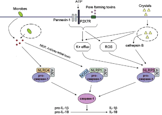
The exact sequence of events leading to inflammasome activation is not well understood for all inflammasomes (Fig.10). It is believed that upon stimulation, the respective inflammasome oligomerizes and recruits pro-caspase-1 directly via a CARD homotypic interaction (e.g., NLRP1 or NLRC4 inflammasomes) or indirectly via the adaptor ASC. In the latter case, NLRP3, for example, interact with ASC which in turn, interacts with pro-caspase-1 via their CARDs (Stutz et al., 2009).
Fig. 10 Inflammasome activation by microbes and danger signals. Several
NLRs can form multiprotein complexes called inflammasomes. Activation of the inflammasome results in activation of the cysteine protease caspase-1 and the resultant processing of pro-1 and pro-18, into biologically active IL-1 and IL-18. Activation of the NLRC4 inflammasome following infection of macrophages with Salmonella typhimurium, Pseudomonas aeruginosa, Shigella flexneri, or Legionella pneumophila requires a functional type III or type IV secretion system. Anthrax lethal toxin and MDP are capable of activating the NLRP1 inflammasome in a manner that may also require NOD2. A wide variety of stimuli including bacterial pore-forming toxins, ATP, DNA, bacterial RNA, and crystals such as silica, asbestos, uric acid, alum, and amyloid-b can activate the NLRP3 inflammasome. NLRP3 activating PAMPs and DAMPs induce a K+ efflux and the generation of mitochondrial-derived ROS that play a role in NLRP3 inflammasome activation by an unknown mechanism. Crystal induced lysosomal damage, and the resultant release of cathepsin B, are also postulated to play a role in NLRP3 inflammasome activation by an unknown mechanism (Modified from Pedra et al., 2009).
Different models of inflammasome activation have been proposed. The first model involves the intracellular level of potassium ([K+]). Pore formation by bacterial toxins might trigger both the entry of unknown ligands for NLRP3 and the K+ efflux. These observations suggest that K+ efflux might be the common event to trigger inflammasome activation (Kanneganti et al., 2007; Petrilli et al., 2007; Aroian and van der Goot, 2007). The second model is mediated by lysosomial destabilization. Endogenous particulates such as MSU, silica, alum or cholesterol crystal, are likely to cause lysosomal rupture leading to the cytoplasmic release of lysosomal contents including cathepsin B that might activate NLRP3 inflammasome (Halle et al., 2008; Hornung et al., 2008). Next model for activation of NLRP3 is mediated by the intracellular ROS, produced responding to infection or tissue injury. A wide range of PAMPs or DAMPs have been shown to induce the production of ROS in an NADPH oxidase (NOX)-dependent or mitochondria dependent manner. It remains poorly defined how ROS affects NLRP3 inflammasome (Cruz et al., 2007; Cassel et al., 2008; Dostert et al., 2008). Furthermore, differences in inflammasome activation have been observed in different cell types. Inflammasome activation in monocytes, macrophages and DCs are different. Two fundamental differences exist for inflammasome activation in DCs, macrophages and circulating monocytes. While monocytes have constitutively activated caspase-1, the activation of caspase-1 in DCs and macrophages needs to be induced (Netea et al., 2009; Novikov et al., 2011; Abdalla et al., 2012; Riteau et al., 2012). Monocytes need a single PRR stimulus for IL-1β secretion, whereas DCs and macrophages need a double stimulation (TLR and NLR stimuli). The reason for this differential regulation is poorly understood. Current evidence suggests an adaptation of each cell type to their respective environment (Chen and Pedra, 2010). Circulating monocytes function in a pathogen-free environment and must promptly respond to a microbial threat. DCs and macrophages are confined to a non-sterile environment and are
constantly exposed to microbial pathogens. Therefore, a second checkpoint mechanism is necessary to avoid deleterious inflammation (Piccini et al., 2008; Netea et al., 2009).
The differences in inflammasome activation do not seem to be restricted to macrophages, DCs and monocytes. In contrast to monocytes and macrophages, in which low intracellular [K+] triggers inflammasome activation, neurons and astrocytes activate caspase-1 by high intracellular [K+] (Silverman et al., 2009).
2.5 Caspase-1: Inflammatory caspase
The caspase family of cysteine proteases occupies critical positions in signal transduction cascades associated with immune responses. In many of these situations, caspase activation culminates in the apoptosis of the cell in which these proteases become activated. However, caspases also play important roles in immune reactions that culminate in cytokine production rather than apoptosis. Caspase-1 is the best described inflammatory caspase. It has been originally identified as a result of attempts to purify the enzyme responsible for processing of the IL-1. In fact it has been originally named IL-1 Converting Enzyme (ICE) (Kostura et al., 1989; Cerretti et al., 1992; Miller et al., 1993; Thornberry et al., 1994; Alnemri et al., 1996). Caspase-1 proteolytically processes the immature IL-1 from a 31kDa precursor to the 17kDa mature form. In addition to processing IL-1, caspase-1 is known to efficiently activate IL-18, and to be required for the efficient production of IL-1. Although caspase-1 appears to be engaged primarily in non cell-death-related context, its role in the pathway that leads to a form of cell death with characteristics of necrosis and apoptosis that has been called “pyroptosis” is well established. How caspase-1 activation is achieved in divergent pathways that culminate in cell death or result in pro-inflammatory cytokine production is still unclear.
Caspase-1 is synthesized as an inactive 45kDa precursor, or pro-caspase-1, which is constitutively expressed in the cytosol. To gain its full activity, pro-caspase-1 has to be recruited to the inflammasomes, and to be auto-processed into a heterotetrameric structure composed by two 20-kDa (p20) and two 10-kDa (p10) active fragments. The catalytic site is formed by aminoacids from both the p20 and p10 subunits, with the active site cysteine located within the p20 subunit (Stennicke and Salvesen, 2000). However, although caspase-1 was discovered more than 15 years ago details of its activation have yet to be fully understood. Intriguingly, proteolytically processed caspase-1 is very difficult to observe in cells that are actively producing mature IL-1. Possible explanations for this include rapid degradation of the enzyme, rapid extracellular secretion of the active caspase-1 or highly stringent controls on caspase-1 activation (Yamin et al., 1996; Salvesen and Abrams, 2004).
3. Innate immune response to Mtb
As discussed above, alveolar resident macrophages are the primary cell type involved in the initial uptake of Mtb. After this first encounter, DCs and monocyte-derived macrophages also take part in the phagocytic process (van Crevel et al., 2002).
As intracellular pathogens, mycobacteria are actively internalized by macrophages. Uptake through phagocytosis requires recognition of mycobacteria by receptor molecules exposed at the macrophage cell surface, which either bind to non-opsonised Mtb or recognize opsonins on the surface of Mtb. Among these, the complement receptors CR1, CR3, CR4 and immunoglobulin fragment carrying the constant region of the heavy chain (Fc) receptors take up opsonised bacteria, whereas the scavenger receptors and the macrophage mannose receptor aid in the uptake of non-opsonised bacteria (Schlesinger 1993; Hirsch et al. 1994;
Aderem and Underhill, 1999). This receptor recognises the mannose-capped LAM (manLAM) present on virulent strains of mycobacteria (Schlesinger et al., 1996; Schafer et al. 2009). Other receptors, including CD14, have roles in the phagocytosis of mycobacteria (Ernst, 1998). Moreover, various mycobacterial proteins and lipids have been individually demonstrated to be involved in TLR-dependent signalling cascades. The mycobacterial 19 kDa lipoprotein (LpqH) has been well characterized and shown to signal through TLR2 to induce cell activation, apoptosis and mycobacterial killing (Aliprantis et al., 1999; Brightbill et al., 1999; Noss et al., 2001; Thoma-Uszynski et al., 2001; Gehring et al., 2004; Pecora et al., 2006). Recent reports indicate that the 38 kDa glycolipoprotein acts through both TLR2 and TLR4 to induce the activation of pro-inflammatory cytokine responses during mycobacterial infection (Jung et al., 2006). Of note, recent work has shown that PE_PGRS33, one member of the PE_PGRS (polymorphic GC-rich sequence) family of Mtb H37Rv, signals through TLR2. The mycobacterial heat shock protein (HSP) 65 signals exclusively through TLR4, whereas HSP70 is known to signal through TLR2 and TLR4 (Bulut et al., 2005).
Following the phagocytosis, the destruction of mycobacteria depends on the intrinsic microbicidal capacity of host phagocytes and virulence factors of the ingested mycobacteria. At this stage, macrophages can exert various microbicidal mechanisms. Nevertheless, Mtb is able to survive within macrophages even though a range of effector mechanisms exist in these host cells to control the growth of invading bacteria.
The macrophage antimicrobial defences comprise both oxidative and non-oxidative mechanisms. In macrophages, nitric oxide is generated by inducible nitric oxide synthase (iNOS), which is a cytosolic enzyme that catalyzes the conversion of L-arginine to L-citrulline and nitric oxide. Nitric oxide is extremely bactericidal, and mice lacking iNOS succumb rapidly to Mtb (MacMicking et al., 1997). Not surprisingly, pathogenic
mycobacteria have evolved ways of countering the destructive effects of nitric oxide and preventing the assembly of iNOS with phagosomes, possibly through rearrangement of the actin cytoskeleton, and this might lead to a reduction in the local concentration of nitric oxide (Miller et al., 2004). Unexpectedly, potential damage by nitric oxide can also be countered by the mycobacterial proteasome (Darwin et al., 2003). The proteasome is a multi-subunit molecular machine that is highly conserved from archaebacteria to humans and is responsible for the proteolysis of cytoplasmic proteins. Mtb has adapted the proteasome machinery to protect itself from the killing effect of nitric oxide (Pieters and Ploegh, 2003).
Non-oxidative mechanisms are also employed by activated macrophages in order to kill mycobacteria. During the process of phagocytosis, microorganisms are taken up into phagocytic vacuoles which eventually fuse with lysosomes resulting in the formation of a phagolysosome. The phagolysosome environment is highly acidic and phagocytosed microbes are degraded by acid hydrolases. Mtb employs various mechanisms to survive in the phagosome such as the modification of the environment it resides in by exclusion of vacuolar H+ ATPases (Wong et al., 2011). The exclusion of vacuolar H+ ATPases from phagosomes containing live mycobacteria leads to the incomplete acidification of the phagosome, thus allowing the bacteria to survive (Fratti et al., 2003; Vergne et al., 2003; Vergne et al., 2005). It has been additionally shown that, mycobacteria inhibit the maturation of the phagolysosome, thus avoiding enzymatic degradation (Walburger et al., 2004). Several Mtb products are thought to be responsible for the inhibition in maturation, including LAM, trehalose dimycolate and sulpholipids, as well as the phosphatase SapM and the kinase PknG (Rohde et al., 2007) and ESAT-6 has been implicated in inhibition of phagosomal maturation during M. marinum infection of macrophages (Tan et al. 2006).
Another mechanism is the inhibition of the macrophage response to pro-inflammatory cytokines including IFN-γ by interfering with
signalling events downstream of the IFN-γ receptor through the 19-kDa lipoprotein (Fortune et al. 2004). Furthermore, Mtb can suppress the expression of MHC class II on macrophages to prevent antigen presentation to CD4+ T cells by blocking transport and processing of the molecules, thereby avoiding an IFN-γ response (Hestvik et al., 2005). Although macrophages serve as the long-term hosts for mycobacteria, Mtb infects iDCs as well and its interaction with these cells is crucial for the development of protective immunity (Jiao et al., 2002, Tian et al., 2005). Considerable evidences show that DCs can phagocytose mycobacteria, resulting in DC function and phenotype being modified (Tsuji et al., 2000). A number of receptors have been implicated in the recognition of mycobacteria by DCs, e.g. the mannose receptor, CD11b and CD11c (Schlesinger 1993; Prigozy et al., 1997; Nigou et al., 2001). One of the C-type lectin receptor extensively characterized with respect to mycobateria is Dendritic Cell-Specific ICAM-3 Grabbing Non-integrin (DC-SIGN), a major receptor of human DCs for Mtb (Tailleux et al., 2003). Functional characterization of DC-SIGN-Mtb interaction has shown that specific targeting of DC-SIGN by Mtb through manLAM is a mechanism to impair DC maturation and to induce production of the anti-inflammatory cytokine IL-10 (Geijtenbeek et al., 2003). Interaction of human DCs with Mtb results in increased surface densities of a number of molecules such as MHC II, CD40, CD54, CD58, CD80, CD83 and CD86 that are involved in interaction with T cells (Kim et al., 1999; Henderson et al., 1997; Giacomini et al., 2001; Hickman et al., 2002). The ability of mycobacteria to survive in DCs is controversial. Both a constrained survival of mycobacteria in DCs (Laochumroonvorapong et al., 1997; Tailleux et al., 2003) as well as increased replication of mycobacteria in DCs when compared to tightly regulated replication in macrophages has been reported (Fortsch et al., 2000; Bodnar et al., 2001; Buettner et al., 2005). Like resting macrophages, DCs provide an environment within which mycobacteria can survive and replicate, albeit to a low extent. It could be attributed to the fact that turnover of
mycobacteria within DCs is not enough to kill the cell and the outcome of slow replication is reflected by constant availability of antigens for presentation to T cells that potentiate the immune response. Following activation with TNF- and IFN-, macrophages kill mycobacteria resident in phagosomes, but this does not occur in activated DCs. Upon stimulation with IFN-, DCs are able to control the replication of mycobacteria, but the bacilli are not killed. Instead, they appear to reside in vacuoles separated from normal recycling pathway. DCs, might, therefore, act as a reservoir for mycobacteria in vivo, particularly within lymph nodes to which they have migrated (Sinha et al., 2007).
3.1 Mtb and inflammasome modulation
While inflammasome activation and subsequent IL-1 and IL-18 synthesis are strongly upregulated in Mtb infected macrophages (Giacomini et al., 2001; Montero et al., 2004), the mechanism by which mycobacteria activate the inflammasome for processing and release of these cytokines has not been examined closely. One report suggests that both Mtb and the vaccine M. bovis BCG actively inhibit inflammasome activation via a zinc metalloprotease (Master et al., 2008). Others described that Mtb actively induces caspase-1 activation for IL-1 release, similar to induction of mature IL-1 by M. marinum, which depends on the region of difference 1 (RD1) locus (encoding part of the ESX-1 secretion system) (Koo et al., 2008). It has been shown that the ESX-1 effector molecule ESAT-6 acts upstream of NLRP3 activation. ESAT-6 functions by disrupting cell membranes and allowing the Mtb protein Ag85 to enter the cytosol and stimulate the inflammasome (Mishra et al, 2010). Notably, in vivo experiments suggest that during murine TB mature IL-1 can be produced in a caspase-1-independent way (Mayer-Barber et al., 2010).
Inflammasome assembly and activation is a hallmark of the innate immune response against intracellular pathogens (Mariathasan and
Monack, 2007; Stutz et al., 2009). Recent studies have demonstrated that caspase-1 activation and subsequent IL-1 synthesis are strongly upregulated in Mtb infected macrophages (Montero et al., 2004), and inflammasome activation occurs upon Mtb infection (Netea et al., 2006; Kleinnijenhuis et al., 2009). A role for the 19 kDa lipoprotein (LpqH) of Mtb has been suggested, as this protein can induce IL-1 secretion (Ciaramella et al., 2000). However, a direct role of LpqH in caspase-1 dependent IL-1 release during Mtb infection is not documented, and the mechanisms regulating these events remain poorly characterized.