-1-
U
NIVERSITÀD
EGLIS
TUDID
IM
ESSINAT
ESI DID
OTTORATO DIR
ICERCA INB
IOLOGIAA
PPLICATA EM
EDICINAS
PERIMENTALEC
URRICULUM INM
EDICINAS
PERIMENTALEXXIX CICLO
SSD BIO/14
The role of mTOR signaling pathway
in Brain and Spinal Cord Injury
Candidata:
D
OTT.
SSAM
ARIKAC
ORDAROCorrelatore: Relatore:
Ch.mo Prof. Ch.ma Prof.ssa
S
ALVATOREC
UZZOCREAE
MANUELAE
SPOSITOCoordinatore:
Ch.mo Prof. SALVATORE CUZZOCREA
-2-
The choice of a young man depends on his inclination,
but also by the good fortune to meet a great teachers.
-3-
INTRODUCTION __________________________________________________________________________________ 6
CHAPTER 1:CENTRAL NERVOUS SYSTEM INJURY _____________________________________________ 9
1.1SPINAL CORD INJURY ____________________________________________________________________________ 9
1.1.1 CLASSIFICATION OF SPINAL CORD INJURY _____________________________________________________ 11 1.1.2 EPIDEMIOLOGY, INCIDENCE AND CAUSES _______________________________________________________ 12 1.1.3 PHARMACOTHERAPY __________________________________________________________________________ 14 1.1.3.1 Methylprednisolone _____________________________________________________________________________ 15 1.1.3.2 GM1 Ganglioside _________________________________________________________________________________ 15 1.1.3.3 Thyrotropin Releasing Hormone _______________________________________________________________ 15 1.1.3.4 Nimodipine _______________________________________________________________________________________ 15 1.1.3.5 Gacyclidine _______________________________________________________________________________________ 16 1.1.3.6 Minocycline _______________________________________________________________________________________ 16 1.1.3.7 Estrogen __________________________________________________________________________________________ 16
1.2TRAUMATIC BRAIN INJURY _____________________________________________________________________ 17
1.2.1 CLASSIFICATION OF TRAUMATIC BRAIN INJURY _________________________________________________ 18 1.2.2 EPIDEMIOLOGY, INCIDENCE AND CAUSES _______________________________________________________ 20 1.2.3PHARMACOTHERAPY ___________________________________________________________________________ 21 1.2.3.1Acetylcholinesterase inhibitors _________________________________________________________________ 22 1.2.3.2 Amantadine ______________________________________________________________________________________ 22 1.2.3.3 Cyclosporine A/FK 506 __________________________________________________________________________ 22 1.2.3.4 Progesterone _____________________________________________________________________________________ 23 1.2.3.5 Simvastatin/other statins _______________________________________________________________________ 23 1.2.3.6 N-acetylcysteine (NAC) __________________________________________________________________________ 23 1.2.3.7 Growth hormone (GH) ___________________________________________________________________________ 24
CHAPTER 2:THE ROLE OF NEUROINFLAMMATION __________________________________________ 25
2.1CYTOKINE RESPONSES TO INFLAMMATION ___________________________________________________ 25
2.1.1 INFLAMMATORY MEDIATOR: ROLE OF TNF α __________________________________________________ 26 2.2MICROGLIA ACTIVATION _______________________________________________________________________ 28
2.3APOPTOSIS _______________________________________________________________________________________ 28
2.4INFLAMMATORY/IMMUNOLOGIC RESPONSE ___________________________________________________ 30
2.4.1 LYMPHOCYTES INFILTRATION _________________________________________________________________ 30 2.5DUAL ROLE OF INFLAMMATION IN SPINAL CORD AND BRAIN INJURY _______________________ 31
CHAPTER 3:AUTOPHAGY AND MTOR _______________________________________________________ 33
3.1 AUTOPHAGY ______________________________________________________________________________________ 33
-4-
3.2.1 MACROAUTOPHAGY (AUTOPHAGY) ____________________________________________________________ 34 3.2.2 MICROAUTOPHAGY ____________________________________________________________________________ 35 3.2.3 CHAPERONE-MEDIATED AUTOPHAGY __________________________________________________________ 35 3.2.4 SELECTIVE AUTOPHAGIES _____________________________________________________________________ 37 3.3 MTOR SIGNALLING PATHWAYS ________________________________________________________________ 38
3.3.1 FUNCTIONS OF MTORC1 ______________________________________________________________________ 39 3.3.2 FUNCTIONS OF MTORC2 ______________________________________________________________________ 39 3.4CROSSTALK BETWEEN AUTOPHAGY AND OXIDATIVE STRESS ________________________________ 40
3.5CROSSTALK BETWEEN AUTOPHAGY AND INFLAMMATION ___________________________________ 43
3.6CROSSTALK BETWEEN AUTOPHAGY AND APOPTOSIS PATHWAYS ____________________________ 45
3.7TARGETING MTOR AS AN EMERGING PHARMACOLOGICAL STRATEGY FOR CNS INJURIES
________________________________________________________________________________________________________ 49
CHAPTER 4: MTOR INHIBITORS ______________________________________________________________ 52
4.1FIRST GENERATION OF MTOR INHIBITORS:RAPAMYCIN AND RAPALOGS __________________ 52
4.1.1. RAPALOGS ____________________________________________________________________________________ 54 4.2SECOND GENERATION MTOR INHIBITORS:KU0063794 ______________________________________ 55
CHAPTER 5:MATERIAL AND METHODS ______________________________________________________ 57
5.1MATERIALS AND METHODS FOR SCI STUDY ___________________________________________________ 57
5.1.1 IN-VIVO PROCEDURES _________________________________________________________________________ 57 5.1.1.1 ANIMALS ___________________________________________________________________________________________ 57 5.1.1.2 SPINAL CORD INJURY ________________________________________________________________________________ 57 5.1.1.3 EXPERIMENTAL GROUPS _____________________________________________________________________________ 57 5.1.1.4 TISSUE PROCESSING AND HISTOLOGY _________________________________________________________________ 58 5.1.1.5 GRADING OF MOTOR DISTURBANCE __________________________________________________________________ 59 5.1.1.6 IMMUNOHISTOCHEMICAL LOCALIZATION OF COX2, INOS, BAX, BCL-2 AND GFAP. ____________________ 59 5.1.1.7 WESTERN BLOT ANALYSIS FOR NNOS, FAS-LIGAND, IL-1β, TNF-Α AND β-ACTIN. _____________________ 60
5.1.2 EX-VIVO PROCEDURES _________________________________________________________________________ 61 5.1.2.1 PREPARATION OF SPINAL CORD ORGANOTYPIC SLICE CULTURES _______________________________________ 61 5.1.2.2 KU0063794 TREATMENTS _________________________________________________________________________ 62 5.1.2.3 VIABILITY OF ORGANOTYPIC CULTURES BY TETRAZOLIUM DYE _________________________________________ 62 5.1.2.4 MEASUREMENT OF NITRITE LEVELS __________________________________________________________________ 62 5.1.3 MATERIALS ___________________________________________________________________________________ 63 5.1.4 STATISTICAL EVALUATION _____________________________________________________________________ 63 5.2.MATERIALS AND METHODS FOR TBI STUDY __________________________________________________ 64
5.2.1 ANIMALS ______________________________________________________________________________________ 64 5.2.2 CONTROLLED CORTICAL IMPACT (CCI) EXPERIMENTAL TBI. ___________________________________ 64
-5-
5.2.3 EXPERIMENTAL GROUPS _______________________________________________________________________ 65 5.2.4 BEHAVIOURAL TESTING _______________________________________________________________________ 65 5.2.5 QUANTIFICATION OF INFARCT VOLUME ________________________________________________________ 66 5.2.6 TISSUE PROCESSING AND HISTOLOGY __________________________________________________________ 66 5.2.7 WESTERN BLOT ANALYSES FOR IκBα, NF-κB, COX-2, INOS, β-ACTIN AND LAMIN A/C ____________ 67 5.2.8 IMMUNOFLUORESCENCE FOR BDNF, NT3, NEUN ________________________________________________ 68 5.2.9 MATERIALS ___________________________________________________________________________________ 69 5.2.10 STATISTICAL EVALUATION ___________________________________________________________________ 69
CHAPTER 6:RESULTS _________________________________________________________________________ 70
6.1RESULTS FOR SCI STUDY _______________________________________________________________________ 70
6.1.1 IN VIVO STUDY ________________________________________________________________________________ 70 6.1.1.1 The severity of tissue damage following SCI is decreased in KU0063794 and Temsirolimus treatment mice ___________________________________________________________________________________________ 70 6.1.1.2 KU0063794 and Temsirolimus modulates COX2 and iNOS expression and nNOS formation after SCI ___________________________________________________________________________________________________ 72 6.1.1.3 Effect of KU0063794 and Temsirolimus on astrocyte activation and cytokines production _____________________________________________________________________________________________________________ 74 6.1.1.4 Effect of KU0063794 and Temsirolimus on apoptosis pathway _____________________________ 76 6.1.2 Ex vivo study ________________________________________________________________________________ 78 6.1.2.1 Effect of KU0063794 on cell viability and nitrite (NO2-) concentration in spinal cord slices _____________________________________________________________________________________________________________ 78
6.2RESULTS FOR TBI STUDY _______________________________________________________________________ 80 6.2.1 Effect of KU0063794 on brain edema, infarction and locomotor activity following TBI _____ 80 6.2.2 Effect of KU0063794 treatment on histological parameters ___________________________________ 82 6.2.3 Effect of KU0063794 on IκBα degradation, NFκBp65 translocation, iNOS and COX-2 expressions _______________________________________________________________________________________________ 84 6.2.4 Effect of KU0063794 on TBI-induced apoptotis _________________________________________________ 86 6.2.5 Effect of KU0063794 on TBI-induced activation of astrocytes and microglia ________________ 88 6.2.6 Effect of KU0063794 on TBI-induced pro-inflammatory cytokines production ______________ 90 6.2.7 Effect of KU0063794 on neurotrophic factor release following TBI _____________________________ 92 6.2.8 Effect of KU0063794 on TBI-induced neuronal loss ______________________________________________ 94
CHAPTER 7:DISCUSSION AND CONCLUSION _________________________________________________ 96
ACKNOWLEDGEMENTS __________________________________________________________________________ 101
-6-
I
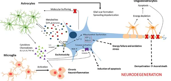
NTRODUCTIONCentral nervous system (CNS) is one of the complex systems in the body that consists of brain and spinal cord. Any disease or traumatic assault may lead to the degeneration of CNS including loss of homeostasis. CNS injuries constitute a major cause of morbidity and mortality includes the life threatening injuries such as traumatic brain injury (TBI) and spinal cord injury (SCI). TBI and SCI are caused by both primary and secondary injuries influencing the cascades of cellular and molecular events, which will cause further damage in the system, and loss of body functions. The consequences of the secondary injury include mitochondrial dysfunction, neurotransmitter accumulation, blood-brain barrier (BBB) and blood spinal cord barrier disruption, apoptosis, excitotoxic damage, initiation of inflammatory, and immune processes which is followed by initial primary mechanical trauma. Secondary injury involves the production of highly reactive species, reactive oxygen species (ROS), reactive nitrogen species (RNS), or free radicals which will cause damage to protein structure, DNA, and cell membrane and leads to oxidative stress which plays a major role in the pathophysiology of CNS injury. The progression of the damage starts from the primary impact on brain or spinal cord and will continue for hours, days, and weeks after the initial mechanical insult which will result in tissue damage (Samantaray et al., 2009; Khalatbary et al., 2010; Bains and Hall, 2012b; Bhalala et al., 2013; Naseem and Parvez, 2014).
Plus the stress response, autophagy is a highly essential cellular response to damage and influences the improvement and progression of post-traumatic disease (Wang et al., 2015). The term autophagy, from Greek “self-eating” refers to a range of processes, including chaperone-mediated autophagy, microautophagy and macroautophagy, which regulated process of degradation and recycling of cellular constituents, participated in organelle turnover and the bioenergetic management of starvation of spinal cord injury.
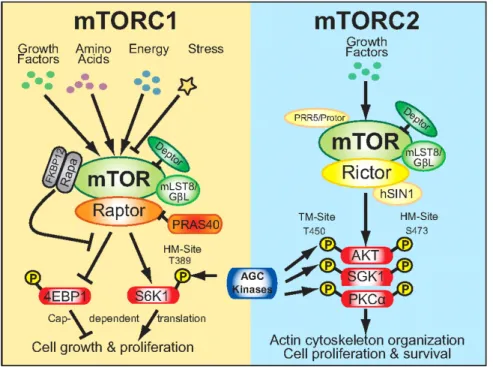
The mammalian target of rapamycin (mTOR), a conserved serine/threonine kinase, is the catalytic subunit of two fundamentally distinct complexes: complexes-mTOR complex 1 (mTORC1) and complexes-mTOR complex 2 (mTORC2) that individually plays an essential role in the control of cell proliferation. Both complexes localized to distinctive subcellular sections, thus affecting their initiation and role (Wullschleger et al., 2006; Betz and Hall, 2013). The mTORC1 stimulate protein synthesis by mRNA translation and cell development by entering the G1 phase of the cell cycle, however mTORC2, firstly identified as a regulator of the actin cytoskeleton, has been indicated to phosphorylate members of the AGC kinase
-7-
family, including Akt, which is linked to several pathological conditions (Menon and Manning, 2008; Foster and Fingar, 2010; Sparks and Guertin, 2010). They have distinctive downstream targets, different biological functions and importantly, different sensitivity to the drug rapamycin. mTORC1 is pharmacologically inhibited by short-term rapamycin management, whereas mTORC2 is resistant to short-term rapamycin treatment, although long-term treatment can prevent mTORC2 complex assembly (Phung et al., 2006; Sarbassov et al., 2006).
One of the most important mTOR inhibitor studied until today was Rapamycin.
Rapamycin, an inhibitor of the mTOR pathway, can extend lifespan and improve age-related functional decline in mice, thereby providing the first proof of principal that a pharmaceutical agent can slow the aging process in mammals. These outcomes have proven robust in repeated studies; however, their potential translational relevance towards a means to slow aging or prevent age-related disease in otherwise healthy humans remains unclear. Part of the challenge in addressing the potential of rapamycin (or its analogs) as a pro-longevity therapeutic lies in its known clinical risks for adverse side effects. Primary amongst these are metabolic defects that include hyperglycemia, hyperlipidemia, insulin resistance and increased incidence of new-onset type 2 diabetes. In healthy rodents, treatment with rapamycin also causes a relatively rapid, dose-dependent impairment of markers of glucose homeostasis. The natures of the metabolic effects/defects caused by rapamycin remain ambiguous regarding their role in longevity and healthy aging. Fang et al. suggested the effects of rapamycin on metabolism depend on the length of treatment with a detrimental effect on glucose metabolism in the short-term whereas mice treated chronically with rapamycin actually became insulin-sensitive. On the other hand, Blagosklonny has proposed that the presumed metabolic impairments caused by rapamycin may simply be a consequence of its action as a “starvation-mimetic” and, further, may be fundamentally required for its pro-longevity effect (Blagosklonny, 2011; Wilkinson et al., 2012; Fang et al., 2013; Miller et al., 2014).
Considering a lot of Rapamycin-induced side effect, in these years, a number of inhibitors of the PI3K/AKT/mTOR pathway has been identified such as Temsirolimus and KU0063794. Temsirolimus was the first mTORC1 inhibitor investigated in clinical trials in the late 1990s in patients with cancer. Is an ester derivative of rapamycin and it is a specific inhibitor of mTORC1 that interferes with the synthesis of proteins that regulate proliferation, growth, and
-8-
survival of tumor cells. Treatment with temsirolimus leads to cell cycle arrest in the G1 phase and stops tumor angiogenesis by reducing synthesis of VEGF (Duran et al., 2006).
KU0063794 is a second-generation mTOR inhibitor targeting mTORC1 and mTORC2, including p70S6K, 4E-BP1 and Akt. Specifically, inhibits the phosphorylation of S6K1 and 4E-BP1, which are downstream substrates of mTORC1, and it inhibits Akt phosphorylation on Ser473, which is the target of mTORC2 (Garcia-Martinez et al., 2009; Zhang et al., 2013). In a recent study it has been demonstrated that KU0063794 decreasing the viability and growth of renal cell carcinoma cell lines, Caki-1 and 786-O, and showed anti-fibrotic activity in Keloid disease (Syed et al., 2013; Zhang et al., 2013). Previously it has been showed that mTOR plays a key role in modulation of macrophage/microglia activation, reduction of IL-1β and TNFα production, expression of nitric oxide synthase, prevention of apoptosis neuronal loss and demyelination both in the first and second phases of the damage after injury (Kanno et al., 2012). Moreover, it has been demonstrated that rapamycin treatment significantly improved the neurological recovery from SCI and increased the number of surviving neurons at the lesion epicenter (Chen et al., 2013b).
However, the mechanism of autophagy related inflammation after SCI and TBI is still unclear. So, in this regard in this thesis we have evaluated the effect of Ku0063794, as potential treatments for inflammation in SCI and TBI models.
-9-
C
HAPTER1:
C
ENTRALN
ERVOUSS
YSTEMI
NJURY 1.1 SPINAL CORD INJURYSpinal cord injury has a significant impact on quality of life, life expectancy and economic burden, with considerable costs associated with primary care and loss of income. In one study, quadriplegics ranked recovery of arm and hand function as a priority, whereas paraplegics rated recovery of sexual function as most important (when measured against recovery of bladder/bowel function, and eradicating autonomic dysreflexia, improving walking movements and trunk stability, regaining normal sensation and eliminating chronic pain)(Anderson, 2004).
The normal architecture of the human spinal cord can be radically disrupted by injury (Bunge et al., 1993; Kakulas, 1999). SCI is heterogeneous in cause and outcome and can result from contusion, compression, penetration or maceration of the spinal cord. SCI leads to the death of cells, including neurons, oligodendrocytes, astrocytes, precursor cells, and any resulting cavities and cysts may interrupt descending and ascending axonal tracts, although circumferential white matter is often spared (Horky et al., 2006). The pathophysiology of SCI comprises both primary and secondary mechanisms of injury.
-10-
The “primary injury” refers to the forces that impart the primary mechanical insult to the spinal cord, which in its mildest form causes a cord concussion with brief transient neurologic deficits and in its most severe form causes complete and permanent paralysis. After the initial insult to the spinal cord, additional structure and function are lost through active “secondary damage” such as apoptosis of oligodendrocytes, loss of myelin, generation of oxidative stress, activation of immune system response and inflammation process (Crowe et al., 1997).
Demyelinated axons are observed up to a decade after human SCI, and the extent to which these axons survive unmyelinated or become remyelinated by central or peripheral myelin is a subject of ongoing investigation (Guest et al., 2005; Totoiu and Keirstead, 2005). Resident and invading inflammatory cells (including neutrophils, microglia, macrophages and T cells) can have a range of destructive and reparative roles. SCI culminates in glial scarring, a multifactorial process that involves reactive astrocytes, glial progenitors, microglia and macrophages fibroblasts and Schwann cells (Bruce et al., 2000; Jones et al., 2002; Jones et al., 2003; Jones et al., 2005). Progressive expansion of the injury across more than one segment (syringomyelia) can also occur over months or years, sometimes proving fatal (Silver and Miller, 2004; Fawcett, 2006).
In contrast to these destructive events, commonly observed pathological features do indicate some spontaneous repair after SCI (Beattie et al., 1997). Whereas there is little or no neurogenesis in the injured spinal cord, proliferation in the ependymal and peri-ependymal canal generates new precursor cells that exclusively differentiate into glial cells (Yamamoto et al., 2001; Azari et al., 2005; Yang et al., 2006). Limited axon sprouting does occur and lesions might even be spanned by trabeculae containing axon sprouts. Sprouting is largely impeded by geometrical and molecular factors, and few axons regenerate over long distances back to their original targets (Beattie et al., 1997; Hill et al., 2001; Pettigrew et al., 2001). However, various forms of cortical, brainstem and spinal plasticity occur that could contribute to limited compensatory recovery (Raineteau and Schwab, 2001; Weidner et al., 2001). After SCI, new spinal circuits can bypass the lesion, including sprouting of injured corticospinal axons onto spared, long descending propriospinal tracts that increase connectivity with lumbar motor neurons (Raineteau et al., 2002; Bareyre et al., 2004). Cortical sensorimotor areas can functionally rearrange and, at the subcortical level, the rubrospinal system can reorganize and compensate for much of the function lost after corticospinal injury (Raineteau and Schwab, 2001; Bareyre et al., 2004; Thuret et al., 2006).
-11-
Therefore, although there is some spontaneous repair after CNS injury, it is incomplete. Further recovery of function will require a combination of effective and safe therapeutic interventions
1.1.1CLASSIFICATION OF SPINAL CORD INJURY
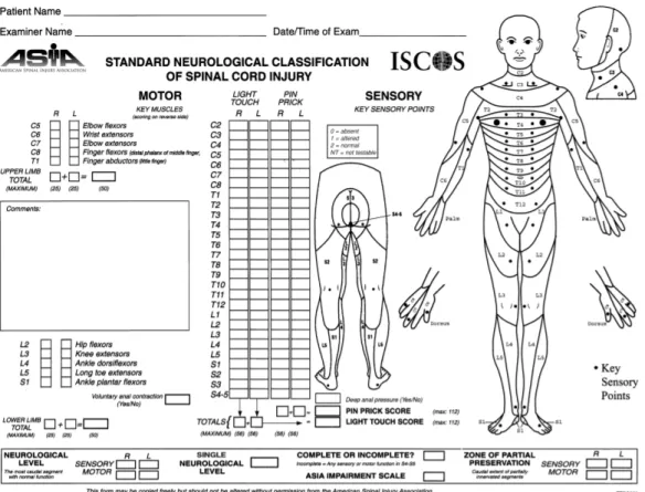
Injuries are classified in general terms of being neurologically “complete” or “incomplete” based upon the sacral sparing definition (Waters et al., 1991). “Sacral Sparing” refers to the presence of sensory or motor function in the most caudal sacral segments as determined by the examination (i.e. preservation of light touch or pin prick sensation at the S4-5 dermatome, DAP or voluntary anal sphincter contraction). A complete injury is defined as the absence of sacral sparing (i.e. sensory and motor function in the lowest sacral segments, S4-5), whereas an incomplete injury is defined as the presence of sacral sparing (i.e. some preservation of sensory and/or motor function at S4-5) (Kirshblum et al., 2011).
The following ASIA Impairment Scale (AIS) designation is used in grading the degree of impairment:
ü A = Complete. No sensory or motor function is preserved in the sacral segments S4-S5.
ü B = Sensory incomplete. Sensory but not motor function is preserved below the neurological level and includes the sacral segments S4-S5, AND no motor function is preserved more than three levels below the motor level on either side of the body. ü C = Motor incomplete. Motor function is preserved below the neurological level, and
more than half of key muscle functions below the single neurological level of injury have a muscle grade less than 3 (Grades 0–2).
ü D = Motor incomplete. Motor function is preserved below the neurological level, and at least half (half or more) of key muscle functions below the NLI have a muscle grade >3.
ü E = Normal. If sensation and motor function as tested with the ISNCSCI are graded as normal in all segments, and the patient had prior deficits, then the AIS grade is E. Someone without a SCI does not receive an AIS grade.
-12-
Figure 2 Standard neurological classification of spinal cord injury
1.1.2EPIDEMIOLOGY, INCIDENCE AND CAUSES
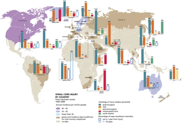
SCI epidemiology has been studied extensively over the past 40 years. Studies focused on descriptive epidemiology, including overall incidence rates, age, gender, race, cause of injury, level and completeness of injury (Kraus et al., 1975; Bracken et al., 1981; Griffin et al., 1985). Published reports of SCI incidence in the United States vary from 25 to 59 new cases per million population per year with an average of 40 per million (Acton et al., 1993; Price et al., 1994; Thurman et al., 1994; Johnson et al., 1997; Surkin et al., 1998). This would translate to approximately 12 400 new SCIs in 2010. The incidence of SCI in the rest of the world is much lower than in the United States (Devivo, 2012). There are several possible explanations for this. One is the relative absence in most countries of SCI because of acts of violence. However, there also appear to be fewer SCIs related to motor vehicle crashes in other countries. Possible explanations for this would be a lower average passenger miles of exposure, greater use of seat belts, or safer driving habits and road conditions. Conversely, lower incidence could also result from greater mortality at the site of the accident. Finally, incomplete case ascertainment may have occurred in many of these studies as they are not
-13-
typically population-based but rather rely on referrals to specialized centers. No studies have addressed the reasons for international variation in SCI incidence.
SCI incidence rates are lowest for the pediatric age group, highest for persons in their late teens and early twenties, and generally decline consistently thereafter, although some studies suggest a secondary increase in incidence rates among the elderly.(Acton et al., 1993; Price et al., 1994; Hagen et al., 2010). Among persons enrolled in the combined US data set, the mean age at injury has increased from 28.3 years during the 1970s to 37.1 years between 2005 and 2008 (DeVivo and Chen, 2011). These figures mirror the increasing median age of the general US population, which were 30 years in 1980 and 36.9 years in 2010.
The average age at injury is a few years higher in most other countries than in the United States (Devivo, 2012). This is likely because of the lower rate of injuries due to violence that typically occur among younger persons, although other factors such as the average age of the general population and differences in other cause-specific incidence rates also likely have a role in raising the average age at injury in other countries. In 2001, 13% of the US population was >65 years, compared with 18% in Japan and 15% in Europe.
Figure 3 Global mapping of spinal cord injury from traumatic
-14-
SCI occurs predominantly among men and will continue to do so in the future. SCI annual incidence rates are typically 3 to 4 times higher for men than women (Devivo, 2012).
However, the percentage of new injuries occurring among men in the combined US data set has declined slightly over time from 80.9% during the 1970s to 77.1% since 2000 (Devivo, 2012). A similar trend has occurred in Norway where the incidence rate was 5.3 times higher among males than females between 1952 and 1956, but only 4.2 times higher between 1992 and 2001 (Hagen et al., 2010). This trend toward a slightly increasing percentage of women among new SCIs should continue because injuries among older persons are increasing, and SCIs among the elderly are more evenly split between men and women than SCIs that occur among teenagers and young adults.
In the United States, motor vehicle crashes are the leading cause of SCI (Acton et al., 1993; Price et al., 1994; Devivo, 2012). Although the percentage of SCIs owing to motor vehicle crashes in the combined US data set has fluctuated over time, it is approximately the same today (48.3% since 2000) as it was during the 1970s (47.6%) (Devivo, 2012). Injuries due to acts of violence peaked in the 1990s (21%) but have since declined dramatically (12% since 2000). 18 Overall, sports-related SCIs have declined slightly from 14.2% during the 1970s to 10.0% since 2000 (Devivo, 2012). Injury prevention initiatives have reduced the occurrence of SCIs in many sports, most notable diving, American football and trampolines. However, SCIs from winter sports such as snow skiing have increased.
Falls are the leading cause of SCI among persons aged >60 (Price et al., 1994).Therefore, it is not surprising that the proportion of new SCIs owing to falls has been increasing steadily as injuries among older persons have become more frequent. During the 1970s, falls accounted for 16.2% of new SCIs in the combined US data set compared with 21.8% since 2000 (Devivo, 2012). This trend is likely to continue, with a corresponding decline in sports and violence-related SCIs that do not typically occur among older persons.
1.1.3PHARMACOTHERAPY
Pharmacotherapy early after SCI is aimed at neuroprotection to minimize the secondary injury. Several trials with different neuroprotective pharmacological agents have been reported in literature. These pharmacological agents block one or more of the mechanisms of secondary injury following trauma to the cord. Unfortunately, till date, no neuroprotective pharmacological agent has been conclusively shown to be clinically effective in human subjects with SCI (Tator, 2006).
-15-
1.1.3.1 Methylprednisolone
Animal studies have demonstrated that methylprednisolone (MP) reduces the secondary cord injury following trauma by inhibiting lipid peroxidation, improving spinal blood flow, enhancing the postinjury activity of Na+/K+-ATPase, and facilitating the recovery of extracellular calcium ions (Young, 1991; Hall et al., 1992). Three MP trials have been reported from North America (Bracken et al., 1984; Bracken et al., 1990; Bracken et al., 1997). The NASCIS 2 trial (Bracken et al., 1990) suggested clinical efficacy that did not translate into effectiveness. Subsequent study from France (Pointillart et al., 2000) and evidence-based analysis (Hurlbert, 2000; Short et al., 2000; Hurlbert, 2001; Hurlbert, 2006) have not supported the routine use of MP in acute SCI. Despite equivocal effectiveness, MP is widely used in many North American centres, for want of a better alternative. The drug is given within 8 h of injury in a bolus dose of 30 mg/kg followed by a maintenance dose of 5.4 mg/kg/h over next 23 h.
1.1.3.2 GM1 Ganglioside
Animal studies have shown that monosialotetrahexosylganglioside (GM-1) ganglioside enhances the functional recovery of damaged neurons (Ferrari and Greene, 1998). The first randomized control trial, with 34 patients, suggested beneficial effects of GM-1 (Geisler et al., 2001). However, a later adequately powered multicenter study failed to establish the effectiveness of this agent in SCI (Geisler et al., 2001).
1.1.3.3 Thyrotropin Releasing Hormone
Thyrotropin releasing hormone (TRH) is a partial endorphin antagonist. Endorphins are released after SCI and postulated to exacerbate posttraumatic ischemia by reducing spinal blood flow secondary to systemic hypotension (Faden et al., 1981). A smaller study (Pitts et al., 1995) suggested beneficial effects of TRH in SCI. A larger study to evaluate the efficacy has not been carried out.
1.1.3.4 Nimodipine
Nimodipine is a Ca2+ channel blocker. It can potentially minimize the injury following SCI by counteracting vasospam, ischemia, and infarction that contribute to secondary damage(Guha
-16-
et al., 1987; Pointillart et al., 1993). A single center randomized trial that compared four treatment arms viz. nimodipine, MP, MP and nimodipine, and placebo failed to demonstrate any significant benefits of nimodipine alone or in combination with MP (Pointillart et al., 2000).
1.1.3.5 Gacyclidine
Gacyclidine is an N-methyl-D-aspartate antagonist. It blocks the toxic effects of glutamate, which is released following SCI. A multicenter RCT failed to establish efficacy of Gacyclidine at 1 year following SCI (Tator, 2006).
1.1.3.6 Minocycline
Minocycline is a tetracycline derivative (a bacteriostatic antibiotic) that is currently in common clinical use for the treatment of acne and chronic periodontitis. Animal experiments have shown minocycline has a neuroprotective effect following SCI. It reduces axonal loss at the site of injury, decreases oligodendrocyte apoptosis, and prevents activation of microglia and macrophages (Lee et al., 2003; Wells et al., 2003; Stirling et al., 2004). A pilot study to evaluate minocycline in patients with acute spinal cord injuries has been initiated in Calgary, Alberta, Canada (Kwon et al., 2005).
1.1.3.7 Estrogen
Early treatment with the estrogen 17 β-estradiol has been shown to have a neuroprotective effect following experimental SCI in rats (Yune et al., 2004; Sribnick et al., 2006). 17β-Estradiol reduces apoptosis in the penumbra around the zone of necrosis, thereby minimizing the secondary damage.
-17- 1.2 TRAUMATIC BRAIN INJURY
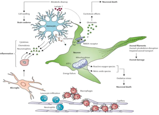
Traumatic brain injury is a nondegenerative, noncongenital insult to the brain from an external mechanical force, possibly leading to permanent or temporary impairment of cognitive, physical, and psychosocial functions, with an associated diminished or altered state of consciousness (Topal et al., 2008). TBI can manifest clinically from concussion to coma and death. Injuries are divided into 2 subcategories: primary injury, which occurs at the moment of trauma, and secondary injury, which occurs immediately after trauma and produces effects that may continue for a long time. Changes to the cerebral environment involve a complex interplay between cellular and molecular processes, in which glutamate-driven excitotoxic effects, oxidative stress, inflammation, ion imbalance, and metabolic disarray are major components. These pathways induce progressive neuronal loss through necrosis and apoptosis (Morganti-Kossmann et al., 2007; Kalia et al., 2008). Also, important are the intracellular changes that are determined by the excessive influx of calcium, which affects mitochondrial integrity, depleting cells of an essential source of energy. The metabolic disarray caused by accumulation of lactate results in cytotoxic swelling of cells, which, together with the increased permeability of the cerebral vasculature, leads to brain oedema, elevated intracranial pressure, and reduced cerebral perfusion(Morganti-Kossmann et al., 2007; Kalia et al., 2008; Bains and Hall, 2012a).
-18-
In addition, regardless of origin, TBI sufferers experience a relatively stereotyped array of symptoms associated with the injury: dizziness, confusion, and sometimes loss of consciousness (especially in severe injury). Even after the initial injury is managed and resolved, approximately 70–80% of TBI patients develop long-lasting effects such as changes in personality and cognition, anxiety and depressive-like behaviors (Whitnall et al., 2006; Kiraly and Kiraly, 2007; Draper and Ponsford, 2008; Ponsford et al., 2008). TBI also increases the risk for certain neurodegenerative conditions. For instance, repeated concussive TBI has been associated with the development of chronic traumatic encephalopathy (CTE) in athletes. Furthermore, both repeated and single TBI show a strong association with increased Alzheimer's disease (AD) risk or earlier AD onset. Correlations with Parkinson's disease and amyotrophic lateral sclerosis (ALS) have also been reported, but the supporting evidence is not as strong as for CTE and AD (Gyoneva and Ransohoff, 2015).
Considering the high prevalence of TBI and its association with serious neurological problems and risk for neurodegenerative diseases, there is a strong impetus to develop new TBI therapies that not only promote cell survival immediately after the injury but also address the development of secondary pathology. Because of the close association between neuropathology and inflammation in space and time, the latter has emerged as an important target for the amelioration of TBI (Das et al., 2012; Giunta et al., 2012; Woodcock and Morganti-Kossmann, 2013).
1.2.1CLASSIFICATION OF TRAUMATIC BRAIN INJURY
Glasgow Coma Scale (GCS) is the most widely used scoring procedure for mental and
neurological status following head injury in the U.S. and most English-speaking countries (Gouvier et al., 1987). Its score is based on the sum of three components: eye opening response, verbal response, and best motor response. For instance, if an individual at the accident scene opened eyes to voice, used inappropriate words, and demonstrated a flexion response to motor stimulation, the scoring would be E + V + M = 3 + 3 + 4 = 10 (see following table).
-19-
This in turn produces a graded score in the moderate severity range. The GCS can be further subdivided into mild injury (GCS = 13 to 15), moderate injury (GCS = 9 to 12), and severe injury (GCS = 3 to 8). The clinical features of mild injury are loss of consciousness for 20 minutes, no focal neurological signs, no intracranial mass lesion, and no intracranial surgery. Regardless of mental state, a focal computed tomography (CT) lesion places the patient into the moderate category. A coma duration of at least 6 h places the patient into the severe category, regardless of mental state.
In terms of outcome, the most commonly used current scales are the Glasgow Outcome Scale and the Rancho Los Amigos Level of Cognitive Functioning Scale (see following table).
Table 1 – Glasgow Coma Scale.
-20-
The Rancho Scale is widely used by rehabilitation facilities after the patient leaves the neurosurgical intensive care unit or neurosurgical floor for postacute care. Generally, a final grading using the Rancho Scale is made prior to the patient’s discharge from a brain injury rehabilitation unit if such is required (Gouvier et al., 1987).
1.2.2EPIDEMIOLOGY, INCIDENCE AND CAUSES
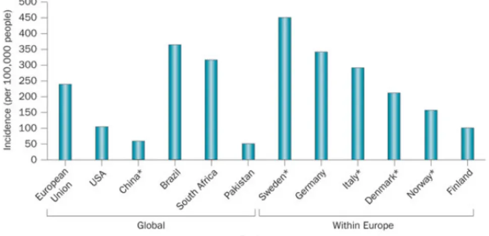
Each year, approximately 30 million injury-related emergency department (ED) visits, hospitalizations, and deaths occur in the United States. Of the injury hospitalizations, approximately 16% included TBI as a primary or secondary diagnosis. Of the injury deaths, approximately one-third included a TBI as a direct or underlying cause of death. In 2010, CDC estimated that TBIs accounted for approximately 2.5 million ED visits, hospitalizations, and deaths in the United States, either as an isolated injury or in combination with other injuries. Of these persons, approximately 87% (2,213,826) were treated in and released from EDs, another 11% (283,630) were hospitalized and discharged, and approximately 2% (52,844) died.
Figure 5 - Estimates of the global incidence of TBI
These figures, however, underestimate the occurrence of TBIs, as they do not account for those persons who did not receive medical care, had outpatient or office-based visits, or those who received care at a federal facility (i.e., persons serving in the U.S. military or seeking care at a Veterans Affairs hospital) (Faul, 2010). Department of Defense data revealed that from 2000 through 2011 235,046 service members (or 4.2% of the 5,603,720 who served in
-21-
the Army, Air Force, Navy, and Marine Corps) were diagnosed with a TBI (CDC, NIH, DoD, and VA Leadership Panel, 2013). In the United States, children aged 0–4 years, adolescents aged 15–19 years, and older adults aged ≥75 years are the groups most likely to have a TBI-related ED visit or hospitalization (Faul, 2010). Adults’ aged ≥75 years have the highest rates of TBI-related hospitalizations and deaths among all age groups. Overall, males account for approximately 59% of all reported TBI-related medical visits in the United States (Faul, 2010). As shown in Table 4, during 2002—2010, the leading causes of TBI-related ED visits were falls, being struck by or against an object, and motor-vehicle traffic crashes. The leading causes of TBI-related hospitalizations were falls, motor-vehicle traffic incidents, and assaults. For TBI-related deaths, the leading causes were motor-vehicle traffic incidents, suicides, and falls (Coronado et al., 2012). The proportion of TBIs occurring during sports and recreation-related activities are undetermined because of limitations of the data source. However, according to the National Electronic Injury Surveillance System – All Injury Program, during 2001–2009 (CDC, 2011) the activities associated with the greatest estimated number of TBI-related ED visits were bicycling, football, playground activities, basketball, and soccer among persons younger than 19 years.
1.2.3PHARMACOTHERAPY
Despite substantial investments by government, philanthropic, and commercial sources over the past several decades, TBI remains an unmet medical need and a major source of disability and mortality in both developed and developing societies.
-22-
1.2.3.1Acetylcholinesterase inhibitors
Central acetylcholinesterase inhibitors (AChEI) increase synaptic acetylcholine by inhibiting its breakdown in the synaptic cleft. Studies of these compounds for the treatment of patients with TBI suggest they may have potentially beneficial effects—particularly in patients with chronic moderate and severe TBI who have persistent cognitive deficits—by increasing synaptic ACh levels (Tenovuo, 2005). Beneficial effects have been reported in pre-clinical TBI studies with AChEI, including positive effects on acute injury processes with reduced TBI-induced neuronal death, preservation of neurons in the CA1 hippocampal region, reduced blood–brain barrier (BBB) disruption, decreased vasogenic brain edema, and preserved neurologic and motor function (Chen et al., 1998; Ballesteros et al., 2008)..
1.2.3.2 Amantadine
Amantadine (1-adamantamine hydrochloride) is a tricyclic amine used for the prophylaxis and treatment of influenza A and was serendipitously discovered to have modest efficacy for the treatment of Parkinson disease (PD). The anti-parkinsonian mechanism of action is not fully understood, but research has suggested that amantadine increases extracellular dopamine (DA) concentrations either by blocking DA reuptake or facilitating DA synthesis (Bales et al., 2009). Amantadine may also have post-synaptic effects on DA circuits by increasing DA receptor density (Gianutsos et al., 1985). One study showed that amantadine treatment, starting 1 day after a closed controlled cortical impact model of TBI in rats and continuing for 18 days after injury, resulted in modest improvement in Morris water maze (MWM) latencies (Dixon et al., 1999).
1.2.3.3 Cyclosporine A/FK 506
Cyclosporine A (CsA) inhibits opening of the mitochondrial permeability transition pore after TBI, thereby maintaining mitochondrial membrane potential. Many studies in animal models of TBI have suggested that this action of CsA confers benefit by preserving mitochondrial function and reducing reactive oxygen species (Brustovetsky and Dubinsky, 2000; Sharov et al., 2007) Inhibition of the protein phosphatase calcineurin via the immunophilin effects of CsA also has beneficial effects on axonal injury and learning and memory (Van Den Heuvel et al., 2004; Setkowicz and Guzik, 2007). Similarly, immunosuppressive effects, also mediated by calcineurin inhibition, may further confer benefit after TBI or mediate potential side effects. The related compound, FK 506, inhibits calcineurin and exhibits
-23-
immunosuppressive effects but does not inhibit opening of the mitochondrial permeability transition pore (Singleton et al., 2001).
1.2.3.4 Progesterone
Progesterone is a steroid that is made in the brain, in addition to its synthesis in the reproductive organs and adrenal glands. Progesterone has pleiotropic effects, and thus has multiple candidates for mechanisms of action with regard to its potential therapeutic efficacy in TBI (Hammond et al., 1983). Multiple pre-clinical models of TBI have demonstrated neuroprotective properties of progesterone and have shown that it enhances behavioral and functional outcomes, decreases cerebral edema, apoptosis, pro-inflammatory cytokines, and other markers of inflammation, and prevents neuronal cell death (Liu et al., 2009). Progesterone also enhances myelination, neurogenesis, and impacts aquaporin expression, and modulates neurotrophin expression, among other actions (Koenig et al., 1995; Liu et al., 2009).
1.2.3.5 Simvastatin/other statins
Statins, 3-hydroxy-3-methylglutaryl coenzyme A (HMGA) reductase inhibitors, reduce serum cholesterol but also have potent effects in the brain relevant to mechanisms of TBI injury and recovery. Such effects target mechanisms that influence both the acute and chronic phases of TBI (Li et al., 2009; Chauhan and Gatto, 2010). There is pre-clinical evidence of beneficial effects including those on acute injury processes such as brain edema, BBB integrity, cerebral blood flow, neuroinflammation, axonal injury, and cell death, in addition to effects on key facets of regeneration such as trophic factor production. A variety of molecular outcomes are influenced including TUNEL staining, CREB, Akt, eNOS, FOXO1, NF-κB, GSK3, cytokines, BrdU labeling, blood vessel formation, and vascular endothelial growth factor (Chen et al., 2009a).
1.2.3.6 N-acetylcysteine (NAC)
NAC is FDA-approved as an antidote for acetaminophen overdose and as a mucolytic for cystic fibrosis and other bronchopulmonary diseases. In animal models of TBI, NAC has shown strong antioxidant activity by increasing glutathione levels and decreasing markers of oxidative damage (Hicdonmez et al., 2006). NAC also showed anti-inflammatory activity by decreasing the activation of NF-κB, while lowering interleukin (IL)-1β, tumor necrosis factor
-24-
(TNF)-α, and intercellular adhesion molecule (ICAM)-1 levels (Yi and Hazell, 2005; Chen et al., 2008). It is unclear how the anti-inflammatory action of NAC is related to its antioxidant activity. NAC has been shown to reduce lesion volume while simultaneously reducing levels of the putative neuroprotective enzyme heme oxidase (Yi and Hazell, 2005; Chen et al., 2008).
1.2.3.7 Growth hormone (GH)
GH is a polypeptide that is synthesized, stored, and secreted by somatotrophic cells within the lateral wings of the anterior pituitary gland. GH deficiency/insufficiency (GHD/GHI) is the most common anterior pituitary abnormality after TBI. Manipulating the GH axis has been shown to improve motor function, enhance learning and memory retention after TBI in rats, and to improve spatial learning and memory in a mouse model of AD (Saatman et al., 1997; Doulah et al., 2009).
-25-
C
HAPTER2:
T
HER
OLE OFN
EUROINFLAMMATION2.1CYTOKINE RESPONSES TO INFLAMMATION
Cytokines are small and nonstructural proteins with no amino acid sequence motif, their biological activities allow us in turn to group them into different classes: exit 18 cytokines called interleukin (IL), some of these promote inflammation and are named pro-inflammatory cytokines such as IL1β and IL1α, IL6, IL8 and TNFα; whereas other cytokines suppress the activity of pro-inflammatory cytokines and are called anti-inflammatory cytokines such as IL-4, IL-10, TGFβ. The hypothesis that some cytokines function primarily induce inflammation while others suppresses inflammation is essential to cytokine biology and to clinical medicine.
Cytokines are secreted by a variety of immune cells such as T-lymphocytes and macrophages, as well as b non-immune cells such as fibroblasts; the physiological effects mediated by cytokines comprise the stimulation or inhibition of cell growth, cytotoxicity/apoptosis, antiviral activity and inflammatory responses. The main function of cytokines is the regulation of T-cell differentiation from undifferentiated cells to T-helper 1 and 2, regulatory T cells, and T-helper 17 cells (Steinman, 2007). These regulatory proteins include ILs, interferons (IFNs) and TNFs. Many of these cytokines have already been shown to be produced by neurons or glia in CNS disorders in which they are notably increased.
-26-
The cytokine class of inflammatory mediators is secreted by microglia and astrocytes and their production is increased in inflammatory states, moreover, they act by modulating the intensity and duration of the immune response. Pro-inflammatory cytokines and chemokines up-regulate microbicidal activity of neutrophils, and they can be considered as additional immunomodulatory agents to treat serious or refractory infections in humans.
Through cytokines IL-1 initiate the immune response, having a crucial role in the onset and expansion of a complex hormonal and cellular inflammatory cascade; the IL-1 family of cytokines includes IL-1α and IL-1β, which generate cell activation upon binding with specific membrane receptors and has been documented that IL-1 play a role in neuronal degeneration. In astrocytes, IL-1 induces IL-6 production, stimulates iNOS activity (Hausmann, 2003), enhances neuronal acetylcholinesterase activity, microglial activation and additional IL-1 production, and astrocyte activation.
Another important pro-inflammatory cytokine is the IL-6, a multifunctional cytokine that plays an important role in host defense (Okada et al., 2004), and possess main effects during the inflammatory response (Roxburgh and McMillan, 2016). IL-6 is associated to the neuropoietin cytokine’s family and it possess direct and indirect neurotrophic effects on neurons (Teng and Tang, 2006); moreover, IL-6 promotes astrogliosis (Morales et al., 2010), activates microglia (Inoue, 2002), and stimulates the release of acute phase molecules.
2.1.1INFLAMMATORY MEDIATOR: ROLE OF TNFα
Through all the cytokines involved in the secondary damage of SCI and TBI, TNF-α plays a crucial role in fact it is release shortly after injury, it can accumulate rapidly at the site of injury and it is produced by a number of different cell populations, such as neutrophils, macrophages and microglia, astrocytes and T cells (Yan et al., 2001). Several cell types are able to produce TNFα, including macrophages after its activation, dendritic cells, monocytes, NK cells, CD4+ T cells, CD8+ T cells, microglia and astrocytes. Macrophages/monocytes are able to produce TNFα in the acute phase of inflammation and this cytokines drives several range of signalling events within cells, leading to necrosis or apoptosis.
Several biological functions are ascribed to the TNFα and for this reason the mechanism of action is somewhat complex; although it inhibits the growth of tumor cells and it has an enhancing effect on the proliferation of normal cells (Mandi et al., 1991) TNF-α is takes part in septic shock, autoimmunity and inflammatory disorders. The major role of TNFα is explicated as mediator in resistance against infections; moreover, it was postulated that TNF
-27-
plays a pathological role in a number of autoimmune pathology such as graft vs host rejection or rheumatoid arthritis. Moreover, TNFα possess potent pro-inflammatory effects that are associated to its capacity to generate endothelial cell adhesion molecules and subsequently support neutrophil adherence to vascular endothelium. Neutrophils are exquisite targets of TNF-α that, under certain conditions, strengthens their expression of adhesion molecules, induces their degranulation and successive release of lysosomal enzymes, causing the production of highly reactive oxygen species. TNF-α-induced the migration of neutrophils mediating the production of chemotactic factors, including IL-8, this testifies cytokine networking involvement in inflammatory cell recruitment and an active role in inflammation. TNFα works by binding and clustering high-affinity receptors that are present in a great numbers on most cell membranes (Loetscher et al., 1991), the ligand/receptor complex is easily internalised via clathrin-coated pits and ends up in secondary lysozymes where it is degraded. Interestingly the binding of TNFα to the 75 kDa TNFR-2 is not sufficient to reach cytotoxicity, but rather binding to the 55 kDa TNFR-1 is sufficient to reach TNFα mediated cell killing. TNFα exerts its effects by activating several secondary proteins that provoke a variety of responses within the cell such as activation of gene transcription and/or production of reactive oxygen or nitrogen radicals (e.g., NO). Activated proteins include Gprotein, transcription factors such as NF-κB and AP-1 and serine and cysteine proteases, known as caspases. Many members of the TNF receptor superfamily have intracellular “death domains,” which represent protein interaction domains each consisting of 65–80 amino acids; these proteins participate in TNFα mediated apoptosis process; many evidence demonstrated that TNF–TNFR interactions are implicated in the pathogenesis of CNS disorders such as EAE and MS. These interactions are able to monitor the disease outcome by modifying immune response and the interactions between CNS-resident cells and effector immune cells in the CNS.
However, recent studies showed a dual nature for TNF-α not only neurotoxic but also it can be neuroprotective; a study conducted with transgenic mice for TNF-α receptors demonstrated that the mice lacking for TNF-α showed more tissue loss and functional deficits compared to wild-type mice, implying that TNF-α mediated a neuroprotective effect (Kim et al., 2001). The beneficial or deleterious effects of TNF-α dependent when it is being released and on cellular populations that is acting on, the conflicting actions of TNF-α described above reflects a growing view of inflammation as a “dual-edged sword,” having neurotoxic and neuroprotective properties (Bethea, 2000).
-28-
Thus, comprehension of their profile, kinetics of expression and interactions between TNF-α ligands and their TNFRs on different CNS residents and infiltrating immune cells, would aid to better design strategies to control neuroinflammation and CNS autoimmunity. Blockers of TNF α have been acknowledged for human use in treating TNF-linked autoimmune and inflammatory disorders. Pathways downstream of receptor ligation supply critical points for interjection for planning new therapeutic strategies.
2.2MICROGLIA ACTIVATION
Moreover, another important mediators of inflammation that respond rapidly to disturbances within the microenvironment by change in morphology are the microglia, the expression ‘activated microglia’ is used to define cells that change their immunophenotype and their morphology after a specific stimuli; the principal role of microglia at the lesion site is a rapid phagocytosis of fragments and induction of apoptosis (Shuman et al., 1997). The different response of microglia in vitro suggest that these cells may elicit unique functional properties, and consequently control the inflammatory response at the injury site. Microglial activation has been well-known in the spinal cord tissue that receive a trauma and has been shown to occur from caudal to lumbar enlargement, based on that there are papers supporting the role of microglia in pain after injury and showing activation of microglia post-SCI.
Microglia activate the innate immune system and are key regulators of inflammatory processes in CNS pathologies such as trauma and neurodegenerative diseases participation in both acute and chronic phase of the inflammatory responses. Activated microglia secrete cytotoxic substances including various cytokines such as TNF-α, IL-1, reactive free radicals, and nitric oxide. However, the principal effects of microglia at the levels of the lesion core are probably rapid phagocytosis of debris rather than induction of apoptosis. Microglia when activated can cause neuronal and glial toxicity through the release of cytokines, free radicals, eicosanoids, activated neutrophils and macrophages (Schwartz, 2000). On the other hand, microglia activation lead to beneficial effects producing growth factors that are fundamental for neuronal and tissue restoration. Moreover, it has been demonstrated that transplantation of peripherally activated macrophages has beneficial effects on spinal cord regeneration.
2.3APOPTOSIS
In the last decade the generation of apoptotic process after SCI and TBI was also confirmed, apoptosis can be triggered by a variety of insults including cytokines, inflammatory injury, free radical damage and excitotoxicity. The apoptotic process after SCI and TBI is activated
-29-
in neurons, oligodendrocytes, microglia, and perhaps, astrocytes; apoptosis in microglia contributes to inflammatory secondary injury.
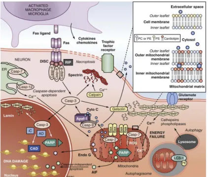
Two main pathways of apoptosis—extrinsic or dependent and intrinsic or receptor-independent—have been well characterized, and both appear to be active in SCI and TBI; the extrinsic or receptor-dependent pathway is mediated by Fas ligand and Fas receptor (Leskovar et al., 2000) and/or inducible nitric oxide synthase production by macrophages (Satake et al., 2000), while intrinsic or receptor-independent pathway is mediated via direct caspase-3 proenzyme activation (Citron et al., 2000) and/or mitochondrial damage, release of cytochrome c and activation of the inducer caspase-9, pathways of caspase-mediated apoptotic death (Hartley et al., 2000).
Receptor-dependent apoptosis is evoked by extracellular signals, the most significant of which is TNF, so the term of “extrinsic” pathway; tumor necrosis factor is known to rapidly accumulate in the injured spinal cord, and activation of the Fas receptor of neurons, microglia, and oligodendrocytes induces a programmed sequence of caspase activation. Moreover, additional control of cell death/survival is provided by the balance between major proapoptotic proteins such as Bax, Bad, and Bid and antiapoptotic proteins such as Bcl-XL and Bcl-2.
Apoptotic process that is activated in the secondary injury in SCI has recently come under close study, and the precise contribution and potential therapeutic implications of apoptosis in
-30-
SCI could be help to generate new therapeutic approach to treat the secondary events associated to spinal cord injury.
2.4INFLAMMATORY/IMMUNOLOGIC RESPONSE
The inflammatory and immunological response to injury within the CNS, is different than that which is occurring in other tissues (Schwartz et al., 1999). The inflammatory and immunologic responses to injury involve activation of innate immune cells that provide immediate defense against inflammatory stimuli and in turn help to recruit cells of the adaptive immune system (i.e., T and B lymphocytes). The activation of immune system is driven by interactions involving presentation of antigen and release of various inflammatory mediators (Ling et al., 2003). Also, cells present in the injury site may sequester debris and carry CNS antigens to secondary lymphoid organs (Karman et al., 2004), where trigger lymphocyte activation. Recent studies in mice showed that the number of activated T and B cells increases in the spleen and bone marrow within 24 hours of trauma (Ankeny et al., 2006).
2.4.1LYMPHOCYTES INFILTRATION
Under normal conditions, activated T cells can cross the BBB and enter the CNS parenchyma. In contrast with other inflammatory cells enrolled after a trauma, the number of lymphocytes remains low (Schnell et al., 1999); however, T-lymphocytes play an important role in the CNS immune system, since on activation, T-lymphocytes may kill target cells and produce cytokines (Kierdorf et al., 2010).
Once lymphocytes enter to the lesion site, they persist indeterminately (Sroga et al., 2003; Ankeny et al., 2006), whereas T and B cell increase in the lesion site at least 9 weeks post-injury (Bilgen et al., 2002; Whetstone et al., 2003), suggesting that cytokine/chemokine gradients exist chronically and regulate integrin expression on endothelia and cells (Lee et al., 2000; Babcock et al., 2003). These chemokine gradients and adhesion molecules represent molecular targets for manipulating the effects of intraspinal lymphocytes after SCI (Eng and Lee, 2003; Gonzalez et al., 2003; Bao et al., 2004); the progressive increase in lymphocyte numbers may also be justified by lymphocyte activation and proliferation within the injured centre of spinal cord.
-31-
Figure 9 - Cellular immune response in SCI
Moreover, induction of immune response could be generating as impaired nerve transmission; increasing production of pro-inflammatory cytokines in chronic phase of SCI could worsen the damage increasing the axonal injury and demyelination. Furthermore, there are evidences that autoreactive lymphocytes promote neuronal survival in vivo through activation not only of autoreactive-T-cell but also through activation other non- CNS-reactive T cells or B cells such as resident microglia and infiltrating macrophages.
Thus, because lymphocytes remains for long term at the site of the lesion, new strategy of treatment could orientate on this cells that posses a fundamental role in regulating degenerative and regenerative processes after injury.
2.5DUAL ROLE OF INFLAMMATION IN SPINAL CORD AND BRAIN INJURY
Based on manifold experimental and clinical studies published in recent years, there is conflicting evidence on the role of the neuroinflammatory response in the injured CNS. Many of the formerly designated “proinflammatory” mediators have shown to possess potential effects in mediating deleterious as well as repair processes in the CNS.
“Classic” inflammatory cytokines like TNF have been historically determined as harmful mediators based on in vitro data of neurotoxicity and in vivo data showing neuroprotection by
-32-
pharmacological inhibition of TNF in various models of neuroinflammation and neurodegeneration (Shohami et al., 1999)
Moreover, many pro-inflammatory mediators have been found to induce neurotrophin production after brain injury, as demonstrated for cytokines (e.g., IL-1β, IL-6), chemokines (e.g., IL-8), and inflammatory complement activation fragments (e.g., C3a) (Stahel, 2004). In addition to neurotrophin induction, the neuropoietic cytokines like IL-6 have been shown to possess additional mechanisms of neuroprotection in TBI and SCI models, by mediating the generation of antioxidants, such as metallothioneins (Penkowa et al., 2003a; Penkowa et al., 2003b).
Altogether, in order to reconcile the apparently conflicting reports of beneficial and deleterious effects of various pro-inflammatory mediators, the exact timing and extent of mediator production and activation must be taken into account, as well as the presence of additional factors which may take over redundant functions, e.g., in neuropathology models with use of genetically engineered mice. Thus, appropriate context of concomitant factors and the kinetics and localization of inflammatory mediator expression and activation will determine the harmful or protective properties in the context of neuroinflammation.
-33-
C
HAPTER3:
A
UTOPHAGY AND MTOR
3.1 AUTOPHAGY
The term ‘autophagy’, derived from the Greek meaning ‘eating of self’, was first coined by Christian de Duve over 40 years ago, and was largely based on the observed degradation of mitochondria and other intra-cellular structures within lysosomes of rat liver perfused with the pancreatic hormone, glucagon (Glick et al., 2010). The mechanism of glucagon-induced autophagy in the liver is still not fully understood at the molecular level, other than that it requires cyclic AMP induced activation of protein kinase-A and is highly tissue-specific (Yin et al., 2008). In recent years the scientific world has ‘rediscovered’ autophagy, with major contributions to molecular understanding and appreciation of the physiological significance of this process coming from numerous laboratories (Mizushima, 2007; Xie and Klionsky, 2007; Levine and Kroemer, 2008; Nakatogawa et al., 2009).
Although the importance of autophagy is well recognized in mammalian systems, many of the mechanistic breakthroughs in delineating how autophagy is regulated and executed at the molecular level have been made in yeast (Saccharomyces cerevisiae) (Xie and Klionsky, 2007; Nakatogawa et al., 2009). Currently, 32 different autophagy-related genes (Atg) have been identified by genetic screening in yeast and, significantly, many of these genes are conserved in slime mould, plants, worms, flies and mammals, emphasizing the importance of the autophagic process in responses to starvation across phylogeny.
-34-
3.2MAJOR TYPES OF AUTOPHAGY
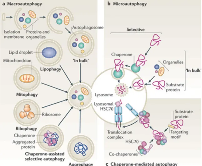
Based on the type of cargo delivery, there are three types of autophagy systems in mammals macroautophagy (autophagy), microautophagy, and chaperone-mediated autophagy.
3.2.1MACROAUTOPHAGY (AUTOPHAGY)
Whole regions of the cytosol are sequestered and delivered to lysosomes for degradation. Cargo sequestration occurs in the autophagosome, a double-membrane vesicle that forms through the elongation and sealing of a de novo generated membrane (Ohsumi and Mizushima, 2004).
This limiting membrane originates from a tightly controlled series of interactions between more than 10 different proteins which resemble the conjugation steps that mediate protein ubiquitinization (Cuervo, 2010). Formation of the limiting membrane also requires the interaction between a protein and a specific lipid molecule, regulated by conjugating enzymes.
-35- 3.2.2MICROAUTOPHAGY
Microautophagy is the direct uptake of soluble or particulate cellular constituents into lysosomes. It translocates cytoplasmic substances into the lysosomes for degradation via direct invagination, protrusion, or septation of the lysosomal limiting membrane. In other words, microautophagy involves direct invagination and fusion of the vacuolar/lysosomal membrane under nutrient limitation. The limiting/sequestering membrane is the lysosomal membrane, which invaginates to form tubules that pinch off into the lysosomal lumen. Microautophagy of soluble components, as in macroautophagy (autophagy), is induced by nitrogen starvation and rapamycin. Microautophagy is controlled by the TOR and EGO signaling complexes, resulting in direct uptake and degradation of the vacuolar boundary membrane (Uttenweiler et al., 2007). Hence, this process could compensate for the enormous influx of membrane caused by autophagy.
It seems that microautophagy is required for the maintenance of organelle size and membrane composition rather than for cell survival under nutrient restriction. Uttenweiler et colleagues have identified the vacuolar transporter chaperone, VTC complex, required for microautophagy (Uttenweiler et al., 2007). This complex is present on the endoplasmic reticulum and vacuoles, and at the cell periphery. Deletion of the VTC complex blocks microautophagic uptake into vacuoles.
3.2.3CHAPERONE-MEDIATED AUTOPHAGY
Chaperone-mediated autophagy (CMA) has been characterized in higher eukaryotes but not in yeast. Because of the particular characteristics of this type of delivery, only soluble proteins, but not whole organelles, can be degraded through CMA (Cuervo, 2010). CMA is dependent on the constitutively expressed heat shock cognate 70 (Hsc70), shares 80% homology with the heat shock protein 70 (Hsp70), and identifies peptide sequences of cytoplasmic substrates; thus, it is more selective than autophagy in its degradation (Hoffman et al., 2012).
CMA serves to balance dysregulated energy, and is maximally activated by nutrient/metabolic and oxidative/nitrosative stresses. Cross talk between CMA and autophagy is likely. CMA differs from the other two types of autophagies with respect to the mechanism for cargo selection and delivery to the lysosomal lumen for degradation. In other words, CMA is involved in the delivery of cargo, which does not require the formation of intermediate vesicles, membrane fusion, or membrane deformity of any type. Instead, the substrates are translocated from the cytosol directly into the lysosomal lumen across the membrane in a