Abstract
Cribriform is a histopathological term used to describe a neo-plastic epithelial proliferation in the form of large nests perforated by many quite rounded different-sized spaces. This growth pattern may be seen in carcinomas arising in different organs, and shows important prognostic implications. Therefore, recent data in liter-ature suggest that cribriform carcinoma is a histologically and clinically distinctive type of tumour that should be separated from other similar tumour types. In this article, the pathology of cribri-form adenocarcinoma of the prostate, lung, breast, stomach, colon, thyroid, and skin is discussed with particular reference to morpho-logic and immunohistochemical features, differential diagnosis, and clinical behaviour.
Introduction
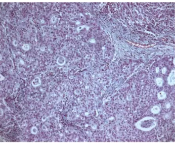
In histopathology practice, the term cribriform (from the ancient Latin cribrum i.e. sieve) refers to neoplasms showing an architectural pattern of growth made up of straight packed glands endowed with unequally distributed lumina, lacking interposed
stromal tissue (Figure 1).¹ A cribriform pattern has been identified in invasive neoplasms arising in different organs.2-7 Recently,
cribriform neoplasms have been reclassified as more aggressive entities than previously reported (e.g., carcinoma of the lung, prostate, stomach, and colon).4,8-11Therefore, the identification of
a cribriform pattern may have practical prognostic implications for pathologists and oncologists.1Our aim is to review
morpho-logical and immunohistochemical features, as well as discuss the differential diagnosis, and prognostic implications of this histolog-ic type.
Cribriform pattern in invasive prostate cancer
Several studies have demonstrated that a cribriform pattern in radical prostatectomy specimens was associated with extraprosta-tic extension, positive surgical margins, distant metastases, and cancer-specific mortality.7,12,13In 2005, the International Societyof Urological Pathology (ISUP) assigned a Gleason grade 4 rather than Gleason grade 3 for a cribriform pattern.8,9In 2009, Lotan et
al showed that there was often histologic continuity between glomeruloid glands and cribriform pattern.14 Glomeruloid pattern
is a term used to describe dilated glands containing intraluminal cribriform structures with a single point of attachment, reminis-cent of a renal glomerulus.7,14Two recent studies suggest that
glomeruloid structures could be considered as an early stage of cribriform Gleason pattern 4.14Moreover, Lotan et al. showed that
84% of cases with glomeruloid glands were associated with Gleason grade 4, or higher, cancer.14 At the 2014 ISUP grading
conference, assigned for glomeruloid pattern was assigned a Gleason grade 4.7Solid nests or cribriform structures with
come-donecrosis are considered Gleason pattern 5.7
The differential diagnosis for atypical cribriform lesions of the prostate has become increasingly complex and includes intraduc-tal carcinoma of the prostate (IDC-P), high-grade prostatic intraepithelial neoplasia (HG-PIN), ductal carcinoma, and acinar cell carcinoma with cribriform pattern.15,16Invasive acinar
adeno-carcinoma may closely mimic IDC-P when it has a cribriform or solid architecture. The key difference between IDC-P and invasive acinar adenocarcinoma with cribriform/glomeruloid pattern is a lack of any basal cell layer present in invasive acinar adenocarci-noma.7,15,17,18 IDC-P must be distinguished from HG-PIN.
Usually, in HG-PIN tumour cells are uniformly atypical with nuclear size 2-3 times normal adjacent nuclei, whereas in IDC–P there is a marked pleomorphism with nuclear size >6 times normal adjacent nuclei.15,17,19,20Moreover, HG-PIN involves few glands,
whereas IDC-P frequently involves >6 glands.15 Comedonecrosis
and mitoses are rarely seen in HG-PIN, whereas they are easily identifiable in IDC-P.15,17-20 Immunohistochemical staining for
PTEN may be useful in difficult cases, as it remains positive in HG-PN and is negative in IDC-P.15,18,20,21Table 1 summarizes the
Correspondence: Giovanni Branca, Department of Human Pathology Gaetano Barresi, AOU Polyclinic G. Martino, Pad. D, via Consolare Valeria, 98125 Messina, Italy.
E-mail: [email protected]
Key words: Cribriform carcinoma; histopathology; immunohistochem-istry; differential diagnosis; prognosis.
Contributions: GB designed the research, provided overall supervision, edited the manuscript; AI provided overall supervision; VB provided overall supervision; GT provided overall supervision; RAC designed the research, provided overall supervision, edited the manuscript, and performed histologic photographs.
Conflict of interest: the authors declare no potential conflict of interest. Received for publication: 19 September 2016.
Revision received: 15 December 2016. Accepted for publication: 6 February 2017.
This work is licensed under a Creative Commons Attribution NonCommercial 4.0 License (CC BY-NC 4.0).
©Copyright G. Branca et al., 2017 Licensee PAGEPress, Italy Oncology Reviews 2017; 11:317 doi:10.4081/oncol.2017.317
An updated review of cribriform carcinomas with emphasis
on histopathological diagnosis and prognostic significance
Giovanni Branca, Antonio Ieni, Valeria Barresi, Giovanni Tuccari, Rosario Alberto Caruso
Department of Human Pathology G. Barresi, University of Messina, Italy
Non-commercial
morphologic and immunohistochemical features that are useful in the differential diagnosis between HG-PIN, IDC-P, ductal carcino-ma and acinar cell carcinocarcino-ma with cribriform architecture.
Invasive cribriform carcinoma of the breast
Invasive cribriform carcinoma is a rare histological type of invasive breast cancer accounting for less than 4% of all breast cancers.3,22,23 Histologically, it is characterized by angulatedepithelial nests composed of mild to moderate atypical cells arranged in a sieve-like pattern, which are surrounded by a fibrosclerotic stroma.3 Typically, neoplastic cells with apical
snouts as well as lumina occupied by microcalcification and mucin-like substance can be recognized.3,24 Immunohisto
-chemically, cancerous nests are positive for cytokeratins, and strongly for estrogen, as well as for progesteron receptors, but myoepithelial markers such as p63 and smooth muscle actin are not expressed.24Breast invasive cribriform carcinoma is
subdivid-ed into three forms: the pure invasive type (whenever >90% of a cribriform pattern of the entire lesion), the invasive type (i.e., crib-riform pattern plus a component of tubular carcinoma <50%), and the mixed type (characterized by 10-49% of a carcinoma with his-totype other than tubular).3Generally, this neoplasm has a better
prognosis compared to invasive carcinoma of no special type (10-year overall survival results between 90% and 100%) even if the mixed type showed a worse prognosis between the two forms.3,24
Pleomorphism is minimal or absent, and nuclear grade I is present in at least 90% of cells. Mitotic figures are unusual. According to biomolecular classification of breast neoplasms, invasive cribri-form carcinoma is mainly of the luminal A-type (luminal B-type has also been described), since estrogen and progesteron receptors are positively immunoexpressed in contrast to a negative hyperex-pression/amplification for Her2 receptors.3,22,24
Table 2 shows the main differential diagnostic considerations on adenoid cystic carcinoma, neuroendocrine tumours, and cribri-form ductal carcinoma in situ and invasive.3,23,25-27
Cribriform pattern in lung adenocarcinoma
Histology-based 2015 World Health Organization (WHO) classification of lung tumours identifies five histotypes: e.g., lep-idic, acinar, papillary, micropapillary, and solid, whereas a cribri-form pattern is considered a rare variant of the acinar adenocarci-noma.5,10Warth et al identified predominant and minor patterns oflepidic, acinar, micropapillary, solid and cribriform components in a series of 674 resected adenocarcinomas.28Only 28 cases (4.2%)
were cribriform predominant, although a minor component was frequently observed (28.6% of all cases).28Moreover, a cribriform
pattern must be distinguished by metastatic cribriform neoplasms of other sites, including breast, prostate, ovary, pancreas, uterus, colon, and thyroid. Histologically, primary cribriform
adenocarci-Table 1. Differential diagnosis among cribriform neoplasms of the prostate.
HGPIN IDC-P Ductal carcinoma Acinar carcinoma (cribriform pattern)
Growth pattern Usually micropapillary Solid/dense cribriform Papillary, solid or cribriform with Cribriform and glomeruloid or loose cribriform. or Loose cribriform/ slitlike spaces pattern (Gleason pattern 4)
micropapillary (endometrioid pattern)
Comedonecrosis No Frequent non-focal Present Present in Gleason pattern 5 (invasive cribriform carcinoma)
Basal cell layer Intact or fragmented At least partial Absent Absent Gland size Similar to adjacent normal glands Expanded, branching Large gland Small glands Cytological features Cuboidal Cuboidal cells Tall columnar cells Cuboidal cells Nuclear features 2-3 times normal >6 times normal Elongated Enlarged Nucleoli Prominent Prominent Prominent Prominent Mitoses Rare Few/frequent Frequent Frequent Immunohistochemistry
PSA Positive Positive Positive Positive AMACR Positive Positive Positive Positive PTEN Positive Negative in 61-84% Negative Data unavailable Basal markers Positive Positive Negative Negative
Figure 1. Gastric carcinoma with a cribriform component. Note nests of epithelial cells with round spaces producing cribriform pattern (Hematoxylin and eosin staining; magnification, x100).
Non-commercial
nomas could show structures similar to: i) cribriform prostatic or breast adenocarcinomas, such as roman bridges and comedonecro-sis; ii) mucinous adenocarcinomas of the pancreas or ovary such as garland-like distribution at the tumour periphery; iii) endometrioid adenocarcinomas such as subnuclear vacuoles and comedonecro-sis; iv) colorectal adenocarcinomas such as dirty necrosis and scat-tered goblet cells; v) papillary thyroid carcinoma such as clearing of nuclear chromatin and overlapping of the nuclei.10 However, a
primary lung phenotype can be confirmed by immunohistochem-istry (i.e., TTF1+/CK7+/napsin A+) and negative results for other markers.5,10,29
The correlation between cribriform predominant pattern and ALK expression, EGFR or KRAS mutations is a controversial issue. Mackinnon was unable to find a specific molecular signature for cribriform predominant carcinomas, whereas Warth et al. showed high rates of KRAS and no EGFR mutations.10,28
ALK-positive pulmonary adenocarcinomas were more frequently observed in tumours characterized by mixed growth pattern including acinar, solid, micropapillary, and cribriform.10,28,30Due
to its rarity and controversy over classification, further studies are needed to verify specific associations of molecular signatures with a cribriform pattern. Recent studies suggest that cribriform pattern is associated with aggressive behavior.5,10Kadota et al. showed
that cribriform predominant adenocarcinoma is characterized by a higher mitotic rate, vascular invasion, and tissue necrosis com-pared to acinar predominant adenocarcinoma.1In particular, Warth
et al. showed that cribriform histotype heavily influences outcome, and propose that a cribriform pattern would be better included in high-grade tumours such as solid and micropapillary patterns rather than in the acinar group, as currently suggested by WHO classification.28Based on promising data demonstrating peculiar
clinicopathologic features, several authors suggest the identifica-tion of a cribriform pattern as a specific histotype in future WHO lung tumours classifications.
Cribriform-morular variant of papillary thyroid
carcinoma
This name is referred to a rare variant of papillary thyroid car-cinoma (corresponding to less than 0.5% of whole papillary thy-roid carcinomas) that occurs sporadically, mainly in young women, or in association with FAP or Gardner syndrome, which, in the last WHO classification of endocrine tumours, is denominat-ed simply cribriform carcinoma.6,31,32The prognosis as well as the
biomolecular alterations (i.e., presence of RET/PTC translocation, and no BRAF mutations) are similar to those discovered in classi-cal papillary thyroid carcinoma.6 Histological findings of this
tumour other than the cribriform pattern of growth are: cuboidal to columnar shaped cells, the observation of intratumoral squamoid islands called morules (although they are squamous in morpholo-gy, they are devoid of keratinization or of cellular bridges), the eosinophilic biotin-containing inclusion of some nuclei localized within the morules, hyperchromatic/pseudostratified nuclei, focal nuclei with hallmark of papillary carcinoma (i.e., clearing, grooves, intranuclear cytoplasmic inclusions), absence or scarcity of colloid, lack of mitotic activity and necrosis, and encapsulated well circumscribed edges with or without capsular and/or vascular invasion.6,31-35Immunohistochemically, neoplastic follicular cells
are stained with antibodies against thyroglobulin (although with variable results), as well as for CK19 and TTF-1.31-33,36
Intriguingly, the morular cells express CA-19.9 and CD10, whilst they are immunonegative for epithelial membrane antigen, high molecular weight cytokeratins, and vimentin.32 An aberrant
nuclear expression for betacatenin evidences an alteration in APC/Wnt pathway.32 The main differential diagnosis is between
the tall and columnar cells variant of papillary thyroid carcino-ma.32,33Tall cell variant shows neoplastic cells with heights at least
three times more compared to their widths, mainly papillary pat-tern of growth, lack of cribriform as well as morular structures, dif-fuse nuclear alterations typical of classical papillary thyroid carci-noma, basally located nuclei, necrotic areas, mitotic activity, dif-fuse immunostaining for thyroglobulin, detection of BRAF muta-tion, major incidence in older men, and, aggressive behavior.6,31-33
Table 2. Differential diagnosis of invasive cribriform carcinoma of the breast.
Histological type Microscopy Immunohistochemistry
Adenoid cystic carcinoma Round-shaped spaces containing dPAS positive basement cKit positive cells;
membrane-like substance surrounded by two cell types p63, calponin, CK 5/6 positive myoepithelial cells (myoepithelial basaloid and eosinophilic cells) Estrogen and progesterone receptors generally negative No ductal carcinoma in situ
Invasive neuroendocrine tumours Nests/trabeculae of spindle, plasmacytoid Synaptophysin and chromogranin positivity or large clear cells
Cribriform adenocarcinoma Absence of dPAS positive cores No cKit positivity
Ductal carcinoma in situ may be present Myoepithelial cells absent on immunohistochemical stains Estrogen and progesterone receptors positive
Cribriform ductal carcinoma in situ Presence of surrounding myoepithelial cells P63, calponin, CK 5/6 positive myoepithelial cells
Figure 2. Cribriform gastric carcinoma with tripolar mitosis (Hematoxylin and eosin staining; magnification, x400).
Non-commercial
Instead, the columnar variant is typically characterized by supra and/or subnuclear vacuolization of pseudostratified hyperchromat-ic elongated cells, resembling those observed in early secretive stage of endometrial glands, which are well decorated by TTF1 antigen, but variably for thyroglobulin.6,31,32Moreover this variant
of PTC is devoid of any cribriform-morular pattern, and shows an aggressive behavior.31,33,34
Gastric carcinoma with cribriform component
A cribriform component in gastric adenocarcinoma morpho-logically similar to mammary intraductal carcinoma, is a rare pat-tern of growth recognized in this organ, which to date has been described only in a small cohort of intestinal type adenocarcino-mas by Lino-Silva et al.4 Moreover this histopathological patternwas not described in the last WHO Classification of Tumours of Gastrointestinal tract.37Gastric carcinoma with a cribriform
com-ponent is mainly diagnosed in adult men in an age similar to that observed for common gastric adenocarcinoma, always associated with an intestinal type carcinoma component, and not correlated with histological hallmarks of Helicobacter pylori infection in the adjacent mucosa (e.g., follicular gastritis, gastric atrophy, intestinal metaplasia).4 Furthermore, gastric carcinoma with a cribriform
pattern shows a major tendency to possess a higher stage, lympho-vascular and perineural invasion, and a lower disease free survival rate, in comparison to conventional gastric carcinoma.4 Figure 2
shows a gastric carcinoma with a cribriform component character-ized by atypical mitosis (Figure 2). From an immunohistochemical viewpoint, tumour cells are diffusely decorated with CK7 and CK19, but focally for CK20.4 Positivity for MUC5AC has also
been described, whereas the estrogen and progesterone receptors, CDX2, MU1, MUC2, and GCDFP-15 are not expressed.4 The
most important differential diagnosis for gastric carcinoma with cribriform component is with a metastatic breast cribriform carci-noma.3,4However, the presence of an in situ adenocarcinoma
com-ponent together with an immunonegative expression for the usual markers of breast carcinoma such as mammoglobin, progesterone and estrogen receptors, and GCDPF-15, are all useful elements for diagnosing primitive gastric adenocarcinoma with cribriform com-ponent.4
Colonic cribriform comedo-type adenocarcinoma
This histotype has been briefly described in the current WHO Classification of Tumours of the Digestive System, and defined as an adenocarcinoma in which is recognized a cribriform pattern with a central area of comedo-like necrosis similar to that observed in mammary intraductal carcinoma.2Recently, a study byLino-Silva et al. has highlighted a negative outcome for this entity.11
Moreover, the majority of the cases enrolled in that study showed adverse prognostic findings such as high stage, lymphovascular invasion, lymph nodes metastasis in >5 of the harvested ones, and lower 5-year survival than conventional adenocarcinomas histo-type.11 Moreover, Kim et al. demonstrated a negative prognostic
impact for those colorectal carcinomas with a cribriform compo-nent characterized by microsatellite instability high (MSI-H), in terms of shorter disease-free survival.38
Primary cutaneous cribriform carcinoma
Cutaneous cribriform carcinoma, recently described in 1998, is
considered as a variant of sweat gland carcinoma, characterized by excellent outcome without recurrence and/or metastasis.39,40The
main histopathological findings of this tumour are the following: i) partially or relatively well-circumscribed, non-encapsulated der-mal nodule; ii) nests of epithelioid cells owning lumina; iii) pleo-morphic elongated nuclei; iv) desmoplastic stroma; v) rare mitoses; vi) none lymphovascular or perineural invasion.39,40
Immunohistochemical stains for CK5/6 and 7, CEA and EMA (luminal), C-kit (CD117) BerEp4, and S100 reveal positivity in tumour cells; CK20 invariably shows negativity.39Primary
cuta-neous cribriform carcinoma should be distinguished from primary cutaneous adenoid cystic carcinoma.39,40,41However, the latter
his-tologic type is characterized by high recurrence risk, infiltrative growth pattern, rare lymph node and lung metastases, perineural infiltration.39,40,41Moreover, the tumour consists of basaloid cells
surrounding hyaline, periodic acid-Schiff positive basement mem-brane-like substance or alcian blue (pH 2.5) positive mucins.41
Immunohistochemistry is not helpful for the differential diagnosis against cribriform carcinoma.
Conclusions
The diagnostic approach of pathologists in their daily practice is based on the analysis of architectural and cytological features, immunohistochemical stains and other ancillary techniques in the contest of knowledge of the clinical background of the case. Recently, cribriform pattern has emerged as a paradigmatic exam-ple of an additional adverse prognostic factor in adenocarcinomas of various organs, such as prostate, lung, stomach, colon. Instead, patients with breast cribriform carcinoma have significantly better survival rates than patients with invasive ductal cancer (not other-wise specified), whereas the prognosis of a cribriform-morular variant of papillary thyroid carcinoma is similar to that of classical papillary thyroid carcinoma. Thus, cribriform carcinoma is not only a morphologic tumour growth pattern, but also a prognostic factor. More research is required to evaluate the prognostic impli-cations of a diagnosis of cribriform carcinoma in other organs such as in the skin. However, practicing pathologists should be aware that cribriform tumours of prostate, lung, stomach and colon are clinically aggressive, in order to diagnose and treat an affected patient correctly.
References
1. Kadota K, Yeh YC, Sima CS, et. al. The cribriform pattern identifies a subset of acinar predominant tumors with poor prognosis in patients withstage I lung adenocarcinoma: a con-ceptual proposal to classify cribriform predominant tumors as a distincthistologic subtype. Mod Pathol 2014;27:690-700. 2. Hamilton SR, Bosman FT, Boffetta P, et al. Carcinoma of the
colon and rectum. In: Bosman FT, Carneiro F, Hruban RH, Theise ND, eds. WHO Classification of Tumours of the Digestive System. Lyon: IARC; 2010. p 138.
3. Rakha E, Pinder SE, Shin SJ, Tsuda H. Tubular carcinoma and cribriform carcinoma. In: Lakhani SR, Ellis LO, Schnitt SJ, Tan PH, van de Vijver MJ, eds. WHO Classification of Tumours of the Breast. Lyon: IARC; 2012. pp 44-5.
4. Lino-Silva LS, Salcedo Hernández RA, Molina-Frías E. Mixed gastric carcinoma with intestinal and cribriform patterns: a dis-tinctive pathologic appearance associated with poor prognosis in advanced stages and a potential mimicker of metastatic
Non-commercial
breast carcinoma. Int J Surg Pathol 2013;2:6-14.
5. Travis WD, Noguchi M, Yatabe Y, et al. Adenocarcinoma. In: Travis WD, Brambilla E, Burke AP, Marx A, Nicholson AG, eds. WHO Classification of tumours of the lung, pleura, thy-mus and Heart. Lyon: IARC; 2015. pp 26-37.
6. Sak SD. Variants of papillary thyroid carcinoma: multiple faces of a familiar tumor. Turk Patoloji Derg 2015;31:34-47. 7. Humphrey PA, Amin MB, Berney DM, et al. Acinar
adenocar-cinoma. In: Moch H, Humphrey PA, Ulbright TM, Reuter VE, eds. WHO Classification of Tumours of the Urinary System and Male Genital Organs. Lyon: IARC; 2016. pp138-62. 8. Kir G, Sarbay BC, Gümü_ E, Topal CS. The association of the
cribriform pattern with outcome for prostatic adenocarcino-mas. Pathol Res Pract 2014;210:640-4.
9. Sarbay BC, Kir G, Topal CS, Gumus E. Significance of the cribriform pattern in prostatic adenocarcinomas. Pathol Res Pract 2014;210:554-7.
10. Mackinnon AC Jr, Luevano A de Araujo LC, et al. Cribriform adenocarcinoma of the lung: clinicopathologic, immunohisto-chemical, an dmolecular analysis of 15 cases of a distinctive morphologic subtype of lung adenocarcinoma. Mod Pathol 2014;27:1063-72.
11. Lino-Silva LS, Salcedo-Hernández RA, Herrera-Gómez A, et al. Colonic cribriform carcinoma, a morphologic pattern asso-ciated with low survival. Int J Surg Pathol 2015;23:13-9. 12. Keefe DT, Schieda N, El Hallani S, et al. Cribriform
morphol-ogy predicts upstaging after radical prostatectomy in patients with Gleason score 3 + 4 = 7 prostate cancer at transrectal ultrasound (TRUS)-guided needle biopsy. Virchows Arch 2015,467:437-42.
13. Kweldam CF, Kümmerlin IP, Nieboer D, et al. Disease-specif-ic survival of patients with invasive cribriform and intraductal prostate cancer at diagnostic biopsy. Mod Pathol 2016;29:630-6.
14. Lotan TL, Epstein JI. Gleason grading of prostatic adenocarci-noma with glomeruloid features on needle biopsy. Hum Pathol 2009;40:471-7.
15. Magers M, Kunju LP, Wu A. Intraductal carcinoma of the prostate: morphologic features, differential diagnoses, signifi-cance, and reporting practices. Arch Pathol Lab Med 2015;139:1234-41.
16. Egevad L, Epstein JI, Hameed O, et al. Ductal adenocarcino-ma. In: Moch H, Humphrey PA, Ulbright TM, Reuter VE, eds. WHO Classification of Tumours of the Urinary System and Male Genital Organs. Lyon: IARC; 2016. pp. 166-7.
17. Guo CC, Epstein JI. Intraductal carcinoma of the prostate on needle biopsy: histologic features and clinical significance. Mod Pathol 2006;19:1528-35.
18. Epstein JI, Oxley J, Ro JY, et al. Intraductal carcinoma. In: Moch H, Humphrey PA, Ulbright TM, Reuter VE, eds. WHO Classification of Tumours of the Urinary System and Male Genital Organs. Lyon: IARC; 2016. pp. 164-5.
19. Bostwick DG, Qian J. High-grade prostatic intraepithelial neo-plasia. Mod Pathol 2004;17:360-79.
20. Bostwick DG, Humphrey PA, Montironi R, Srigley JR. High-grade prostatic intraepithelial neoplasia. In: Moch H, Humphrey PA, Ulbright TM, Reuter VE, eds. WHO Classification of Tumours of the Urinary System and Male Genital Organs. Lyon: IARC; 2016. pp. 162-3.
21. Lotan TL, Gumuskaya B, Rahimi H, et al. Cytoplasmic pten protein loss distinguishes intraductal carcinoma of the prostate from high grade prostatic intraepithelial neoplasia. Mod Pathol 2013;26:587-603.
22. Liu XY, Jiang YZ, Liu YR, et al. Clinicopathological
charac-teristics and survival outcomes of invasive cribriform carcino-ma of breast: a SEERPopulation-Based Study. Medicine (Baltimore) 2015;94:e1309.
23. Cong Y, Qiao G, Zou H, et al. Invasive cribriform carcinoma of the breast: a report of nine cases and a review of the litera-ture. Oncol Lett 2015;9:1753-8.
24. Zhang W, Zhang T, Lin Z, et al. Invasive cribriform carcinoma in a Chinese population: comparison with low-grade invasive ductal carcinoma-not otherwise specified. Int J Clin Exp Pathol 2013;6:445-57.
25. Sapino A, Sneige N, Eusebi V. Adenoid cystic carcinoma. In: Lakhani SR, Ellis LO, Schnitt SJ, Tan PH, van de Vijver MJ, eds. WHO Classification of Tumours of the Breast. Lyon: IARC; 2012. pp. 56-7.
26. Bussolati G, Badve S. Carcinomas with neuroendocrine fea-tures. In: Lakhani SR, Ellis LO, Schnitt SJ, Tan PH, van de Vijver MJ, eds. WHO Classification of Tumours of the Breas. Lyon: IARC; 2012. pp 62-3.
27. Schnitt SJ, Allred C, Britton P, et al. Ductal carcinoma in situ. In: Lakhani SR, Ellis LO, Schnitt SJ, Tan PH, van de Vijver MJ, eds. WHO Classification of Tumours of the Breast. Lyon: IARC; 2012. Pp. 90-4.
28. Warth A, Muley T, Kossakowski C, et al. Prognostic impact and clinicopathological correlations of the cribriform pattern in pulmonary adenocarcinoma. J Thorac Oncol 2015;10:638-44. 29. Pelosi G, Fukuoka J, Hiroshima K, Marchevsky AM. Metastases to the lung. In: Travis WD, Brambilla E, Burke AP, Marx A, Nicholson AG, eds. WHO Classification of tumours of the lung, pleura, thymus and Heart. Lyon: IARC; 2015. pp. 148-9.
30. McLeer-Florin A, Moro-Sibilot D, Melis A, et al. A Dual IHC and FISH testing for ALK gene rearrangement in lung adeno-carcinomas in a routine practice: a French study. J Thorac Oncol 2012;7:348-54.
31. LiVolsi VA, Albores-Saavedra J, Asa SL, et al. Papillary carci-noma. In: DeLellis RA, Lloyd RV, Heitz PU, Eng C, eds. World Health Organization Classification of Tumours. Pathology and Genetics of Tumours of Endocrine Organs. Lyon: IARC; 2004. pp 57-66.
32. Cameselle-Teijeiro J, Menasce LP, Yap BK, et al. Cribriform-morular variant of papillary thyroid carcinoma: molecular characterization of a case with neuroendocrine differentiation and aggressive behavior. Am J Clin Pathol 2009;131:134-42. 33. Chikkamuniyappa S, Jagirdar J. Cribriform-morular variant of
papillary carcinoma: association with familial adenomatous polyposis - report of three cases and review of literature. Int J Med Sci 2004;1:43-9.
34. Cameselle-Teijeiro J, Ruiz-Ponte C, Loidi L, et al. Somatic but not germline mutation of the APC gene in a case of cribriform-morular variant of papillary thyroid carcinoma. Am J Clin Pathol 2001;115:486-93.
35. Nikiel B, Chekan M, Jarzab M, Lange D. Endogenous avidin biotin activity (EABA) in thyroid pathology: immunohisto-chemical study. Thyroid Res 2009;2:5.
36. Subramaniam MM, Putti TC, Anuar D, et al. Clonal character-ization of sporadic cribriform-morular variant of papillary thy-roid carcinoma by lasermicrodissection-based APC mutation analysis. Am J Clin Pathol 2007;128: 994-1001.
37. Lauwers GY, Carneiro F, Graham DY, et al. Gastric carcinoma. In: Bosman FT, Carneiro F, Hruban RH, Theise ND, eds. WHO Classification of Tumours of the Digestive System. Lyon: IARC; 2010. Pp. 52-4.
38. Kim JH, Bae JM, Oh HJ, et al. Pathologic factors associated with prognosis after adjuvant chemotherapy in stage ii/iii
Non-commercial
microsatellite-unstablecolorectal cancers. J Pathol Transl Med 2015;49:118-28.
39. Arps DP, Chan MP, Patel RM, Andea AA. Primary cutaneous cribriform carcinoma: report of six cases with clinicopatholog-ic data and immunohistochemclinicopatholog-ical profile. J Cutan Pathol 2015;42:379-87.
40. Cacchi C, Persechino S, Fidanza L, Bartolazzi A. A primary cutaneous adenoid-cystic carcinoma in a young woman.
Differential diagnosis and clinical implications. Rare Tumors 2011;3:e3.
41. Requena L, Kutzner H, Hurt MA, et al. Malignant tumours with apocrine and eccrine differentiation. In: LeBoit PE, Burg G, Weedon D, Sarasain A, eds. World Health Organization Classification of Tumours. Pathology and Genetics of Skin Tumours. Lyon: IARC; 2006. pp 134-35.