UNIVERSITÀ DEGLI STUDI DI MESSINA
TESI DI DOTTORATO DI RICERCA IN BIOLOGIA APPLICATA E MEDICINA SPERIMENTALE XXXIII CICLO
SSD BIO/19
Alzheimer's Disease: IgGs against β-
amyloid conformational epitopes exposed
by engineered phages as novel
biomarkers for state/stage diagnosis
Candidato:
Dott.ssa Maria Giovanna Rizzo
Relatore:
Chiar.mo Prof. Salvatore P. P. Guglielmino
Coordinatore:
Chiar.ma Prof.ssa Nunziacarla Spanò
INDEX
BACKGROUND ... 4
CHAPTER 1 ... 5
Alzheimer's Disease: definition, epidemiology, pathophysiology and diagnosis ... 5
CHAPTER 2 ... 10
β-amyloid in Alzheimer's Disease: amyloid polymorphism and structural basis... 10
CHAPTER 3 ... 14
Antibodies and Neurobiological Relevance ... 14
CHAPTER 4 ... 18
Phage display technology ... 18
AIM OF THE THESIS ... 25
E.S. - EXPERIMENTAL SECTION ... 30
I. Conformation dependent antibodies... 31
R.S. Results Section ... 33
II. “Double binding” phage display selection ... 36
R.S. Results Section ... 43
III. Phage ability to interact with Αβ42 in vitro test ... 46
R.S. Results Section ... 48
Enhancement of 12III1 phage inhibition of Αβ-42 cytotoxicity in vitro ... 48
IV. Ex-vivo immunostaining analysis ... 50
R.S. Results Section ... 52
V. Phage diplay mediated Immuno-PCR for Alzheimer’s diagnosis ... 56
R.S. Results Section ... 62
VI. Micro array system in CDC Biochip ... 73
R.S. Results Section ... 75
CONCLUSION ... 77
ACKNOWLEDGEMENTS AND COLLABORATIONS ... 80
4
5
CHAPTER 1
Alzheimer's Disease: definition, epidemiology,
pathophysiology and diagnosis
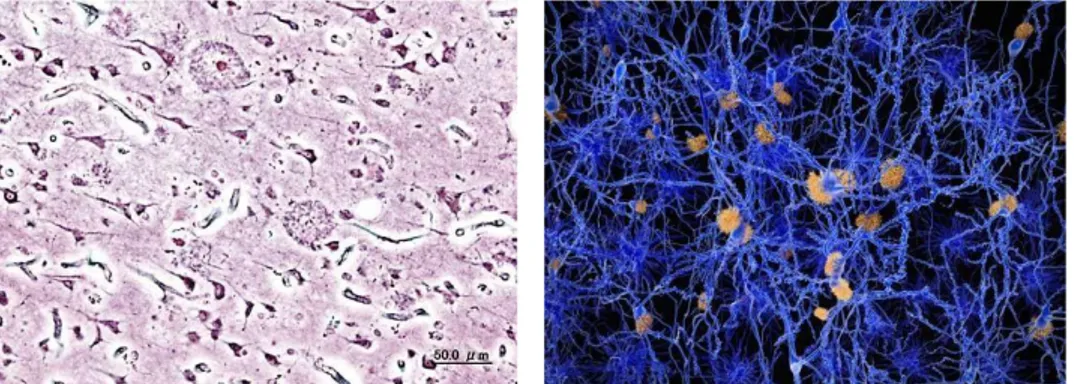
Alzheimer's disease (AD) is the most common neurodegenerative disease, accounting for approximately two thirds of all cases of dementia and affecting up to 20% of individuals older than 80 years [1]. It results in a progressive and irreversible decline in memory and a deterioration of various other cognitive abilities. The disease is characterized by the destruction of nerve cells and neural connections in the cerebral cortex of the brain and by a significant loss of brain mass (Fig.1).
Fig. 1 (a) Histopathologic image of neuritic plaques in the cerebral cortex in a patient with Alzheimer
disease; (b) A neuron network affected by amyloid plaques.
An estimated 40 million people, mostly older than 60 years, have dementia worldwide, and this fi gure is projected to double every 20 years, until at least 2050. Projected increases in the prevalence of dementia are proportionally much higher for developing countries with young populations than for western Europe and the USA, which already have a much older population (Dementia statistics, Alzheimer's Disease International).
Multiple processes have been implicated in AD including: abnormal β-amyloid (Aβ) production [2–8], tau hyperphosphorylation and neurofibrillary tangles (NFTs) [9, 10], synaptic pathology [11–13], oxidative stress [14–16], inflammation [5, 17–20], protein processing or misfolding [21, 22], calcium dyshomeostasis [16, 21–27], aberrant reentry of neurons into the cell cycle [28, 29], cholesterol synthesis [30, 31],
6
hormones [24, 32] or growth factors [18, 33]. However, the factors that promote these processes still remain unclear.
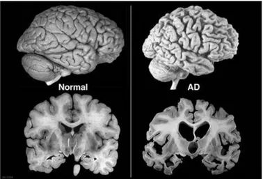
Macroscopic and microscopic markers are known and may help the understanding of the disease pathogenesis and in the development of possible strategies [34, 35]. At the macroscopic level (Fig. 2) there is the atrophy of the hippocampus and cerebral cortex, which in AD appears more sharply [36, 37]. Microscopically it is possible to observe the formation of amyloid plaques, or senile plaques, which are amorphous structures of Aβ, and accumulation of hyperphosphorylated Tau protein which implies the formation of neurofibrillary tangles, and extensive neuronal loss [34, 35, 38-41].
Fig. 2 Macroscopic changes. Atrophy of the hippocampus and cerebral cortex
Although the course of the disease is different for each individual patient, AD has a number of common symptoms that can be classified into three main phases:
Mild: phase characterized by a slight loss of memory on sporadic episodes of daily
life (on-going memory) and of prospective memory; states of confusion can also occur.
Intermediate: In this phase the subject has greater difficulty in remembering recent
events, begins to lose retrograde memory and manifests the first forms of aggression and / or passivity.
Advanced: This phase is characterized by speech difficulties, severe cognitive
deficits and violent, anxious and paranoid attitudes. There are also motor alterations which are followed by the loss of autonomy.
7
As a rule, the disease is often anticipated by the so-called mild cognitive impairment (MCI), a slight decline in the performance of various cognitive functions related to memory, orientation and verbal skills.
The major neuropathological characteristics of Alzheimer's appear to be correlated with the formation of neurofibrillary plaques and tangles. The plaques appear to develop first in brain areas associated with cognition and as disease progresses in cortical areas. Senile plaques consist of insoluble deposits of amyloid β-peptide (Aβ), a fragment of the amyloid precursor protein (APP). The Aβ peptide is generated by two consecutive cleavage events. There appear to be two types of Aβ: a longer species, Aβ42, and a shorter species, Aβ40. Aβ42 appears toxic to the neuron causing inflammation or increasing the production of free radicals. Accumulation of neurofibrillary tangles in neurons is a second distinguishing feature of AD. Neurofibrillary tangles are mostly formed from chemically altered (abnormally folded and phosphorylated) tau proteins, a protein involved in the formation of microtubules. The formation of nodes is related to the severity of the disease; the more advanced the stage of the disease, the more tau becomes entangled in the brain.
Diagnosis
AD was identified for the first time in 1901 by Dr. Alois Alzheimer, a German psychiatrist who identified the symptoms of this neurodegeneration in a patient. In 1984, the Association of Alzheimer's Patients established the NINCDS-ADRDA (National Institute of Neurological and Communicative Disorders and Stroke and Alzheimer's Disease and Related Disorders Association) diagnostic criterion, later updated in 2007, which requires that the presence of cognitive deficits and a suspected one dementia syndrome must be confirmed by neuropsychological tests. To date, neuropsychological and cognitive assessments, allow tests to characterize the state of the disease. In particular, are examined: memory, language, perceptual ability, attention, constructive ability, orientation, problem solving and functional skills. Moreover, the application of advanced biomedical imaging systems such as computed tomography (CT), magnetic resonance imaging (MRI), single photon emission tomography (SPECT), positron emission tomography (PET), were used for differential diagnosis of dementia, through Lewi bodies generalized in the cortex and identification of markers of β-amyloid plaques deposited in neuronal tissue. Also
8
laboratory blood tests can be used in support for diagnosis of AD as: complete blood count, serum urea, creatinine, thyroxine (T4), Thyroid Stimulating Hormone (TSH), albumin, liver enzymes (SGOT, SGPT, gamma GT), vitamin B12, calcium, serologic tests for syphilis, complete HIV serology. In particular, cerebrospinal fluid exams are used to analyze the changes in two biomarkers: reduced Aβ 42 which is a component of neuritic plaques and increased levels of Tau protein (total and phosphorylated) that is related to neuronal decay [42].
For global cognitive function, the main clinical examination established by NINCDS-ADRDA is the Mini-Mental State Examination (MMSE). The MMSE is one of the most used tests in the world. The test consists of 30 questions, which refer to seven different cognitive areas: orientation in time, orientation in space, recording of words, attention and calculation, recall, language, constructive practice.
The total score is between a minimum of 0 and a maximum of 30 points. A score equal to or less than 18 indicates a severe impairment of cognitive abilities; a score between 18 and 24 indicates moderate to mild impairment, a score of 25 is considered borderline, and a score of 26 to 30 indicates cognitive normality. The indications are however indicative, since there are calibration factors linked to the age and schooling of the subject.
However, a confirmed diagnosis of AD can only be obtained through post-mortem identification of neurofibrillary tangles and/or abnormal plaque deposits in the brain (Fig.3) known to be associated with AD [43].
9
As the number of people developing AD is expected to increase sharply in the next decades, there is an urgent need to develop diagnostic tests applicable to living people. The confirmatory diagnosis of AD in early stages could facilitate early therapeutic intervention and treatment of the disease. Compared to biomarkers that require brain imaging and collection of the cerebrospinal fluid (CSF), the blood- based biomarkers are particularly desirable because they are less expensive, relatively non-invasive, and easily accessible [44, 45]. Through the search of such blood-derived biomarkers, (e.g. antibodies-autoantibodies) have emerged as potentially effective bio-markers for AD.
10
CHAPTER 2
β-amyloid in Alzheimer's Disease: amyloid
polymorphism and structural basis
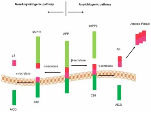
Pathogenic Aβ peptides are produced by a proteolytic cleavage of amyloid precursor protein (APP) by enzyme complexes β- and γ-secretases. APP cleavage occurs via two distinct pathways (Fig.4).
Fig. 4 Pathways to produce β-amyloid peptide
The non-amyloidogenic pathway that involves cleavage of APP by α-secretase and
generate two fragments: an 83 amino acid C-terminal fragment (C83) that remains in the membrane and an N-terminal ectodomain (sAPPα) released into the extracellular medium. The cleavage of APP by α-secretase occurs within the Aβ domain and consequently prohibits Aβ peptide production. C83 membrane fragment subsequently cleaved by γ-secretase produce a short fragment called P3 peptide and an APP intracellular domain (AICD).
The amyloidogenic pathway leads to neurotoxic Aβ generation. β-secretase (BACE1)
produce a large N-terminal ectodomain (sAPPβ) into the extracellular medium. A 99- amino acid C terminal fragment (C99) remains in the membrane [46, 47]. The exposed C99 N-terminus corresponds to the first amino acid of Aβ. Sequential cleavage of this fragment by γ-secretase (between residues 38 and 43) releases the
11
Aβ peptide. Most of the Aβ peptides are 40 residues in length (Aβ 1–40), with a minor percentage containing 42 residues (Aβ 1–42). Aβ 1–42 is considered the more neurotoxic form because the extra two amino acids provide a greater tendency to misfold and subsequently aggregate [48]. Infact, higher plasma levels of Aβ 1–42 have been correlated with Alzheimer’s disease [49].
Conformational variation of β-amyloid
The role of Aβ in AD and the lack of correlation between Aβ in the brain and the cognitive ability of patients it is not known. Infact, some patients with Aβ deposits show no symptoms of AD [50,51].
The heterogeneity to Alzheimer’s disease may lie in structural variations of Aβ, which can form polymorphic Aβ oligomers in a process known as segmental polymorphism. The segments that form beta sheets vary between different fibril structures [52–54].
Therefore, forms of structurally distinct Aβ might be deposited in places and state different in the brains of patients with Alzheimer’s disease. However, it remains unclear which types of deposit are more closely linked with the cognitive symptoms of the disease [55].
Amyloid Polymorphism and structural basis
Recent studies highlight the medical importance of amyloid self-propagation in neurodegenerative diseases [56 – 68]. Polymorphism is another important property of amyloid fibrils. For Aβ peptide associated with AD, fibrils formed by a single peptide or protein can be polymorphic, and exhibit multiple distinct forms as shown in transmission electron microscopy (TEM) images (Fig.5). Although, amyloid polymorphs could be different bundled arrangements of the same basic amyloid ‘‘protofilament’’ structure, ssNMR measurements show that amyloid polymorphs contain distinct molecular structures, and that each molecular structure can propagate itself [69-73].
12
Fig.5. Polymorphism of Amyloid Fibrils and Aggregation Intermediates, (A) striated ribbon
morphologies. (B) twisted morphologies in TEM images.
Aggregation intermediates can be neurotoxic types in neurodegenerative diseases. The toxicity of fibrils in cell cultures depends on their state of self-association and/or their lengths [73;74]. The relationship between inherent cytotoxicity and molecular structure (e.g., cross-β versus non-cross-β structure, parallel β sheet versus antiparallel β sheet structure) is not yet clear. In animal models, indication exists that oligomeric aggregates have antagonistic effects on neuronal function and memory [75-78]. In the case of AD, immunohistochemical studies show that definite oligomeric species exist in human brain tissue [79, 80]. In AD, both fibrillar and non- fibrillar aggregates contribute to neurodegeneration by different mechanisms.
Experiments by Petkova et al. showed that Aβ40 fibril morphologies can be controlled reproducibly by subtle variations in growth conditions in vitro [73]. Specifically, Aβ40 fibrils grown at 24°C and pH7.4 with moderate agitation of the Aβ40 solution during the growth period have a predominant ‘‘striated ribbon’’ morphology (Fig. 5A), whereas fibrils grown under the same conditions except without agitation have a predominant ‘‘twisted’’ morphology (Fig. 5B). These results demonstrate that distinct fibril morphologies correspond to distinct molecular structures. In general, molecular structures within amyloid fibrils formed in vitro are not determined individually by the amino acid sequences of amyloid-forming peptides and protein. Instead, they are determined by the precise details of growth conditions.
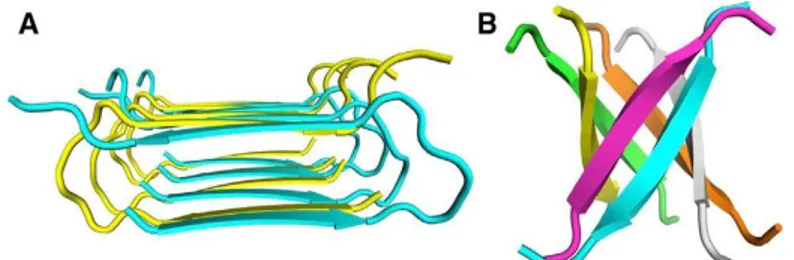
Full molecular structural models for striated ribbon and twisted Aβ40 fibrils, shown in Fig. 6A and 6B, were developed from combinations of structural constraints from ssNMR and electron microscopy [81, 82]. The principal difference between the two
13
polymorphs is their symmetry, with the striated ribbon protofilament containing two cross-b subunits, related by 2-fold rotational symmetry on the fibril growth axis, and the twisted fibril containing three cross-b units, related by approximate 3-fold rotational symmetry. In addition, the detailed conformations of the bend segments differ in the two polymorphs. The bend segment within 2-fold symmetric striated ribbon protofilaments is bridged by an electrostatic interaction between oppositely charged side chains of Asp23 and Lys28. This interaction is absent in 3-fold symmetric twisted fibrils.
Fig. 6 (A) Fibrils grown in vitro: striated ribbon morphology (B) twisted morphology (C) Fibrils derived
from brain tissue of a patient with AD. Data from ssNMR and Electron Microscopy.
The ratio of Aβ40 to Aβ42 is around 5:1 in cerebrospinal fluid of normal individuals [83], the insoluble Aβ in AD brain tissue is frequently predominantly Aβ42 [84]. Aβ42 fibrils prepared in vitro have similar morphologies to Ab40 fibrils, contain parallel b sheets that interact through similar hydrophobic contacts, and may also exist as both 2-fold symmetric and 3-fold symmetric polymorphs [85-89]. Fig. 7 represent the generic motifs for protofibrillar and non-fibrillar intermediates in amyloid formation.
Fig. 7 Molecular Structural Models for Two Types of Aggregation Intermediates (A) Protofibrils
formed antiparallel cross-β structure identified by ssNMR (B) Cylindrin oligomer the 3-fold symmetry axis of the cylindrin lies vertically in the phage.
14
CHAPTER 3
Antibodies and Neurobiological Relevance
Several studies indicates that the immune system is involved in the progression of AD, and that particular antibodies may have the potential to serve as diagnostic/prognostic biomarkers for AD. Some may contribute to the pathogenesis of AD, and others may play a protective role, thus facilitating the development of effective immunotherapies for AD.
Autoantibodies against Aβ
Although the pathogenesis of AD is not fully understood, it is widely accepted that accumulation of Aβ in the brain, especially the more amyloidogenic Aβ42, due to overproduction or impaired clearance initiates the pathogenic cascade, ultimately leading to neurodegeneration and dementia [90]. The potential of using Aβ as a blood-based biomarker for AD has been investigated extensively but it technical challenges and the results are inconsistent [91-92]. Earlier studies indicated that AD patients had significantly lower levels of unbound serum Aβ-autoantibodies than healthy age-matched individuals [93-97]. Other studies showed that both AD and control subjects had low and variable concentrations of Aβ-autoantibodies and that neither the presence nor the levels of Aβ-autoantibodies were correlated with the status of AD [98-100]. In circulation, Aβ-autoantibodies may exist either as unbound form or as antigen-antibody complexes, which could affect the capture efficiency for Aβ-autoantibody and the accuracy of assays as well as the sample preparations could also affect the assay resultsand may explain the wide-range of discrepancy in different studies [101]. Using an other approach, Mruthinti et al. observed that the levels of Aβ-autoantibodies were significantly higher in AD patients than in controls [102]. Gruden et al. showed that the levels of autoantibodies reacting with oligomers of a short but neurotoxic fragment of Aβ, Aβ(25-35), were significantly higher in AD patients than in the control group who had undetectable autoantibodies to the Aβ fragment. Further analysis showed that there was a biphasic relationship between autoantibodies to aggregated Aβ(25-35) and the stage of dementia, with the level of
15
the autoantibodies rising during the mild to moderate phase and then descending within the moderate to severe stage [103].
Using acidic dissociation approach to measure both the bound and unbound antibodies, Gustaw-Rothenberg et al. also observed that the levels of total serum Aβ autoantibodies were significantly higher in AD patients than the aged-matched control subjects [101-104]. However, there are concerns about this acidic dissociation approach as it has been shown that exposure of sera to low pH resulted in partial denaturation of antibodies and caused an artifactual increase in apparent anti-Aβ antibody titers [105]. To further clarify the biomarker value of the bound Aβ-autoantibodies, Maftei et al. developed a new strategy to specifically determine the antigen-bound Aβ-autoantibodies (intact Aβ-IgG immune complexes) in the serum and CSF from AD patients and control subjects [106]. They found that both serum and CSF levels of Aβ-IgG immune complexes were significantly higher in AD patients compared to control subjects. Moreover, the levels of Aβ-autoantibody immune complexes were negatively correlated with the cognitive status across the groups, declining cognitive test performance accompanied by the increasing levels of Aβ- autoantibody complexes [106]. These findings most likely indicate that there is a decrease in clearance of Aβ-IgG immune complexes in AD rather than an increase in the level of free autoantibodies to Aβ. Indeed, consistent with many previous studies, a recent study showed that the levels of unbound autoantibodies to Aβ were significantly reduced in the serum of patients with AD compared to those of healthy controls, especially in individuals over 65 years of age [107].
These studies demonstrate that Aβ-autoantibodies show promise as an effective blood biomarker for AD. However, due to different methodologies, varied sample sizes and disease stages, and the existence of bound and unbound forms, the measurements of Aβ-autoantibodies were highly variable and the conclusions were sometimes inconsistent.
A uniform procedure needs to be applied so that results can be compared across studies with subjects from different populations. The additional value of using the level of Aβ-autoantibodies for diagnosing AD, predicting/tracking disease progression, and monitoring treatment efficacy in a panel of blood- derived biomarkers warrants further investigation.
16
Conformation dependent monoclonal antibodies
Information of a more qualitative nature about structures of aggregation intermediates has also been obtained from experiments with conformation-dependent antibodies that preferentially recognize definite classes of structures [79-90]. These antibodies have been used to identify the presence of both nonfibrillar and fibrillar oligomers [108-110] in AD brain tissue.
Asa Hatami et. al. [111] have investigated the humoral immune response to Aβ42 fibrils and produced 23 OC-type monoclonal antibodies recognizing distinct epitopes differentially associated with polymorphic structural variants: parallel, in-register β- sheets, antiparallel β-sheets, β-solenoids, β-barrels, and β-cylindrins. These groups share many common properties, some amyloid sequences form polymorphic variants. These mOC antibodies defined at least 18 different immunological profiles represented in amyloid-β (Aβ) aggregates, mainly recognizing amyloid aggregates versus monomers, indicating that they recognized conformational epitopes.
The antibodies that recognized linear Aβ segments reacted with fibrils formed from unrelated amyloid sequences, indicating that reactivity with linear parts of Aβ does not was sequence-specific. The antibodies display altered patterns of immunoreactivity in Alzheimer disease and transgenic mouse brain and recognized spatially and temporally unique amyloid deposits. These results indicated that immune response to Aβ42 fibrils was correlated by the structural polymorphisms in fibrillar amyloid. These polymorphisms may contributed to differences in toxicity and at the effects of pathological processes.
Rakez Kayed et al., [112] immunized rabbits with a morphologically homogeneous population of Aβ42 fibrils and showed that immune serum (OC) recognizes fibrils, but not random coil monomer or prefibrillar oligomers, indicating fibrils display a distinct conformation dependent epitope absent in prefibrillar oligomers. The fibril epitope is also displayed by fibrils of other types of amyloids, indicating that the epitope is a generic feature of the polypeptide backbone. This proved that the fibril specific antibodies were conformation dependent, sequence-independent.
Rakez Kayed et al., [113] have produced several monoclonal antibodies that recognize prefibrillar oligomers and do not recognize amyloid fibrils, monomer or natively folded proteins. Immunological analysis of different prefibrillar Aβ
17
oligomer preparations showed that structural polymorphisms exist in Aβ prefibrillar oligomers that can be distinguished on the basis of their reactivity with monoclonal antibodies.
The need for conformation-specific antibodies
The biomedical importance of Aβ structural variation, conformation-specific, will play a central role in the future of Alzheimer’s research.
Research in humans has shown the clinical relevance of Aβ structural variation. Tissue taken from two Alzheimer’s disease patients with distinct clinical histories revealed that each patient had a predominant Aβ fibril structure [115].
Furthermore, the complexity of Aβ structure is correlated with the immune system, the antibodies produced in response to Aβ fibrils are diverse, reflecting their structural variation [114,116]. Considering all the studies, it is becoming increasingly clear that a single antibody will not be enough to study or target all the possible pathological aggregates of Aβ contributing to Alzheimer’s disease. This makes conformation-specific Aβ antibodies an essential tool for the future of Alzheimer’s disease research [117-120].
18
CHAPTER 4
Phage display technology
Phage display allows the exposition of several peptides on the surface a filamentous phage M13, which leads to the selection of peptides and proteins, including antibodies, with high affinity and specificity to target. The technology involves the introduction of exogenous peptide sequences into a site in the genome of the phage capsid proteins. The encoded peptides are expressed on the phage surface as a fusion product with one of the phage coat proteins. Random insertion of peptides into the amino-terminal portion of the pVIII major coat protein allows the formation of large molecular libraries, one billion of different exposed peptides, which can be used to discovery "new biomarkers", epitope mapping and sequence selection that can mimic the characteristics of many biological molecules.
M13 Bacteriophage
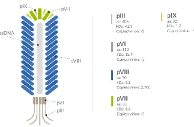
Several bacteriophage can be utilized for phage display, as the T4, lambda, the filamentous M13 bacteriophage [121]. The M13 phage only infect Escherichia coli strains that express the F pilus as the adsorption of the phage to the bacterium needs binding of a phage coat protein to the tip of the F pilus [122]. The phage contains a genome of single-stranded DNA (ssDNA) with a length of 6407 bp [123] that consists of nine genes encoding 11 different proteins. Five of these proteins are coat proteins, and the remaining six proteins are involved in replication and assembly of the phage. The M13 phage has a length of 900 nm and a width of 6.5 nm [124]. The most abundant of the coat proteins is the capsid protein G8P, which forms an envelope of approximately 2700 protein units (Fig.8).
19
Fig. 8 Schematic representation of M13 Bacteriophage
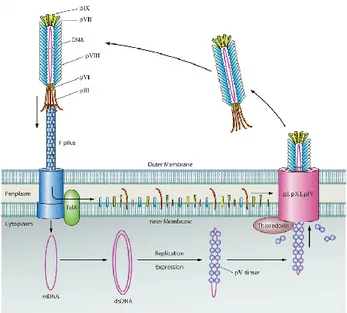
The first step of the M13 infection consists in the the adsorption process, binding of the N2 domain of the G3P coat protein to the F pilus on the surface of E. coli [125– 127] (Fig. 9). Contact between the G3P-N2 domain and the F pilus mediates the allocation of the G3P-N1 domain and allows it to bind TolA, co-receptor on the surface of the bacterium. Three Tol proteins in E. coli are essential for depolymerization of the phage coat and translocation of the ssDNA into the bacterium. Upon infection, the single stranded (+) chromosome of M13 is converted into the double stranded replicative forme RF. Replication of the M13 chromosome uses the replicative form and carried out by rolling circle replication. The 3′ end that is generated from the nick will then be elongated by DNA polymerase using the (−) strand as a template. G5P binds the ssDNA, dimerizes in a back-to-back conformation leading to the conversion from the circular appearance of the ssDNA to a more rodshaped appearance. The ssDNA bound by the G5P protein determine to phage assembly.
20
Fig. 9 Life cycle of Phage M13
Assembly of the M13 phage particles consists in an complex of pre-initiation formed by G1P, G11P, and G4P that interacting in the periplasmic domains. A multimeric complex of G1P and G10P form a channel [128]. During the elongation step, the G5P bound to the phage genome is replaced with G8P for translocation of the DNA through the membrane-spanning channel. The translocation continues until the phage genome has become completely coated with G8P, at which point G3P and G6P will collaborate in the release of the phage from the bacterium. During pre-termination, membrane-embedded G3Ps complexed with G6Ps are incorporated at the terminal end of the phage particle. Termination involved the realease of the phage, brought about by a conformation in the G3P-G6P compelx [129-131].
Phage display technology step
The key advantage of phage display is the possibility to identify target-binding proteins from a library of zillions of different random peptides without the need to screen each molecule individually. Phage display cycle can be describes in three steps: library creation, biopanning and selected clone analysis.
Library Creation: to construct an phage display library, a phagemid vector pc89 is
used for the visualization of peptides on the main coating protein pVIII of the filamentous bacteriophage, in which the expression of the fusion of the peptides on the pVIII is subjected under the control of an unducible promoter. A library is generated by inserting randomly synthesized oligonucleotides “degenerate” near the
21
N terminus of the pVIII protein. A library contain zillions of DNA clones harboring sequences of interest.
A typical random peptide library has about a 1014 – 1019 different phage clones.
Biopanning: a typical phage library is used for screening through an Biopanning
affinity-based. This process involves a repeated series of selection, elution and amplification cycles to isolate phages that expose peptides on the capsid able to interact with a specific target protein (antibody, receptor, enzyme, etc. [132].
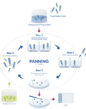
Biopanning is characterized by the following stages (Fig.10): - Location of the target: the target on which to carry out the selection can be of different nature and size, in fact it can be represented by protein molecules, peptides, antibody, nucleic acids, carbohydrates, or whole cells of prokaryotic and eukaryotic origin. The immobilization of the target can be carried out in various ways: on a solid support through the biotin-streptavidin interaction, or by passive adhesion on magnetic beads or micro plates. In the case of insoluble materials, biopanning can be performed in a suspension.
- Interaction with the phage library: The phage display random peptide library is added to the target present that allows its stability. It is important to start the first cycle with a large and diverse library to have more possibilities to isolate the peptides of interest.
- Removal of the unbound phage: at the end of the first cycle of biopanning, between the phage library and the target of interest, phage clones that have not bound will be eliminated, through consequential washes.
- Elution of the phages binding the target: following washing to eliminate the unbound phages, the phages that are instead linked to the target of interest will be recovered, through a process called "specific elution" which is generally carried out with solution acids in order to disassemble the bond established between the specific clones, binding the target, and the target itself.
- Amplification of the related phages: promotes the multiplication of the selected phages, and therefore the growing number of phage clones that bind to the target at each cycle.
22
Fig. 10 Biopanning step
Screening procedure consists of several rounds of biopanning (generally 2 or 3 rounds) to be carried out before being able to characterize the phage clones and obtained the amino acid sequence of the peptide.
To make a good selection it is necessary to consider two parameters in particular: -stringency: condition in which the most specific peptides are favored during the selection over the less suitable ones.
-yield: fraction of viral particles that remains at the end of the selection.
Finally, a step of extraction and subsequent analysis, by Sanger sequencing, of DNA is carried to identify the aminoacid sequences of the foreign peptides displayed by the selected phage clones.
Selected clone analysis
Typically, standard selective tests are the so-called phage enzyme-linked immunosorbent assay (phage-ELISA), immune-screening and/or western blot, these can evaluate the affinity and specificity of individual clones or entire eluate populations after biopanning. Those save the cost of synthesis of peptides which may have been enriched due to artifacts in the screening process described above. Successively, to assure a reasonable significance, 50–100 phage clones should be sequenced and frequency of each amino acid and the distribution in the peptide fragments evaluated. All the data lead to identify the peptides ability and the
23
contribution in the bound with targets of their amino acids The find peptides can be “epitope”, that is the part of a ligand with which the receptor makes specific contact, when show ability against the target. These epitopes will be “continuous,” if the sequence motif in the selected peptides matches with the primary amino acid sequence of the natural ligand, or “discontinuous” when carry of critical binding residues that are distant in the primary sequence but close in the folded native conformation. Geysen and colleagues introduced for the first time the term “mimotope” to refer at small peptides that specifically bind a receptor’s binding site without matching the natural epitope in the amino acid sequence, considering just cases of these “conformation-dependent” epitopes or where the natural ligand is non proteinaceous [133].
Phage Applications
The phage display technology born for identification novel interacting protein- proteins. In this prospective, phage libraries is used to predicted the potential binding residues that involved in different conditions such as between: bacterial membrane transport proteins; intracellular interactions of distinct protein domains (molecular mapping) [134; 135]; to map epitope involved in specific interaction receptor/antibody or receptor/intrabody [136]. In enzymology, phage display is used to develop modulators positive or negative of both the active and allosteric sites of the enzyme or for structure and function of receptors.
Recently, phage display is applied in the identification of protein and/or amino acid involved in the interaction with inorganic materials [137]. Therefore, phage has been applied in a several fields such as biosensing [138], cellular imaging [139], vaccine development [140] and drug and genedelivery [141].
Moreover, recent advances describe the use of phage display in the discovery of novel bioactive molecules to produce proteins and peptides able to minimize the use of antibodies, antibiotic and cancer drug.
Final Remarks
Applications of phage display technology are being actively explored with the aim of finding and producing numerous and diverse biomarket peptides that may prove useful for basic and clinical research. The use of antibody detection with new phage
24
diplay procedures would allow them to be exploited in diagnostic practices, targeted therapies improving the basic conditions of numerous diseases. In this context, phage screening can be widely used as a new alternative for treatment and diagnosis in humans.
25
26
Alzheimer's Disease (AD) is a chronic and progressive neurodegenerative disease and is characterized by the presence of neurotoxic Aβ plaques in the brain. These plaques are formed by monomeric Aβ spontaneously assembling into soluble oligomers, which cluster together to form insoluble fibrils. The solubility of Aβ, and the quantity of Aβ in different pools, may be more closely related to disease state. The composition of these pools of Aβ reflects different populations of amyloid deposits, and has definite correlates with the clinical status of the patient [142]. It is the most common cause of dementia of the elderly population in developed countries: at present it is estimated that about 5% of the population will be affected over the age of 65 and about 20% of over-85 or older, although in many cases can also occur an early onset around the age of 50.
Currently, the diagnosis of Alzheimer's Disease is based on the analysis of clinical history, neurological examinations and neuropsychological tests.
According to the NINCDS/ADRDA criteria (National Institute of Neurological and Communication Disorders/Alzheimer's Disease and Associated Disease Association), the absence of other symptomatic disorders and a progressive worsening of memory loss are necessary for the correct diagnosis.
To assess changes in a patient's cognitive abilities, neuropsychological tests, such as the Mini-Mental State Examination (MMSE), are widely used. In addition, single photon emission tomography (SPECT) and positron emission tomography (PET) can be very useful in association with mental status assessments. However, a confirmed diagnosis of AD can only be obtained through post-mortem identification of neurofibrillary tangles and/or abnormal plaque deposits in the brain. The confirmatory diagnosis of AD in early stages could facilitate early therapeutic intervention and treatment of the disease.
Many studies have been focused on biomarkers discovery in a non-invasive way in different body fluids, such as cerebrospinal fluid (CSF), saliva, urine and blood. Serum levels of antibodies specifically binding Aβ (Aβ-autoantibodies) in AD and in non-AD control subjects are extensively investigated as potential blood biomarkers applied to AD diagnosis [143-144-145-146].
These results appeared inconsistent due to several factors affecting the detection of specific IgGs against Aβ42, including nonspecific bindings [147], low avidity and
27
level in serum [148], incorrect diagnosis [149], the circulation of Aβ-autoantibodies both in free and antigen-bound form [150-152], and, not least, the structural conformation of Aβ42 [153].
It has been hypothesized that the inhomogeneity of data about the specificity of the antibodies revealed with the different methods of analysis may depend on the fact that different and distinct variants or multimers of Aβ, involved in AD, can be recognized as conformational antigens, generating antibody populations that remain largely unknown, as well as their specific function. The structural plasticity of amyloid is partially based on its polymorphism, the ability to form aggregates of different structures [154-156]. The different deposition of Aβ-42 in size, shape and structure different from the native form, could generate discontinuous and conformational epitopes.
Consequently, during the immune response, antibodies could be directed not only against the primary structure of the protein, but also against its misfolding.
Many of the antisera and monoclonal antibodies produced against oligomeric and fibrillar forms of Aβ-42 are directed against conformation specific epitopes that are specifically associated with the aggregated state and are absent in normal proteins [157-160]. Conformation-dependent antibodies have been reported to recognize a generic epitope that is specific to many types of amyloid fibrils and not soluble monomer regardless of their sequences [161,162].
It has been investigated [114] the humoral immune response against Aβ-42 fibrils and it has been reported that 23 different OC-type monoclonal (mOC) antibodies, recognizing distinct epitopes differentially associated with polymorphic structural variants, were produced. These mOC antibodies define at least 18 different immunological profiles represented in aggregates of Aβ. All of the antibodies strongly prefer amyloid aggregates over monomer, indicating that they recognize conformational epitopes. It is possible to hypothesize that different aggregational states are also present in patients with AD and that these evoke a conformation- dependent antibody response.
However, it is not possible to precisely define the aggregation state of the protein and its misfolding. About that, various possible conformational misfondings of proteins similar to Aβ already present have been searched, eg. in bacterial proteins.
28
Thus, in our work [163] a set of proteins having conformational motifs homologous to Aβ-42 through bioinformatics tools have been screened. Among microbial proteins, the epitopic region of Yersinia pestis F1 capsular antigen (Caf 1) showed the most significant structural homology with the fibrillar form of Aβ. In order to search for IgG autoantibodies in AD sera against a potential “generic conformational antigen” of Aβ-42 in vivo and to evaluate the possibility to use them as state/stage biomarkers of AD, monoclonal antibodies directed against Caf 1 were used in alternate biopanning cycles, the so-called “double binding” phage display selection. Thanks to the phage display technology it is possible to select “conformational mimotopes” that can be recognized by conformation-dependent antibodies. It uses engineered M13 filamentous bacteriophage, in which the major coat protein pVIII (2700 copies) displays a foreign peptide specific for a target.
The reactivity of the phage clones isolated were evaluated in ELISA assay against monoclonal Ab that recognized Caf1 of Y. pestis and IgGs of AD-patients. From the selected clones, one clone, named 12III1, detected a significant level of IgG for discrimination between AD and non-AD subjects. Furthermore, this phage clone was able to interfere with Aβ-42 fibrillation in vitro and to promote its disaggregation in SH-SY5Y cells.
The data obtained suggested that the peptides displayed, although selected also by the monoclonal antibody specific for Caf1, probably mimicked several conformational epitopes recognized by circulating IgG from patients with AD. Thus, in order to investigate the immune response for a state/stage diagnosis of AD, different phage clones resulting from the same double binding selection were investigated. The ability to interact with amyloid plaques in AD mouse brain sections with high significance, confirmed that the peptides were amyloid mimotopes recognized by conformation-dependent antibodies.
Thus, to detect conformational antibodies present in the different stages and states of the disease, the peptides were used in a new combinatorial diagnostic micro-array besed on Phage mediated Immuno PCR CDC Biochip, able to discriminate AD from healthy sera.
The data would pave the way for the use of these Aβ-42 conformational mimotopes to determine the status and stage of AD. This could be very useful for a personalized
29
pharmacological treatment, avoiding the progression of the disease. Since these peptides were able to interact with amyloid plaques in brain sections, they could also be used as detection probes for imaging. Furthemore, their ability to interact and disaggregate Aβ-42 fibrils could lead to their use in therapeutic practices.
Workflow of thesis:
I. Conformation dependent antibodies research
II. Double binding phage display selection
III. Phage ability to interact with Αβ42 in vitro test
IV. Ex-vivo immunostaining analysis
V. Phage diplay mediated Immuno PCR for Alzheimer’s diagnosis
30
31
I. Conformation dependent antibodies
AD is associated with misfolding and aggregation states of Aβ-42. Significant structural polymorphism among fibrillar amyloid aggregates are thought to be responsible for the brain degeneration observed in AD patients [164-167].
On the other hand, conformation dependent antibodies have been reported to recognize a generic epitope common to amyloid fibrils regardless of their amino acid sequences, as they also bind other disease related amyloid fibrils and amyloid like aggregates derived from other proteins of unrelated sequence. These antibodies do not bind the native amyloid protein precursors, nor other kinds of protein aggregates, indicating that fibrils display a distinct conformation dependent and sequence independent epitope that is absent in prefibrillar oligomers. In addition, similar amyloid oligomer conformations were found in several bacterial proteins resulting immune positive to conformation dependent antibodies.
At this purpose, conformational mimotopes in unrelated amyloidogenic proteins able to recognize IgG autoantibodies in AD sera against a potential “generic conformational antigen” of Aβ-42 were searched. Among microbial proteins, suitable “surrogates of Aβ 42” were screened by bioinformatics tools.
E.S. I.1 Conformational similarities research by tool bioinformatics
"Conformational similarities" between different forms of Aβ-42 and other amyloid- like proteins have been screened using bioinformatics tools.
Template proteins were searched among proteins with similar conformation or self- assembly, using UniProtKB tool (https://www.uniprot.org/). For the structural similarity, structures having a β strand, basic component of amyloid have been searched. Then it has been started: i) field: UniProtKB AC Term: beta strand. ii) field: structure; 3D structure available. iii) field: NOT Homo sapiens (Human) [9606]. iv)field: binary interaction. v) field: NOT function Function CC, enzyme classification, activity regulation, catalytic activity. The use of UniProtKB tool has allowed the identification of 47 proteins.
32
From selected proteins, F1 capsule antigen (UNIPROT ID: P26948) was considered to the following steps in 3D alignment recognition to verify the match with Aβ amyloid structures.
First of all, amino acid sequences of Aβ-42 and F1 antigen were aligned using Clustal X2.167 (Gonnet 250 Protein Weight Matrix, Gap opening 10, Gap extend 0.1) tool to identify similar regions based on physico-chemical properties of side chain amino acids.
Then, alignment of 3D protein structure was performed to verify the conformational match of F1 protein with Aβ amyloid structures. The PDB IDs associated to 3D structure of the F1 capsule antigen (1p5u, 1z9s, 3dos, 3dpb, 3dsn) were obtained from Uniprot KB, through the ENTRY code P26948 and excluding those which were not in the mature conformation.
EMBL-EBI tool was used in PDB&fold section (http://www.ebi.ac.uk/msd- srv/ssm/), setting SSM submission form to obtain the alignment with all the structures. The 3D alignment with Aβ amyloid in fibril form (PDB ID 2nao) was carried out using Sequence & Structure Alignment tool available in https://www.rcsb.org/pages/analyze_features#Sequence, with algorithm JFATCAT rigid.
33
R.S. Results Section
Bioinformatics analyses
Using UniProtKB 47 proteins were identified.
Among them, the Caf1 of Yersinia pestis F1 antigen (UNIPROT ID:P26948) was chosen as target of analysis in 3D alignment to verify the conformational match with Aβ amyloid structures.
Caf1 is a beta-structural protein that in the polymeric form has very high conformational stability. The 2.2 Å resolution crystal structure of the ternary Caf1M:Caf1:Caf1N complex revealed that Caf1 is an incomplete β-sandwich immunoglobulin-like fold. The final stable fold is the result of replacement of the chaperone parallel β-strand by the “spare” anti-parallel β-strand from the N terminus of the subsequent subunit, thus linking them to form a chain. Similarly, it is known that the 3D structure of the Aβ-42 protofilament is formed by two stacked, intermolecular, parallel, in-register β-sheets that perpetuate along the fibril axis [168]. A recent study by cryo–electron microscopy to 4-Å resolution, complemented by solid-state nuclear magnetic resonance experiments, showed the β strands are staggered with relation to one another in a zipper-like manner [169]. On the other hand, it is well known that Aβ exists in several polymorphs with varying width and helical pitch, different cross-section profiles and different interactions between the monomers.
Linear alignment between the F1 protein and Aβ-42 was initially conducted. On Caf1 the regions mainly involved in alignment (Fig. 11) fall within two B (84-101; 121- 144) and one T (102-116) cell epitopes, the immunodominant and highly immunogenic sequences of F1 protein, which is recognized by a high amount of antibodies raised against the native F1 antigen [170,171].
The other regions involved in the alignment were on Aβ(1–16), epitopic for immunoglobulins and involved in the formation of proto-filaments [172,173], and the region Aβ (21–37), recognized by “fibril-inhibiting” Aβ-autoantibodies [174,175]. Moreover, it is known that Aβ (4–10) is epitopic for “plaque-specific” antibodies inhibiting both Aβ fibrillogenesis and cytotoxicity [176,177].
34
Fig.11 Sequence alignment between Aβ42 and Caf1 by Clustal X2.1
In particular, the sequence overlap between the C-terminal of β amyloid and the immunodominant region of Caf1 observed in linear alignment (50% similarity, 29% identity) concerned the GxxxG, GxxxxG and GxxxxA repeating motifs [178] (Fig. 11), which conferred the particular conformation to the proteins and are recently demonstrated as primarily responsible for both Αβ self-assembly and neurotoxicity [179].
Conformational similarity analyses were then conducted, based on identifying residues occupying an equivalent geometric shape in space.
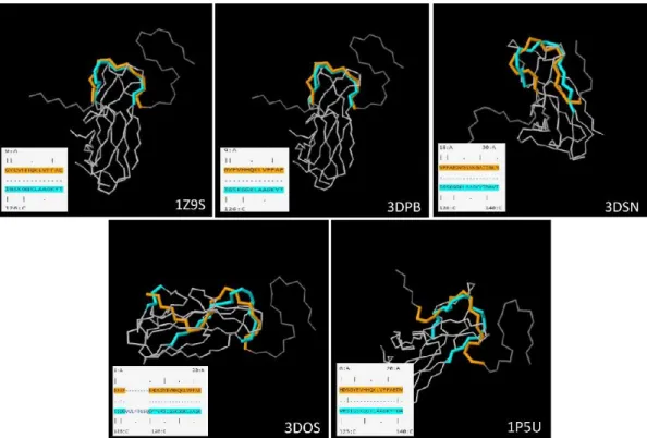
3D structure similarity analyses confirmed that conformational alignment fell within the same immunodominant region as evoked by the primary sequence alignment. In addition, RCSB Sequence & Structure Alignment tool was used in order to perform the 3D alignment of fibrillar amyloid (PDB ID 2nao) with the single specific chains of F1 antigen (identified by PDB identification numbers 1p5u, 1z9s, 3dos, 3dpb, 3dsn) (Fig. 12). The Aβ-42 regions involved in 3D alignment are the following: 18-VFFAEDVGSNKGAIIGLM-35 (with 1p5u.C, 3dsn.C), 9- GYEVHHQKLVFFAE-22 (with 1z9s.C, 3dpb.C) and 1-DAEF---
RHDSGYEVHHQKLVFFAE-22 (with 3dos.C). Intriguingly, on Caf1, in all the PDB analyzed, conformational alignment with Aβ-42 fibril falls within the immunodominant region (from 121-F to 143-T).
35
Fig. 12 Alignment of Amyloid fibrils (PDB ID 2nao) with F1 capsule antigen using “Sequence and
Structure Alignment” (https://www.rcsb.org/pages/analyze_features#Sequence)- algorithm JFATCAT rigid
The conformational homology found between the different forms assumed by Caf1 and Aβ-42 fibrils confirmed the non-probability that these were randomly similar. Based on the bioinformatic analysis, F1 was considered as a protein with an amyloid oligomeric conformation such as the Aβ-42 protein. Moreover, a monoclonal antibody - mAb-YPF19 - of Y. pestis recognized the common trait of interest between the immunodominant regions of Caf1 and Aβ-42.
36
II. “Double binding” phage display selection
It is known that antibody ligands with structures partly or completely similar to the native antigen can be isolated by phage display library screening. In this section, peptide libraries have been successfully screened for “conformational mimotopes” that bind to target-specific antibodies.
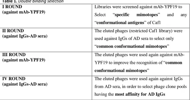
pVIII M13 phage display libraries were then screened against anti-Caf1 monoclonal antibody, and against IgGs of AD-patients, in alternate biopanning cycles of a so- called “double binding” selection. In the first round, the phage library was screened against the monoclonal Ab that recognized Caf1 to select specific mimotopes of the Caf1 and any “conformational antigens”. Then eluted phages were used in the second round against a pool of sera from patients’ AD, in order to select only “common” conformational mimotopes. A third round was carried out, with the eluted phages from the second selection round, against the mAbYPF19 again, and finally a fourth against IgG-AD, in order to select phage pools having more affinity for the latter. [International patent application has been advanced No. PCT/IB2017/057422 Nov
27, 2017: Distretto Tecnologico Sicilia Micro e Nano Sistemi S.C.A.R.L.
“Conformational mimotopes for detecting specific antibodies”
(IT/28.11.16/ITA10201600012020].
E.S. II.1 Phage M13 libraries
Phage M13 libraries were used, kindly donated by Prof. Franco Felici, expressing random peptides, exposed on the pVIII protein, based on the phagemid vector pC89 on which random oligonucleotide sequences were inserted in the region 5' of the VIII gene present in the vector, under the control of the LacZ promoter. The digestion with the restriction enzymes EcoRI and BamHI linearized the vector and allowed the insertion of the oligonucleotides with random sequences which, flanked by the same restriction sites, allow the recircularization of the vector through ligase reaction. For the selection, four types of peptide libraries were used, expressing twelve amino acids in the pVIII amino terminus region: pVIII-12aa and pVIII-12aa-cys, and nine amino acids in the pVIII amino terminus region: pVIII-9aa and pVIII-9aa-cys, wherein the latter have a cysteine-cysteine constriction expressed in the peptide, so
37
as to stabilize the structure thereof. The amplitude of each library is comprised between 10 and 100 million independent clones.
II.2 “Double binding” phage display selection
The epitopic region of Yersinia pestis F1 capsular antigen (Caf 1) showed the most significant structural homology with the fibrillar form of Aβ. This portion was recognized by a monoclonal antibody mAb-YPF19, that recognized only this specific portion of Y.pestis and not other species such as Y. enterocolitica and Y.
pseudotubercolosis. Then, a “double binding” biopanning screening of pVIII M13
phage display libraries was applied, using a monoclonal antibody against Caf 1 and a pool of AD sera, to identify a “common conformational” peptide to be used as target for serum circulating IgGs of AD patients.
Libraries were screened against antibodies immobilized on paramagnetic beads. Phages binding the targets were eluted and amplified in the bacterial host and reused in the next phage display biopanning cycle, as described in the following:
Table 1. Double binding selection I ROUND
(against mAb-YPF19)
Libraries were screened against mAb-YPF19 to Select “specific mimotopes” and any “conformational antigens” of Caf1
II ROUND
(against IgGs-AD sera)
The eluted phages (restricted Caf1 library) were used against IgGs of AD sera to select only “common conformational mimotopes” III ROUND
(against mAb-YPF19)
The eluted phages were used again against mAb- YPF19 to improve the recognition of “common conformational mimotopes”
IV ROUND
(against IgGs-AD sera)
The eluted phages were used again against IgGs from AD sera, in order to select phage clone pools having the most affinity for AD IgGs
As a source of antibodies directed against possible conformational epitopes of human polymorphic beta-amyloid 1-42 (Aβ42 a pool of 5 human sera from patients with AD (IgGAD) (mean age of 77.4 years, mean value of MMSE = 15.2) has been used. On the other hand, the monoclonal antibody YPF19 (AbDSerotec® A Bio-Rad Company, IgG1-9820-5007) anti-Yersinia pestis F1 (reacting to Y. pestis F1 capsular antigen, Uniprot code P26948) was used to screen for putative conformational
38
mimotopes homologous to Caf1 of Yersinia pestis which results to be of interest in the conformational structure similarity with Aβ-42 as evoked by bioinformatics analysis.
For the immobilization of the antibodies, Dynabeads® Protein G (Thermo Fisher scientific) superparamagnetic beads of 2.8 μm were used. Buffers and reagents were purchased at Sigma Aldrich. 50 μl of Dynabeads® Protein G were washed 3 times with citrate-phosphate buffer under stirring for 10', separated with a magnetic device for 1-2', then incubated with respective sera pools containing IgG-AD diluted 1:10 and 5 μg of mAbYPF19, for 60' at room temperature (RT) under mild stirring.
Dynabeads, functionalized with IgG-AD (DYN-IgG-AD) or mAbYPF19 (DYN- mAbYPF19), were separated with a magnetic device for 2', washed 4 times with Conjugation Buffer (20 mM sodium phosphate, 0.15 M NaCl, pH 7-9), separated with a magnetic device for 2' and resuspended in 250 μl of 5 mM BS3 (bis (sulfosuccinimidyl) suberate) at RT for 30’. Cross-linking reaction was blocked by adding 12.5 μl of Quenching Buffer, from 25 mM to 60 mM Tris, incubated at room temperature for 15' with inclination/rotation. DYN-IgG-AD and DYN-mAbYPF19 were washed 3 times with 200 μl PBST and finally resuspended in 1 ml of storage solution (PBS + 1% BSA + 0.01% Tween 20 pH 7.4). Before using for phage display selection, beads were blocked for 1 h at room temperature with PBS pH 7.4-5% not fat milk-0.05% Tween 20.
II.3 Biopanning
100 μl of each library (1×10¹²
viral particles) were added to 50 μl of Dynabeads® Protein G and resuspended in 190 μl of TBS-Tween 0.1%. After 30' incubation, separate with a magnetic device for 1-2'; the supernatant is recovered and used to carry out again the two preceding steps twice before the use of the library for the selection.
In the first round of selection, 500μl of DYN-mAb YPF19 were incubated with 100 μl of each of the four phage libraries with a titer of 1012
for 3-4 h at room temperature under mild stirring. The beads were washed 3 times in PBS-0.05% Tween 20 and separated with a magnetic device for 1-2' to eliminate supernatants containing unbound phages. Selected phage clones were eluted from antibodies with 500 μl of eluting buffer, 0.2 M of glycine-HCl (pH 2.2) + 0.1% BSA, neutralized
39
immediately with 1 M Tris–HCl pH 9.6. The enriched phage pools were amplified by infecting TG1 E. coli, purified twice by PEG precipitation, titrated and used as the input for further panning. Biopanning affinity selection was repeated in the second round against DYNIgG- AD, then in the third round against DYN-mAb YPF19 again, and finally a fourth selection round was carried out as the second one.
II.4 Phage amplification
In order to exploit the phage clones in the experimental plan, these were amplified and then quantized in phage titration (TU/mL). Tenfold serial dilutions of M13 or engineered phage clones were dispensed in 90 µL of E. coli TG1 cultures. Then, incubated at 37 °C for 15 min in static condition and for 20 min in shaking (250 rpm) conditions. 100 µL of each engineered phage clones/E. coli TG1 suspension were dispensed into Luria-Bertani medium containing ampicillin (50 µg/mL) agar plates. All plates were incubated at 37 °C overnight. Plates that had between 30–300 colonies were considered, and the phage concentration was determined as Transducing Unit per milliliter (TU/mL), according the following equation (1):
TU = (number of colonies)
volume (0.1 ml)×dilution factor (Equation 1)
II.5 M13 phage production
The protocol used is according to Kay et al. (1996), with some modification for our conditions. Briefly, a culture of E. coli strain TG1 (Kan-, Amp-, lacZ-) at OD600 =
0.7 was infected with M13K07 (Kan+), then incubated at 37 °C in static condition for 15 min, followed by shaking (250 rpm) for 20 min. The cells were collected by centrifugation, at 8000 x g for 15 min, transferred in 500 mL of Luria–Bertani (LB, Oxoid) medium containing kanamycin (100 μg/mL). Phages were produced by growing the culture overnight (o.n.) with shaking at 37 °C. The infected culture was centrifuged at 8000 × g for 20 min at 25 °C. The supernatant was mixed with 25% (v/v) of PEG/NaCl solution, containing 200 g PEG-8000 (polyethylene glycol; Sigma) and 150 g NaCl per liter, cooled in ice for 4 h and precipitated by centrifugation, at 15000 × g, for 45 min at 4 °C. The pellet was resuspended in 10% (v/v) of TRIS buffered saline (TBS, 7.88 g/L of Tris hydrochloride and 8.77 g/L of NaCl in deionized water), mixed again with 25% (v/v) of PEG/NaCl, cooled in ice
40
for 4h, and the solution was centrifuged as above. The pellet, containing phage particles, was suspended in 10% (v/v) of TBS, filtered through 0.22 μm- pore size membrane (Millipore), and stored at 4 °C.
II.6 Engineered phage clone production
Protocol used is according to Kay et al. (1996), with some modification for our conditions. E. coli strain TG1 (Kan-, Amp-, lacZ-) broth culture (OD600= 0.7) was
infected with engineered phage clone (Amp+), then incubated at 37 °C in static condition for 15 min, followed by shaking (250 rpm) for 20 min. After incubation, suitable aliquots of culture were plated onto agarized Luria–Bertani (agar 20 g/L) plates containing ampicillin (50 μg/mL) and incubated at 37 °C in static condition. One colony of E. coli strain TG1, containing phage clones, was inoculated into 10 mL of LB medium containing ampicillin (50 μg/mL) and incubated at 37 °C with shaking (250 rpm) until reaching OD600= 0.2. Then, the culture was added with
isopropylthio-β-galactoside (IPTG, 40 μg/mL) and helper phage M13K07 (Kan+) (109 TU/mL), incubated at 37 °C in static condition for 30 min, and gently shaken for 30 min. The cells were harvested by centrifugation at 8000 xg, transferred to 500 mL of LB medium containing ampicillin (50 μg/mL) and kanamycin (10 μg/mL), and incubated overnight with shaking at 37 °C. The infected culture was centrifuged 8000 × g for 20 min at 25 °C, the supernatant was then mixed with 25% (v/v) of PEG/NaCl solution, cooled in ice for 4h, and precipitated by centrifugation at 15000 × g for 45 min at 4 °C. The pellet was resuspended in 10% (v/v) of TBS, mixed again with 25% (v/v) of PEG/NaCl, cooled in ice for 4 h, and the solution was centrifuged as above. The pellet, containing phage particles, was suspended in 10% (v/v) of TBS, filtered through 0.22 μm-pore size membrane, and stored at 4 °C.
II.7 Reactivity of phage clones isolated
The reactivity of phage clones isolated were evaluated in ELISA assay with YPf19 and IgG-AD.
1012 TU/ml phage preparations were dispensed in duplicate (100 μl/well) into a 96- well microtiter plate (Multisorp, Nunc, Roskilde, Denmark). Plates were left overnight at 4°C, blocked for 2 h at room temperature with blocking buffer (PBS - Tween 20 0.05% - 6% Not Fat milk), and washed in PBS-0.05% Tween 20. 100 μl of
41
mAb YPF19 diluted 1:100 in dilution buffer (PBS - Tween 20 0.1% - 1% Not Fat milk) or 100 μl of AD sera pool diluted 1:50 in dilution buffer were added in duplicate into wells and incubated 1 h at 37°C under stirring. The plates were washed 10 times as described above and exposed to (HRP)-conjugated anti-human IgG (IgG Fc AP113P) diluted 1:15000 in dilution buffer, or anti-mouse diluted 1:50000, for 1 h at 37°C under stirring. The plates were washed 5 times as described above and developed with TMB substrate, incubating in the dark for 30'-45' at room temperature, and stopped with 100 μl of 1N HCl. Optical absorbance was recorded at 450 nm (Labsystem Multiskan Bichromatic). TBS was used as a negative control (antigen non-coated wells) for the evaluation of the background noise reaction caused by hydrophobic binding of immunoglobulin components in sample specimens to solid surfaces. Wild-type vector pC89 (containing no insert) was used as an internal control for evaluation of nonspecific binding through M13 coat proteins interaction [163].
II.8 Sequence analysis of selected phage clones
The selected clones which simultaneously show greater reactivity against the two antibody categories, IgG present in the serum of AD subjects and mAbYPF19 (specific monoclonal antibody for the identified protein F1 capsular antigen having a high conformational similarity degree for fibrillar Αβ42), were amplified and sequenced to determine the amino acid sequences of the displayed peptides.
The sequencing primers M13-40Rev (5’-GTTTTCCCAGTCACGAC-3’) and E24Fw (5’- GCTACCCTCGTTCCGATGCTGTC-3’) were obtained from Proligo, Sigma (Milan, Italy). A sample (1μl) of the suspended colony was added to the PCR reaction tube, containing 49 μl of PCR mixture. The mix was then denatured in a thermal cycler for 10' at 95°C, and then 0.25 μl of my TAQ were added. Each sample was subjected to the following PCR cycles: 4' at 94°C; 25 cycles of: 30" 94°C/ 30" 52°C/ 30" 72°C; 7' 72°C. PCR products were analyzed by agarose gel electrophoresis (1%, w/v agarose, Sigma, Milan, Italy), and 35 μl were purified with the extraction Kit Nucleo Spin® (Macherey-Nagel), and sequenced by DNA
sequencing service of BMR Genomics (Padova, Italy) using the primer M13-40Rev. The DNA sequences were translated into amino acids by using the “translate”
42
Analysis System (ExPASy http://www.expasy.ch/). Sequence alignments were performed by using the CLUSTAL X sequence alignment program (available at http://www.ebi.ac.uk/clustalw/). GeneDoc (http://www.psc.edu/biomed/genedoc/) was used as a tool for visualizing, editing, and analyzing multiple sequence alignments of the peptides. Each cluster of similar peptides was then aligned as a group with the amino acid sequence of F1 antigen and Aβ42 to identify regions with amino acid composition similar to that of the peptides.
II.9 Peptide analysis: functional motif and % epitopicity
The individual peptides exposed by the phage have been screened for a study of the functional motif and their degree of epitopicity. The functional significance of the motifs has been highlighted as the repeated presence of the typical traits present in many neurofibrillary isoforms described in AD, as well as in terms of amino acid residues involved in the formation of sheet structures in amyloids.
The degree of epitopicity was assessed using the bioinformatic tool LBtope: Prediction of Linear B-cell Epitopes, a web server LBtope for predicting and designing B-cell epitopes (https://webs.iiitd.edu.in/raghava/lbtope/mutant.php). In particular, Prediction of Epitopes in Mutated Peptides has been used. This module generate all possible mutants of a peptide. It also predicts B-cell epitope in these peptide mutants. This subroutine is important for identification of minimum mutation in a peptide in order to make it strong B-cell epitope.
Furthermore, to correlate the degree of epitopicity of the individual peptides with the regions of the Aβ, the amino acid sequences in FASTA format were aligned through ClustalW multiple sequence alignment program. It has been provided an integrated environment for performing multiple sequence and profile alignments and analyzing the results. The sequence alignment has been displayed in a window on the screen. A versatile coloring scheme has been incorporated allowing to highlight conserved features in the alignment.