UNIVERSITÀ DEGLI STUDI DELLA TUSCIA DI VITERBO
DIPARTIMENTO DI SCIENZE ECOLOGICHE E BIOLOGICHE
CORSO DI DOTTORATO DI RICERCA BIOTECNOLOGIA DEGLI ALIMENTI - XXIV CICLO
Phytoestrogens: characterization and biological effects
BIO/10
Coordinatore: Prof. Marco Esti
Tutor: Prof. Nicolò Merendino
2
TABLE OF CONTENTS
i ABSTRACT
………...ii SUMMARY
………§ 1. INTRODUCTION
………1.1 Phytoestrogens: general description
………...1.2 Lignans
………...1.2.1
Origin and structure
………...1.2.2
Food sources and dietary intake
………...1.2.3
Bioavailability and metabolism
………1.2.4
Biological activities and health effects
…...§ 2. COLORECTAL CANCER
…………..……….2.1 Lignans and colorectal cancer studies
………..§ 3. BUCKWHEAT AND BIOACTIVE COMPOUNDS...
§ 4. AIMS OF THE STUDY
.………...§ 5. MATERIALS E METHODS
………...§ 6. RESULTS
………...§ 7. DISCUSSION
………...§ 8. CONCLUSIONS AND FUTURE PERSPECTIVES
§ 9. REFERENCES
………...§ 10.GLOSSARY
………..ABBREVIATIONS
………APPENDIX
………..ACKNOWLEDGEMENTS
………...Pag. 3
Pag. 4
Pag. 5
Pag. 6
Pag. 14
Pag. 14
Pag. 16
Pag. 18
Pag. 22
Pag. 28
Pag. 35
Pag. 38
Pag. 46
Pag. 47
Pag. 50
Pag. 64
Pag. 67
Pag. 68
Pag. 87
Pag. 90
Pag. 91
Pag. 92
3
i. ABSTRACT
In this PhD thesis, some of the biological effects of phytoestrogens, in particular of lignans and the selection and characterization of buckwheat seeds and sprouts as potential rich food sources of polyphenol and pytoestrgens are reported. The effects in vitro of lignans on proliferation of human colon cancer cells lines (HT29 and HCT8) showed that lignans inhibit cellular proliferation and induce arrest of cell cycle in the G1-phase on both cellular lines. In order to study the mechanism of action we observed the behaviour of cyclin D1 and p-21 proteins. The results showed that cyclin D1 decreased expression while p-21 protein level resulted increased in cells treated. Further we measured the intracellular ROS (reactive oxygen species) level in cells treated. The analysis showed a reduced concentration of ROS especially after 24 h compared to 48 h. In addition procedure for growth and characterization of buckwheat sprouts and seeds, as preliminary step for a future setting up of a proof-project for formulation of functional food rich in biologically active components like phytoestrogens was studied. Hence hydroponic culture of Buckwheat sprouts was produced and after evaluated the yields of harvest we analyzed the amount of total phenolic contents both in sprouts and in seeds. Tartary buckwheat sprouts and seeds show higher content of phenolic contents than Common buckwheat. The research produced promising results which open horizons on the possibility to produce innovative food that provide health benefit using, for the first time, powder of Tartary buckwheat sprouts.
4
ii. SINTESI
In questa tesi di Dottorato sono stati analizzati alcuni degli effetti biologici dei fitoestrogeni, in particolare dei lignani ed è stata effettuata la selezione e la caratterizzazione dei germogli e dei semi di grano saraceno come fonte alimentare potenzialmente ricca in polifenoli e fistoestrogeni. Gli effetti in vitro sulle linee cellulari tumorali di colon (HCT8 e HT29) hanno mostrato che i lignani inibiscono la proliferazione cellulare e inducono l’arresto nella fase G1 del ciclo cellulare in entrambi le linee cellulari. I risultati dell’analisi dell’espressione proteica hanno mostrato che il blocco del ciclo cellulare in G1 porta ad una diminuzione della ciclina D1 ed a un relativo aumento della proteina P21. E’ stata inoltre valutata la concentrazione intracellulare dei ROS (reactive oxygen species) e si è osservato che i lignani riducono lo stress ossidativo delle cellule trattate in confronto a quelle controllo specie nei trattamenti a 24 ore. La ricerca si è poi rivolta alla selezione di semi di grano saraceno poi impiegati per la produzione di germogli da colture idroponiche e dopo l’essiccamento in stufa dei germogli, è stata valutata la resa a partire da grammi definiti di semi iniziali. E’ stato poi valutato il contenuto dei polifenoli totali sia nei germogli essiccati sia nei semi. I germogli ed i semi di grano saraceno Tartary hanno mostrato un più alto contenuto di polifenoli totali in confronto al grano saraceno Common. Nel complesso questi risultati guardano vantaggiosamente all’impiego dei germogli essiccati di grano saraceno Tartary come materiale di partenza ricco in componenti bioattivi per lo sviluppo futuro di un alimento funzionale avente un effetto antiossidante ed un’attività antiproliferativa.
5 § 1. INTRODUCTION
The tenet "Let food be thy medicine and medicine be thy food," espoused by Hippocrates nearly 2,500 years ago, is receiving renewed interest. In particular, there has been an explosion of consumer interest in the health enhancing role of specific foods or physiologically-active food components, so-called functional foods. Clearly, all foods are functional, as they provide taste, aroma, or nutritive value. Within the last decade, however, the term functional as it applies to food has adopted a different connotation that of providing an additional physiological benefit beyond that of meeting basic nutritional needs. The term functional foods was first introduced in Japan in the mid-1980s and refers to processed foods containing ingredients that aid specific bodily functions in addition to being nutritious. “Functional foods”, “nutraceuticals” or “pharmaconutrients” are all terms, which indicate nutrients or nutrient enriched foods that can prevent or treat diseases (Hardy, 2000). These foods and food components represent the fastest growing segment in food industry. In this contest, in the last decade, cereal, buckwheat, soy, flaxseed and their products are notified the most common foodstuff based functional food and nutraceuticals (Andlauer et al., 2002). Overwhelming evidence from epidemiological, in vivo, in vitro and clinical trial data indicates that a plant-based diet can reduce the risk of chronic disease, particularly cancer. In 1992, a review of 200 epidemiological studies (Block et al., 1992) showed that cancer risk in people consuming diets high in fruits and vegetables was only one-half that in those consuming few of these foods. It is now clear that there are components in a plant-based diet other than traditional nutrients that can reduce cancer risk and cardiovascular disease, stroke, Alzheimer disease, cataracts, and some of the functional declines associated with aging. Steinmetz and Potter (1991) identified more than a dozen classes of these biologically active plant chemicals, now known as "phytochemicals." The isolation, identification and quantification of phytochemicals in foods and the evaluation of their potential benefits to human health has now become a major research topic. Actually, there are more than thousand known phytochemicals. Some of the best known phytochemicals are carotenoids (such as beta carotene, lutein, lycopene and zeaxanthin), flavonoids (such as quercetin, anthocyanins and hesperidin), limonene, indole, ellagic acid, allium, sulphoraphane, glucosilonates, phenolic acids. Within the
6 polyphenols group are phytoestrogens and this thesis deals with the study of their biological properties (Carratù et al., 2005).
1.1 Phytoestrogens: general description
Phytoestrogens are defined by the British Working Group on Phytoestrogens of the Committee of Toxicity of Chemicals in Food, Consumer Products and the Environment of the Food Standards Agency (FSA, 2003) as any plant substance or metabolite that induces biological responses in vertebrates and can mimic or modulate the actions of endogenous oestrogens usually binding to oestrogen receptors. The majority of phytoestrogens belong to a large group of substituted phenolic compounds known as flavonoids. Three classes of flavonoid, the isoflavones, coumestans and prenylated flavonoids are phytoestrogens that possess the most potent oestrogenic activity. The major bioactive isoflavones are genistein and daidzein, which are derived from the precursors biochanin A and formononetin, respectively. Coumestrol is the most important form of coumestan consumed by humans. A class of non-flavonoid phytoestrogens, the lignans has also been identified (Figure 1). The scheme in Figure 1 may not be an exclusive list as other phytoestrogens may be identified as constituents of food in the future.
Fig. 1 Various groups of phytoestrogens and members of these groups
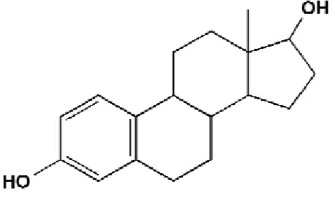
The phytoestrogens classes mentioned above have a similar structure to oestradiol and are able to bind the estrogen receptor (ER), preferably the ERβ, although their binding affinity is lower than that of endogenous estradiol. All the structures of the
7 phytoestrogens possess the phenolic (bottom, left) and hydroxyl (top, right) moieties of the oestradiol structure (Figure 2) and the distances between the two groups in each compound are similar.
Fig. 2 Structure of oestradiol
As regards estrogenicity of the phytoestrogens, their potency is dependent on the assay used to determine the value and varies considerably. Overall, the phytoestrogens with the highest receptor binding potency is coumestrol, which is ± 10-500 times less potent than the endogenous ERβ. Isoflavones are about 20,000-100,000 times less potent than ERβ. Despite this weak activity, concentrations of phytoestrogens in the body are 100 to 1,000-fold higher than peak levels of endogenous estradiol in premenopausal women. In fact, the isoflavones metabolites genistein and daidzein have been shown to exert estrogenic effects even greater than endogenous estradiol at high concentrations in vitro, though these are outside the range of concentrations typically found in humans. Phytoestrogens show a complex mode of action via interaction with the nuclear estrogen receptor isoforms ERα and ERβ, exhibiting either estrogen-agonist or estrogen-antagonist effects. Their final biological activity, assessed by cell culture assay systems, animal studies and clinical trials, depends on multiple factors such as the chemical structure of the phytoestrogens, the kind of tissue and cell type, the intrinsic estrogenic status, the route of administration, the metabolism as well as the time and the level of exposure. They are characterized by high tissue specificity and dose-dependent activity. Compounds which antagonize the estrogenic effects (antagonists) in some tissues, such as breast and uterus, while mimicking the estrogens effects (agonists) in other tissues, such as bone, brain and cardiovascular cells, are known as selective estrogen receptor modulators (SERMs). (Adlercreutz et al., 1992) (Mei et al., 2001) (Ren et
8 al., 2001) (Bhathena et al., 2002). The main classes of phytoestrogens and their common dietary sources are shown in Table 1 and Table 2, which suggest only the isoflavones and the lignans are commonly found in a Western diet (high in animal fats, refined grains and sugar, low in fruits and vegetables). It is possible that other phytoestrogens compounds are present in foods, which have not been detected. Until recently, most of the available information on concentrations of phytoestrogens in foods is related to isoflavone aglucones (not bound to glucose). This is due to the limitations in the analytical methods used. Data on the concentrations of isoflavone glucosides or glucones (i.e. bound to glucose), prenylated flavonoids, coumestans and lignans are more limited. Isoflavones are primarily found in legumes where they often occur as glucosides. Soybeans and soy-based foodstuffs are a particularly rich source of isoflavones, especially genistein and daidzein and to a lesser extent glycitein. Biochanin A and formononetin (which are derivatives of genistein and daidzein) are generally less prevalent in soy and are found mostly in clover and alfalfa sprouts. The coumestans of which coumestrol is the most common form, have been found in high concentrations in clover and fresh alfalfa sprouts as well. Prenylated flavonoids have been found in high concentrations in hops, which are used in some beers. The levels measured in beer are low. Note that Table 2 is a selection of published data and not a complete list of concentrations analyzed in foodstuffs. More complete lists can be found in the constructed databases, such as the United States Department of Agriculture (USDA)-Iowa State University Isoflavones Database (http://www.nal.usda.gav/fnic/foodcomp/Data/isoflav/isoflav.html) (Murkies et al., 1998) (Adlercreutz et al., 2002) (Benassayag et al., 2002) (Duffy et al., 2007). There is a considerable variation of phytoestrogens concentrations in different plants. The concentration of these compounds can be influenced by a number of factors including species, strain, crop year and environmental conditions. The concentrations can vary by ~2-3 fold for each factor. Processing can also alter the phytoestrogen content of foodstuffs. For example fermentation of soy into products such as tempeh, miso and bean paste reduces the isoflavone content. Cooking has also been shown to reduce phytoestrogens concentrations. However, baking or frying does not appear to alter the total isoflavones content of foodstuffs. Isoflavones and lignans are ingested mainly as glucosides and are hydrolyzed by gut bacterial and mammalian enzymes, which releases the deglycosylated compounds daidzein, genistein and glycitein (among others). These may be absorbed or further
9 metabolized by the gut bacteria to many specific (more potent) metabolites, including equol from daidzein and enterodiol and enterolactone from lignans. Research data suggest that once absorbed, isoflavones and lignans are extensively conjugated to glucuronides and sulfates in the liver and excreted in the bile or urine. This inhibits their ability to bind to the oestrogen receptors.
Tab. 1 Phytoestrogens content of Foods as consumed (Wet Weight) per serving (μg)
Phytoestrogens class Examples of dietary source
Isoflavones Legumes, lentils, chickpeas, soybean Coumestans young sprouting legumes and cereals
Lignans Most cereals, linseed, fruit and vegetables Prenylated flavonoids Some beers (hops)
Tab. 2 Phytoestrogens and common dietary sources
As a consequence, the oestrogenically active parent compounds have relatively low concentrations in the blood. Factors influencing absorption and metabolism of phytoestrogens include diet and gut microflora. The phytoestrogens can be measured in urine, plasma, feces, semen, bile, saliva, and breast milk and the concentration of
10 their metabolites differ widely among individuals. In human subjects, even those on controlled diets, there is large interindividual variation in the metabolism of isoflavones and lignans, particularly in the production of equol (only 30% to 50% of adults excrete equol). This is due to gut microflora, antibiotic use, bowel disease, gender difference and concomitant dietary intake. The foods ingested with phytoestrogens can affect their bioavailability as well. Fiber intake has been shown to correlate positively with serum and urinary levels of phytoestrogens attained in women. The sum of the main phytoestrogens in plasma can reach 2000 nmol/l in people on a traditional Asian diet and 50 nmol/l in people on a standard Western diet. In effect, various studies report that isoflavones are widely consumed by Asian populations, predominantly in the form of soy. The typical concentration of genistein in soy foods is 1 to 2 mg per g of protein, and Asians consume 20 to 80 mg of genistein per day in the usual diet. By contrast, the average American ingests only 1 to 3 mg per day (Jacobson et al., 1983) (Adlercreutz et al., 1987) (Barnes et al., 1995) (Adlercreutz et al., 1997) (Ginsburg et al., 2000) (Rowland et al., 2000) (Eden et al., 2001). Breast cancer, prostate cancer, menopausal symptoms, osteoporosis and heart disease share a common epidemiology in that they are rare in South East Asian populations eating traditional diets containing soy products. In the early 1980s the possible beneficial effects of phytoestrogens in cancer prevention and other hormone-related diseases were first published. Since then literature on possible health benefits of phytoestrogens has expanded exponentially. The prevalence of symptoms of the menopause, like hot flashes, is lower in South East Asian women as compared to Western women, which may be due among others to differences in the diet. Given the demonstrated risks to conventional HRT (hormone replacement therapy), many women and their practitioners have been in search of alternatives. As a consequence, a large number of studies have been performed into the effect of soy-based products or isoflavones on menopausal symptoms. The results of these studies are inconclusive. In general, data support a benefit of soy isoflavones (30-104 mg/d) on hot flash frequency and severity, but the data are ambiguous, as positive results are often not statistically significant and strong placebo responses are observed (FSA, 2003). Kurzer (2003) mentions that only 10-20 % of the effect is due to the isoflavones per se, the rest is due to the placebo effect. Many risk factors, such as high blood pressure and diabetes are associated with cardiovascular disease. However, the underlying basis for cardiovascular disease is a combination of
11 atherosclerosis (excessive accumulation of lipids and smooth muscle cells in the artery) and thrombosis (development of fibrinous clots). Hormonal status is known to play a role in the development of cardiovascular disease. The similarity of phytoestrogens to oestrogens (which can decrease cholesterol levels) and the lower cardiovascular disease mortality rates in populations consuming soy suggested that phytoestrogens are protective against cardiovascular disease. Indeed, there is a considerable body of evidence to indicate that the intake of soy can have beneficial effects on low-density lipoprotein (LDL) and total cholesterol levels, but this requires that the isoflavones be consumed intact in soy protein. There is some evidence that flaxseed-fibre also has a beneficial effect on cholesterol. The effects of phytoestrogens on other factors important in the risk of cardiovascular disease such as blood pressure, thrombosis or atherosclerosis have not been extensively investigated. Osteoporosis is characterized by low bone mass and micro-architectural deterioration of bone tissues with a consequent increase in bone fragility and risk of fractures. Animal studies suggest that dietary isoflavones may exert benefits on bone mineral density and bone turnover. Epidemiological studies suggest that intakes of phytoestrogens are associated with higher bone mineral density in populations consuming relatively large amounts of soy. There have been very few intervention studies in humans in this area but results suggest small protective effects in the lumbar spine. Further studies are needed to establish whether these effects are sustained over long periods of time. Isoflavones have been shown to significantly inactivate rat and human thyroid enzymes in vitro, fortification of the diet with genistein did not change the thyroid function in vivo. So, although there is a clear effect of isoflavones on thyroid enzymes, additional factors appear necessary for isoflavones to cause overt thyroid toxicity. Data from human studies suggest that dietary soy or isoflavones are unlikely to affect the thyroid function in normal individuals with adequate iodine intake. However, for individuals with a lack of dietary iodine and/or very low thyroid activity it is possible that their thyroid function may be adversely affected by the consumption of soy-based or phytoestrogens rich foodstuffs and supplements. Also, it is possible that soy-based infant formulae may have the capacity to inhibit thyroid function in infants. However, it is not clear whether the concentrations in these formulae are high enough to realize this inhibition. It is reported that children fed soy formulae have a higher incidence of autoimmune thyroiditis but this finding may be biased because
12 children put on soy formula may be more likely to have autoimmune disorders, such as food allergies. Finally, limited epidemiological evidence suggests that phytoestrogens exposure is not associated with thyroid cancer risk. Oestrogens are active in the central nervous system (CNS) and are thought to influence behavior, movement and cognition (among others). They are also involved in the development and maintenance of normal immune function. Studies suggest the blood brain barrier effectively restricts phytoestrogens transfer to the CNS in adult rodents. However, despite this, relatively high dietary exposures to isoflavones in rodents have been shown to alter protein concentrations and structures in the brain as well as induce behavioral effects. The implications of these findings for humans are unclear. Studies on the effect of isoflavones on cognitive function in humans suggest small effects, but reports are conflicting. Within past few years, phytoestrogens have attracted considerable attention for their potential anticancer activity. Since almost all anticancer drugs have serious side effects, there is search for "natural" alternatives or complements to traditional therapy. Further, the increased enthusiasm in phytoestrogens as potential anticancer agents is evidenced by the published data. The population-based studies show that the mortality due to breast, ovarian, prostate, and colon cancer has a negative correlation with the phytoestrogens and cereal intake in the diet. It has been observed that the rate of breast cancer is lower in women from some populations in Asia compared with Western populations. This has led to the suggestion of a possible reverse association between breast cancer and dietary phytoestrogens intake. Studies in premenopausal women have thus focused on the potential cancer protective effects of phytoestrogens. The idea is that phytoestrogens increase the menstrual cycle length, which is thought to reduce the risk of breast cancer by decreasing the lifetime exposure of women to endogenous oestrogens. Data from studies on premenopausal women suggest that supplementation of the diet with phytoestrogens produces weak hormonal effects. However, the nature of these effects is inconsistent. The animal data on breast cancer is conflicting. A number of studies have shown that genistein has a protective effect in animal models of chemically induced cancer. However, similar experiments using (in vitro) tumor implant models showed that genistein stimulated the growth of established breast cancer cells. At lower concentration, they tend to stimulate the proliferation of two ER-α-dependent breast cancer cell lines whereas, at high concentration, they exert strong cytotoxic effect. In men, increased phytoestrogens consumption has been
13 associated with a reduced risk of prostate cancer. However, reports of hormonal effects in men from dietary phytoestrogens supplementation are inconsistent, showing no or weak hormonal effects. It has been suggested that exposure to oestrogens of phytoestrogens during development in utero, in infancy or in childhood may play an important role in the programming of hormonal homeostasis and influence the risk of developing cancer later in life. This may, in part, explain why the relatively low risk of certain cancers observed among migrant populations from South East Asia increases in subsequent generations. Long-term studies should be undertaken to establish the clinical efficacy of phytoestrogens in these conditions in humans. Due to this, there is difficulty in making wide spread recommendations about dietary intake of phytoestrogens. Thus, more research is required to establish the role of phytoestrogens in above discussed conditions. Evaluation of benefits and risks of phytoestrogens is a complex task due to interindividual variation and complexity in absorption and metabolism. Overall, it is correct to assume that consumption of phytoestrogens may be good. On the other hand, inappropriate or excessive use may be detrimental. Before making widespread recommendations for phytoestrogens intake, extensive data on specific intracellular effects, duration of exposure and disease, and results from prospective randomized studies in humans is essential. It is also necessary to determine the potential side effects of phytoestrogens. Among various phytoestrogens, isoflavones (genistein and diadzein) have been most studied. Studies on lignans are few and for coumestans very few. This might be due to lack of industrial funding and problems in analytical techniques. Study of effects of individual compounds in various clinical conditions is the need of the hour. Based on dietary phytoestrogens, structure activity relationship studies should be carried out and more synthetic and semisynthetic compounds (like ipriflavone) should be evaluated. Genetic modification of soybean and other plants and improvement in food technology to enhance phytoestrogen production is inevitable (Cassidy et al., 2000) (Chun-Sen et al., 2001) (Maggiolini et al., 2001) (Ariyo et al., 2002) (Doerge et al., 2002) (Horn-Ross et al., 2002) (Kurzer, 2003) (Cornwell et al., 2004) (Crisafulli, et al., 2004) (Dixon et al., 2004).
14 1.2 Lignans
1.2.1 Origin and structure
Lignans, a type of phytoestrogens, are constituents of many plants and form the building blocks for the formation of lignin in the plant cell wall. They are ubiquitous in the woody portions of plants, in plant roots, seeds, coat of seeds, the bran layer in grains, stems, leaves and fruits and they have been suggested to be involved in plant defense. Plants contain a large group of non-nutrient (poly)phenolic compounds which are synthesized to protect plants from photosynthetic stress, reactive oxygen radicals, wounds and attackers such as microbes and herbivores. Plants (poly)phenols like lignans are derived from phenylalanine units, and they have in common at least one aromatic ring structure with hydroxyl group(s). They are both derived from monolignols, p-coumaryl, coniferyl and sinapyl alcohols via their respective pathways. They are characterized by the coupling of two C6C3 units by a bond between positions C8 and C8’. They comprise a whole class of compounds with a similar basic skeleton, but with large variations in substitution patterns. Several hundred different lignans structures have been identified in various plants. The two most important lignans type phytoesentrogens were identified as trans-hydroxy-benzyl)-γ-butyrolactone (enterolactone, ENL) and 2,3-bis(3-hydroxybenzyl)butane-1,4-diol (enterodiol, END) which have been described as the major lignans present in serum, urine, bile and seminal fluids of humans and animals (Fig.3). Because these two compounds are produced in animals as opposed to plants they are usually termed the mammalian lignans to distinguish them from lignans from plants. The mammalian-derived lignans differ from plant-derived lignans in possessing phenolic hydroxy groups only in the meta-position of the aromatic rings.
Fig. 3 Structures of lignans-type phytoestrogens END (2,3-bis(3-hydroxybenzyl)butane-1,4-diol ,enterodiol) and ENL (trans-2,3-bis(3-hydroxy-benzyl)-γ-butyrolactone, enterolactone)
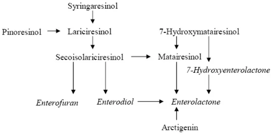
15 After ingestion, some plant lignans can be converted to the enterolignans, enterodiol (END) and enterolactone (ENL) by the intestinal microflora and absorbed into the body. It has long been assumed that only secoisolariciresinol diglucoside (SDG), secoisolariciresinol (SECO) and matairesinol (MAT) are precursors of enterolignans, but recently it has been shown that also lariciresinol (LARI), pinoresinol (PINO) and syringaresinol (SYR) can be efficiently converted. Enterolignans are thought to be the biologically active metabolites of plant lignans (Fig. 4) (Setchell et al., 1980) (Setchell et al., 1981) (Ayres et al., 1990) (Liggins et al., 2000) (Moss et al., 2000) (Begun et al., 2004).
16 1.2.2 Food sources and dietary intake
At the time of discovery of enterolignans in mammals, around 1980, the presence of lignans in plants was already known, but plant lignans had not yet been established as the dietary precursors of the enterolignans. Because urinary lignans excretion is correlated with dietary fiber intake, it was hypothesized that enterolignans precursors are present in fiber-rich foods, such as grains, nuts and legumes. The dietary origin of enterolignans could soon be confirmed, and the plant lignans SECO and MAT were identified as their precursors. Mazur et al. (1998) were the first to developed an analytical method for the quantification of these lignans in foods. In this method, lignans are released using acid and enzymatic hydrolysis, and quantified using GC-MS. Subsequently, they have reported lignans contents of a variety of Finnish foods on a dry weight basis (μg/100 g dry weight). SECO and MAT were reported to be present in several seeds and flaxseeds was identified as a rich source of SECO. In general the amount of SECO in foods was higher than that of MAT. The richest source of lignans are flaxseeds (approximately 301 mg/100 g), which contained mainly SECO (also the amount of MAT was relatively high). In general the most commonly analyzed lignans in edible plants are SECO and MAT. Also, the lignans concentrations in sesame seeds (approximately 29 mg/100 g, mainly pinoresinol and lariciresinol) were relatively high. For grain products, the lignin concentration ranged from 7 to 764 μg/100 g. Concentrations in other oilseeds such as, sesame, clover, sunflower, caraway, poppy, and peanut were much lower. Lignans concentrations in whole grain were 48-112 μg/100 g. In grain brans the concentrations were higher than in whole grain 63-299 μg/100 g whereas in flour they were lower 8-32 μg/100 g. The total lignans concentrations for nuts were 96-261 μg/100 g, for vegetables 16-3874 μg/100 g, for fruits and berries 5-1510 μg/100 g and for legumes 0-476 μg/100 g. In addition, relatively high lignans concentrations were reported for tea leaves 770-3050 μg/100 g and coffee powder 393-716 μg/100 g. The total lignans content of cereals species can be in the following order: rye > wheat > oat > spelt wheat > Japanese rice > wild rice > buckwheat > barley > amaranth > corn > millet > red rice > brown rice. Lignans contents in beverages ranged from 0 for cola to 91 μg/100 ml for red wine. Although the difference of lignans content maybe at least partly due to difference in analytical methods, it is also possible that the content of some lignans may vary within the same species depending on factors such as genetic factors or
17 growth conditions. Studies report that lignans values in boiled vegetables were on average 25% lower than those in raw vegetables, whereas after frying, lignans concentrations were on average 30% higher. The increased lignans concentrations after frying can be explained mainly by the decreased moisture content of the fried foods. On a dry weight basis, the amount of lignans after frying decreased with 25%, comparable to what it saw for boiling. The vegetables were fried in margarine, which we have shown also to contain some lignans. However, it was calculated that the maximum contribution from lignans in the margarine was less than 1%. The effects of food preparation on lignans contents have only been reported for baking of bread, thermal treatments of olive oil and roasting of pumpkin seeds. Muir & Westcott (2000) reported that SECO diglucoside (SDG), purified or as flaxseeds, added to wheat flour before the preparation of bread, was stable in the bread making process. Besides, they found that SDG could withstand the higher temperatures in the core during baking. Brenes et al. found that microwave heating of olive oil for 10 min did not change the amount of lignans. Even after 25 h (simulated) frying at 180°C, only 20-50% of the lignans were lost, whereas other phenolics compounds were almost completely destroyed. When olive oil was boiled with water (at pH 4-6) for 30 min, a large proportion of lignans leached into the water phase, but the total decrease in the lignans concentration was only 30%, irrespective of the pH. Murkovic et al. (2004) reported that SECO in pumpkin seeds was completely destroyed after 20 min of roasting. Thus, a further evaluation of the effects of food processing might increase the reliability of lignans intake estimations (Mazur et al., 1996) (Ross Barcelo, 1997) (Mazur et al., 1998) (Mazur et al., 1998a) (Liggins et al., 2000) (Muir et al., 2000) (Heinonen et al,. 2001) (Brenes et al., 2002) (Cornwell et al., 2004) (Dixon et al., 2004) (Murkovic et al., 2004) (Penalvo et al., 2004) (Branca et al., 2005) (Penalvo et al., 2005) (Raffaelli et al., 2006) (Smeds et al., 2007).
18 1.2.3 Bioavailability and metabolism
Consumption of lignans and subsequent exposure to enzymatic and bacterial activity in the mouth, stomach, intestines, colon and cecal generates lignans metabolites (Fig. 5) discovered in human urine already in the 1980s. In foods, lignans occur as glycosidic conjugates, which are hydrolysed by bacterial β-glucosidases in the gut and then released as aglycones (Fig.6, Fig.7). When these foods are consumed, the chewing action of the mouth physically breaks down lignans into small particles. The first step of metabolism may involve removal of the attached sugars in the lignans glycosides a reaction catalyzed by glycosidase. Glycosidase activities can occur in the food itself (endogenous or added during processing) or in the cells of the gastrointestinal mucosa or can be secreted by the facultative anaerobic colon micro flora. Colon microflora is also important in lignans fermentation process that occurs in the distal end of the digestive system. Gut microflora can metabolize plant lignans further into mammalian lignans (END and ENL) and enterodiol can be further oxidized to enterolactone. The importance of microflora in the metabolism of lignans has been demonstrated both in germ-free animals and in humans by the administration of antibiotics, which prevented the production of mammalian lignans. In vitro studies have also demonstrated the efficient production of mammalian lignans from the dietary precursors by human fecal flora. These suggest that the primary site of their production is the cecum and colon. Two bacterial strains, Peptostreptococcus sp. Strain SDG-1 and Eubacterium sp. Strain SDG-2, capable of demethylation and dehydroxylation, respectively, were isolated from a human fecal suspension. These findings further suggest that the formation of END and ENL is not the result of spontaneous chemical reactions but due to the metabolic reaction of viable intestinal bacteria under anaerobic conditions. In vitro and in vivo experiments have revealed the plant precursors of the mammalian lignans enterodiol and enterolactone to be secoisolariciresinol, matairesinol, lariciresinol, cyclolariciresinol (isolariciresinol), pinoresinol, syringaresinol, 7 hydroxymatairesinol, arctigenin and its glycoside arctiin. In addition, 7-hydroxyenterolactone and enterofuran have been identified as mammalian lignin metabolites in human urine. An in vitro study showed that 7-hydroxyenterolactone was a metabolite of 7 hydroxymatairesinol, and enterofuran a metabolite of lariciresinol, pinoresinol and secoisolariciresinol. Some differences in proportions of different lignin metabolites emerge when comparing
19 results from in vitro and in vivo experiments. For example, 7-hydroxymatairesinol was converted to both 7 hydroxyenterolactone and enterolactone in an in vitro incubation with faecal flora, whereas orally administered 7-hydroxymatairesinol was mainly metabolized enterolactone in rats. The fermentation of dibenzylbutane and methylenedioxybridged furanofuran lignans has been found to consists of 4 steps: deglucosylation, demethylation, dehydroxylation and dehydrogenation (Stitch et al., 1980) (Axelson et al., 1981) (Setchell et al., 1981) (Borriello et al., 1985) (Liggins et al., 2000) (Heinonen et al., 2001) (Saarinen et al., 2002) (Saarinen et al., 2002a) (Wang et al., 2002). Absorption of lignans occurs as aglycones. Wood-originated lignans are already in unconjugated form and thus can be absorbed in the upper parts of the small intestine. Lignans can be reconjugated in the intestinal epithelium during absorption or in the liver by UDP-glucuronosyltransferases and sulphotransferases.
Fig. 5 Metabolic pathways of plant lignans to the mammalian lignans enterofuran, enterodiol, enterolactone and 7-hydroxyenterolactone
Additional metabolism beyond glucuronidation or sulphation may also occur in the liver; enterolactone and enterodiol with extra hydroxyl groups have been identified in human urine after flaxseeds ingestion. Oxidative metabolism has been suggested to be a means of disposing of lignans from the mammalian body. Plasma lignans circulate either as glucuronide and sulphate conjugates or as free forms. Lignans and excess unabsorbed lignans metabolites, are excreted in the urine and via bile into fecal matter. ENL and END are excreted in the urine mainly as glucuronides, sulphatases, and to a minor extent as free aglycones (Adlercreutz et al., 1995 ) (Jansen et al., 2005).
20 Fig. 6 Enterohepatic circulation of plant lignans
Fig. 7 Intestinal conversion of secoisolariciresinol diglycoside (SDG) from phenolic complex and absorption and excretion of SECO, END and ENL.
21 Conjugated lignans, which are excreted through bile, undergo enterohepatic recycling by hepatic phase II enzymes (lignans are re-excreted via the bile duct into the intestinal tract, deconjugated by the bacterial β-glucuronidases and sulphatases, and reabsorbed by the intestinal cells) or they are flushed away in feces in free form. Most of the lignans are eventually excreted via feces and urine in mammalian organisms. A study reported that after ingestion of secoisolariciresinol diglycoside, over 50% of the lignans were excreted in feces and 30% were present in urine. In tissues, the greatest concentrations were in those tissues involved in lignans metabolism, like intestinal, hepatic, and renal tissues and in blood. Moreover, some lignans were found in the uterus. The presence of lignans in human semen, saliva, breast aspirate or cyst fluid, prostate fluid and amniotic fluid has also been reported. In humans, average levels of ENL in serum/plasma are usually less than 10-30 nmol/l. However, values up to 1000 nM have been reported for specific groups, such as vegetarians, and after intervention with lignans-rich products. Pharmacokinetics relates to the rate of availability and elimination of mammalian lignans from different organs within the body; inter-individual variation is large and in individuals consuming lignan-containing foods, serum/plasma ENL levels may be considerably over 100 nmol/l. High concentration of metabolite lignans therefore, might be achievable in the enterocytes of the gut lumen where lignans are hydrolyzed by intestinal flora and then absorbed (range μM). Various studies reported big intraindividual variations, affected by a range of different factors such variation in the microbiota, intestinal transit time, structures of the lignans and composition of the diet and food matrix. Serum ENL concentrations and a single measurement of ENL is insufficient to estimate the basal level in human subjects. In effect, the stereochemical structure of SDG and SECO has been shown to determine the chirality and the composition pattern of END and ENL and their oxidation products. Further crushing or grinding of whole food matrix has been shown to increase the levels of plasma END and ENL in humans compared with whole food. In in vitro fermentation models, the formation of END and ENL is increased by high amounts of carbohydrates, dietary fibre and xylanase treated rye bran. An increase in fat content in the diet decreases the urinary excretion of lignans in both rats and humans. (Axelson et al., 1981) (Adlercreutz et al., 1987) (Bannwart et al., 1989) (Rickard et al., 1998) (Jacobs et al., 1999) (Cassidy et al., 2000) (Muir et al., 2000) (Rowland et al., 2000) (Horn-Ross et al., 2002) (Scalbert et al., 2002) (Wang et al., 2002)
22 (Kilkkinen et al., 2004) (Stattin et al., 2004) (Aura et al., 2005) (Hongyan et al., 2005) (Kuijsten et al., 2005) (Walcott et al., 2005).
1.2.4 Biological activitiesand health effects
Lignans possess a range of biological activities in vivo and in vitro systems, including antioxidant, antitumor, weakly estrogenic, and anti-estrogenic properties, and inhibition of enzymes involved in the metabolism of sex hormones. The protective effects of mammalian lignans may be due to their ability to compete with E2 for the type II estrogen receptor, to inhibit enzymes involved in the metabolism of sex hormones and act as antioxidant. Lignans may affect enzymes involved in the formation of estrogens, such as aromatase, and 5α-reductase, 17β-hydroxysteroid dehydrogenase and may to enhance the synthesis of sex hormone binding globulin (SHBG) which may subsequently to modulate the binding of sex hormones (free estradiol). For exemple, ENL has been proposed to affect sex hormone production in vitro by inhibiting the action of steroid-metabolizing enzymes such as aromatase, an enzyme converting testosterone and androstenedione to 17β-oestradiol and oestrone, respectively. An ER independent pathway may be involved as well. Human studies have demonstrated non-consistent effects of flaxseeds on endogenous sex hormone production and metabolism. A study reported no change in plasma total or free testosterone levels or SHBG level in men consuming 13.5 grams of flaxseed per day for six weeks. Three studies with postmenopausal women reported that consumption of flaxseed changed sex hormone levels in the urine or serum (Saarinen et al., 2002a) (Shigang et al., 2008). Most of lignans containing hydroxyl group was suggested to exhibit antioxidant action mechanisms according to the number or position of hydroxyl group especially in cells not expressing estrogen receptors. In Trolox-equivalent antioxidant activity (TEAC) and chemiluminescence (CL) assays, 8-hydroxypiniresinol glycoside and 8-hydroxypinoresinol showed high antioxidant properties. The aglycone hydroxypinoresinol displayed more powerful antioxidant activity than pinoresinol. Likewise, aglycone 9- hydroxypinoresinol was more potent than its precursor, petaslignolide A. Thus, the antioxidant action of pinoresinol derivatives depends on the number of hydroxyl group in the structure. The antioxidative function of sesamin on exercise-induced lipid peroxidation was observed in animals using strenuous physical exercise as a trigger for oxidative stress. Sesamin, scavenging free radicals, exerted a strong protective effect against
23 exercise induced lipid peroxidation. Separately, syringaresinol and sesamin, isolated from Chinese propolis, were observed to inhibit lipid peroxidation in rat liver microsomes potently. Consistent with this, sesamin exhibited an antioxidative effect on lipid and alcohol metabolism in the rat liver. Further, sesamin and sesaminol elevated tocopherol concentration and decreased thiobarbituric acidreactive substance (TBARS) level in the blood plasma and liver of rats. In a separate experiment, sesamin was more effective than sesamolin in reducing serum and liver lipid levels while sesamolin is stronger in increasing hepatic fatty acid oxidation. The antioxidant activity of some plant lignans and of the enterolignans has been evaluated in several in vitro test systems at concentrations ranging from 10 100 μM. They did not show to have prooxidant activity in this concentration range. In the FRAP (Ferric Reducing Antioxidant Power) assay, SECO and MAT had high antioxidant activity compared to ascorbic acid. Some lignans, with antioxidant activity, were observed to express a neuroprotective action in excitotoxin-induced neurotoxicity in rat cortical or hypoxic neuronal cells. Furthermore, the antioxidant action of lignans from edible plants was extended to their neuroprotective action in animal experiments. Oral administration of 9-hydroxypinoresinol and its glycoside, petaslignolde A, showed a protective effect on the seizure and mortality caused by kainic acid. In addition, these lignans successfully prevented the loss of the GSH peroxidase activity and the lipid peroxidation in brain tissue, which was exposed to kainic acid, an excitotoxin. In comparison, 9-hydroxypinoresinol, a metabolite of petsalignolide A, was more effective than its precursor glycoside, petaslignolde A in preventing kainic acid induced neurotoxicity. Under the same condition, quercetin or pinoresinol, despite their antioxidant action, showed no significant effect on the seizure and mortality caused by kainic acid. Thus, peatslignolide A and its aglycone, 9 hydroxypinoresinol seems to have antioxidant activity in brain tissue, and therby exert a neuroprotective effect. Thus, the extract containing 9- hydroxypinoreinol derivative may be usefully used in the prevention and treatment of neurodegerative diseases. Taken together, antioxidant action of lignans is supposed to be responsible for various bioactivities of lignans, since cellular oxidative stress is intimately linked to disease states such as carcinogenesis, inflammation or atherosclerosis. However, other bioactivities of lignans are not necessarily related to the number of hydroxyl group, suggesting that the antioxidant action may not be necessarily required to the expression of various bioactivities (Setchell et al., 1981) (Adlercreutz et al., 1987)
24 (Welshons et al., 1987) (Ruiz-Larrea et al., 1997) (Andreasen et al., 2001) (Hou et al., 2003) (Ghafoorunissa et al., 2004) (Ikeda et al., 2004) (Kiso et al., 2004) (Eklund et al., 2005) (Hu et al., 2007). The structure of lignans, as phytoestrogens, is similar to that of endogenous estrogens, such as 17-β estradiol. Both enterolignans and plant lignans may bind to the estrogen receptors α and β, but at low affinity compared to endogenous estrogens. They may act as a weak estrogen agonist or antagonist depending on the endogenous estrogen concentration. There is considerable evidence from epidemiological studies that correlate high concentrations of lignans in body fluids, due to dietary intake, with a low incidence of hormone-dependent tumors (breast, ovary and prostate) and non hormone-dependent cancer (colon) (Heinonen et al., 2001). Various in vitro experiments suggested END and ENL significantly inhibited the growth of human colon tumor cells and the E2 (17-β-estradiol) induced proliferation of MCF-7 breast cancer cells was inhibited by ENL. In human breast cancer cell line (MCF 7), ENL at 10 nM significantly inhibited the growth of cells. At a lower dose (0.5-2 nM), the effect was stimulatory for cell proliferation; the dose amount used is the same as the levels of estrogen hormone estradiol circulating under normal conditions (1 nM). This and other studies suggested that ENL is agonist towards estradiol receptors in stimulated MCF-7 breast cancer cells at a low dose but antagonist at higher doses, hence indicating a possible mechanism by which it affects growth of estrogen sensitive cells. In prostate cancer cell lines (PC-3, DU-145, LNCAP), 10-100 μM ENL and END significantly inhibit growth of all cell lines. Mammalian lignans can stimulate proliferation and DNA/protein synthesis of breast cancer cell lines at 1-10 mM concentrations (low oestrogenic activity). In vivo they show to reduce epithelial-cell proliferation and the number of aberrant crypt foci in various animal models (Wang et al., 1997) (Kitts et al., 1999). High ENL and END concentrations can also inhibit the growth of ER positive and ER-negative breast cancer cells. The interplay of lignans and ER α/β might result in competition with endogenous oestrogens, thus modulating the biological activity of oestrogens in target tissues. Interestingly, women with breast cancer or with a high risk for breast cancer excrete lower amounts of lignans in urine compared to healthy women they have the same diet. Anticancer effects of SDG, ENL, END and 7 hydroxymatairesinol may, in part, be associated with their antioxidant capacities, as observed in vitro assays. In most studies, plant and mammalian lignan concentrations were at micromolar levels, which can be over one hundred/thousand-fold the
25 concentrations generally found in the plasma of human subjects. Although the action of lignans action has been postulated to occur via ERs, conclusive evidence for oestrogen-like activity in vivo has not been found. Early studies with ENL showed no clear oestrogenic effects on uterine weight or uterine RNA synthesis in female mice and rats. In the latter study, ENL inhibited oestrogen-stimulated RNA synthesis in the rat uterine only when administered 22 hours before oestradiol. Saarinen et al. subsequently reported that ENL, 7-hydroxymatairesinol, MAT and SECO had no oestrogenic/antioestrogenic activity or aromatase-inhibiting capacity in immature rats, as judged by uterine growth. Furthermore, END and ENL at concentrations < 1 mM did not show any activation of a reporter gene via α- and β type oestrogen receptors. In these studies, the mechanism of the action of lignans in DMBA-induced mammary tumours remained partly unknown. Several studies with young and adult rats have shown that flaxseed supplementation, pure SDG and nortrachelogenin may cause some oestrogen-like effects in rats, but the effect is dependent on the life stage. Thus, short-term experiments with flaxseeds have yielded no conclusive evidence for oestrogenic action of lignans. Similarly, no clear changes in the metabolism of sex hormones or SHBG, which has suggested to be in part responsible for lowering the risk of hormone-dependent cancers, were detected. Besides the estrogenic and antioxidant activities of lignans, there are several other mechanisms, which might explain the health effects of lignans. Some examples of other mechanisms are interaction with other receptors like the PXR-receptor, inhibition of enzyme expression, for example the enzymes involved in blood pressure regulation, or inhibition of particular enzyme activity. However, because these mechanisms are not extensively studied, they are not discussed further (Sung et al., 1998) (Thompson et al., 1998) (Lee et al., 2004) (Jacobs et al., 2005) (Thompson et al., 2005) (Sok et al., 2006) (Cui et al., 2007). There has been increasing interest in phytoestrogens due to a wider awareness of their possible beneficial effects on human health included the modulation of immune system. Actually little is known about the impact and the mechanism of lignans and other phytoestrogens on the immune system. From in vitro models it is know that isoflavone genestein dose-dependently affects lytic activity of NK cells and inhibits cell proliferation as well as the production of cytokines, leukotrienes and oxide nitric (Prasad K, et al. 1991). Recently, the carrageenan-induced rat paw oedema formation has been frequently employed in the screening of inflammatory agents. LAR and isolariciresinol expressed their
anti-26 inflammatory activities by significantly inhibiting carrageenan-induced hind paw edema in mice. These were supported by their potent in vitro inhibitory effect on the production of TNF-α, a proinflammatory cytokine. Lariciresinol glycoside, pinoresinol, pinoresinol glycodiside and syringaresinol glycoside also showed anti-inflammatory effects. Macrophages and lymphocytes, playing an important role in host immune responses, are proliferated and activated by inflammatory signal compounds, such as lipopolysaccharide (LPS). As a result, they secrete proinflammatory mediators such as cytokines (TNF-α, ILs) and lipid mediators (prostaglandin E and leukotriene B), as well as reactive oxygen and nitrogen intermediates. Isolariciresinol, lariciresinol glycoside, pinoresinol, pinoresinol glycodiside and syringaresinol glycoside were found to significantly inhibit TNF-α production from mouse macrophages. In addition, pinoresinol and syringaresinol glycoside showed significant suppressive effects on NO production triggered by LPS. However, lignin compounds seemed to interfere with biosynthetic pathway for TNF-α production, rather than NO formation, in activated macrophages. In related experiment to see the effect of lignan on concanavalin A or interleukin-2 induced lymphocyte proliferation, syringaresinol glycoside potently inhibited T lymphocyte proliferation induced by concannavalin A or interleukin-2. In addition, pinoresinol showed a significant inhibitory effect on cytokine production from LPS (or phytohemagglutinin)-stimulated human peripheral mononuclear cells (Prasad et al., 1997) (Cho et al., 2001) (Küpeli et al., 2003) (Cicala et al., 2007) (Sandra et al., 2008). Lignans complex from flax seed was suggested be beneficial in preventing atherosclerosis and reducing risk factors for coronary artery disease and stroke. Concerned with this, there are epidemiological studies on the associations between enterolignan concentrations in biological fluids or the intake of plant lignans and chronic disease risk. In case control studies, there was an inverse associations of serum lignans with cardiovascular diseases in Finnish studies. The lignans SDG from flaxseeds has been shown to be effective in decreasing serum cholesterol and reducing the extent of atherosclerosis in the hypercholesterolemic rabbit. Additionally, flaxseed lignans SDG was suggested to prevent and alleviate hypercholesterolaemic atherosclerosis by inducing adiponectin mRNA expression and showing beneficial effects on lipid metabolism in diet-induced obesity in mice. Nonetheless, it was reported that flax lignans complex failed to produce regression of atherosclerosis (Prasad et al., 2000). SDG was shown to reduce total serum
27 cholesterol in rabbits and it had antihypertensive and angiogenic activity in rats. In several human intervention studies, consumption of flaxseed could reduce total and LDL cholesterol, without an influence on HDL or total triglycerides. However, besides lignans flaxseed also contains relatively high amounts of α-linolenic acid and soluble fiber, so it is not clear whether these results can be attributed to the lignans present in flaxseed (Ward, 1993) (Vanharanta et al., 1999) (Heinonen et al., 2001) (Vanharanta et al., 2003). Diabetes mellitus, a disorder caused by defects in insulin secretion, sensitivity, or both, is characterized by hyperglycaemia and it is followed by different complications in the vascular system and in some tissues and organs. In experimental animal models of diabetes, a preventive or delaying effect of lignans on the development of diabetes mellitus has been obtained. A few studies on glucose metabolism have been performed with flaxseed or the phenolic complex. Postmenopausal women (n=25) with hypercholesterolaemia given food rich in lignans like flaxseed showed reduced glucose and insulin levels. In humans with hypercholesterolaemia, the phenolic complex had a reducing effect on fasting plasma glucose levels. In another human intervention study using type 2 diabetic hypercholesterolaemic postmenopausal women (n=30), the subjects showed modest improvements in long term glycaemic control, measured as reduction in glycosylated haemoglobin, after eating lower amounts of the phenolic complex for eight weeks, but there was no effect on fasting glucose and insulin sensitivity. Another flaxseeds component, the mucilage given to young healthy humans, has previously been shown to reduce postprandial glucose levels in blood plasma. These studies indicate that SDG and/or other component/s in flaxseeds might have lowering effects on glucose levels. Flaxseeds and SDG are suggested to protect against renal diseases. Renal function in animal models or in humans has been shown to improve with flaxseeds treatment or SDG (Cunnane et al., 1993) (Clark et al., 2000) (Prasad et al., 2001) (Lemay et al., 2002) (Velasquez et al., 2003) (Bouché et al., 2004) (Prasad et al., 2005) (Prasad et al., 2007) (Zhang et al., 2007).
28 § 2. COLORECTAL CANCER
“Colorectal cancer is a cancer that forms either in the tissues of the colon, the longest part of the large intestine, or in the tissues of the rectum, the last part of the large intestine before the anus” (definition taken from National Cancer Institute, U.S.). Colorectal cancer is the third most common cancer (9.4%) worldwide after lung and breast cancers. It ranks fourth in mortality (7.9%), after lung, stomach and liver cancers. Colorectal cancer ranks among the three most common cancers in terms of both cancer incidence and cancer-related deaths in most Western industrialized countries, every year nearly one million people worldwide develop colorectal cancer. Lifetime risk of colorectal cancer may reach 6% of the population in the Western industrialized countries. Incidence rates of colorectal cancer in countries of the European Union (EU) according to the 2006 estimates varied by a factor of 3 for men and a factor of 2 for women, with the lowest age standardized incidence rates to be observed in Greece (31/100.000 for men and 21,3/100.000 for women) and the highest incidence rates to be observed in Hungary (106/100.000 for men and 50,6/100.000 for women). Incidence rates estimates for the EU are 59/100.000 for men and 35,6/100.000 for women. The age-specific incidence of colorectal cancer rises sharply after 35 years of age, with approximately 90% of cancers occurring in persons older than 50 years, for this individuals aged from 50 to 74 years old are invited every two years for bowel screening. In Italy the number of deaths from colorectal cancer represents about 11% of the cancer mortality in men and 14% of the cancer mortality in women. The most recent data from Italian population-based cancer registries show a relevant variation in colorectal cancer burden by sex and geographical area. Colorectal cancers are the third most prevalent cancers in the United States and accounts for 10% of cancer deaths in both men and women. Large variations in incidence rates were observed with the lowest incidence rate to be observed in Africa (incidence rate in middle Africa: 2,3/100.000 for men and 3,3/100.000 for women) and the highest to be observed in Australia, North America and Europe (highest incidence rate in Australia / N. Zealand: 48,2/100.000 for men and 36,9/100.000 for women). There is no clear trend in global age standardized incidence rates of colorectal cancer. In countries of relatively low-income economy, which have recently made a transition to a higher-income economy (e.g. eastern and southern European countries, Japan, Singapore), a rapid increase in incidence rates
29 has been observed. Migrant studies, where populations migrate from low-risk to high risk areas, have demonstrated that the colorectal cancer incidence among the immigrants quickly (within one generation) approach the incidence of the native population of the host country with the largest increase occurring in risk of cancer in the distal colon. The large international variation in incidence rates and the shift in sub-site distribution (proximal or distal segment of colon) after migration, indicate the importance of environmental factors and life style factors as a part of colorectal carcinogenesis. Lifestyle factors including diet, overweight, low physical activity and smoking may account for 70% of colorectal cancers. Diet is a major but controllable factor that affects colorectal carcinogenesis. Other important factors are alcohol intake, non steroidal anti-inflammatory drugs (NSAIDs) intake and hormone replacement therapy (HRT) in post-menopausal women (the two last are associated with a reduced risk of colorectal cancer) (Schottenfeld et al., 1996) (Parkin et al., 2002) (Stewart et al., 2003) (Crocetti et al., 2004) (Ferlay et al., 2004) (Weitz et al., 2005) (Grande et al., 2007) (Verdecchia et al., 2007). Age, personal history of previous colorectal cancer or adenomatous polyps, family history of colorectal cancer, chronic bowel inflammatory disease, and presence of either HNPCC (Hereditary Non-Polyposis Colorectal Cancer or Lynch syndrome) or FAP (Familial Adenomatous Polyposis) are considered as established risk factors of colorectal cancer to. According to the American Cancer Society, individuals that have a personal history of colorectal cancer, a personal history of adenomatous polyps and have a family history of colorectal cancer, are at increased risk of developing colorectal cancer. Germline mutations in the mismatch repair genes, including MLH1, MSH2 and MSH6, cause Lynch syndrome, and the syndromes’ penetrance is approximately 80% for colorectal cancer. Microsatellite instability was described as a hallmark of Lynch syndrome. In addition have a history of inflammatory bowel disease (including ulcerative colitis and Crohn’s disease) of significant duration or have one of the two hereditary syndromes (HNPCC or FAP), are at high risk of developing colorectal cancer. For individuals at increased and high colorectal cancer risk screening and surveillance techniques should be provided to decrease incidence and mortality rates. Adenomatous polyps are neoplastic benign epithelial tumours and most adenocarcinomas of the colon and rectum arise from pre-existing adenomatous polyps via the adenoma–carcinoma sequence. Most cancer-causing mutations occur in somatic cells, and sporadic colorectal carcinomas arise from a
30 multistage process in which epithelial cells harbour multiple genetic changes in tumour-suppressor genes and proto-oncogenes. Some of the genetic changes underlying intestinal tumour progression are summarized in Figure 8. The adenomacarcinoma sequence is widely accepted to be one of the main pathways, and the APC tumour-suppressor gene to be one of the first genes mutated during intestinal neoplasia. The APC gene is mutated in 80% of sporadic colon cancers and in all patients with FAP, and is followed by mutation or loss of other tumour suppressor genes (p53 and SMAD2/4) and mutation of the KRAS protooncogene. In addition to a mutation in one allele, inactivation of tumour-suppressor genes requires losses of a part of the chromosome carrying the wild-type allele (loss of heterozygosity, LOH). The APC-related pathway of transformation is typical of distal (left) colorectal cancers, whereas proximal (right) colorectal cancers more often possess microsatellite instability and defects in mismatch repair genes. A differential pattern of gene expression between the proximal and distal colon may, in part, cause the tissues’ susceptibilities to certain pathways of tumorigenesis. Mutations in the APC gene lead to the accumulation of hypophosphorylated β catenin protein in the cytosol and later in the nucleus, where β-catenin together with transcription factors (T cell factor/lymphoid enhancer-binding factor) constitutively activate the expression of target genes such as C-myc and cyclin-D1. Numerous studies have demonstrated that these patients have a higher risk of recurrent adenomas (associated with the size and number of the initially detected adenomas) and / or of developing colorectal cancer than the general population. According to a meta-analysis of 116 studies, the overall prevalence of colorectal cancer in patients with ulcerative colitis is 3.7%. In addition, an estimation of the cumulative colorectal cancer risk according to the duration of ulcerative colitis was calculated to be 2% at 10 years, 8% at 20 years, and 18% at 30 years. The evidence for the link between Crohn’s disease and colorectal cancer is less clear than for ulcerative colitis. According to a meta-analysis conducted in 2007, patients with Crohn’s disease were found to have a 2.4-fold increase in risk of colorectal cancer, which was however associated with significant heterogeneity (Teppo et al., 1985) (Nishisho et al., 1991) (Powell et al., 1992) (Aaltonen et al., 1993) (Kinzler et al., 1996) (Tetsu O et al., 1999) (Eaden et al., 2001) (Lynch et al., 2003) (Winawer et al., 2003) (Mitchell et al., 2008) (Rapuri et al., 2008) (Xie et al., 2008).
31 Fig. 8 Colorectal carcinomas arise from a multistage process in which epithelial cells harbour multiple genetic changes in tumour-suppressor genes and protooncogenes. APC, adenomatous polyposis coli; KRAS, proto-oncogene coding a signalling protein; LKB1, serine/threonine kinase gene; LOH, loss of heterozygosity; MMR genes, mismatch repair genes; p53, a gene coding a multifunction tumour-suppressor protein; PTEN, a tumour-suppressor gene coding a protein that attenuates signals originating at tyrosine kinase receptors, e.g. insulin-like growth factor 1 receptor; SMAD3/4, genes coding signaling proteins downstream of transforming factor b.
Hormonal factors also seem to play a role in the progression of colorectal cancer in humans since hormone-replacement therapy in postmenopausal women decreases the risk of colorectal cancer. The protective mechanism is unclear, but a decrease in secondary bile acid concentration by oestrogen or a direct effect of oestrogen on epithelial cells has been suggested. A more recent hypothesis involves ERβ, which was cloned in 1996 and found also to be present in the intestinal mucosa of the colon. Endogenous oestrogen was also demonstrated to protect against Apc induced tumour formation, and this protection was associated with a relative increase in ERβ and a decrease in ERα in intestinal tissues. Later the same study group showed that treatment of ovaryectomized Min mice with 17-βestradiol (E2) and the phytooestrogen coumestrol, but not the soy isoflavone genistein, resulted in a significant reduction in tumour number. The data reported thus far suggest that both endogenous oestrogen and functional ERβ may protect against colon cancer. The proposed actions of lignans in colon tumorigenesis might be direct (inside the
32 intestinal lumen) or indirect (systemic via blood), possibly being mediated though the ER. However, ER-independent mechanisms may also be involved. (Grodstein et al., 1999) (Foley et al., 2000) (Hawk et al., 2004) (Javid et al., 2005).
Environmental factors are likely to cause damage to DNA through direct binding of metabolites (adduct formation) or oxidative stress, whereas repair of such lesions and defence against oxidative stress could be crucial. Single nucleotide polymorphisms result in substantial variation in the capacity of these mechanisms and may be important biomarkers of susceptibility to cancer. Several life style factors and dietary components are suggested to be associated with risk of colorectal cancer. The associations may possibly be due to an increasing level of DNA adducts and oxidative DNA damages. Air pollution is not an established risk factor for development of colorectal cancer in humans, although several studies have shown higher risk among workers exposed to diesel exhaust. DNA adduct levels are increased following occupational exposure among foundry and coke oven workers and among workers exposed to diesel exhaust, while among fire-fighters, traffic exposed policemen and aluminium workers, no associations between occupational exposures and DNA adducts have been found. Exposure to ambient air particles and benzene has consistently been associated with oxidative DNA damages, e.g. high levels of 8-oxoG in lymphocytes (Nielsen et al., 1996) (Goldberg et al., 2001) (Kyrtopoulos et al., 2001) (Sorensen et al., 2003)
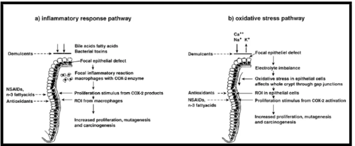
According to numerous studies, diet has a very important role in the prevention and causation of colorectal cancer. It has also been thought that the role of diet in colorectal carcinogenesis is particularly important when a poor diet is combined with a generally unhealthy lifestyle, consisting of excess calorie intake and weight gain, physical inactivity and consumption of alcohol. The roles of several foods and nutrients in colorectal carcinogenesis have been investigated. Evidence regarding the positive association between colorectal cancer and intake of red and processed meat is quite consistent. Various review, meta-analysis and observational analytical studies showed a positive association of intake of red meat and processed meat with risks associated with colorectal cancer. For example a meta-analysis of the cohort data showed that every 50g/day increase of red meat intake was associated with a 15% increase in colorectal cancer risk. The elevated risk may be due to an increased endogenous production of N-nitroso compounds (NOC), which may enhance the colonic formation of the DNA adduct O6-carboxymethyl guanine. Cooking meat at
33 high temperatures, intake of charbroiled or smoked meat lead to the formation of polycyclic aromatic hydrocarbons (PAHs) and heterocyclic amines (HCAs) and increased levels of DNA adduct. The levels of some HCAs and PAHs are comparable for red meat, fish and poultry smoked or cooked at high temperatures. Intake of red meat, but not of fish and poultry, increases the luminal contents of N-nitrosocompounds (NOCs) in colon. The increase in endogenous N-nitrosation can be attributed to heme iron, which is 10-fold higher in red meat than in white meat. An increase in the ratio of the consumption of red meat to consumption of fish/chicken was associated with an increase in colorectal polyp risk. Colorectal cancer risk may be negatively associated with fish intake. Intake of fish are reported to be negatively associated with DNA adduct levels, although another study found no effect. The protective effect of fish intake are suggested to be due to the content of n-3 poly-unsaturated fatty acids in fish. High intake of dietary fat has been associated to an increased risk of proximal cancers, while high intake of protein has been associated with an increased incidence of distal cancers. A statistically significant lower risk for colorectal cancer associated with high-fibre intake was showed from the European Prospective Investigation into Cancer and Nutrition (EPIC; 1,721 cases, nine European countries) and in other research. Intake of fruit, vegetables or antioxidant vitamins have been shown to be negatively associated with DNA adduct levels, although some studies found no effect and one study found an effect of increased vitamin intake only in females. The significance of dietary fibres as a protective factor against colorectal cancer remains controversial. However, a large study of European populations (the EPIC study) including 519,978 individuals have confirmed intake of dietary fibres to be protective. To my knowledge no studies has been published concerning intake of fibres and the level of oxidative DNA lesions or DNA adduct formation. Studies in cancer epidemiology and experimental carcinogenesis provided basis for possible mechanisms relating diet and colorectal cancer risk. Investigators initiated animals with carcinogens and then attempted to accelerate or inhibit promotion by modifying the diet. Two possible pathways to explain these puzzling results are explained as shown in Figure 9 in a local irritation. The irritation produces a focal inflammatory response that activates COX-2 (cyclooxygenase) and generates prostaglandins from arachidonic acid. This activates inflammatory cells which generate reactive oxygen intermediates (ROIs) that are mutagenic and mitogenic and promote carcinogenesis.