University of Messina
Department of Human Pathology
PhD Course in Biology and Molecular Biotechnology
XXX Cicle
2014-2017
Hepatitis B Virus affects Dendritic cell activation and
their cross-talk with Natural Killer cells
Tutor
PhD Student
Prof.re Raimondo Giovanni Claudia De Pasquale
Co-tutor
Sommario
ABSTRACT ... 3
INTRODUCTION ... 4
INNATE IMMUNITY IN VIRAL INFECTION ... 5
Dendritic cells: antigens recognition and immunological functions during microbial invasion ... 8
DC tissue distribution and migratory properties ... 10
DC pathogen recognition and activation of immune responses ... 13
Natural Killer cells: antiviral response and regulation of cytolitic activity ... 17
NK Distibution and trafficking ... 17
Activator and inhibitory Receptors ... 20
NK-DC COOPERATION DURING VIRAL INFECTION ... 26
HEPATITIS B VIRUS:ROLE OF INNATE IMMUNITY IN CONTROLLING HBV INFECTION AND ITS INVOLVEMENT IN VIRAL PATHOGENESIS ... 29
AIM OF THE STUDY ... 37
MATERIALS AND METHODS ... 38
CELL ISOLATION AND GENERATION OF HUMAN MONOCYTE-DERIVED DENDRITIC CELLS ... 38
DENDRITIC CELL IN VITRO STIMULATION AND ANALYSIS ... 38
HEPATITIS BVIRUS PARTICLES PURIFICATION ... 39
CYTOKINE MEASUREMENT ... 39
CYTOTOXICITY ASSAY ... 39
NK CELL PROLIFERATION ASSAY ... 40
RT-PCR ... 40
FLOW CYTOMETRY MAB ... 41
STATISTICAL ANALYSIS ... 41
RESULTS ... 42
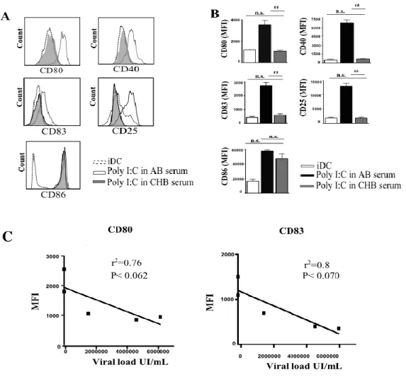
DC MATURATION IS DRASTICALLY AFFECTED IN THE PRESENCE OF SERUM FROM CHRONIC HEPATITIS B INFECTED PATIENTS. ... 43
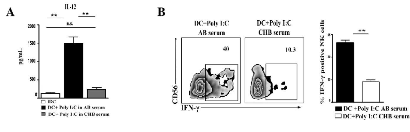
IL-12 PRODUCTION BY DCS IS DRASTICALLY IMPAIRED IN THE PRESENCE OF CBH SERUM, AFFECTING IN TURN IFN-Γ SECRETION BY NK CELLS FOLLOWING DC STIMULATION. ... 44
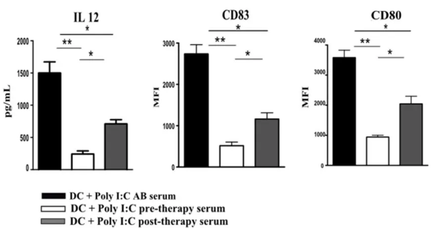
FUNCTIONS AND IL-12 PRODUCTION BY DCS ARE PARTIALLY RECOVERED IN THE PRESENCE OF POST-THERAPY CHB SERUM ... 45
HBV PARTICLES DIRECT AFFECT DC FUNCTION IN A TIME AND DOSE-DEPENDENT MANNER, IMPAIRING IN TURN PROLIFERATION AND CYTOKINE PRODUCTION BY NK CELLS ... 48
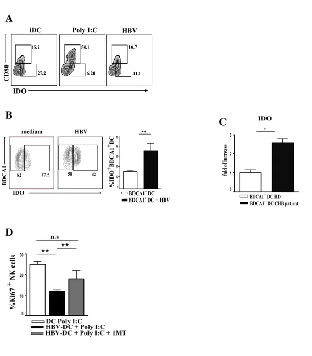
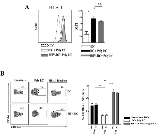
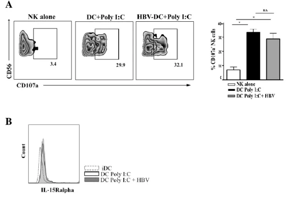
HBV SIGNIFICANTLY UP-REGULATES IDO ENZYME ON IN VITRO AND EX-VIVO DCS, AND BLOCKING EXPERIMENTS CONFIRM IDO INVOLVEMENT IN INHIBITION OF DC-MEDIATED NK CELL PROLIFERATION ... 51 HBV DOES NOT AFFECT HLA-I EXPRESSION ON DCS PROTECTING THEM FROM NK CELL -MEDIATED KILLING .. 53 HBV-CONDITIONED DCS ARE ABLE TO STIMULATE NK CELL CYTOLITIC ACTIVITY AGAINST HEPATOCARCINOMA CELL LINE ... 55 THE EXPRESSION OF SPECIFIC IMMUNOPROTEASOME SUBUNITS IS SIGNIFICANTLY IMPAIRED ON
HBV-CONDITIONED DCS ... 56 CONCLUSION AND DISCUSSION ... 57 BIBLIOGRAPHY ... 59
Abstract
The interplay between Natural killer (NK) cells and Dendritic cells (DCs) represents a first line of defence against viral infections. NK/DC interaction results in reciprocal cell activation and production of Interferon-γ (IFN-γ), a major antiviral cytokine. Chronic Hepatitis B (CHB) virus infection is characterized by a marked immune dysfunction that involves also NK cells, since IFN-γ production by NK cells is significantly reduced in both peripheral blood and liver of CHB infected patients.
Considering the crucial role of DCs in stimulating NK cell response, we sought to determine whether DCs could be responsible for NK cell impairment, as a consequence of HBV infection.
To this purpose, DCs were cultured in the presence of highly viremic sera from CHB patients and their phenotype and functions analysed.
In the presence of HBV serum, a drastic inhibition of DC maturation occurred, as demonstrated by the low expression of maturation markers, co-stimulatory molecules and IL-12, the latter representing a key cytokine for IFN-γ production by NK cells. Accordingly, DCs conditioned by the serum were significantly impaired in inducing both IFN-γ and TNF-α production by NK cells as well as in inducing NK cell proliferation. Similar data were obtained by adding HBV particles to DC cultures, suggesting that HBV has a direct role in the functional impairment of DCs.
In addition, the inhibitory role of HBV on DCs was apparently also exerted by a significant up-regulation of the tolerogenic enzyme Indoleamine 2 3-dioxygenase (IDO), which played a major role in the inhibition of NK cell proliferation induced by DCs.
Altogether, our data revealed that serum from chronic HBV patients significantly impairs DC functions and, in turn, both proliferation and IFN-γ production by NK cells upon DC/NK interactions. HBV is directly responsible for the phenotypic and functional alterations observed in DCs, suggesting that, during HBV chronic infection, abnormalities in the interactions between DCs and NK cells might occur, thus resulting in a reduced antiviral response.
Introduction
Hepatitis B virus (HBV) is a hepatotropic virus that causes variable degrees of liver disease in humans.. Despite the availability of a prophylactic vaccine, HBV is estimated to infect around 400 million people worldwide. Chronically infected patients are at risk of developing HBV-related diseases such as liver cirrhosis and hepatocellular carcinoma. However, HBV replication itself is not directly cytotoxic to cells, so to gain the name of stealth virus. Therefore, inadequate immune response toward HBV contributes to the establishment and progression of Chronic hepatitis B virus (CHB) infection (1).
Innate immunity is important in controlling infection immediately after contact with the pathogen, in order to limit the spread of the infection and to initiate an efficient development of adaptive immune response.
The immune system evolved to recognize specific structures on pathogens and eliminate foreign antigens exploiting specific receptor on cell surface. Among these, Toll like Receptors (TLR) play an important role in identification of viral antigens. Dendritic cells (DC), well equipped in TLRs, are extremely potent in initiating a primary immune response due to their ability to process foreign antigens in order to present them to effector cells. Additionally, DCs are able to activate natural killer cells (NK), orchestrating NK cell mediated innate immune responses. The outcome of NK/DC interaction during viral infection, is the activation of NK cell cytotoxicity and IFN-γ production, alike activated NK cells enhance DC maturation and IL-12 production. NK cells exert their non-cytolitic anti-viral function mainly by IFN-γ production (2).
Accumulating evidences suggested that an efficient induction of innate responses occurs during HBV acute infection by non-cytolytic mechanisms, advising that the innate immune response has the potential to contain the virus during early phases of HBV infection. However, studies indicate that NK cells from Chronic HBV infected patients are impaired in their non-cytolitic antiviral activity, as shown by the significant reduction of IFN- γ release. Still, NK cytotoxic function is maintained in peripheral blood and liver wondering that NK cells might also be implicated in disease pathogenesis.
The HBV-infected patients display large amounts of circulating HBV particles, which interact with DCs. Although, it has been demonstrated that HBV does not replicate into DCs, it has been proposed that HBV may cause alteration in DC functions probably interfering with signaling pathways involved in maturation and cytokine production (3)
Because DCs are potent activators of NK cells, remains to be clarified whether any DC dysfunction may affect NK functions. Therefore, in this study we wondered to investigated
whether the presence of HBV might alter dendritic cell activity, thus impairing NK/DCs cross-talk, resulting in a less efficient NK cell anti-viral response.
Innate Immunity in viral infection
Innate immunity have an important role in controlling infection immediately after contact with the pathogens, in order to limit the spread of the infection and to initiate an efficient development of adaptive immune response. Immune system is divided in two branches: Innate and Adaptive immunity, that work cooperatively to defend the host against infections. These two aspects of immunity differ with respect to how quickly it responds and for how long it responds to pathogens, for effectors cell types and for specificity to different classes of microbes.
Furthermore, the innate responses do not require prior exposure to the pathogen. On the contrary, adaptive immune responses are characterized by specificity and by the expansion of lymphocyte populations from a vast repertoire of lymphocytes bearing antigen-specific receptors that are generated through a mechanism generally known as gene rearrangement.
The innate immune response relies on recognition of evolutionarily conserved structures on pathogens, termed pathogen-associated molecular patterns (PAMPs), that are characterized by being invariant among entire classes of pathogens, essential for the survival of the pathogen. PAMPs are distinguishable from “self” through a limited number of germ line-encoded pattern recognition receptors (PRRs). PRRs upon recognition of pathogen structures trigger pro-inflammatory and antimicrobial responses by activating a multitude of intracellular signaling pathways, including adaptor molecules, kinases, and transcription factors. The signal transduction pathways ultimately result in the activation of gene expression and synthesis of a broad range of molecules, including cytokines, chemokines, cell adhesion molecules, and immuno-receptors, which together orchestrate the early host response to infection and at the same time represent an important link to the adaptive immune response.
Several and different pathogens, including viruses, bacteria, fungi, and protozoa, are recognized by PRRs that though are slightly different, display similar and overlapping mechanisms of action.
During viral infection, host PRRs detect viral components, such as genomic DNA, single-stranded (ss) RNA, double-single-stranded (ds) RNA, RNA with 5′-triphosphate ends and several viral proteins. Currently, different classes of PRRs have been shown to be involved in the recognition of virus-specific components in innate immune cells, among these Toll-like receptors (TLRs), that has been studied most extensively, together with retinoic acid-inducible gene I (RIG-I)-like receptors (RLRs) and NOD-like receptors (NLRs). Detection of viral components by RLRs and TLRs in immune cells activates intracellular signaling cascades, leading to the secretion of type I
IFNs, pro-inflammatory cytokines and chemokines. Type I IFNs activate intracellular signaling pathways via a type I IFN receptor, and regulate the expression of a set of genes. The IFN-inducible genes are involved in eliminating viral components from infected cells, inducing apoptosis of infected cells and conferring resistance to viral infection on uninfected cells. Type I IFNs are produced not only by professional innate immune cells, including dendritic cells (DCs) and macrophages, but also by non-professional cells, such as fibroblasts. Pro-inflammatory cytokines and chemokines are also critical for eliminating virus infection by provoking inflammation and recruiting innate and acquired immune cells. TLRs are transmembrane proteins suitable for detecting viral components outside of cells as well as in cytoplasmic vacuoles after phagocytosis or endocytosis. Among the at least ten known TLRs, present in mammals, TLR2, TLR3, TLR4, TLR7 and TLR9 are involved in the recognition of viral components. TLR2 and TLR4, present on plasma membrane, are involved in the recognition of viral envelope proteins on the cell surface, while TLR2 and TLR4 are critical for the recognition of bacterial components, lipoproteins and lipopolysaccharide, respectively. In contrast, TLR3, TLR7 and TLR9 are localized on cytoplasmic vesicles, such as endosomes and the endoplasmic reticulum, and recognize microbial nucleotides. TLR3 recognizes dsRNA, while TLR7 and TLR9 recognize ssRNA and DNA with CpG motifs, respectively. While TLR3 recognizes dsRNA in conventional DCs and possibly epithelial cells, TLR7 and TLR9 are highly expressed in plasmacytoid DCs (pDCs).
All TLRs, except TLR3, activate a common signaling pathway leading to the production of pro-inflammatory cytokines via MyD88 signaling pathway. Following the downstream of signaling transduction, finally NF-κB and MAP kinases are activated resulting in induction of genes involved in inflammatory responses.
Alternatively, in response to stimulation with dsRNA, TLR3 recruits another adaptor protein, TIR domain-containing adaptor inducing IFN-β (TRIF) that leads to TRAF6 activation and, finally, this latter is responsible for activating NF-κB leading to the expression of proinflammatory cytokines (4-7).
TLR3, as previously described, recognizes dsRNA, a universal viral molecular pattern, and thus, is involved in antiviral host immune responses (8). A synthetic ligand, Poly (I:C), structurally similar to double-stranded RNA, can also mediate responses through TLR3 triggering. The role of TLR3 was investigated in different types of viral infection and it was found to play a role in viral recognition and in promotion of various immune responses. For instance, it has been described a role for TLR-3 during Rhinoviruses infection, the major cause of the common cold, viral replication induces expression of TLR3 mRNA and its surface protein expression. TLR3 mediates protective antiviral activity in human bronchial epithelial cells infected with Rhinovirus
as confirmed by several impairment of antiviral response upon blocking of TLR-3, resulting in increased Rhinovirus replication (9).
Interestingly, TLR3 plays an important role in the pathogenesis of West Nile virus, a ssRNA flavivirus, mediating penetration of West Nile virus across the blood-brain barrier and induces neuronal injury. Another interesting feature of TLR3 is that it promotes cross-priming to virus-infected cells. To this regard, it has been proposed that TLR3 may have evolved to permit cross-priming of cytotoxic T cells against viruses that do not directly infect dendritic cells (10).
It has been described a role for TLR-3 also in course of Hepatitis B (HBV) infection. Wieland et al proposed that TLR3 ligand poly I:C induces intrahepatic IFN-β production , quite able to inhibits HBV replication by non-cytolytic mechanisms (11).
Additionally, Isogawa et al. (12) tested the ability of different TLR ligands to inhibit HBV replication in the HBV transgenic mouse model injectioning a single-dose of TLR3, TLR4, TLR5, TLR7, and TLR9 ligands able to suppressed HBV replication in the liver in an IFN-α/β-dependent manner. TLR3 activation by Poly I:C of hepatic Non-Parenchymal Cells (NPCs) such as Kupffer cells, could lead to release of IFN-β production and antiviral cytokines which are able to inhibit HBV replication in an in vitro co-culture mouse model (13).
Interestingly, several studies have suggested that HBV seems able to inhibit pattern recognition receptor (PRR) and IFN signaling. HBV surface and “e” antigen (HBsAg, HBeAg) and HBV particles could inhibit the activation of NPCs by TLR3 ligands by trigger IL10 production on hepatic cells and thereby attenuates the TLR3-mediated activation of NPCs. Additionally, at high amounts of HBV, even TNF-α and IL-6 expression in NPCs induced upon TLR triggering was suppressed, suggesting that mechanisms employed by HBV may counteract induction of an efficient innate response (14).
It also been reported that TLR3-mediated functions are impaired in patients with chronic HBV infection and may recover partially under successful antiviral treatment (15). In the woodchuck model of infection, peripheral blood mononuclear cells (PBMCs) from animals with chronic WHV infection show reduced responses to Poly I:C stimulation (16). All together these information confirming that the interaction of HBV or viral components of HBV with the innate immune system is complex, leading both to activation and inhibition of host innate responses.
As previously discussed, an effective immune response requires recognition of pathogen and consequent induction of innate and adaptive immune systems. The innate immune responses to infection are rapid and mostly dependent from PRRs that recognize PAMPs. Some of these receptors are present on the surface of professional phagocytic cells such as macrophages and
neutrophils, where they mediate the uptake of pathogens, and delivered them into the lysosome for degradation.
Additionally, other cells type take part to promote the implementation of innate response since, asserting innate immune system as crucial in the initiation and subsequent direction of adaptive immune responses. Natural Killer (NK) cells and Dendritic Cells (DCs) represent two central components of the innate immune system, and both play a key role in fighting early infection. DCs and NK cells interact each other through cell to cell contact and soluble factors in periphery or in secondary lymphoid tissues. The bidirectional crosstalk between NK cells and DCs results in maturation, activation, and cytokine production by both cells (17).
Dendritic cells: antigens recognition and immunological functions during microbial
invasion
Dendritic cells (DCs) are professional antigen-presenting cells and essential mediators of immunity and tolerance. In 1973 Ralph Steinman discovered in the mouse spleen a rare population of cells characterized by stellate morphology and extended veils. He further studied what he named dendritic cells to provide evidences of the emerging functional specializations of these cell types. Over the following years, DCs was found to be critical in shaping innate and adaptive immunity, thanks to their ability in mounting immune responses to foreign antigens, and its contribution to the induction of tolerance to self-antigens. Consequently, subsequent studies focused on the potential therapeutic benefits of modulating DCs for vaccines or suppressive therapies against pathogens, tumors, and/or autoimmune diseases (18). Soon after the identification of DCs in lymphoid organs, and the discovery of epidermal Langerhans cells (LCs) led to the idea that more than one branch to the DC family might exist (19). Consequent studies had revealed the presence of cells with a similar phenotype in most non-lymphoid tissues that, upon antigen encounter, migrate through the lymph to lymphoid organs, where they localize in the T cell zone and present antigens to T lymphocytes. More recently, a further major division in the DC family occurs thanks to the identification of a population of cells that morphologically resemble plasma cells but, upon exposure to viral stimuli, produce enormous amounts of interferon IFN-α. Importantly, these cells also differentiate upon stimulation into immunogenic DCs that can prime T cells against viral antigens and were named plasmacytoid DCs (pDCs) (20). To distinguish pDCs from Steinman’s DCs, the latter were renamed conventional DCs (cDCs), and remain so today.
DCs initiate an immune response by presenting the captured antigen, in the form of peptide– major histocompatibility complex (MHC) molecule complexes, to naive T cells in lymphoid
tissues (21). When compared with other APCs, such as macrophages, DCs are extremely efficient. In peripheral tissues, DCs capture antigens through several complementary mechanisms (22). Antigen-loaded DCs then migrate into the draining lymph nodes through the afferent lymphatics. Meanwhile, they process the proteins into peptides that bind to both MHC class I molecules and MHC class II molecules.
Immature non-activated DCs can also present self-antigens to T cells (23, 24), which leads to immune tolerance either through T cell deletion or through the differentiation of regulatory (T regulatory cells, Treg) or suppressor T cells. By contrast, mature activated and antigen-loaded DCs can start the differentiation of antigen-specific T cells into effector T cells with unique cytokine profiles and function.
Antigens can also directly reach lymph resident DCs through the lymph (25). Lymph node-resident DCs that acquired antigen directly from the lymph are the first to present peptides to naive CD4+ T cells, which results in T cell priming. T cells primed by activated DCs produce
high amount of interleukin-2 (IL-2), which in turn facilitates T cell proliferation and clonal expansion in LN. CD4+ T cells and CD8+ T cells upon interaction with DCs differentiate into
antigen-specific effector T cells with different functions.
Additionally, DCs also have an important role in controlling humoral immunity but the mechanism by which DCs address antigens into compartment where they do not undergo degradation, that results in the presentation of unprocessed antigens to B cells is poorly characterized (26).
However, these DC properties allow the activation of both arms of the adaptive immune system. Following Ag encounter, DCs undergo maturation, that is associated with several events, such as the downregulation of antigen-capture activity, the increased expression of surface MHC class II molecules and co-stimulatory molecules, the increased ability to secrete cytokines, as well as the acquisition of CCR7, which allows migration of the DC into the draining lymph node (22).
Conventional DCs (cDCs) can be divided into at least two main subsets characterized by either CD8α and CD103 or CD11b expression Both subpopulations can be found in lymphoid tissue, including spleen, lymph node, and bone marrow (BM), as well as most non-lymphoid tissue. Heterogeneity within the DC population was first demonstrated by both the Shortman and Steinman groups, including the discovery of a CD8α-expressing DC subset in murine lymphoid organs (23). An equivalent population also exists in non-lymphoid tissues, although these cells do not express CD8 but are instead identified by the CD103 integrin marker (29, 30) and they also appear to be conserved through evolution. In both human and mice DC subset was discovered a potential common marker, the chemokine receptor XCR1, found by transcriptome profiling studies (32, 33). Development of CD8α+cDCs and the nonlymphoid tissue equivalent, the
binding 2 (Id2), interferon regulatory factor 8 (IRF8), basic leucine zipper ATF-like 3 transcription factor (BATF3), and the nuclear factor interleukin 3 regulated (NFIL3). CD11b+cDCs are the most abundant cDCs in lymphoid organs except for the thymus and can also
be found in nonlymphoid tissue. In contrast to CD8α and CD103 DCs, the population currently defined as CD11b+cDCs is heterogeneous and remains less well characterized.
Splenic CD8α+cDCs are functionally specialized in cross-presenting exogenous Ags on MHC-I
molecules to CD8+ T cells (38) and stimulation of TLR signaling on the CD8α+ and CD103+cDC
lineage induces prominent secretion of “bioactive” IL12p70. In contrast in lymphoid organs, CD11b+ DCs are not efficiency to cross-present and produce specific cytokines, such as IL-12. It
has however been noted that CD11b+cDCs are, as compared to CD8α+cDCs, superior in the
induction of CD4+ T cell immunity, potentially because of their prominent expression of MHC-II
presentation machinery (35, 36). This specialization of CD11b+cDCs was recently attributed to
their expression of the transcription factor IRF4 (37) CD11b+ DCs can also be characterized by
their cytokines production, such as IL-6 (38) and IL-23 (39). Furthermore, splenic CD11b+cDCs
were shown to be prominent producers of proinflammatory chemokines after TLR ligand exposure, such as CCL3, CCL4, and CCL5 (40).
cDCs generally display a short half-life of approximately 3–6 days and are constantly replenished from BM precursors in a strictly Flt3L-dependent manner. Myeloid and lymphoid branches of the immune system bifurcate early during hematopoiesis into common myeloid and lymphoid precursors (CMPs, CLPs). It has been proposed that CD8α-positive and -negative subsets, are of myeloid origin.
DCs thus initially share their origin with monocytes. Althought, a pre-DC population was subsequently identified in lymphoid tissues and in the blood. Pre-DCs were found to populate lymphoid organs via the circulation and give rise to cDCs in lymphoid and non-lymphoid tissues (41, 42, 43, 44)
DC tissue distribution and migratory properties
DC progenitors are not restricted to the BM but can be found in multiple locations, including the thymus, blood, lymph, and most visceral organs. DCs reside in lymphoid and selected non-lymphoid tissues. Lymphoid tissue–resident cDCs differentiate in non-lymphoid tissues, such as splenic compartment, where they spend their entire lives. Lymph Node also include non-lymphoid tissue migratory cDCs.
The spleen harbors different populations of resident cDCs: Among these, CD8α+cDCs represent
about 20%–30% of the total splenic DC compartment and are localized in the marginal zone where they sample lymph- and blood-borne Ags and pathogens. CD8α+cDCs furthermore
efficiently uptake apoptotic or necrotic cells from peripheral blood (45) and are thus able to present exogenous tumor- or virus-derived Ags. Further functional properties of splenic CD8α+cDCs are their highly specific expression of the double-stranded RNA sensor
In Lymph node (LN), cDCs can be subdivided into CD8α− and CD8α+ subsets (46) but harbor in
addition migratory DCs that entered via the afferent lymphatics from associated non-lymphoid tissues. The latter probably import pathogen antigens for T cell stimulation.
In contrast to tissue-migratory cDCs that arrive in the LNs in a mature state, lymphoid tissue CD8+cDCs are phenotypically immature in the steady state (47). Activation to a phenotypically
mature state occurs upon stimulation with microbial products or when cDCs are isolated from the lymphoid tissue and cultured in vitro. In LN, CD11b+cDC represents another subset that
predominates the lymphoid-resident cDC population; they proliferate in situ in response to Flt3L and are characterized by the lacking of CD8 marker. CD11b+cDCs crucial feature is the high
production the CD4+ T cell attractant chemokines CCL17 and CCL22.
Like lymphoid organs, most non-lymphoid tissues contain at least two major subsets of cDCs that often share the αE integrin CD103 marker but can be distinguished according to CD11b expression. Non-lymphoid tissue DCs are in contact with body surfaces, such as the skin, lung, and intestine and have different specializations. For istance, Langherans Cells (LC) residing in the skin epidermis represent the principal skin DC population and play a critical role in the defense against external threats. Indeed, LCs efficiently phagocytose pathogens after epidermal cell injury and switch their chemokine expression pattern from CCR6 to CCR7, which allow them to migrate toward cutaneous LNs. In contrast to most DCs, LCs develop independently of Flt3 (48). Compared with dermal cDCs, LCs are characterized phenotypically by lower MHC-II levels, intermediate CD11c levels, and very high levels of the C-type lectin langerin, CD207, which is responsible for the generation of Birbeck’s granules, the ultra-structural hallmark formed by LCs. LCs that are transiting through the dermis or that have migrated to skin-draining LN can be identified based on expression of Langerin, EpCam and CD11b and the absence of CD8 and CD103 expression.
In contrast, dermal DCs are discriminate from Langherans Cells and most often identified in the dermis and LN based on expression or the absence of Langerin, CD103 and the absence of CD11b, CD8 and EpCam expression. Importantly, careful examination of subset ontogeny has clearly identified that LCs and Langerin+dDCs are distinct DC subsets (49).
Additionally, other markers help to identified DCs: among these HLA-DR and DC-SIGN (DC specific intercellular adhesion molecule-3 ICAM-3), a c-type lectin receptor, that recognize high-mannose-containing glycoproteins on viral envelopes acting as receptor for several viruses such as HIV and Hepatitis C. Moreover, it is used as a marker for immature DCs.
Recently, the human DC subset-specific markers termed: blood DC Ag-1 BDCA-1 (CD1c), BDCA-2 (CD303), BDCA-3 (CD141), and BDCA-4 (CD304) have been also identified (50).
DCs are sparsely distributed through the liver, and immunohistochemical studies of patient liver biopsies indicate that they are primarily found in the portal regions and occasionally in the parenchyma.
The DC population in the liver can be further divided into two major functionally and phenotypically distinct subsets. In mice, the hepatic CD11chiMHC-IIhi DC population contains a
more prevalent CD103−CD11bhi population and a rarer CD103+CD11blow population. CD11b
expression on the CD103− DC population tends to be heterogeneous, and the CD103−CD11blow
subset may represent a less mature population. Corresponding counterparts to the CD11bhi and
CD103+ DC populations can be further identified in human livers by the markers CD1c (BDCA1)
and CD141 (BDCA3) (51)
Recent studies suggest that CD141+ DC subset may play a specialized role during hepatic viral
infections. The frequency of CD141+ DCs is higher in the liver than in the peripheral blood and
has been found to further increase in the setting of hepatitis C virus infection along with high levels expression of TLR3 (52). Remarkably, DC population present in human liver, produces higher levels of IL-10, compared to DCs from the blood, spleen and skin, which accounts for their reduced allogeneic stimulatory capacity. In normal liver, DCs reside as “immature” expressing low levels of surface MHC and costimulatory molecules (CD40, CD80, CD86) necessary for T cell activation. However, these immature DCs are extremely well-equipped for Ag capture, processing, and loading onto MHC class II molecules for export to the cell surface.
Circulating DCs and their precursors circulate the blood and traffic in response to tissue-specific recruitment signals that are expressed on vascular epithelial cells. These signals include pro-inflammatory chemokines, and chemotactic cytokines, that are produced from sites of inflammation or from normal tissues, able to recruit DC precursors. DCs express specific adhesion molecules and maturation-dependent chemoattractant receptors that allow them to respond to a variety of ligands (53). Immature DCs enter to non-lymphoid peripheral tissues and travel within them, by utilizing specific chemokine receptor-ligand pathways, such as CCR2-CCL2 (54, 55), CCR5-CCL5 (56), and CCR6-CCR2-CCL20 (57). When DCs encounter Ags becoming mature and downregulate their responsiveness to these inflammatory chemokine pathways and traffic to the draining LNs by upregulating CCR7, which responds to two ligands, CCL19 and CCL21 (48, 53). These chemokines are expressed by peripheral lymphatic endothelial cells as well as LN stroma cells and guide DCs to downstream LNs. Leukocyte extravasation occurs in a series of distinct steps including tethering, rolling, activation by a chemoattractant, firm adhesion, and diapedesis. Mainly for circulating DCs, tethering and rolling are primarily mediated by one
or more of the three members of the selectin family: integrins, in particular LFA-1 (αLβ2), VLA-4 (αVLA-4β1), Mac-1 (αMβ2) and αVLA-4β7 mediate arrest of the rolling cells by binding to members of the immunoglobulin superfamily, including ICAM-1 (ligand for LFA-1 and Mac-1), ICAM-2 (ligand for LFA-1), VCAM-1 (ligand for VLA-4 and weakly for α4β7) and MAdCAM-1 (ligand for α4β7) (58). A large variety of pro-inflammatory stimuli induce DCs to migrate from peripheral tissues to LNs, such as chemical (e.g. contact sensitizers and irritants), physical (e.g. UV radiation or trauma) or biological stimuli (e.g. microbial or tissue necrosis). At the site of injury, DC antigen-uptake occurs, followed, within hours, by maturation process whereby DCs regain their motility, re-arrange their chemokine receptor repertoire, upregulate their Ag presentation machinery and eventually migrate to LNs (59).
DC pathogen recognition and activation of immune responses
DCs process and present antigen to activate both CD4+ and CD8+ T cells. This appears to be the
most prominent role for DCs, since only DCs are capable of activating naïve T cells. Dendritic cells, present throughout peripheral tissues, constitutively sample the environment for the presence of pathogens that upon recognition are internalized and processed into peptides. Peptides can be generated either by lysosomal proteases in the endocytic pathway, or by proteasomes. Thus, generated peptides may associate intracellularly with either MHC class I or MHC class II molecules, and in that context can be transferred and displayed at the plasma membrane. DCs migrate to lymphoid tissues, thus MHC–peptide complexes can be recognized by T cells. Antigen specific CD8+ cytotoxic T cells are activated by recognition of MHC-I on
DCs that load Ag, helping in elimination of infected and malignant cells. In contrast to MHC-II, MHC-I is expressed by nearly all cell types, and in non-professional antigen-presenting cells is exclusively loaded with peptides that are generated from cytosolic proteins by the ubiquitin-proteasome system. Cytosolic peptides, upon processing, can be translocated into the lumen of the endoplasmic reticulum for loading onto MHC-I. Peptide-loaded MHCI is then transported out of the ER via the Golgi apparatus to the plasma membrane, where it is stably exposed. Infected cells that display pathogen-derived peptides on MHC-I can be killed by cytotoxic T cells that specifically recognize relevant MHC-I/peptide complexes (60).
In contrast, MHC class II molecules are normally found only on antigen-presenting cells such as dendritic cells, mononuclear phagocytes, some endothelial cells, thymic epithelial cells, and B cells.
The antigens presented by class II peptides are derived from extracellular proteins. Loading of a MHC class II molecule are loaded with peptides upon phagocytosis; extracellular proteins are endocytosed, digested in lysosomes, and the resulting peptide fragments are loaded onto MHC class II molecules. Peptide-loaded MHC II is thus presented to CD4+ T cells.
DCs have evolved specific surface molecules to uptake pathogens, in addition to expressing a number of specific receptors for certain viruses such as CD4 and CXCR4 for HIV, the previously named DC-SIGN and its close relative DC-SIGNR, or L-SIGN found on liver endothelium, are some of the most intensively studied pathogen receptors (61). Additionally, DCs sense the environment through both surface and intracellular receptors, which comprise several families, including cell surface C-type lectins (CLRs), surface and intracellular TLRs, and intracellular helicases. The helicases are a very large family of molecules, including retinoic acid-inducible gene I (RIGI), which recognize nucleic acids.
The different signals that are provided by different microbes either directly or through the surrounding immune cells induce DCs to acquire distinct phenotypes. DC maturation varies according to different microbes because microbes express PAMPs that trigger distinct DC molecular receptors. Strikingly, although most microbes activate DCs, same of these can block DC maturation. As previously mentioned, viral products, pro-inflammatory cytokines, bacterial or double-stranded RNA trigger the maturation of immature DCs and in this context cytokines milieu play a critical role.
Concerning DC cytokines profiles it is dependent from signals occurs upon pathogens encounter. It is now clear that the interaction between TLRs and PAMPs plays a key role in enhancing the release of cytokines, such as interleukin-10 (IL-10), IL-12 and type I interferons. Production of IL-12 and IL-10 by mature murine DCs can be elicited by many pathogens or their products (62-64) In human system, Gram-negative bacteria, but not Gram-positive bacteria, prime DCs to produce the IL-12 (65). In contrast, Mycobacterium tuberculosis blocks DC maturation and induces IL-10 release by targeting DC-SIGN (66).
Considering DC role in directing the development of T-cell responses, the pattern of cytokines that they release upon their activation play an important role in determining the T-cell response. Importantly, DCs are also able to produce type I interferon in response to viral infection or following interaction with T cells. It is now well established that IL-12-secreated DC drive T helper type 1 (Th1) responses whereas IL-10 inhibits them, promote Th2-type responses. Also, Type I interferons have been shown to regulate T-cell differentiation, but their role has not yet been clearly resolved.
The major events in DC maturation are probably the up-regulation of MHC and costimulatory molecules on their surface. Pattern of costimulatory molecules is well known. A variety of inflammatory or pathogen-derived molecules rapidly up regulate expression of very early costimulatory signals. Members of B7 family,such as CD80/CD86 are expressed on DCs and constitutes the most important costimulatory pathway in T cell activation since through binding of CD28 on T cells is promoted the production of IL-2, a factor that supports expansion and survival of T cells. Importantly, CD80/CD86 by binding with CD28, also strongly interferes with tolerogenic properties of immature DC. Interestingly, the same costimulatory molecules are also
responsible for shutting down T cell activation exploiting the upregulation of an inhibitory molecule, CTLA-4, on T cell surface. Briefly, CTLA-4 binds with higher affinity to CD80/CD86 than CD28 and thereby competes for interaction with both costimulatory molecules provides a very simple negative feedback loop (67). Human immature DCs constitutively express intermediate amounts of CD86 and lack CD80. Hence, for characterization of human DC maturation, CD80 is considerably more reliable, as it is exclusively induced on mature DC while CD86 is already present on immature DC and further up-regulated upon stimulation (68).
The ligation of the co-stimulatory receptor CD40 (also known as TNFRSF5) is an essential signal for the differentiation of immature DCs into fully mature DCs.
CD40 ligation on DCs, indeed, increases expression of costimulatory, adhesion and MHC molecules and promotes the production of T cell stimulatory cytokines such as IL-12. Recombinant CD40 ligand therefore is often used to induce DC maturation (69).
It is important to note that in the absence or low levels of costimulation signal, including IL-1, IL-12, TNF-α, CD40, CD80, CD86, the cross-linking between T cell receptors and MHC-bound peptides expressed on the surface of DCs leads to anergy or apoptosis of antigen-specific T cells. The functional properties of DCs are thus mainly dependent on their status of maturation and activation. Accordingly, subsets of immature DCS induce and maintain peripheral T cell tolerance whereas differentiated mature DCs efficiently induce the development of effector T cells. To this regard, it is well stated that a crucial role for DCs in maintaining immuno-tolerance also occurs. Several studies have shown that DCs in the steady state are able to have negative effect on T cell survival. Certain subpopulations of DCs in the periphery were found to induce CD4+CD25+ Treg cells, a subsets well known to display regulatory functions in vitro and in vivo
among these subsets liver-derived DC, IL-10-producing DC or lymphoid-derived DC. In the absence of inflammation thus in homeostatic conditions, circulating immature DC have the primary function to migrate in Lymph-node and induce the differentiation of naive or resting T cells into Treg cells.
A key factor that alters the maturation process of DC is IL-10. The immunosuppressive properties of IL-10 on DC result in a reduced expression of MHC class II molecules as well as co-stimulatory and adhesion molecules. Steinbrink et al (70) demonstrated that human DC from the peripheral blood, matured in the presence of IL-10, induce anergic T cells, inhibiting IL-2 production and T cell proliferation. Therefore, the presence of IL-10 during DC-maturation inhibits the development of immunostimulatory DCs.
Upon pathogens encounter, DCs undergo maturation process, resulting in DC maturation by phenotypical and functional change that include the up-regulation of MHC I and co-stimulatory molecules and progressive variation in Ag-processing machinery (APM) component expression. The proteasome is a central element of APM and can be expressed in two forms, constitutive and
inducible also called immunoproteasome, endowed with different protein cleavage specificities. The switch of constitutive proteasome in the immunoproteasome form occurs in sites of infection and suggests that, during the peak phase of viral or bacterial elimination, this last form is more efficient. Immunoproteasome adopts a distinct manner to cleave proteins thus generating more peptides capable to better bind to MHC class I molecules.
Immunoproteasome proteins were significantly upregulated also in response to the major immunomodulatory cytokine, interferon-gamma (IFN-γ).
The constitutive proteasome is a four-ring structure; the outer rings contain seven non-catalytic α-type subunits, whereas the inner rings contain seven β-α-type subunits, three of which have catalytic properties (delta/β1, Z/β2, and X/β5). The immunoproteasome, inducible by TNF-α and IFN-γ, contains alternative forms of the catalytic subunits (LMP2, MECL1, and LMP7), that replace the corresponding constitutive homologs β1, β2, and β5. LMP2 and LMP7 were located within the major histocompatibility complex (MHC) class II region where they are clustered with the TAP-1 and TAP-2 genes, while MECL-1 is encoded outside the MHC class II region. After proteolysis of a ubiquitinated proteins in the immunoproteasome core, peptides are further cut by a battery of aminopeptidases, and bind to TAP-1 and TAP-2 for transport into the ER where the N-terminus of peptides were further cleaved and loaded into the MHC class I complex. The rising MHC- I complex is formed chaperoned by calnexin (CNX), tapasin (TPN), and calreticulin (CRT). Peptides generated by either partial or complete degradation of proteins can be loaded into MHC class I molecules for recognition by CD8 T cells as part of immune surveillance process (71).
The location of the LMP2 and LMP7 immunoproteasome subunits in the MHC region, and their production in response to proinflammatory cytokines, suggested their role in antigen processing. Study with KO mice for one or both of these subunits help to investigate subunit-specific properties. It was found that mice deficient in LMP7 exhibited a modest reduction in surface expression of MHC class I, and resulted in mice with a reduced response to antigens. Deficiency in LMP2 was found to result in a reduced of CD8 T cell activation. Additionally, LMP2-deficient cells were less able to activate NF-κB pathway (72). Findings derived from the use of these KO mice, have demonstrated and confirmed that iproteasome subunits have a roles in antigen-specific interactions with microbial challenges. Importantly, van Helden and colleagues (73) reported that MECL-1 and LMP7 deficiency resulting in a reduced MHC class I expression, made those cells susceptible to natural killer cell-mediated killing in mice whose immune system had been activated by a viral infection.
Natural Killer cells: antiviral response and regulation of cytolitic activity
Natural killer (NK) cells interact with DCs to reciprocally activate and influence subsequent effector functions. The intricate cross-talk between NK cells and DCs serves to modulate the anti-viral immune responses. Although specific NK cell responses depend on viral context, they require other cells to coordinate an effective antiviral response.
NK cells are the prototypic innate lymphoid cell (ILC) and are considered crucial components of innate immune system given their pivotal role in the first line of defense.
NK cells are characterized by a wide spectrum of effector functions such as killing and controlling of target cells, mainly tumor and virally-infected cells, the ability to influence various steps of the immune response (74-76).
NK cells comprise 5–10% of human peripheral blood lymphocytes, however, this proportion can vary with age. The traditional cell surface phenotype defining human NK cells; analyzing the lymphocyte gate by flow cytometry NK cells are characterized by the absence of CD3 and expression of CD56, the 140-kDa isoform of neural cell adhesion molecule (NCAM) found on NK cells and a minority of T cells.
Human NK cells can be subdivided into different populations based on the relative expression of the markers CD16 (or FcγRIII, low-affinity receptor for the Fc portion of immunoglobulin G) and CD56: CD56bright CD16− (50–70% of CD56bright), CD56bright CD16dim (30–50% of CD56bright),
CD56dim CD16−, and CD56dim CD16bright subset (77).
The two major subsets are CD56bright CD16dim/− and CD56dim CD16bright, respectively. The
CD56dim CD16bright NK cells represent at least 90% of all peripheral blood NK cells and are
therefore the major circulating subset (78, 79). A maximum of 10% are CD56bright NK cells. NK Distibution and trafficking
One of the challenges in identifying tissue NK cells is the discrimination of these cells from other ILCs, since they share several markers. NK cells are present in healthy skin and gut, in the liver, in the lungs, and are abundant in uterus during pregnancy. In addition, human NK cells were investigated also in other tissues such as the kidney (80), joints (81), and breast under pathophysiological conditions. In the normal intestinal mucosae, NK cells (82) are found predominantly as intraepithelial lymphocytes and within the lamina propria, but are rarely associated to lymphoid aggregates, although they can be found in the parafollicular region of cecal lymphoid patches, Peyer’s patches, and mesenteric lymph nodes.
NK cells in healthy human liver strikingly account for almost 20–30% of all human hepatic lymphocytes and are found among the non-parenchymal cells that populate this organ (83).
However, in steady-state, NK cells are preferentially located in the hepatic sinusoids, often adhering to the endothelial cells (84). A particular subset of NK cells is found in the placenta, where it regulates specific developmental processes at the fetal–maternal interface. During the first trimester of pregnancy, NK cells represent a subpopulation with unique phenotypic and functional properties, representing about 50–90% of the lymphoid cells infiltrating in this tissue. Carrega et al. (85) showed that NK cells expressing NKp46marker populate the normal lung, counting for ~10% of lymphocytes present in this tissue. In addition, it has been described that the majority (~80%) of lung-NK cells belong to the CD56dimCD16+ subset. The localization of
NK cells in different tissues indicate that they could migrate to various organs then reside there, where they acquire peculiar activities. Although, different theories argue the possibility that NK cells could re-circulate constantly through the tissues.
At date, NK cell subsets, mostly represented by CD56bright populate gut, liver, lung, and other
different human solid tissues thus indicating that specific homing signals are important to drive the localization of NK cells to the different tissues. Importantly, it has been described that NK cells are present in in human afferent lymph draining peripheral tissues suggesting that NK cells might even exit the organ and traffic through tissues in normal conditions (86). Concerning NK trafficking, it has been demonstrated that the expression of CCR7 and L-selectin (CD62L), drive NK cell trafficking.
NK cells continuously traffic toward tissue through a combination of stimuli able to promote their mobilization, such as chemokines. Chemokines bind with high efficiency to physiologic L-selectin ligands on peripheral LN high endothelial venules (HEVs) thus promoting a conformational change in the receptor, that trigger intracellular signals, to finally drive cell polarization, migration, and adhesion, thus resulting in the induction of leukocyte trafficking and homing. NK cell subsets display a differential pattern of chemokine receptor expression. CD56bright NK cells are targeted to lymph nodes via CCR7, preferentially express CXCR3 and
have higher CXCR4 expression levels as compared with CD56dim cells. CD56dim NK cells, in
contrast, uniquely express CXCR1, and CX3CR1. CXCR3 ligands are expressed at low levels in
homeostatic conditions, but their expression can be upregulated (87). It has been described that in multiple myeloma patients with active disease, to an up-regulation of CXCR3 ligand, CXCL10, corresponded to marked down-regulation of CXCR3 expression levels by BM NK cells, an event that was linked to reduction of NK cell localization in the BM in multiple myeloma-bearing mice (88). CXCL10/CXCR3 axis is also involved in hepatic trafficking of NK cells, in which a truncated form of ligand can bind to CXCR3 without signaling, thus preventing NK cells to migrate into infected liver but they instead accumulate in the peripheral circulation (89).
The in vivo NK cell developmental pathway has remained somewhat of a mystery in contrast to the pathways for B cell and T cell development, it was generally accepted that NK cells develop
exclusively within the bone marrow similar to most other leukocyte populations. BM ablation results in NK cell deficiency in mouse models, and human NK cells may be derived in vitro from BM-derived CD34+ hematopoietic precursor cells (HPC) (90). Moreover NK cell commitment
requires the expression of transcription factors such as nuclear factor IL-3 regulated (NFIL3) and thymocyte selection-associated HMG box factor (TOX). Interestingly, NK cell precursors are normally detected in the circulation, and recent data indicate that specific CD34+ NK cell
precursors are selectively enriched in extramedullary tissues where unique subsets of mature NK cells reside, suggesting that the latter may derive locally in situ (91).
Scientific consensus at date, suggest that NK cells derived from CD34+ hematopoietic progenitor
cells (HPCs). However, the site of maturation and details of the process are only now beginning to emerge. Human T cells develop in the thymus and human B cells develop in the bone marrow, and the intermediate populations can be isolated in situ from their respective maturational sites. In contrast, the characterization of the full NK cell developmental pathway from CD34+ HPCs
within the bone marrow or in the thymus needs to be further clarified. A first clue that NK development might not occur wholly in the bone marrow came from the observation that CD56bright could be isolated from lymph nodes and tonsils or secondary lymphoid tissue (SLT).
CD56bright NK cells are relatively dominant in SLT compared with their more abundant CD56dim
NK counterpart found in bone marrow, blood, and spleen (92).
Further studies have suggested that the CD56dim NK-cell subset is derived directly from the
CD56bright NK subset. Interestingly, one of these recent studies also revealed an important role for
the CD56 molecule itself in promoting this terminal maturational step. Using an in vitro co-culture system consisting of purified CD56bright NK cells and human fibroblasts, Studies have
demonstrated that antibody blockade of the interaction between CD56 and fibroblast growth factor receptor-1 significantly inhibited the generation of CD56dim NK cell (93). Supporting these
data, it was found that the CD56bright NK subset is the major NK cell population that is derived
early in vitro when CD34+ HPCs are cultured in NK development supportive conditions, whereas
CD56dim NK cells develop later over time. Additionally, CD56brightappear to accumulate earlier in
the blood following bone marrow or stem cell transplantation, display longer telomeres compared to CD56dim NK cells.
Additionally, within the SLT relative to blood or bone marrow was found a selective enrichment of both CD34+CD45RA+ pre-NK cells and CD56bright NK cells in close contact with an
abundance of dendritic cells (DCs) and other antigen presenting cells (APCs) that express membrane-bound IL-15. Il-15 is required for NK-cell maturation, suggesting that SLT may be a site, although not preferentially, for NK-cell development in vivo.
Among multiple growth factors likely facilitate the development of NK cells, IL-15 is considered the most important NK cell homeostatic cytokine. IL-15 promotes the proliferation and survival
of mature NK cells and is also capable of inducing the differentiation and maturation of CD34+
HPC into CD56bright NK cells in stroma-free medium (94, 95). IL-15 mostly produced by
dendritic cells, is typically provided in soluble form in vitro, yet in vivo it is presented in trans as a membrane bound ligand in association with the IL-15 receptor alpha chain (IL-15Rα) (96, 97). In order to respond to IL-15, NK cells must express the common gamma chain (CD132) as well as the IL-2Rα (CD122) (98). As such, human NK cell precursors have been traditionally defined as CD34+CD122+ cells (IL-2Rα).
Caligiuri et al, described five putative stages of human SLT NK cell development according to the differential expression of CD34, CD117, CD94, and CD16. Stage 1 cells (Lin−CD34+CD117−CD94−CD16−) lack expression of the common IL-2/IL-15 receptor beta chain (IL-2/15Rβ, CD122) and are thus not responsive to exogenous soluble IL-2 or IL-15 ex vivo. However, they can generate NK cells when cultured in IL-15 plus other cytokines, such as Flt3 ligand and c-Kit ligand (KL) that likely induce CD122 expression and hence IL-15 responsiveness. In contrast, stage 2 cells (Lin−CD34+CD117+CD94−CD16−) constitutively express CD122 and a functional high affinity IL-2 receptor, including the IL-2Rα subunit (CD25), can generate functionally mature NK cells in vitro in the presence of exogenous soluble IL-15 in media without other cytokines or support cells. Stage 3 cells (Lin−CD34−CD117+CD94−CD16−) lacked T cell and DC developmental potential and were proposed to represent committed NK cell precursors (99, 100).
The sequential acquisition of receptors and functional capabilities occurs during NK cell differentiation. Immature, not yet cytolytic NK cells acquire activating receptors such as NKp46, NKG2D, and DNAM-1 and the complex CD94/NKG2A, first inhibitory receptor to be expressed. Concomitantly to CD94 acquisition, immature NK cells down-regulate CD117. These events mark the achievement of a mature phenotype reminiscent of peripheral blood CD56bright NK cells.
In peripheral blood, CD56dim NK cells, thought to represent the most mature NK cell population
in humans, are capable of robust natural cytotoxicity and target-induced cytokine production. In contrast, a small population of NK cells in the blood shows bright CD56 expression and a relatively higher capacity for ex vivo proliferation and cytokine production but relatively lower capacity for natural cytotoxicity in comparison to the CD56dim NK subset. A recent study also
shows that peripheral CD56dim NK cells produce low levels of IFN-γ earlier than CD56bright NK
cells (91)
Activator and inhibitory Receptors
The regulation of NK cell responses is managed by a balance between signals derived from activating and inhibitory receptors. It is important to note that NK cells respond rapidly to activation signals and, through perforin and granzymes, thus they can directly exploit their
cytolytic activity without the requirement for transcription or cell proliferation. However, an inappropriate NK cell activation may present a danger to healthy thus, it is important that the process of NK cell activation must be tightly regulated.
Many inhibiting NK cell receptors interact with major histocompatibility complex (MHC) class I proteins, which are ubiquitously expressed on the surface of all nucleated cells. Because of the abundant expression of MHC-I, NK cells remain non-responsive to healthy tissue. But when cells have a decreased expression of MHC-I, which can occur during certain viral infections or in tumors, they can become target for NK cell killing. The process by which NK cells detect cells with aberrant MHC-I expression has been termed by Kärre et al. as “missing-self” detection (101).
However, further studies have indicated that NK cell activation may be determined, not only by lack of MHC class I expression, but also the expression of ligands for NK cell-activating receptors. The ‘induced self’ model of NK cell activation describes the recognition of cellular stress ligands, induced upon malignant transformation or viral invasion.
Additionally, for a correct development of functional NK cells in the bone marrow, interactions between inhibiting receptors and MHC-I are necessary (102). This process is called NK cell “education” and determines the threshold for activation in mature NK cells. Depending on the strength of the inhibitory signals received during development, NK cells balance their activation threshold (103).
Inhibiting NK cell receptors is characterized by the presence of immunoreceptor tyrosine-based inhibitory motifs (ITIM) in their cytoplasmic tail that can decrease the state of activation (104) Commonly, Src homology 2 domain containing phosphatases (SHP1 or 2) are recruited after phosphorylation of a tyrosine residue. Dephosphorylation and specific phosphorylation of intracellular components are thought to be the mechanism by which Inhibitory receptors interfere with activating signaling. The interference in activating signaling can prevent not only NK cell-mediated cytotoxicity, but also interfere with adhesion of NK cells to target cells. In contrast, activating receptors lack ITIMs, but contain a positively charged amino acid (arginine or lysine) in their transmembrane region, and are associated with signaling adaptor molecules containing immuno-receptor tyrosine-based activating motifs (ITAM) (105). After phosphorylation, the Src homology 2 domain containing kinases (Syk or ZAP70) are recruited, leading to a signal cascade, which results in degranulation and transcription of cytokine and chemokine genes. Some activating receptors are associated with adaptive molecules DAP10 or DAP12 which results in different signaling events: DAP-12 signaling results in cytokine secretion and cytotoxicity and DAP-10 signaling results only in cytotoxicity.
Among the activating receptors is a specialized group of receptors called natural cytotoxicity receptors NCRs, which play a key role in recognition and killing of tumor and virally infected cells. Comprising the NCRs are the NKp44, NKp30, and NKp46 receptor. Binding of one or more of these receptors with a specific ligand induces strong NK cell activation and cytotoxicity. NCRs belong to the Ig-superfamily. In humans, NCRs NKp46, NKp80 and NKp30 are expressed on activated and resting NK cells, but NKp44 is upregulated upon IL-2 stimulation of some NK cells. The NCRs were originally believed to be strictly activating NK cell receptors. However, NKp44 and NKp30 have recently been shown to exhibit both inhibitory and activating functions.
Nkp44 (106): is restricted to activated NK cells, its expression is responsible for a dramatic
increase in NK cell killing activity, actually the cross linking the receptor results in the release of cytotoxic granules, IFN-γ, and TNF-α. Reported ligands for NKp44 and also forNKp46, include viral hemagglutinins.
Interestingly, the cytoplasmic tail of NKp44 contains a tyrosine sequence resembling an ITIM, the latter is functional and inhibits the release of cytotoxic agents and IFN-γ, thus conferring to this receptor both activating and inhibitory functions. NKp44 surface expression is dependent on its association with the ITAM containing DAP-12 accessory protein. Upon recognition of activating ligands, signaling transduced through the ITAMs in Dap-12 result in release of cytotoxic agents, tumor necrosis factor-α, and IFN-γ (28-40).
While only found on activated NK cells in circulation, NKp44 is constitutively expressed by a specialized subset of NK cells in the decidua, implicating a role for NKp44 during placentation. Decidual NK cells (dNK) make up 50–90% of lymphocytes in the uterine mucosa during pregnancy and constitutively express NKp44 (107). Trophoblast cells and maternal stromal cells of the decidua both express unidentified NKp44 ligands. As an inhibitory ligand for NKp44 that can inhibit NK cell effector function, the extracellular proliferating cell nuclear antigen, PCNA, over expressed in trophoblast cells during the first trimester expression on trophoblast cells could be a candidate, thus explaining the diminished ability of dNK cells to lyse trophoblasts despite low levels of classical HLA I expression (108).
NKp44 is implicated in recognition and killing of numerous types of cancer: neuralblastoma, choriocarcinoma, pancreatic, breast, lung adenocarcionma, colon, cervix, hepatocellular carcinoma, Burkitt lymphoma, diffuse B cell lymphoma, prostate. While NK cells utilize NKp44 to recognize and kill targets, tumors may also exploit NKp44 to escape NK cell recognition. By engaging NKp44, as well as the other NCRs, tumors can induce NK cell death via up regulation of Fas Ligand on the NK cell, inducing Fas-mediated apoptosis. Tumors may also down-regulate NKp44 surface expression by shedding soluble MHC Class I chain-related molecules or by releasing indoleamine 2, 3-dioxygenase (IDO) and prostaglandin E2 (109).
Recognition of tumor cells is partially mediated through charged-based binding of NKp44 with heparan sulfate proteoglycans (HSPGs) on the surface of tumor cells. Of note, recognition of HSPG only evokes IFN-γ release by NK cells, not cellular cytotoxicity Truncated isoform of mixed-lineage leukemia-5 (MLL5) is an activating cellular ligand for NKp44. This MLL5 isoform contains a specific exon encoding a C-terminus, which interacts with NKp44. Typically located only in the nucleus, MLL5, at date, is considered a possible NKp44 ligand; it is a lysine methyltransferase implicated in hematopoietic differentiation and control of the cell cycle. Contrary to normal MLL5, the isoform recognized by NKp44 is not found in the nucleus but in the cytoplasm and endoplasmic reticulum, destined to be expressed at the cell surface. While MLL5 is expressed in normal tissue, the isoform recognized by NKp44 is only present on tumor and transformed cells. NKP44 recognizes the C terminus of the MLL5 ligand. Due to the dual nature of NKp44 signaling, it will be of interest to determine whether the modulation of NK cell activity via the NCRs, in particular NKp44 could depend on the recognition of the DAMPs molecules, either PCNA or MLL5 (110).
NKP30: The NKp30 activating receptor has emerged as a promising therapeutic target in multiple tumors. Downregulation is observed in patients with cervical cancer and high-grade squamous intraepithelial lesions. In lymphoma and leukemia models, ligation of NKp30 has been shown to activate human NK cells, trigger degranulation, and increase cytotoxicity. In patients with gastrointestinal sarcoma, the NKp30 isoform predicts the clinical outcome; patients with the immunostimulatory NKp30a and NKp30b isoforms have increased survival relative to patients with the immunosuppressive NKp30c isoform. The ligand for NKp30, B7-H6, was highly expressed in neuroblasts, and the serum soluble form of B7-H6 correlated with tumor load and disease dissemination. Concerning NK and DC interaction it is important to note that NKp30 has been reported to have a unique role in determining the fate of immature DCs (iDCs) during their interactions with NK cells. Under certain conditions, NK cells can kill autologous iDCs via ligation of NKp30 (111). Alternately, instead of inducing iDC killing, NK cells can also mediate the maturation of iDCs via engagement of NKp30 and the release of TNF-α and IFN-γ (20). The mechanism controlling the dual roles of NKp30 is not clear, although the ratio of NK cells to DCs is thought to be important (112).
Nkp46: It represent an important regulator of NK cell function and was found recently expressed on some ILCs and a small subset of T cells. Engagement of the CD335 receptor on NK cells results in increased cellular activation, in terms of increased cytokine production and release of cytolytic granules. Although the identity of an endogenous ligand for CD335 is not yet known, the receptor can confer tumor cell recognition activity NK cells and can specifically bind viral hemagglutinins, supporting roles for NK cell CD335 expression in antitumor and antiviral immunity. Normal NK cells in humans and in rodents uniformly express CD335, which is
upregulated during NK cell maturation following commitment to the NK cell lineage. Sivori et all found that NKp46 triggering strongly induces the NK cell-mediated cytolytic activity (113).
As a tumor immunosuppressive mechanism, the surface expression of NKp46 on NK cells can be down-modulated by exposure to l-kynurenine, a m catabolism product generated by IDO enzyme in tumor microenvironments (114). Even though NKp46 is associated with ITAM-bearing subunits, stimulation of primary resting NK cells with NKp46 Abs was not sufficient to activate degranulation (115). However, when combined with signals from any one of the receptors 2B4, DNAM-1, NKG2D or CD2, NKp46 induced degranulation. This stands in contrast to signaling by CD16, which is sufficient to activate degranulation.
NKp80, the most recent discovered receptor, was expressed at the cell surface as a dimer of approximately 80 kDa (NKp80). In polyclonal NK cells, mAb-mediated cross-linking of NKp80 resulted in induction of cytolytic activity and Ca2+ mobilization (116). Recently, Freud et al
demonstrated that NKp80 expression is closely correlates with NK cell functional maturity in SLTs suggests that NKp80 may fulfill an important regulatory role during the maturation process in SLTs, potentially related to the acquisition of cytotoxic and cytokine-production. NKp80, that seems have activating properties, stimulates Sykphosphorylation, but the signals downstream of Syk are yet not known.
Among inhibitory receptors the leukocyte immunoglobulin-like receptors (also known as LIR, ILT or CD85) are expressed on NK cells and bind MHC class I molecules. The function of LIR in the regulation of NK cell activation is unclear, as leukocyte immunoglobulin-like receptor receptors are able to inhibit NK cell activation, although inhibitory KIR and CD94–NKG2 receptors are thought to be more dominant. KIRs have evolved from the Ig-superfamily and consist of type 1 transmembrane glycoproteins with two or three Ig-like domains and possess either a short or long cytoplasmic tail. The overall KIR repertoire is determined by KIR genotype. The repertoire of KIR genes expressed within one individual forms a KIR haplotype (117).
KIR are characterized by two (KIR2D) or three (KIR3D) extracellular immunoglobulin domains. In addition, they have either short (S) or long (L) intracytoplasmic tails which transduce activating or inhibitory signals, respectively. The known ligands for inhibitory KIRs are all represented by MHC class I molecules: HLA-C is recognized by KIR2DL1, KIR2DL2 and KIR2DL3; HLA-B by KIR3DL1 and HLA-A by KIR3DL2, while HLA-G is recognized by KIR2DL4. The HLA determinants that bind to inhibitory KIRs are known as KIR epitopes. In particular, HLA-C allotypes have either the C1 epitope, recognized by KIR2DL2/3, or the C2 epitope that are the ligands for KIR2DL1. Similarly, all HLA-B allotypes have either the Bw4 or Bw6 epitope, but only the Bw4 epitope is a ligand for KIR, its cognate inhibitory receptor being KIR3DL1. Concerning the role of KIR in viral infection, several studies reported association