University of Ferrara
DEPARTMENT OF BIOCHEMISTRY AND MOLECULAR BIOLOGY
PhD Program in Biochemistry, Molecular Biology and Biotechnology
Cycle XXII
Director Prof. Francesco Bernardi
Molecular mechanism and RNAi correction of a
dominant-negative von Willebrand Factor gene deletion
SSD BIO/11
PhD candidate: Tutors:
Dr. Caterina Casari Dr. Mirko Pinotti
Prof. Francesco Bernardi
To my family and my nephews,
to my best friends,
most important people in my life.
CONTENTS
LIST OF ABBREVIATIONS ...3
INTRODUCTION...4
- VON WILLEBRAND FACTOR IN HAEMOSTASIS AND COAGULATION...5
- VON WILLEBRAND FACTOR SYNTHESIS AND STRUCTURE...7
- VON WILLEBRAND DISEASE...12
• VWD 2A subtypes IIC, IID and IIE/F...14
- RNA-INTERFERENCE: CORRECTION APPROACH TO DOMINANT DISEASES...16
BACKGROUND INFORMATION OF THE p.P1105_C1926delinsR GENE DELETION AND OUTLINE OF THIS THESIS...20
MATERIAL & METHODS...24
• Creation of expression vectors...25
• Expression of recombinant VWF in eukaryotic cells...27
• Silencing...27 • mRNA study...29 • Antibodies...30 • Analysis of recombinant VWF...30 • Immunofluorescence microscopy...31 RESULTS...33
• Creation of the deleted expression vector...34
• Expression of the in-frame deleted VWF...36
• Dominant-negative effect of the VWF deletion – cellular model...38 • Interfering with dimerization of the in-frame deleted protein
• Interfering with dimerization of the in-frame deleted protein
by RNA silencing - Correction strategy...47
GENERAL DISCUSSION...51 REFERENCES...56 ABSTRACT...69 RIASSUNTO IN ITALIANO...70 Curriculum vitae & Acknowledgement...71
LIST OF ABBREVIATIONS:
CK - “cysteine knot” domain dsRNA - double stranded RNA ER - Endoplasmic Reticulum FVIII - factor VIII
HMWM - high molecular weight multimer LMWM - low molecular weight multimer PC - pachyonychia congenita
PCR - polymerase chain reaction
pDEL - (pP1105_C1926delinsR) expression vector for the deleted VWF pDEL/C2773R - expression vector for the double mutant VWF
pWT - (pSVHVWF) expression vector for wild type VWF (with mutated SacI restriction site at position 8876)
RISC - RNA-induced silencing complex RNAi - RNA interference
RT - reverse transcription shRNA - short hairpin RNA siRNA - short interfering RNA
TTP - thrombotic thrombocitopenic purpura UTR - untranslated region
WPB - Weibel-Palade body VWD - von Willebrand diseases VWF - von Willebrand factor
VWF:Ag - von Willebrand factor antigen assay
VWF:CB - von Willebrand factor collagen-binding activity wt - wild type
VON WILLEBRAND FACTOR IN HAEMOSTASIS AND COAGULATION
Haemostasis is a tightly regulated and dynamic process that maintains the integrity of the mammalian circulatory system under normal conditions. The ultimate goal is to maintain the blood in a fluid state within the vascular compartment but allowing it to coagulate in case of trauma. It refers to the complex interaction between vessels, blood cells, coagulation factors, coagulation inhibitors and fibrinolytic proteins.
In the resting state the integrity of blood vessels allows endothelial cells to inhibit platelet adhesion/activation and thus blood coagulationʼs starting. In addition, the synthesis of anticoagulant proteins (as activated protein C) was continuously promoted by the presence of basal levels of procoagulant molecules: the maintenance of this equilibrium prevent clots formation1.
After vessel injury, the balance between these processes is disturbed, and quick, localized and subtle regulated events occur to restore the
equilibrium.
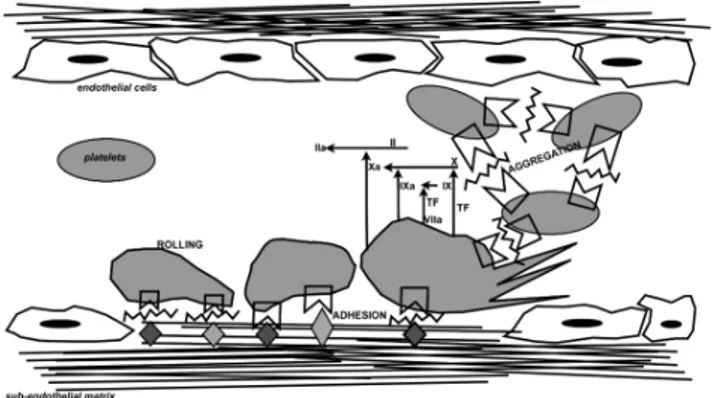
Figure 1: Schematic overview representation of processes that occur after vessel injury, in which VWF is actively involved.
First, vasoconstriction narrows the blood vessel thereby minimizing the vessel diameter and slowing bleeding.
Damaged endothelial cells display negatively charged phospholipids and release procoagulant proteins2, underlying tissue exposes collagen
allowing platelets to adhere and aggregate in the primary hemostatic plug that temporary blocks blood loss3. Consequently platelet were made active
and the coagulation cascade is initiated and amplified at each step finally producing fibrin, which is responsible of the platelet plug stabilization4.
After vessel repair, fibrinolysis results in fibrin, and thus clot, dissolvement5,6.
In these regulated series of events, a major role is held by VWF. This multimeric glycoprotein circulates in plasma in a globular, inactive state but, at site of vascular injury, the high shear stress and subendothelial matrix exposition convert VWF into its active conformation, which allows platelet adhesion and aggregation. Since platelets are unable to bind to matrix components in high shear stress condition in the absence of VWF7,the
bridge-function of VWF is essential for the platelet plug formation.
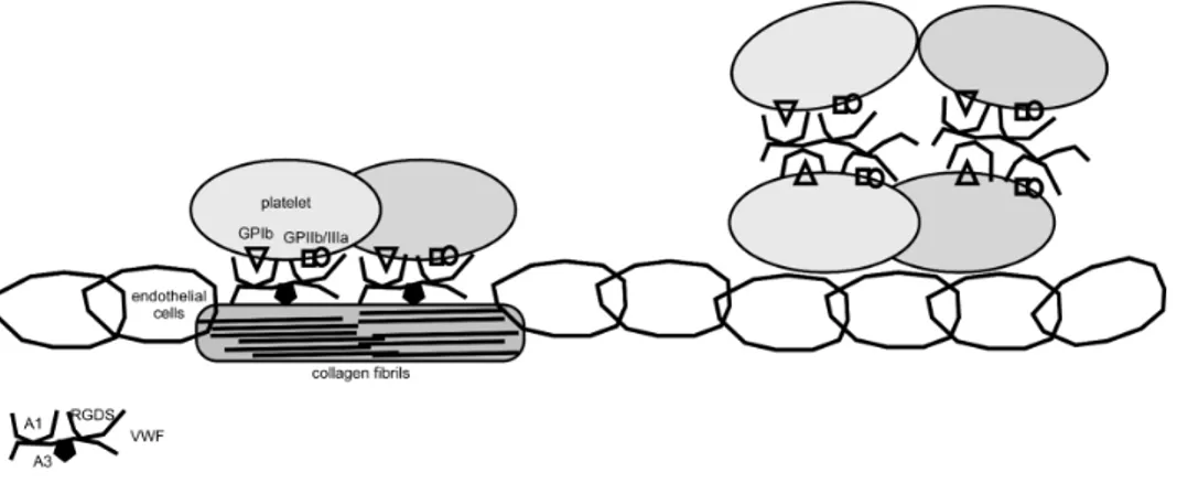
Figure 2: Easy description of major roles of VWF in platelet plug formation. First (left) VWF binds to sub-endothelial collagen exposed after vessel injury and allow platelet rolling and adhesion. Subsequently VWF allow
platelet aggregation (right) and thrombus formation.
The VWF carries another important function, related to the maintenance of the haemostasis, as carrier of FVIII8-10 that is an important and essential
factor involved in the coagulation cascade11. VWF-FVIII complex formation
prevents premature clearance of FVIII from circulation11,12.
VON WILLEBRAND FACTOR: BIOSYNTHESIS & STRUCTURE
VWF is synthesized by endothelial cells and megakaryocytes13,14.
VWF gene (178 kb) is located on the short arm of chromosome 12 (12p13.3)7,15,16 and a not functional pseudogene (21-29 kbp) partially
overlapping with full-length gene (exons 23-34) is situated on chromosome 22g11.117,18.
The primary mRNA (9 kb) product (52 exons) is first translated in a single pre-pro-polypeptide chain of 2813 amino acids and include a signal peptide (22 residues), a large pro-peptide (741 residues) and the mature subunit of 2050 residues (Figure 3). Almost entire pro-protein (>90%) has highly repetitive structure and is composed of four types (A-D) of repeated domains shared with others proteins (integrins, components of the complement system, thrombospondin and collagens). These motifs are arranged in the sequence: D1-D2-D’-D3-A1-A2-A3-D4-B1-B2-B3-C1-C2-CK7.
Figure 3: Schematic representation of the pre-pro-VWF domain organization. Scissors indicate sites where VWF is cleaved during synthesis (before Ala23signal peptidase site of cleavage and Ser764
furin site of cleavage) or in plasma in the A2 domain (after Tyr1605 -ADAMTS-13 site of cleavage-). The diagram is not on scale.
It is remarkable that 234 residues (8.3%) of the preproVWF are cysteine, which are abundantly clustered in the amino and carboxyl terminal region and rare in the A motifs. In the secreted VWF all Cys are paired in intra- or inter-molecular disulfide bonds.
The mature subunit is extensively glycosylated (12 N-linked and 10 O-linked oligosaccharides)7.
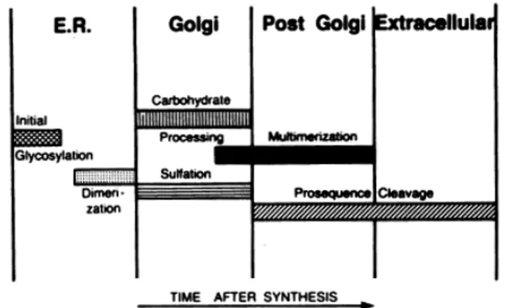
Formation of large VWF multimers is a complex and thinly regulated series of events (Figure 4).
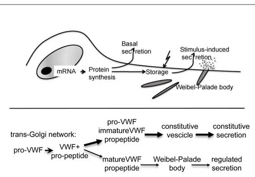
Figure 4: Schematic representation reported by Wagner DD et al19 that
indicate the biosynthetic steps of VWF synthesis and the cellular compartment where they occur.
After removal of the signal peptide by a signal peptidase, proVWF is translocated to the endoplasmic reticulum (ER) where it undergoes initial N-linked glycosylation and dimerization19 (Figure 5). This event occurs
through disulfide bond formation between C-terminal “Cysteine Knot” (CK) domain7,20-22. It has been demonstrated that mutations in the Cys2773
(within sequence Cys2771-Ser-Cys-Cys2774) abrogate dimerization23 and
subsequent studies indicate that this residue is involved in the inter-chain disulfide bond with the corresponding residue in a second subunit24.
ProVWF dimers are then transported to the Golgi apparatus where N-linked glycosylation is completed and O-linked glycosylation is applied25 (these
modifications add 19% to the weight of the protein)26-28. Moving to the trans
Golgi network, the furin-mediated29-32 pro-peptide (D1-D2 domains)
cleavage occurs and, consequently, facilitate the multimerization through D’D3 amino-terminal regions of dimers33,34. Multimers, which can rinse
20000 kDa, are designated to constitutive secretion (≃ 95%) or to storage in specialized cellular compartment as rod-shaped Weibel-Palade body (WPB) in endothelial cells and α-granules in platelets35-37 (Figure 5-6).
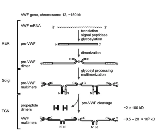
Figure 5: Schematic representation of the processing steps involved in the biosynthesis of von Willebrand factor. ✭= non-covalent interaction; S =
disulphide bonds. The molecular mass of the different VWF species is given on the right38,39.
WPBs appear to be composed by longitudinally oriented tubules surrounded by a membrane and multimerization itself is considered the triggering event for WPB-granules formation40. The largest polymers, i.e.
ultra-large multimers, are internalized and VWF secretion from these storage dispenses are related to secretagogues stimuli. Sorting from granules requires the pro-peptide that function as catalyst in multimerization and as a molecular chaperone in translocation to storage granules. The stoichiometry of the pro-peptide and mature VWF in WPB is 1:119,41,42. The release of these forms of VWF at sites of vascular damage
would deliver the most thrombogenic molecules directly where needed. Introduction
Figure 6: Partitioning of VWF propeptide and pro-VWF between the constitutive and regulated secretory pathway by vascular endothelial cells. The pro-VWF molecule is processed in the trans-Golgi network and results in heterogenous forms of VWF, ranging from dimers to large polymers. The
largest species, consisting of partially processed and immature VWF, is secreted through the constitutive pathway. The remainder, largest, fully processed and functional VWF, is sorted into the Weibel-Palade bodies and
secreted via the regulated pathway. This model proposes that the majority of VWF is secreted through the constitutive pathway38,39.
In vivo, VWF and pro-peptide plasma levels are 10 and 1 µg/ml, with 12 h and 2 h half-life, respectively. About 66% of total variation in plasma VWF levels is genetically determined and approximately 30% of this genetic component can be explained by ABO blood group. Average VWF plasma levels for persons carrying blood group O are 25% to 35% below that of persons with other blood groups43-47.
When secreted in blood flow, VWF multimer size is well regulated by the enzymatic activity of a metallo-protease named ADAMTS-1348-50 (Figure 3).
A deficiency in the ADAMTS-13 protease activity results in the presence of ultra-large VWF multimers that may cause thrombotic thrombocytopenic purpura51 (TTP), which is manifested by thrombocytopenia,
microangiophatic hemolytic anemia, bleedings and other symptoms52.
Several VWF domain-specific functions have been identified and elucidated. D domain contains FVIII and heparin binding site and, in addition, D’D3 may contain P-selectin site of interaction. P-selectin appears
to anchor released ultra-large multimers to the surface of activated endothelial cells and contribute to present ADAMTS-13 cleavage site53-55.
A1 domain contains the binding site for platelet receptor GPIbα, heparin, sulphated glycolipids and snake venom botrocetin. It’s still controversial if this domain contain also a binding site for collagen56. Scientific researchers
agree that collagens type I and III binding site are located within the A3 domain. Integrin αIIβ3 binds to RGD sequence located within C1 domain. The A2 domain contains the ADAMTS-13 cleavage site. Both the A1 and A3 binding sites and their accessibility seem to be dependent on the fluid dynamic forces exerted on the molecule as well as its immobilization on a surface40.
The majority of binding sites for numerous ligand are located within A domains, in accordance to the evolutive properties of this kind of modules, characteristic of adhesive proteins57. On the other hand, amino and
carboxy-terminal ends are more implicated in the VWF structure determination than in ligand binding.
Neither VWF structure nor its adhesive properties are dispensable to allow all VWF-dependent function at sites of vascular injury. This notion is supported by severe VWD due to mutations in either A and D-C domains.
VON WILLEBRAND DISEASE
Von Willebrand Disease is an inherited bleeding disorder due to deficiency and or disfunction in von Willebrand factor protein, and is a relatively common cause of bleeding although the precise prevalence is very difficult to estimate ranging between very different values (0.0023%-1,3%)58.
Considering only VWD due to genetic mutations occurring in the VWF genes, there is no evidence for a single major gene rearrangement associated to a large fraction or more severe cases58. Since the 192659,
when a new bleeding disorder different from haemophilia was described, and from the 1950s when the plasma factor was first named von Willebrand factor, up to 300 (http://www.ragtimedesign.com/vwf/mutation/) different mutations were founded throughout the 178 kb of DNA of the VWF gene. It is not known if the extremely heterogeneity observed for VWD is based on the features of VWF as multifunctional protein, or on its complex biosynthesis with extensive post-translational processing and distinct pathways of intracellular transport and secretion. Faults could occur at any of this levels and could be associated with specific VWD types60.
VWD was classified in three major categories61: type 1 is the most frequent
(70-80 % of the cases), groups partial quantitative deficiencies (1-40% of normal VWF levels) and leads to highly variable phenotypes62-64, type 2
collect all qualitative deficiencies and type 3, the most rare (approximately 1/1 million), give rise to total deficiencies65-67.
Mutations related to decreased synthesis, impaired secretion, increased clearance or a combination of these conditions lead to low VWF plasma levels in type 1 patients. Several studies showed that in more severe VWD type 1 cases, genetic changes are common within the VWF gene and highly penetrant. Genetic determinants in milder VWD type 1 cases involve factors in- and outside the VWF gene68,69.
Mutations causing type 2 VWD are often within VWF gene and interfere with protein structure or function64. Type 2 is divided into 4 subclasses: type
2A, 2B, 2M and 2N.
2A is the most common qualitative abnormality of VWF and is associated with selective loss of large and intermediate-sized VWF multimers paralleled with decreased platelet-dependent function. The reduction of larger multimers is caused by either increased sensitivity to proteolysis by ADAMTS-13 or impaired multimer assembly within the Golgi70,71.
2B is also characterized by loss of larger multimers. However, in VWD type 2B this is due to increased affinity of mutated VWF for the platelet GPIb-IX-V complex72. Gain-of-function mutations in the VWF-A1 domain lead to
VWF with an abnormal conformation, which 'spontaneously' reacts with circulating platelets73,74.
2M includes variants in which platelet adhesion is impaired, due to abrogated binding to GPIbα (i.e. the opposite of the VWD type 2B mutation). However, the VWF multimer distribution in these patients is normal75.
Mutations in the VWF gene responsible for VWD type 2N cause defective binding to FVIII76,77. In these patients FVIII is not stabilized by VWF and
clinical symptoms are similar of those of Haemophilia A78.
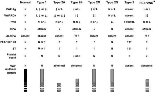
Figure 7: Schematic representation of expected lavoratory values in VWD types (Montgomery RR http://www.nhlbi.nih.gov/guidelines/vwd/
3_diagnosisandevaluation.htm)
In spite of great efforts to precisely describe and define single groups of mutations, many variations are controversially designated in different subtypes (Multiple patophysiologic mechanism) for example the C1149R change61 and the “Vicenza” mutation58 due to the complexity of the
phenotype, and to discrepant interpretation of laboratory testing displaying an ample variation58,61.
VWD TYPE 2A and subtypes IIC, IID and IIE/F
VWD 2A includes all patients displaying lack or decreased HMWMs paralleled with decreased platelet-dependent funtion. However the molecular mechanism resulting in the loss of HMWMs and the mode of inheritance could be different61.
VWD due to defective assembly of VWF structure can be produced by mutations in the propeptide, C-terminal “Cysteine Knot” or D3 regions. In recessive type IIC VWD, homozygous or coumpound heterozygous mutations in the VWF propeptide preventing multimerization in the Golgi apparatus are associated with characteristic simple multimer pattern that is essentially devoid of satellite bands and pronounced first band. The propeptide has an essential role in promoting multimer formation, possibly by catalyzing disulfide interchanges resulting in the interdimeric disulfide bonds60,61,79. The presence of CGLC sequnce both in the D’ and D2
domains (159-162 and 521-524 positions), which are homologous to the sequence of the catalytic site -CGXC- of the ER disulfide isomerase, support this notion. Mutations introduced at these positions result in unfeasible multimerization of recombinant VWF79.
In type IID VWD, a multimer assembly defect is associated with heterozygous mutations in the CK domain preventing dimerization in the ER. As a result both proVWFmonomers and wild type dimers arrive in the Golgi where the binding of a mutated monomer to a normal dimer terminates the polymerization process at that extremity. The multimer pattern shows, in addition to normal multimer bands, less intense and odd intervening sub-bands located between normal oligomers containing small multimers made by normal dimers and mutated monomers60,61,79.
In type IIE/F VWD, the cause of the multimerization inefficiency, have to be found in heterozygous mutations located within the D3 domain, often affecting Cys residues and interfering with inter-subunit disulfide bond formation in the Golgi during dimers association to form multimers. The resultant multimer pattern is often “smeary” that suggests an heterogeneous disulfide bond structure. Some other mutations in the D3 domain are not associated to aberrant disulfide bond formation and result in different phenotype (i.e. type 2M “Vicenza VWD)60,61,79.
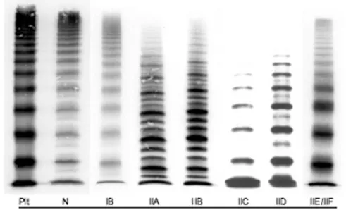
Figure 8: Multimer analysis of VWF from the plasma of patients with different VWD types compared with normal control reported by Schneppenheim R et al. 60 (gel concentration 1.5%). Plt = platelets’s VWF,
displaying the characteristic presence of `supranormal' multimers and the lack of a triplet structure. Normal multimer pattern (N) displays a typical
triplet structure and the high molecular weight multimers appear pronounced. Type IIC is associated to the lack of HMWMs and absence of
a triplet structure of individual multimers. LMWMs and especially the first band, are markedly pronounced. Intervening bands as in VWD IID, representing multimers with an odd number of monomers, are absent. The
similarity between types IID and IIC is based on the absence of a triplet structure, the lack of high molecular weight multimers, and the pronounced
bands of smaller multimers, albeit not as marked as in type IIC. The intervening bands result from the dimerization defect.The type IIE/F multimer pattern is characterized by a lack or relative decrease of HMWMs
and the absence of the outer sub-bands of the normal triplet structure. Inner sub-bands, closer to the central band, are present, which give the
individual multimer a broader appearance60.
RNA-INTERFERENCE: CORRECTION APPROACH TO DOMINANT DISEASES
RNA interference (RNAi) is a recently discovered mechanism in which gene expression is regulated by small double-stranded RNA (dsRNA) molecules that inhibit gene expression with complemetary nucleotide sequences80.
The RNAi machinery acts by either suppressing transcription (transcriptional gene silencing) or activating a sequence-specific RNA degradation process (post-transciptional gene silencing)81.
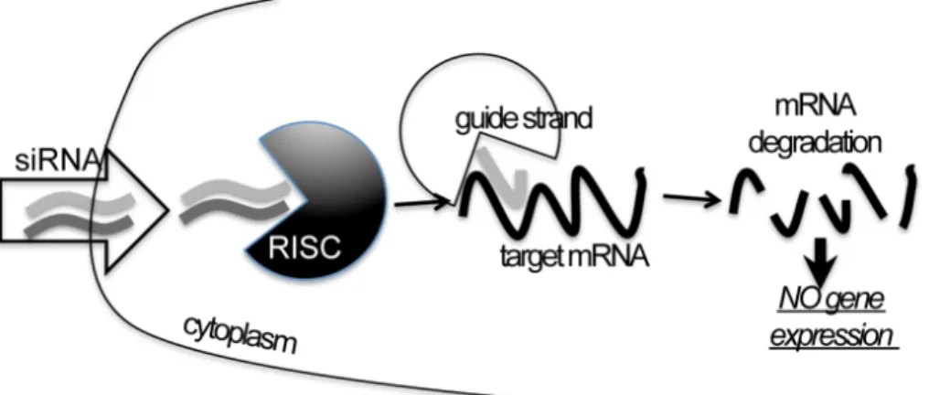
Essentially RNAi (Figure 9) is divided in two major steps. First, in the “RNAi-initiating step” large dsRNA are bound by RNA nuclases and cleaved in discrete 21- to 25-nucleotide RNA fragments called small dsRNAs. In the second step, these short dsRNA molecules (called miRNAs if derived from endogenously cleaved long dsRNA, or siRNAs if exogenously synthesized and introduced inside cells) are associated in the cytoplasm with a protein complex defined as RISC (RNA-induced silencing complex), one of the two RNA strand is degraded and the remaining guide strand mediates the sequence-specific degradation of the corresponding mRNA (in the case of siRNAs) and/or translational repression by binding to the 3’ untraslated region (UTR) in the case of miRNAs80-82.
The existence of the RNAi machinery, that mediate gene expression regulation and/or, in some cases, protection from dangerous (i.e. viral) external RNAs, also give the opportunity to take advantage on this endogenous process to induce the silencing of virtually any gene of interest80.
For this reason RNAi-mediated silencing is recently become an extremely used and useful tool in the reverse genetic analysis of gene functions and it also represents a potential novel drugs for the treatment of diseases80.
Due to the apparent simplicity of the mechanism, which offer almost unlimited silencing applications based only on the sequence availability of the mRNA you want to silence, a large and increasing number of studies have reported fascinating application of the RNAi strategy to numerous disease for which effective treatment are currently unavailable or sub-potential, i.e. viral infection, cancer, autoimmune- neurodegenerative- and cardiovascular-diseases,... although some important and not omissible limitations have not yet been resolved80.
Figure 9: Simple schematic representation of the RNAi mechanism focusing on the exogenous siRNAs silencing action.
The major impediment for the clinical use of siRNAs is their poor cellular uptake and short serum half-life. Although great progress has been made on this regard, researches are currently open to find the most selectively, safe and potent delivery system for siRNAs.
In spite of this, numerous preclinical studies allowed the establishment of some clinical trials: phase I-II for age-related macular degeneration (AMD) and respiratory syncytial virus (RSV)80.
The primary aim of human gene therapy is to correct the genetic disorder caused by mutation specific effects. Basically recessive disorders are approached with replacement therapy that, introducing wild type allele, restores gene function and improves disease pathology. On the other hand dominant phenotypes may be caused by haploinsufficiency (reduction of the wild type protein level), by gain of function mutation or both. The suppression of the detrimental effects of the mutated allele together with the maintenance of the wild type allele expression are necessary to overcome the effects of a gain of function mutation. These features make dominant inherited diseases perfect candidates for RNAi-based gene therapy83.
Several dominant disorders have already been approached, in vitro or in animal models, with RNAi such as Huntington’s disease (HD), spinocerebellar ataxia type 1 (SCA1), amyotrophic lateral sclerosis (ALS),
Alzheimer’s disease, myotonic dystrophy (DMI) all caused by triplet expansion.
When dominant mutations occur within gene coding for protein which needs to aggregate to function properly, a dominant-negative effect is expected. In this case a severe phenotype is often observed, because wild type and mutant proteins interact and also the wild type molecule is not available to carry out its function.
Mutations with dominant-negative effect are classically detectable in structural proteins i.e. collagen or nonstructural proteins that dimerize or oligomerize i.e. transcription factors of the b-HLH-Zip family, which bind the DNA as dimers, or ion channels located in cell membranes, which are often multi-protein complex84.
Recently are reported that RNAi-mediated gene silencing could be a promising treatment for the pachyonychia congenita85,86 (PC), which
represent one of the numerous skin disorder associated to one of the 54 human keratin genes, and is associated to dominant-negative mutations. PC represent a suitable disease to test RNAi treatment because offer some advantages such as the presence of recurrent mutations, and the external location and focal nature of the plantar skin lesions permitting minimally invasive localized treatments to be performed on patients. The discover of potent and selective siRNA tergeting the most common mutation causing PC resulted in the initiation in 2008 of a clinical trial for this skin disorder. VWF displays extraordinary adhesive features: it polymerizes with itself and exploits its function through the binding with collagen, platelet receptors and other ligands. For these reasons it is expected that mutations in the related VWF gene could have dominant-negative effect. Furthermore VWF is synthesized in endothelial cells and megacaryocyte only, suggesting that, in the future, the treatment could be localized to these cell types.
Finally VWD is the most prevalent hemostatic disorder and the majority of its cases are dominantly inherited and could be potentially approached with RNAi treatment.
However, on the contrary to PC, over 300 mutations are associated to VWD and none of these is demonstrated to be the most prevalent. This would imply that very specific siRNAs have to be designed and tested for each VWD-associated mutations that you want to correct.
VWF is also a large protein that undergoes numerous and regulated post-trancriptional modifications and the setting studies to found effective siRNAs molecules need to be careful and rigorous.
For the sake of clarity some important advises for RNAi study design are reported below81,83.
The use of siRNAs to silence gene expression in mammalian cells involves a careful consideration of selecting the siRNA sequences in the target gene to avoid non specific complementariry to unrelated genes and to avoid the choice of target site not accessible for the binding of the siRNA. It may be necessary to screen several sequences, concentrations, combinations and delivery systems to identify the most efficacy molecule for each cellular model. Today are available helpful on-line software, that provided RNA structure models to give information on the site accessibility or offering automated researches for siRNAs sequences with more probability to be effective that can help in the choice of siRNAs molecules. In addition it is essential to accurately optimize the transfection protocol depending on: the target cell type, the selected means such as siRNas or vector-driven shRNAs and the delivery system.
BACKGROUND INFORMATION OF THE
p.P1105_C1926delinsR GENE DELETION AND OUTLINE
OF THIS THESIS
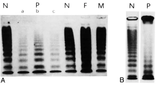
We have previously described a large, de novo and heterozygous deletion in VWF gene associated to a variant type 2 VWD. The propositus showed a severe VWF deficiency, as indicated by reduced ristocetin cofactor activity (3%) and prolonged bleeding time (20 minutes), which produced a number of bleeding symptoms requiring blood transfusion [Bernardi 1990 Blood]. The altered multimer pattern (Figure 10) was characterized by reduced intermediate- and no HMWMs in plasma and platelets, and by abnormal morphology of triplets with a broad band apparently containing a fast-migrating component. Antigen (VWF:Ag) levels (24%) and FVIII coagulant activity (29%) were also reduced87. The gene deletion boundaries were
mapped within introns 25 and 3488 and the new junction between exons 25
and 35 was sequenced after reverse transcription of the platelet RNA (Figure 11). At the protein level Cysteine 1104 should be connected in-frame with mutated codon 1926 (cysteine to arginine).
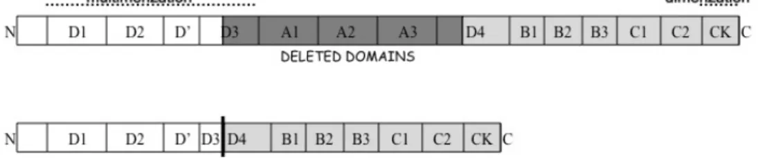
The encoded protein (Figure 12) would lack the last part of the D3 domain, the A1, A2 and A3 modules, and the first part of the D4 domain, which would virtually abolish, in addition to the A domain-associated adhesive function89-92, the D3 domain-associated inter-dimer disulfide bond
formation93,94 and thus multimerization. However, the dominant-negative
attitude of the deleted protein still needs to be demonstrated, and its molecular features dissected. Dimerization and multimerization, distinct property of VWF producing extremely large and adhesive molecules, provide the rationale for the dominant inheritance caused by the interaction of wild type and mutant VWF monomers during polymerization. The strength of the negative effects produced by dominant variants would be associated with the ample variation of residual VWF level and function, and would participate in the phenotypic penetrance and severity of VWD types inherited as dominant traits, which is rather high95. These are major open
questions in VWF biology and VWD molecular bases definition.
In this study we constructed a cellular model of this unique disease variant as a tool to demonstrate the presence, and evaluate the efficiency, of the dominant-negative effect of the deleted protein in the biosynthetic steps of VWF maturation and secretion. This cellular model mimics patient’s plasma VWF phenotype and highlights the key role of heterodimer between normal and deleted VWF molecules. We also propose a silencing approach to correct the dominant-negative effect that partially restored VWF levels.
Figure 10: Multimer analysis of plasma (A) and platelet (B) and densitometric scans (C) reported by Bernardi F et al.87
N: normal control; F and M. father and mother of the propositus (P). In lane b a double amount of plasma of the propositus was loaded and in lane c an
antiproteolytic mixture was added as anticoagulant. (C) Densitometric analysis of lanes reported in (B).
N
P
F
M
B
bleeding time (min)
5-8
20
5
7
8
FVIII:C
60-170
29
98
70
75
VWF:Ag
60-150
24
98
102
72
Ri:Cof
60-150
3
93
76
70
Table 187: Hemostatic parameters in members of the propositus’s family as
in Bernardi et al. 1990 Blood. N = normal range, P = propositus, F = father, M = mother, B = brother.
Figure 11: Schematic representation of the deletion location within VWF gene and the resultant deleted mRNA. The box indicates the new junction
that connected in-frame exons 25 and 35 thus mutating Cys 1926 to Arg.
Figure 12: Schematic representation of the wild type and deleted VWF protein domains. Dark-grey motifs (1105-1925 amino acids) are missed in
the deleted VWF.
Creation of expression vectors
The expression vector pSVHVWF1 for human VWF, having the full-length VWF cDNA cloned into the pSV7d plasmid, was a generous gift from Prof. J.E. Sadler (Washington University in St. Louis)25.
Figure 13: Schematic representation of wild type, deleted and double mutated VWF-expression vectors. Black arrow within pDEL and pDEL/ C2773R indicates the breakpoint. Black bordered flags indicate siRNAs complementary regions. Squared brackets indicate epitopes recognized by
antibodies utilized in Western blotting experiments.
To create the pSVHVWF-P1105_C1926delinsR96,97 (Figure 13) deleted
expression vector (defined as pDEL) the SacI restriction site at position 8876 was initially abolished by site-directed mutagenesis through the mutagenic primers 5’CCACAATAAAGGCTCAGCTCTTATCTTGC3’ (change
underlined) and 5‘GCAAGATAAGAGCTGAGCCTTTATTGTGG3’ by using
the QuikChange XL Site-Directed Mutagenesis Kit (Stratagene, La Jolla, CA). The transient expression of the pSVHVWF variant in monkey kidney cells (COS-1) resulted in secreted VWF antigen and VWF collagen-binding activity levels indistinguishable from that of the pSVHVWF1, and pSVHVWF was subsequently used as wild type VWF expression vector (pWT).
To reproduce the P1105_C1926delinsR deletion we exploited the overlapping PCR strategy98,99 (Figure 14) with appropriately designed
primers. Primers 5’CAATGACCTCACCAGCAGCAACC3’ (VWF-25F) and 5’TGCCTGTGCACACGCGGCACAATGTGGCCGTCCTCC3’ (VWF-25R),
having a 15 bp-tail complementary to nt 6153-6167 in exon 35, were used to amplify the region upstream of the breakpoint. Primers
5’CGGCCACATTGTGCCGCGTGTGCACAGGCAGCTCC3’ (VWF-35F),
having a 15 bp-tail complementary to nt 3675-3689 in exon 25, and
5’TCTTCAGGGACACAGCTGCC3’ (VWF-35R) were used to amplify the
region downstream of the breakpoint. The partially overlapping regions were amplified using the pWT as template (30 cycles; 30’’ at 94°C, 30’’ at 55°C, 50’’ at 72°C). Amplicons were then purified and mixed for a third PCR reaction using the VWF-25F and VWF-35R external primers. (30 cycles; 30’’ at 94°C, 30’’ at 50°C, 1’ at 72°C). The resulting 1279 bp fragment was then cloned into the pWT vector utilizing SacI restriction sites at positions 3395 and 6986.
Figure 14: Schematic representation of the overlapping strategy applied to produce the deleted vector.
To create the pDEL/C2773R (Figure 13) vector the C2773R mutation was inserted into the pDEL vector by site-directed mutagenesis using primers
5 ’G A G A C G C T C C A G G A C G G C T G T G A T A C T C3 ’ a n d 5’GAGTATCACAGCCGTCCTGGAGCGTCTC3’.
The correct sequence of vectors, and the absence of undesired changes, was confirmed by direct sequencing.
Expression of recombinant VWF in eukaryotic cells
COS-1 cells were used for transient expression of VWF variants25. Cells
were grown in Dulbecco modified Eagle’s medium (D-MEM) supplemented with 2 mM L-glutamine, 50 IU/ml penicillin, 50 µg/ml streptomycin, and 10% (vol/vol) fetal bovine serum and seeded at 60-70% confluence into 6-well plate for transfection. Single plasmids, or combination of them in the relative molar proportion reported in the figures were transfected into COS-1 cells by using Lipofectamine2000 (Invitrogen, Carlsbad, CA) and in the presence of Opti-MEM serum-free medium (Invitrogen). The total amount of DNA in each transfection was set to 4.5 µg by adding, when needed, a gutted pUC18 plasmid. Seventy-two hours after transfection the conditioned media were collected and cells were lysed for total RNA isolation (NucleoSpin RNAII isolation Kit, Macherey-Nagel GmbH&Co. KG, Duren, Germany) or intracellular VWF investigations (lysis buffer: NaCl 150 mM, Triton X-100 1%, Tris HCl pH 7.4 50 mM, PMSF 1 mM, Benzamidine 10 mM).
Silencing
The siRNA 852 (sense, 5’CCUCGGACCCUUAUGACUUUU3’; antisense 5’AAGUCAUAAGGGUCCGAGGUU3’), to specifically silence the wild type
V W F e x p r e s s i o n , a n d t h e s i N C ( s e n s e ,
5 ’C C U C A G U C C U A U A G C G C U U U U3 ’; a n t i s e n s e 5’AAGCGCUAUAGGACUGAGGUU3’), as negative control, were designed
by the Invitrogen software BLOCK-iT™ RNAi Designer (https:// rnaidesigner.classic.invitrogen.com) and purchased (Figures 13-14).
Conversely, none of the on-line available software programs for the siRNA design were able to predict a siRNAs targeting the naturally occurring breakpoint region of the mutated VWF variant, and hence able to selectively inhibit the pDEL expression.
To overcome this limitation we first evaluated the VWF mRNA conformation at the breakpoint region and its theoretical accessibility to complementary siRNA with the Mfold100 software (http://mfold.bioinfo.rpi.edu/).
Figure 15: Example of RNA fold made by the mfold on-line software. The figure showed a small part of the entire RNA structure of the deleted molecule surrounding the breakpoint (bold line). In this simplified image the
availability of the site is evident.
The si3681 (sense 5’CAUUGUGCCGCGUGUGCACUU3’; antisense 5’GUGCACACGCGGCACAAUG UU3’) and the si3675 (sense, 5’CGGCCACAUUGUGCCGCGUUU3’; antisense, 5’ACGCGGCACA
AUGUGGCCGUU3’) oligoribonucleotides complementary to the breakpoint
region were then designed (Figure 16-17) in the respect of the siRNA design rules and purchased from Invitrogen.
siRNAs (10-80 nM) were transfected, with VWF expression vectors in COS-1 cells by Lipofectamine2000 (Invitrogen). Medium was replaced with fresh Opti-MEM 4 hours after transfection. Conditioned media and cells were collected as above mentioned.
Preliminary studies were conducted to achive the most efficacy silencing activity of the si3675 and si3681 and their combination toward the pDEL expression, and the appropriate experimental conditions to avoid adverse effects. Treatment with 40 nM of si3681, complementary to the breakpoint sequence exactly in the middle of its sequence, was chosen as the most efficient.
Figure 16. Deleted VWF sequence fragment and siRNAs.
Position and sequence of the mRNA and of the siRNAs (flags) directed to the mRNA breakpoint (3’ of exon 25 and 5’ of exon 35). Nucleotides at the
junction (arrow) are indicated by bigger/bold letters.
Figure 17: Schematic representation of siRNAs targeting region (across the breakpoint) within pDEL.
mRNA study
Reverse-transcription (RT) was performed using “SuperScript III First-Strand Synthesis System” (Invitrogen) and oligo (dT). PCR amplification (30 cycles; 30’’ at 94°C, 30’’ at 50°C, 30’’ at 72°C) was performed with forward primer 5’CAGTGACGTCTTCCAGGAC3’ and reverse primers 5’AGGGTCACTGGGATTCAAGG3’ and 5’TGCCTGTGCACACGCGGC3’
designed on pWT and pDEL vectors, respectively. Capillary electrophoresis was performed on the Experion automated electrophoresis station101
(Bio-Rad Laboratories, Hercules CA, USA) to detect amplification products. This method applies a combination of microfluidic separation technology and sensitive fluorescent sample detection to perform, in our cases, RNA automated analysis. All gel-based electrophoretic steps are automatically performed to generate reproducible separation and quantitative results, which may be reported as an electropherogram gel image (Figure 31A) or scheduled.
Antibodies
Polyclonal Rabbit anti-human VWF (A0082), polyclonal rabbit anti-human VWF/HRP (P0226), polyclonal swine anti-rabbit immunoglobulins/TRITC (R0156) and polyclonal Goat Anti-Mouse IgG/HRP antibodies were purchased from DAKO (DAKO, Glostrup, Denmark). The mouse monoclonal [2G11] antibody to Mannose 6-Phosphate Receptor (late endosomes marker) (ab2733) and the mouse monoclonal [1D3] to PDI (ER marker) (ab12225) were purchased from Abcam (Cambridge, UK). The FITC-conjugated AffiniPure goat anti-mouse IgG (H+L) (115-095-146) was from Jackson ImmunoResearch Laboratories (Baltimore, USA). Monoclonal anti-VWF M31 and M13 were obtained as described102,103.
Analysis of recombinant VWF
VWF:Ag in conditioned media and cell lysates was determined by an ELISA assay performed with above mentioned Dako antibodies (Polyclonal Rabbit anti-human VWF and polyclonal rabbit anti-human VWF/HRP). The ELISA assay described by Federici et al.104 was tested and partially
modified to obtain better results. Essentially the blocking solution is based on PBS instead of TBS because, although both showed good response to scalar dilutions of a pool of normal plasma, the first one showed lower values of background. In order to increase the relative absorbance of each samples several primary antibody concentrations are tested but all of these gave similar good results. When different concentrations of secondary antibody are used a good improvement in the absorbance values was obtained doubling (1 µg/ml) the proposed concentration (0.5 µg/ml). Intra-assay coefficient of variation was ≃ 6.5% and the inter-Intra-assay was ≃ 15%. Sensitivity was ≃ 2 ng/ml.
VWF:CB was measured in conditioned media using the “CollagenBinding Assay kit” (Life therapeutics, Clarkston, USA).
Multimer analysis: the multimeric structure of VWF was detected in sodium dodecyl sulfate (0.1%) agarose gel (1.5-3%) electrophoresis105,106 followed
by Western blotting and detection with immunoenzymatic stain (polyclonal Goat Anti-Mouse IgG/HRP antibodies, Dako) or with radioactive detection (125I-anti-vWF antibody103).
Western blotting of VWF present in media and lysates were performed in polyacrylamide gel (NuPAGE4-12% Bis-Tris Gel, Invitrogen) in reducing condition. The electrophoresis was performed in MOPS 1x buffer (Invitrogen) for approximately 5 hours at 200 V, followed by transfer in TRIS (25 mM)-glycine (192 mM) buffer with methanol (10% v/v) and sodium dodecyl sulfate (0.7 mM) at 300 mA for 2 hours. The nitrocellulose membranes were reacted with different anti-VWF monoclonal antibodies followed by incubation with anti-mouse Immunoglobulins/HRP (Dako). Bands were visualized by autoradiography with Amersham Hyperfilm ECL (GE Healthcare Limited, Pollards Wood, UK) using “Super Signal West FEMTO Maximum Sensitivity Substrate” for media and “Pierce ECL Western Blotting Substrate” for lysates (Pierce, Rockford, USA).
Immunofluorescence microscopy
Cells for immunofluorescence labelling were cultured at sub-confluence in 6- or 12-wells on a coverslip of 12 or 24 mm of diameter. Cells were transfected with Lipofectamine2000 lipid reagent (Invitrogen) using a 2:1 ratio for Lipofectamin/vectors. 24 hours after transfection cells were fixed in methanol for 7 minutes at -20°C and saturated with 4% Bovine Serum Albumin (BSA)/PBS. Subsequent incubation with primary and appropriate secondary antibodies was performed for 1 hour at room temperature. Primary antibodies were diluted 1:100 in 4% BSA/PBS and secondary antibodies were diluted 1:50 in the same solution.
Images were acquired with a Nikon fluorescence microscope Eclipse 50i (Nikon Instruments, Sesto F.no, Firenze, Italy) equipped with DS-5M digital camera and DS-L1 control unit. Objectives used were Nikon Plan 20x/0.5 and 40x/0.75 and Nikon 100x/0.5-1.25 Oil Iris.
Confocal images were acquired as previously described107 with a Zeiss
LSM 510 microscope equipped with a Fluar 40x/1.3 oil immersion objective (Carl Zeiss, Arese, Italy). Briefly, 488 and 543 nm excitation wavelengths were provided respectively by Agron/2 and HeNE laser sources at a 5% intensity.
Superimposition of red and green fluorescence, not specifically due to co-localization, was excluded by applying to the green channel a beam path admitting the acquisition of fluorescence comprised among 505 and 550
nm and to the red channel a light path excluding all fluorescence below 560 nm. Pinhole values were ranging from 40 to 80 µm. Where required, image magnification was obtained digitally with the LSM examiner software (Carl Zeiss).
Creation of the recombinant deleted expression vector (pDEL)
To c r e a t e t h e d e l e t e d e x p r e s s i o n v e c t o r p S V H V W F -P1105_C1926delinsR96,97 (pDEL), the overlapping PCR method was used.
First we identified a fragment of the pSVHVWF1 vector containing the region to be deleted and delimited by SacI restriction sites. Dowstream of the deletion, near the end of the VWF cDNA in the pSVHVWF1 vector, a third Sac I recognition site was present thus complicating the subsequent cloning step. To overcome this limitation we abolished the last Sac I (position 8876) restriction site through site-directed mutagenesis and produced the pSVHVWF (pWT) expression vector.
The transient expression of the pSVHVWF variant in COS-1 cells resulted in secreted VWF antigen and collagen-binding activity levels indistinguishable from that of the pSVHVWF1, and pSVHVWF was subsequently used as wild type VWF expression vector (pWT) (Figure 18).
Figure 18: VWF:Ag (light grey) and VWF:CB (dark grey) levels in conditioned media form cells transiently trasfected with pSVHVWF or pSVHVWF1. Mean values of three replicate of pSVHVWF were used as 100% (570.6 ng/ml for antigen value and 152.3% of normal standard as CB
value) and the mean of three replicate (±SEM) was used to evaluate pSVHVWF1 levels. t test was applied to exclude significative differences.
The overlapping PCR98,99 was be performed on this mutagenized vector.
First two separate PCR reaction were performed with primers 25F-25R and 35F-35R, the pWT vector was used as template (Material & Methods Figure 14 and Figure 19).
Figure 19: Eletrophoretic separation of partially overlapping fragments amplified in the two separate PCR step performed with 25F-25R and 35F-35R set of primers.
Figure 20: Electrophoretic separation of the 1279 bp fragment that connected residues 3689 and 6153 thus “containing the deletion.
The amplicons obtained, which partially overlap (Material and Methods, Figure 14) were electrophoresed on agarose gels, excised, purified and used as template for the third PCR amplification reaction performed with external 25F and 35R primers (Figure 20).
Figure 21: Electophoretic separation of pSVHVWF and pSVHVWF1 expression vectors digested with Sac I restriction enzime. The arrow indicate the fragment excised and purified for the
subsequent ligation step. 1116bp M8 1279bp 25F-35R M8 25F-25R 35F-35R !800bp !400bp 883bp 404bp Results
The 1279 bp fragment carrying the desired junction between nucleotides 3689 and 6153, contained two Sac I recognition sites at each extremity and was then digested with Sac I restriction enzyme. Accordingly, the pWT vector that contained two recognition site, at positions 3391 before the breakpoint, and at position 6892 downstream of the breakpoint, was digested with the Sac I enzyme to produce compatible ends with the above described fragment containing the desired junction (Figure 21).
Finally the ligation between the “7.8 Kb fragment” and the digested “1279 bp” fragment was performed.
The resulting pDEL expression vector, which would produce a protein containing the expected junction between exons 25 and 35, was used to transfect COS cells alone or in combination with pWT vector.
Expression of the in-frame deleted VWF
In order to investigate the molecular mechanism through which the heterozygous deletion P1105_C1926delinsR produced the severe VWD phenotype we first studied the expression of the deleted VWF both in media and cell lysates in the absence of the wild type protein (Figure 22). For this purpose two antibody pools102,103 were used in Western blot
analysis. The M31 pool (Figure 22A, left panels) reacts with VWF residues 2244-2456, downstream of the breakpoint (Material and Methods, Figure 13), and would recognize only domains with a normal reading frame. The M13 pool (Figure 22A, right panels) reacts with VWF residues 1394-1473, located within the deletion boundaries and thus only with the wild type VWF. The comparison of blots supported that the pDEL vector efficiently drove the biosynthesis of the in-frame deleted molecule. The deleted VWF appeared to be only partially secreted (Figure 23), and its electrophoretic migration was compatible with the protein size (≈ 160 kDa) inferred from the extent of the deleted domains (821 amino acids).
Whereas traces of the altered pro-VWF were detectable in lysates, only the mature form of the deleted VWF was secreted. Pro-wild type VWF was abundant both in media and lysates, in accordance with published data25,29,30. Antigen and collagen-binding levels in media from cells
expressing the pDEL vector (Figure 22B) were very low and undetectable, respectively. The multimer analysis in high resolution gels (Figure 25 -page 40) showed the presence of a broad band with increased mobility as compared to wild type, and did not reveal the presence of tetramers.
Figure 22. Expression of the deleted VWF.
(A) Western blot analysis of wild type and deleted VWF performed on conditioned media (upper panels) and cell lysates (lower panels) with monoclonal antibody pool M31 and M13102,103. The M13 antibodies,
targeting VWF A1 domains (right panels), recognize only wild type VWF. The M31 antibodies (left panels), targeting the C-terminal domains, indicate
the in-frame translation of the deleted VWF. Migration of the mature wild type (matureWT-VWF) and deleted (matureDEL-VWF) proteins, and of
forms containing the propeptide (proWT-VWF and proDEL-VWF), are indicated. In cell lysates additional bands containing the pro-wild type VWF
and the pro-deleted VWF are detectable. The asterisks indicate (*)
over-exposure or (**) under-exposure of gels. (B) VWF:Ag (upper panel) and
VWF:CB (lower panel) levels in conditioned media of COS-1 cells. Results (ng/ml) of three independent experiments are reported as Mean ± SEM. The right y axis indicates the percentage of wild type (100% VWF:Ag, 570.6
ng/ml; 100% VWF:CB, 152.3 of normal standard). Statistical significance (* P< 0.001) was evaluated by one-way ANOVA with Bonferroni post-test.
n.d., not detectable.
Figure 23: Intact Western blot containing the entire panels displayed in Figure 22, Figure 24 and Figure 31. The relative molar amounts of vectors
and siRNA are indicated in the table. Antibodies are indicated as in figure 22. Conditioned media (panel A) were tested by the monoclonal antibody
pool M31. Cell lysates (panels B and C) were tested by monoclonal antibodies M31 and M13.
Dominant-negative effect of the VWF deletion – cellular model
To mimic the heterozygous condition in COS-1 cells, the pWT vector was co-expressed with increasing amounts of the pDEL, which progressively decreased the levels of wild type VWF (Figure 24). Equimolar amounts of both plasmids produced 20% (112.3 ± 4.1 ng/ml, Figure 24A, upper panel) of wild type antigen levels (570.6 ± 26.5 ng/ml), and 2% of collagen-binding activity (Figure 24A, lower panel). Accordingly, the ratio between VWF:CB and the antigen values dropped to 1/10 of that calculated for the wild type. A double proportion of the deleted vector decreased the VWF:Ag levels to
12% (70.6 ± 4.5 ng/ml) of wild type, and abolished the collagen-binding activity.
The Western blotting, performed in reduced conditions (Figure 24B) and thus able to evaluate the proportion of the normal and deleted VWF molecules, indicated that in co-expression experiments the altered VWF prevailed. Taking into account the pro-peptide and mature forms, the densitometric analysis in media and lysates indicates that the ratios between the altered and normal VWF were 1.8 and 3.5, respectively. Moreover, the absolute amounts of the deleted protein appeared to be increased in media from cells expressing both vectors (Figure 23).
Figure 24. Dominant-negative effect of the in-frame deleted VWF variant. (A) VWF:Ag (upper panel) and VWF:CB (lower panel) levels in conditioned media of COS-1 cells. The relative molar amount of pWT and pDEL vectors
is reported in the table. The right y axis and statistical analysis as in Figure 22. (B) Western blot experiments in conditioned media (upper panel) and
cell lysates (lower panel) with antibody pool M31.
(C) Multimer pattern detected with
immunoenzymatic stain from media obtained as described in the table. The inset reports samples tested by 125I-labelled-anti-vWF
antibody to better visualize the deleted VWF form.
The multimer analysis (Figure 24C and 25) of VWF in media from co-transfected cells indicated that the pDEL expression impaired secretion of oligomers, particularly the HMW forms, in a dose dependent manner. A detailed interpretation of the observed pattern is complicated by the similar extent of the deleted (821 amino acids) and propeptide region (741 amino acids), which causes co-migration of dimers (D) and tetramers (T) with different subunit composition (i.e. homodimers DA and heterodimers Dc,
Figure 26). In spite of this limitation, the presence of heterodimers (Db)
between the wild type and deleted proteins was clearly detectable. As a matter of fact, co-expression of pWT and pDEL vectors produced the extra-band Db, the migration of which is faster than that of wild type and slower
than that of the deleted homodimers Da (Figure 25).
Figure 25. High resolution multimer analysis of VWF in media. Based on VWF:Ag level evaluation 1 ng of VWF was loaded in each lane
(A and B). Due to the low protein concentration in media from pDEL expressing cells 0.25 ng were loaded in C, and accordingly the gel was over-exposed. PNP was diluted 1/200. The relative molar amounts of each vector and siRNA are reported in the table. Distinguishable dimers (D) and tetramers (T) are indicated together with sub-band composition (DA-C and
Da-c; TA-E and Ta-d) in accordance with the schematic representation (Figure
26). Faint bands are tentatively defined and indicated in Italics.
Figure 26: Schematic representation of dimers and tetramers -in accordance to Schward et al 2000108 and Turecek et al 2002109- reports the
combinations of wild type pro- and mature-VWF (subscript uppercase letters) and mature-deleted-VWF (subscript lowercase letters). Proteins with different subunit composition but similar molecular weight, and thus
co-migrating, are represented on the same line. For the sake of clarity the DEL/C2773R VWF monomer, which is not expected to form polymers, is
reported at the bottom of the scheme.
The comparison between media from pWT and pWT+pDEL expressing cells in the tetramer region (T) enabled us to gain additional information about heterodimer polymerization. The fast-migrating and smeared band observed after expression of both vectors supported the presence of a number of heterotetramers, which could stem from the combination among pro-wild type, mature wild type and mature deleted VWF.
Immunofluorescence staining of intact cells was used to investigate the biosynthesis and intracellular localization of normal and deleted protein, and the dominant-negative effect of the altered VWF. Transfected cells were stained with polyclonal anti-human VWF antibodies and monoclonal antibodies directed to specific markers of subcellular compartments. Antibodies recognizing the protein disulfide isomerase and the Mannose 6-phosphate receptor were used to investigate endoplasmic reticulum and late endosomes110, respectively. In the first compartment, we did not detect
differences in the fluorescence patterns between the wild type and in-frame deleted VWF expressing cells (Figure 27).
Figure 27: Intracellular distribution of recombinant VWF in the ER. Cells transfected with the vectors indicated above the images (A-F) were
co-immunostained for VWF (TRITC, red) and protein disulfide isomerase (FITC, green) as ER marker. Images were acquired with Nikon Eclipse 50i
equipped with DS-5M digital camera, DS-L1 control unit and Nikon Plan 20x/0.5, 40x/0.75 and Nikon 100x/0.5-1.25 Oil Iris objectives. Images were
taken at room temperature on fixed cells mounting in glycerol/1,4 diazabicyclo[2.2.2]octane/DAPI medium.
Differently, the analysis of late endosomes (Figure 28) revealed that the degree of overlapping between the VWF (red)- and Mannose 6-phosphate receptor (green)-specific fluorescence was i) high in cells expressing the pWT vector (panels A-B), ii) barely detectable in cells expressing the pDEL (C-D), and iii) intermediate in cells expressing both vectors (E-F). Although the deleted protein was inefficiently transported to the late endosome compartment, the strong red fluorescence in pDEL expressing cells (C-D) indicated that the protein was efficiently synthesized, in accordance with data obtained in the reticulum (Figure 27), with Western Blot analysis (Figures 23 and 24B) and with ELISA antigen assay on cell lysates (Figure 29).
Figure 28: Intracellular distribution of recombinant VWF in late endosome compartment. Cells transfected with the vectors indicated above the images (A-H) were co-immunostained for VWF (TRITC, red) and mannose
6-phosphate receptor (FITC, green) as late endosome marker. Images were taken with Carl Zeiss LSM 510 equipped with a Fluar 40x/1.3 oil
immersion objective at room temperature on fixed cells mounting in glycerol/1,4 diazabicyclo[2.2.2]octane/DAPI medium. The superimposition
of red and green fluorescence not specifically due to co-localization was excluded as described in materials and methods. Images in the upper
panels were digitally zoomed in twice the original size with the LSM examiner software (Carl Zeiss, Arese, Italy).
Immunofluorescence microscopy experiments did not suggest accumulation of the deleted protein inside co-transfected cells, an observation confirmed by Western blots and VWF antigen levels of cell lysates.
VWF:Ag levels in cell lysates (Figure 29) of pWT and pWT+pDEL expressing cells were not significantly different in spite of a remarkable decrease of VWF:Ag levels in conditioned media due to the addition of the pDEL vector. These observation was also true comparing pWT and pDEL or pWT and pWT+2xpDEL expressing cells.
Figure 29: VWF:Ag evaluation in conditioned media (grey bars) and in cell lysates (black bars). Results of five independent experiments are reported
as Mean ± SEM. Statistical significance (°P< 0.01; *P< 0.001) was evaluated by one-way ANOVA with Bonferroni post-test.
Taken together these experiments demonstrated the ability of the in-frame deleted VWF to interact with wild type VWF in the biosynthetic process, thus producing quantitative and qualitative alteration of secreted VWF, and particularly the impairment of HMWM formation.
Interfering with dimerization of the in-frame deleted protein by a natural mutation affecting VWF dimerization (C2773R)
A key step for the dominant-negative effect is the ability of the in-frame deleted protein to form heterodimers with the wild type VWF. To investigate the interaction between the deleted and wild type protein we exploited natural missense changes interfering with VWF dimerization (type 2A IID VWD)111,112, and among those the C2773R23. Cysteine 2773 was
also selected because molecular modelling and expression of norrin have supported its key role in dimerization through intersubunit disulfide bond formation24. The insertion of this missense change in the double mutant
vector pDEL/C2773R (Material and Methods, Figure 13) was aimed at Results
impairing dimerization of the deleted VWF, when co-expressed with the wild type protein.
Figure 30: Dominant-negative effect of the double mutant pDEL/C2773R VWF variant. (A) VWF:Ag (upper panel) and VWF:CB (lower panel) levels in conditioned media. The relative molar amounts of pWT, pDEL and pDEL/
C2773R vectors are reported in the table. Statistical significance (* P< 0.001; ■ P< 0.05) and analysis as in Figure 22. The right y axis indicates
the percentage of wild type (100% VWF:Ag, 531.7 ng/ml; 100% VWF:CB, 117.9 of normal standard) (B) Multimer pattern and densitometric profile (GS 700 Imaging Densitometer and Quantity One® software, Bio-Rad). 1.7
ng of VWF was loaded in each lane.
Comparison of media from cells co-transfected with pWT + pDEL or pWT +pDEL/C2773R (Figure 30A) indicated that the combination of the deletion and point mutation produced low antigen levels (13.6 ± 2.7% of wild type) and appreciable collagen-binding activity (4.9 ± 0.7% of wild type). On the other hand, multimer patterns obtained by loading equal amounts of VWF
(Figure 30B) revealed that the relative amounts of HMWMs produced by the pWT +pDEL/C2773R co-trasfection appeared indistinguishable from that of wild type. These findings were further detailed by multimer analysis performed in high resolution gels (Figure 25B). Cells expressing the pWT + pDEL/C2773R vectors produced normal oligomers (DA-B-C and TA-B-C-D-E)
and did not secrete detectable amounts of heterodimers or heterotetramers. Moreover, the wild type, mature homodimers (DA) were
better represented in these cells than in those expressing the pWT vector only (Figure 25A, B). It appeared that, in co-transfected COS cells, the propeptide cleavage of the normal protein was relatively improved in the presence of the double mutant VWF, and of the consequent slowdown of the VWF biosynthetic process.
Taken together co-expression experiments supported the notion that reducing the efficiency of heterodimer formation, mediated by the C2773R change in the in-frame deleted VWF molecules, relatively improves the ability of wild type VWF to produce HMWMs.
Interfering with dimerization of the in-frame deleted protein by RNA silencing - Correction strategy
We investigated whether siRNAs (Material and Methods, Figure 13), specifically directed to the VWF mRNA breakpoint in the pDEL, were able to rescue wild type VWF biosynthesis and secretion.
To find the most selective and efficient silencing molecules we tested in preliminary experiments two siRNAs complementary to the breakpoint sequence. Evaluation at the VWF mRNA level by RT-PCR and denaturing capillary electrophoresis led us to select the si3681 molecule, which at 40nM was able to substantially and selectively decrease the deleted VWF mRNA level (Figure 31A).
The positive effects of the si3681 were detectable in the fluorescence pattern of treated cells, which appeared similar to that of wild type VWF expressing cells (Figure 28, panels G-H vs A-B).
Figure 31. Counteracting the dominant-negative effect by allele specific silencing. (A) Wild type and deleted mRNA before and after si3681 treatment detected by capillary electrophoresis of RT PCR products. (B) Western blot analysis of conditioned media (upper panel) and cell
lysates (lower panel) with M31 antibody pool.
Western blotting results (Figure 31B) demonstrated at the protein level that the silencing treatment substantially modified the proportion between secreted wild type and deleted molecules, favouring the wild type. This occurred both in the extracellular space (Figure 31B, upper panel) and in the intracellular compartments (Figure 31B, lower panel). The densitometric analysis indicates that the ratios between the altered and normal VWF decreased from 3.5 to 0.5 in lysates, and from 1.8 to 0.3 in media respectively. The si3681 treatment produced a three-fold increase in antigen levels (up to 74.8 ± 5.0 % of wild type, Figure 31C, upper panel). Moreover, the collagen-binding activity, undetectable before the treatment, improved to 28.0 ± 3.3% of wild type (Figure 31C, lower panel). As control the si3681 did not influence (VWF:Ag, 96.5%) wild type expression.
Figure 31(C): VWF:Ag and VWF:CB levels and from conditioned media. The relative molar amount of pWT and pDEL vectors, and concentration of siRNAs are reported in the tables. Statistical significance (* P< 0.001; ° P<
0.01) and analysis as in Figure 22. The right y axis indicates the percentage of wild type (100% VWF:Ag, 233.0 ng/ml; 100% VWF:CB, 53.8
% of normal standard).
Moreover, a negative control siRNA (siNC) did not induce appreciable alterations of VWF levels in pWT+pDEL expression and a positive control (si852), appropriately designed to silence the expression of wild type VWF, produced a quantitative defect characterized by markedly reduced VWF:Ag and VWF:CB levels (P< 0.001). No changes in cell morphology or cell density were observed by bright-field microscopy after siRNA treatment (not shown).
The si3681-mediated amelioration of antigen and collagen-binding activity in media of co-transfected cells were reflected in the increase in amount of
HMWMs (Figure 31D). Moreover, the high resolution gels (Figure 25B) indicated that the bands containing wild type homodimers (DB-C) and
homotetramers (TD-E) were increased in intensity.
Taken together these data support both quantitative and qualitative improvement induced by the siRNA treatment.
Figure 31(D): Multimer analysis of condition media from transiently transfected cells. Left panel, equal volume of media; right panel, equal amount of VWF (1.7 ng). The relative molar amount of vectors and siRNAs
were indicated in the table.