University of Messina PhD Program XXXI Cycle Medical and Surgical Biotechnologies Coordinator: Prof. Giovanni RAIMONDO
EVALUATION OF BREATHING PATTERN AND SERUM
LEVELS OF INSULIN-LIKE GROWTH FACTOR-1 AFTER
RAPID MAXILLARY EXPANSION IN GROWING PATIENTS:
A MULTICENTRIC RANDOMIZED CLINICAL TRIAL
PhD Thesis of:
Dr. Rosamaria Fastuca
INDEX
ABSTRACT ... 3
BACKGROUND AND PREFACE ... 4
REVIEW OF LITERATURE ... 6
MAXILLARY EXPANSION ... 6
History of RME ... 6
Skeletal Response and Stability ... 7
Midpalatal suture response ... 9
Side Effects ... 25
Treatment Timing ... 28
Distant Skeletal Effects ... 29
SKELETAL DIMENSION AND AIRWAY ... 30
Breathing pattern and cranio-facial growth ... 30
Role of hormones on cranio-facial growth ... 31
Maxillary Constriction and Apnea ... 32
OSA, breathing function and body growth ... 38
Maxillary expansion and breathing pattern: where are we now? ... 43
AIM ... 59
METHODS ... 60
SAMPLE SIZE CALCULATION ... 61
RANDOMIZATION PROCEDURE ... 61
INTERVENTION ... 63
OUTCOMES (PRIMARY AND SECONDARY) ... 65
STATISTICAL ANALYSIS ... 65
RESULTS ... 66
DISCUSSION ... 67
CONCLUSIONS ... 72
FUTURE PERSPECTIVES ... 73
REFERENCES ... 81
A
BSTRACTBackground No evidence has been found on metabolic or hormonal changes involving growth hormone and its mediators after rapid maxillary expansion. The aim of the present randomized clinical trial was therefore to determine changes in apnea/hypopnea index, oxygen saturation and serum levels of insulin-like growth factor-1 in growing patients treated with rapid maxillary expansion.
Methods Treated group comprised 16 patients (10 males and 6 females) with a mean age of 9.50±1.39 years and control group comprised 16 patients (11 males and 5 females) with a mean age of 9.9±0.92 years. All patients of the treated group underwent maxillary expansion with Hyrax-type expander while control group underwent no treatment. Polysomnography exam and blood samples for the evaluation of serum levels insulin-like growth factor-1 were collected at baseline and after 12 months.
Results Apnea/hypopnea index significantly decreased of -1.82 events/hour in the treated group compared to the control. Insulin-like growth factor-1 also showed significant increase of 20.68 ng/mol in the treated group compared to control group. No significant differences were reported for oxygen saturation.
Conclusions Growing patients presenting mild OSA showed significant improvement of AHI and increase of insulin-like growth factor-1 after RME when compared to control, while no significant differences were reported for oxygen saturation.
B
ACKGROUND ANDP
REFACEThe present thesis will include the results of an experimental study (RCT) performed during the whole PhD program starting with a general brief introduction on the background of the topic. Then the introduction will be enhanced with a review of literature which will examine the current literature on the topic underlining the Author’s contributions published over the PhD program that were performed as parallel studies to better explore different aspect and phases of the main topic. The Author’s contributions will be underlined at the beginning of the
paragraphs where needed. The description of the RCT will follow in details and future perspectives will be explored.
The implications and the effects of breathing on craniofacial growth have been widely debated as a controversial issue within orthodontics for decades (Caprioglio et al., 1999; Flores-Mir et al., 2013). The influence of breathing on mandibular growth were investigated suggesting accelerated mandibular growth and closure of the mandibular plane angle, although with a large variation, after adenoidectomy to facilitate nasal breathing (Linder-Aronson et al., 1986). In all individuals, muscular activity is reduced and upper airway resistance increased during sleep compared when awake (Worsnop et al., 2000). This does not have a notable effect on breathing in anatomically and functionally healthy individuals. On the other hand, reduction of muscular tone in children with large adenoids and tonsils, or with other underlying abnormal upper airway anatomy, may lead to airway obstruction and eventually to obstructive sleep apnea (OSA) (McNamara et al., 2015).
Along with breathing issues and craniofacial characteristics, body growth retardation was frequently observed in OSA patients (Bar et al., 1999; Nieminem et al., 2002). The complex
mechanisms behind growth retardation are still unclear. OSA might interrupt slow-wave sleep, when growth hormone (GH) is preferentially secreted and this phenomenon was observed also in chronic snorers not diagnosed for OSA. GH modifications were often related to serum levels of insulin-like growth factor-1 (IGF-1) and IGF-binding protein 3 (IGFBP-3) (Gümüssoy et al., 2009) which are related to diurnal GH secretion and reflect its anabolic role on tissues, especially muscle and bone. IGF-1 resulted significantly increased in children after tonsillectomy and adenoidectomy (T&A) surgery in previous studies thus suggesting a relevant role of the IGF-1 axis in growth retardation of children with upper airway obstruction and the consequent growth catch-up (Nieminem et al., 2002; Gümüssoy et al., 2009).
Rapid maxillary expansion (RME) is an orthodontic procedure commonly used to perform skeletal expansion of maxillary bones and it has been reported to be effective also for the upper airway increase due to the direct force exerted on the nasal area in young individuals (Caprioglio et al., 2014, 2017, 2017; Fastuca et al. 2015, 2015, 2017). Polysomnography (PSG) examinations showed improvement of the breathing pattern not only in the short-term, but also after 24 months follow-up (Villa et al., 2011).
No evidence has been found on metabolic or hormonal changes involving GH after RME. Since breathing pattern improvement was showed after treatment in some patients undergoing RME (Fastuca et al. 2015; Villa et al., 2011), the hypothesis of associated changes in GH levels might be worth investigating along with the changes in breathing pattern as showed in severe OSA patients after T&A.
R
EVIEW OF LITERATUREMaxillary Expansion
History of RME
RME is a commonly used non-surgical maxillary expansion technique (Ekstrom et al., 1977) for the correction of maxillary width deficiency and posterior cross bite by increasing the width of the dental arch (Haas,1970) and of the nasal cavity (Enoki et al., 2006). Emerson C. Angell described the first clinical use of RME in 1860 reporting a case of a fourteen year old girl in whom a jackscrew across the roof of the mouth with its ends bearing against the first and second bicupsids of one side to the other corrected the maxillary transverse deficiency (Angell, 1860). Despite initial arguments against this novel technique based on the possibility of inducing serious disturbance in the surrounding hard and soft tissue, RME was attempted with varying degree of success by several practitioners during the late 1890’s through the late 1920’s. The earliest report of RME to specifically enhance breathing dates back to 1903 when G. Brown observed that the nasal width increased after separating the maxilla in young individuals. A few years later, a RME study evaluating the intranasal changes revealed that the distance between the lateral walls of the nasal cavity below the inferior concha increased and the subjective intranasal respiration improved (Wright 1912). During the 1930’s and 1940’s the use of maxillary expansion was almost completely abandoned in the United States due to the widespread acceptance of the functional theory advocating bone growth in presence of vigorous function and proper dental relations.
Over a century after the first RME publication, Haas re-introduced the concept of RME based on a successful pilot animal study followed by a human case series consisting of 45 subjects with maxillary or nasal insufficiency. The expansion was accomplished by activating the jackscrew 0.5mm per day (0.25 mm in the morning and 0.25 mm in the evening) for 21 consecutive days followed by a retention phase of 3 months. Pre, post and follow up records (frontal, lateral cephalometric X rays, dental casts and patient’s subjective opinion) demonstrated the existence of a significant expansion between the mid palatal sutures, between lateral walls of the nasal cavity and the maxillary intermolar distance along with unanimous subjective improvement in nasal respiration. In addition, a triangular pattern of maxillary suture opening with the base towards the palate and the apex towards the nose, an initial forward and downard movement of the maxilla, mesial drift of the maxillary incisors after initial diastema formation, and uprighting of the mandibular teeth were also reported. Haas postulated that the initial gross reaction of the maxillary expansion was a lateral bending of the alveolar processes followed by a gradual opening of the midpalatal suture and that the zygomatic buttresses caused the separation of the maxillary halves to be wedged shaped with the apex towards the nasal cavity (Haas 1961, 1970). Interestingly, fifty years after Haas documented his findings, very little additional information has been added to this topic other than confirming what has already been reported.
Skeletal Response and Stability
RME can be achieved through the use of tooth-tissue borne or tooth-borne appliances that are fixed to the teeth either by bands or chemical bonding which are capable of producing heavy forces in the range of 15 to 50 Newton (Lagravere et al., 2005). Originally, RME was thought
to provide mostly orthopedic movement of the maxillary bones with minimal orthodontic tooth movement (OTM). However, OTM continues during the retention phase until bone stability is reached, by 4 months true orthopedic maxillary transverse width gain accounts for about half the gained expansion while the remaining comes from the lateral dental movements on their supporting bone (Proffit, 2007). In a cone beam computed tomography (CBCT) study evaluating 3 months post RME skeletal response in 30 consecutive orthodontic patients, the maxillary 1st inter-premolar (P1) and 1st inter-molar (M1) width measured from each buccal plates increased 6 mm and 6.6 mm respectively. However, when the expansion was further analyzed, the sutural orthopedic expansion accounted for only 55% and 38 % at P1 and M1 respectively of the total expansion. The remaining expansion was derived from a significant dental tipping accounting for 39% and 49% at P1 and M1 respectively and a minor contribution from the alveolar plate expansion added 6% and 13% at P1 and M1 respectively. The combined data clarified how the maxillary expansion actually occurs and also demonstrated that a decreasing orthopedic skeletal effect and increasing orthodontic tipping and alveolar bending effect exist from anterior to posterior (Garrett et al., 2008). Slow maxillary expansion on the other hand, consists of expanding the palate at a much lower rate using smaller expanding forces (0.5 mm per week) equivalent to the maximum rate at which the tissues of the midpalatal suture can adapt (Proffit, 2007). A study analyzing the long term effects of maxillary expansion from initial, post treatment and post retention dental casts measuring the points intersecting the lingual groove and the gingival margin of the maxillary first molars revealed that both: slow maxillary expansion (SME) and RME techniques were efficient in correcting the transverse discrepancy. The arch width for the SME group increased by 3.4 mm with 0.29 mm relapse while the RME group increased by 5.95mm and relapsed 0.46 mm at 10 year post retention follow up. Unfortunately, a direct comparison of maxillary
expansion efficiency could not be reached due to the decision of using SPE or RPE based on the severity of the transverse discrepancy preferring SPE when smaller transverse discrepancies were present (Filho et al., 2008).
Midpalatal suture response Author’s contributions
Giuliani A, Mazzoni S, Mangano C, Zecca PA, Caprioglio A, Vercellini N, Raspanti M, Mangano F, Piattelli A, Iezzi G, Fastuca R. Osteo-regeneration personalized for children by rapid maxillary expansion: an imaging study based on synchrotron radiation microtomography. BMC Oral Health. 2018 Jul 25;18(1):125.
Caprioglio A, Fastuca R, Zecca PA, Beretta M, Mangano C, Piattelli A, Macchi A, Iezzi G. Cellular Midpalatal Suture Changes after Rapid Maxillary Expansion in Growing Subjects: A Case Report. Int J Mol Sci. 2017 Mar 11;18(3).
Dimensional changes in the midpalatal suture produced by RME in growing subjects have been investigated by conventional radiographic techniques (Da Silva Filho et al., 1995) and CBCT (Acar et al., 2015; Franchi et al., 2010). Radiographic studies showed significant density reduction along the midpalatal suture at the end of active expansion and an increase in sutural density after 6 months of retention, indicating reorganization of the midpalatal suture (Acar et al., 2013). Along with radiographic investigations, morphologic and histologic studies in animals have been performed in order to assess changes in the midpalatal suture due to expansion procedures. Histologic investigations in animals showed that mechanical separation
at the midpalatal suture gained by RME is a multiple step healing process, characterized by new bone and connective tissue formation, followed finally by remodeling (Storey, 1955; Cleall et al., 1965; Murray et al., 1971; Ohshima, 1972). Remodeling seems to be continuous, but Storey reported that even after 3–4 weeks the normal serrated inter digitated form of suture had not yet been reconstituted. Melsen (1972, 1975) performed the first and only investigation on human beings by collecting bioptic samples at the midpalatal sutures of eight children ranging from 8 to 13 years of age, at various stages during and following RME, starting from 3 weeks after the end of the active expansion phase with autoptic samples as controls. Melsen (1972, 1975) found that a true stimulation of sutural growth was reported only in children before the pubertal growth spurt. When the patient become older several microfractures in the sutural region were found, influencing healing processes and preventing further growth within the midpalatal suture. Deepening knowledge of histologic response to RME treatment and healing processes is indeed helpful in order to better understand the timing for starting treatment since younger patients seemed to have a more favorable response. Moreover, important information might be derived from the investigation of healing time and phases in order to be oriented in choosing correct retention time intervals and preventing relapse. To our best knowledge, only Melsen (1972) investigated these important aspects in human beings, arising then the need for further confirmation of previously reported results. Furthermore, considering that a long time has passed since the publication of the previously cited article, an update might be useful. In any case, Melsen did not enroll bioptic controls since the investigation used autopsy material as the control. The present Author recently published two reports on the effects of RME on midpalatal suture (Caprioglio et al., 2017; Giuliani et al., 2018). The reported case report aimed to investigate changes in midpalatal suture in humans 7 and 30 days following RME with bioptic control samples.
Three patients (1 female and 2 males, mean age 8.3 ± 0.9 years) were enrolled in the study who presented a supernumerary tooth located at the maxillary midline which had caused anomalies in the position of the upper incisors and for this reason need to be surgically removed. Indeed, the sample was enrolled for the presence of a median maxillary supernumerary unerupted tooth (mesiodens) in mixed dentition, which had to be removed since causing eruption problems to the upper incisors in each single case. Two patients (1 female, subject 1 and 1 male, subject 2) presented maxillary transverse deficiency that needed to be corrected with RME treatment before the supernumerary tooth extraction thus facilitating surgical procedure by reducing the amount of bone around the extraction site. The third patient did not need RME treatment but was enrolled as control (subject 3) since the supernumerary tooth on the maxillary midline was present. Each patient underwent CBCT recording prior to the surgical treatment to accurately plan the surgery (Fig. 1).
Figure 1. Volume rendering of the pretreatment cbct: (a and b) treated patients; (c) control patient.
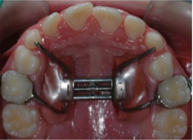
RME was performed until dental overcorrection. The expander was then kept on the teeth as a passive retainer and the patients underwent no further orthodontic treatment during retention (Fig. 2).
Figure 2. Modified Haas-type expander anchored to deciduous teeth.
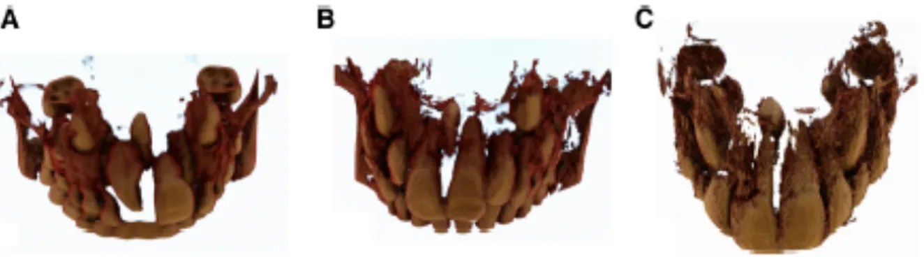
The samples were harvested with the clinical aim to remove bone for the supernumerary tooth extraction. When possible, maxillary suture and bone margins were both included in the sample. All the biopsies were processed for histology in the first investigation (Fig. 3-15) (Caprioglio et al., 2017) and evaluated by complementary imaging techniques, namely
Synchrotron Radiation-based X-ray microtomography (microCT) and comparative light and electron microscopy, in a second investigation (Giuliani et al., 2018).
Midpalatal suture changes after RME were reported to be of great interest (Liu et al., 2015) since a deeper knowledge of this processes might help in treatment options and modalities. The reported case report aimed to investigate immediate histologic changes in midpalatal suture in humans following RME compared to control and was conducted on selected patients who had to perform surgery in the palatal area for other clinical reasons. A similar previous investigation did not enroll biopsy controls (Melsen, 1972) which were instead enrolled in the cited case report. Despite the young age (8.3+/-0.9 years), the subjects exhibited suture gap but with inter-digitations in the midpalatal suture, as previously reported in patients around the age of 10 (Melsen, 1972). At this age, changes in suture morphology from squamosity to sinuosity were observed (Melsen, 1972), supporting the cited report since remodeling of the bone margins of the palatal suture was evident with different maturation stages of the newly-formed bone areas characterized by wide osteocyte lacunae. Moreover, the morphology of the suture at this age (subject 3) presented a parallel orientation of the collagen fibers related to the suture long axis similar to lamellar bone, which is traditionally considered as a stress-strain resistant type of bone. RME performed in pre-pubertal age might avoid fracture of inter-digitations due to immature stages of growth and bone remodeling (Liu et al., 2015). Seven days after the end of expansion (subject 1) newly-formed bone with osteoid matrix undergoing mineralization was evident not only on the bone margins, indicating mineralization processes from within the center of the suture, which seemed to be an original finding of the investigation, as was the peculiar fishbone appearance of the trabecular bone. Moreover, newly-formed bone showed collagen fibers in a transversal orientation related to the suture long axis in comparison to the control sample, where a longitudinal orientation was observed.
This orientation was suggested to be related to the response to mechanical forces as shown in mice and this finding has never been reported in humans (Hou et al., 2007). Within the suture few areas of blood clot with several red blood cells were observed, probably caused by the trauma of maxillary expansion, but no inflammation cells were reported. This result is in contrast with Melsen who reported hyperemia and inflammation associated with osteoblastic activity near to the bone margins in the midpalatal suture 3 weeks after expansion. Inflammation cells were not evident at 7 and 14 days after expansion also in several animal investigations, meanwhile the osteoblastic activity was reported right after the midpalatal splitting (Murray et al., 1971). Cleall et al. (1965) found predominantly disruptive, inflammatory processes and osteoclastic activity in monkeys. The initial inflammatory aspect was reported only in these investigations, meanwhile the hyperemia seemed to be present in all the performed studies on animals (Storey, 1955; Cleall et al., 1965) and human beings (Melsen, 1972) in the first few days after midpalatal suture separation. In the case report no inflammation cells nor osteoclastic activity was noted. Although inter digitations were present in the suture at this stage of growth the suture morphology was still immature. At 30 days (subject 2) there was a 29% increase of newly-formed bone which showed thicker trabeculae and the peculiar fishbone appearance with parallel orientation and confluent in several areas. Diffused osteoblastic activity was evident also at this stage but also vascular activity was noted. Similar results were reported by Melsen (1972) who found newly-formed bone along the margins and in the middle of the suture with confluent islands in cross-section bony extensions 4 weeks after the end of expansion but no angiogenesis was reported and the normal serrated inter-digitated shape of suture had not yet been reconstituted. The results of the cited investigation are limited to the restricted number of patients, since bone biopsy is not ethically justified only to assess treatment outcomes. Therefore, only patients who had to
undergo surgery for clinical reasons could be selected. Furthermore, the results are limited to the appliance that was tooth-anchored on primary teeth. Moreover, the observation time was limited to 30 days after RME but it would be very useful to perform long-term investigations. The quantity of RME might affect the suture since rapid expansion protocols were shown to create midpalatal suture separation followed by the filling of the defect with new bone. Once the midpalatal suture separation is obtained, the amount of expansion might affect the healing time of the bone that starts after the midpalatal separation and keeps going for several months after. The sample harvested from subject 2 showed more advanced healing processes compared to subject 1.
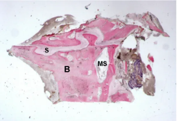
Figure 3. Control. Mature bone (B) with small marrow spaces (MS) was observed. The gap of the palatal suture (S) appeared characterized by inter-digitations. Toluidine blue and acid
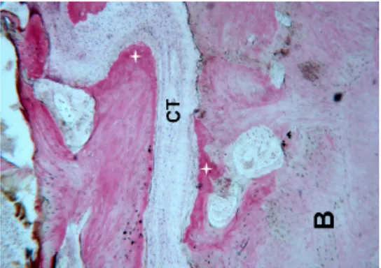
Figure 4. Control. Connective tissue (CT) in the middle of the suture as well as mature bone (B) and newly formed bone (+) was observed. Toluidine blue and acid fuchsin were used.
Original magnification 40x
Figure 5. Control. The bone margins of the suture (S) were irregular. New bone with wide osteocyte lacunae (O black arrows) was observed. Toluidine blue and acid fuchsin were used.
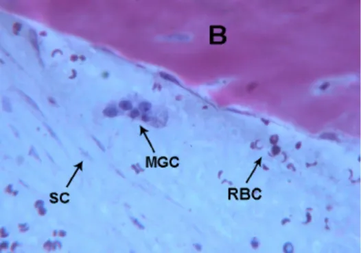
Figure 6. Control. Multi-nucleated giant cells (MGC) near the marginal bone, spindle cells (SC) and red blood cells were observed (RBC). Bone (B).Toluidine blue and acid fuchsin were
used. Original magnification 400x.
Figure 7. Mature bone with small marrow spaces (B) and, in the gap (G) after rapid maxillary expansion, trabecular new bone (NB) with storiform appearance was observed. Toluidine blue
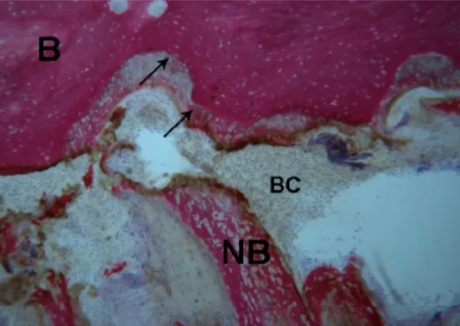
Figure 8. Test 7 days. The bone margins of the suture were characterized by inter-digitations (black arrows), where the newly-formed osteoid matrix undergoing mineralization directly was also observed. Inside the suture, newly-formed trabecular bone (NB) was detected. In the suture gap a blood clot (BC) was present. Toluidine blue and acid fuchsin were used. Original
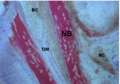
Figure 9. Test 7 days. The trabecular newly formed bone (NB) had a parallel trend and was surrounded by osteoid matrix (OM) undergoing mineralization. In a few areas of the blood clot (BC) numerous red blood cells were present. Toluidine blue and acid fuchsin were used.
Original magnification 100x.
Figure 10. Test 7 days. Newly-formed spicules (NB) close to large (V) and small vessels (V and black arrow were observed. Toluidine blue and acid fuchsin were used. Original
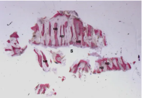
Figure 11. Test at 30 days. The newly-formed bone trabeculae (NB) were oriented perpendicularly to the long axis of the suture (S) and run parallel to each other. Toluidine
blue and acid fuchsin were used. Original magnification 9x.
Figure 12. Test at 30 days. A small portion of the bone margins of the palatal suture was evident, lined by osteoblasts (black arrows) producing osteoid matrix (OM). Toluidine blue
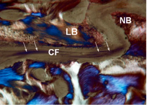
Figure 13. Control. Under polarized light, parallel lamellar bone (LB) and newly-formed bone (NB) without parallel collagen fiber orientation were observed. In the middle of the suture collagen fibers (CF white arrows) oriented parallel to the long axis of the suture were
seen. Toluidine blue and acid fuchsin were used. Original magnification 40x.
Figure 14. Test at 7 days. Under polarized light, newly-formed bone (NB) and collagen fibers (CF) with a storiform orientation (white arrows) were observed. Toluidine blue and acid
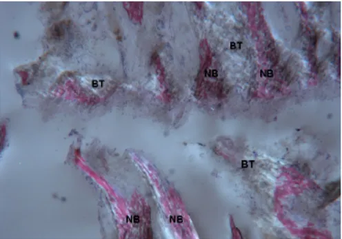
Figure 15. Test at 30 days. Under polarized light, newly-formed bone (NB) undergoing mineralization could be observed. Not yet organized newly-formed bone trabeculae (BT) were
also evident. Toluidine blue and acid fuchsin were used. Original magnification 40x.
As documented in literature (Giuliani, 2016), it is often suggested to couple 2D conventional microscopy with advanced 3D quantitative analysis. Indeed, with the use of microCT, it is reasonable to get significant morphometric results on a statistical sample sometimes narrower than the number of patients involved in the histologic study (Suresh et al., 2012). In the second study, microCT allowed to achieve significant quantitative results (Fig. 16-18) in spite of including a single subject for comparisons at 7 days, 30 days after RME, and a control. Indeed, the previous case report (Cparioglio et al. 2017) on the same subjects was only descriptive and exclusively based on 2D data. In agreement with histological findings, this microCT study detected a relevant amount of newly formed bone both 7 and 30 days after RME. Furthermore, as previously reported (Caprioglio et al., 2017), it was observed a progressive mineralization with the peculiar in-plane fishbone appearance of the trabecular bone. As reported in literature
(Storey, 1955), the suture mineralization and morphology were confirmed in 3D to be still immature respect to the control, also 30 days after RME. However, the microCT analysis did not confirm in 3D another finding observed in 2D by light and electron microscopy, i.e. that the newly formed bone trabeculae were oriented perpendicularly to the long axis of the suture and run parallel to each other. Several microCT data contributed to denying in 3D this observation: the calculated value of DA, both 7 and 30 days after the RME, suggests a rather isotropic and poorly oriented structure; the combined significant increase in the number of trabeculae and their connectivity is not compatible with a structure consisting of parallel trabeculae.
Figure 16. Portion of the “intensity vs. gray levels” profile. The grey levels are proportional to the linear attenuation coefficient µ that, in turns, is nearly proportional to ρ, the bone mineral density (BMD). The integrated areas of the represented peaks correspond to the newly
formed mineralized bone volume in RME-treated midpalatal sites and in the control. b-d Representative 2D sections of the treated palatal sites 7 days (b) and 30 days (c) after RME,
and of the palatal control (d). The thickness of the suture channel was similar to that of the control suture (400–700 µm, yellow arrows), showing that the storiform way of
Figure 17. 3D microCT rendering of the biopsies retrieved 7 days (a) and 30 days (b) after the RME. Both the specimens clearly showed the meshwork of the bone perforated by non-mineralized spaces. The direction indicated by the red arrows corresponded to the section plane of histological and SEM micrographs. The right image offers a better view of the canals
Figure 18, a-c Biopsy retrieved 7 days after RME: (a) 3D microCT reconstruction; (b) Study in 3D of the thickness distribution basing on a color map; (c) 2D sampling color mapped slice. d-f Biopsy retrieved 30 days after RME: (d) 3D microCT reconstruction; (e) Study in 3D
of the thickness distribution basing on a color map; (f) 2D sampling color mapped slice. Thickness scale for the color map at the bottom-center position. (g )Histogram of the distribution of the newly formed bone thickness in both the RME-treated midpalatal biopsies. These data demonstrate that there was a slight (not significant; p > 0.05) increase in thickness
of the struts from 7 days to 30 days after RME.
Further case reports should be focused on the investigation in different timepoints in order to better clarify when the bone healing process might end and give important guidelines on retention and relapse.
Side Effects
Secondary effects of RME relate to the heavy forces produced by the RME appliance which could produce bite opening, microtrauma of the midpalatal suture and temporo-mandibular joint structure and root resorption among others (Lagravere et al., 2005). Periodontal involvement is the most commonly cited side effect of RME due to the possibility of damaging the buccal cortical plates and developing gingival recessions when high forces are
directed towards the banded teeth. A retrospective study analyzing the periodontal effect of RME in 17 growing patients demonstrated that immediately after the expansion the first maxillary molar buccal plate thickness is reduced by 0.5 mm. However, at 6 months post expansion, only the lingual bone plate thickness of both first molars was significantly increased with no differences in the ratio between intermolar widths at the apex and crown levels (Ballanti et al., 2009). In a CBCT study evaluating the buccal alveolar bone changes 3 months after the end RME activation with Hyrax appliance, it was found that the buccal bone thickness decreased 1.1 mm and 1.2 mm for the 1st premolars and 1st molars respectively while the buccal marginal bone level decreased by 4.5 mm and 2.9 mm respectively. This study suggested that the buccal movement of teeth may potentiate the probability of buccal bone dehiscence at the maxillary 1st premolar due to the increased buccal marginal bone loss associated with apical narrowing at this level (Rungcharassaeng et al., 2007). Although periodontal consequences may be present after RME, available literature demonstrate that buccal bone thickness returns to normal level and no periodontal concern should be raised if the patient had an initially normal buccal bone thickness.
The present Author recently reported original findings (accepted in March 2018, publishing in January 2019) on periotontal effects of RME with different appliances. Dislocation of teeth outside their alveolar process, in fact, can damage the periodontium and for this reason maxillary expansion using deciduous teeth as anchorage in mixed dentition might be suggested. The aim of the reported study was to compare changes of buccal bone plate thickness on upper first permanent molars after rapid maxillary expansion performed in mixed and permanent dentition with different anchorage. Two groups of patients were evaluated with CBCT before (T0) and after (T1) rapid maxillary expansion. Group E (21 patients) underwent rapid maxillary expansion using deciduous teeth as anchorage, Group 6 (16 patients)
underwent rapid maxillary expansion using permanent teeth as anchorage. According to the results RME performed in mixed dentition with the appliance anchored to deciduous teeth did not reduce the buccal bone plate thickness of the upper first permanent molars, except for the mesial roots on both sides. Meanwhile, RME performed in permanent dentition produced a reduction of the buccal bone plate thickness of upper first permanent molars when they act as anchor teeth. The range of reduction of the buccal bone thickness of upper first permanent molars when they act as anchor teeth showed statistically significant differences when compared to the overall absence of bone reduction when RME is performed in mixed dentition. Nevertheless the bone loss (ranging between 0.73 mm and 1.25 mm) is not clinically significant. Important consideration should then be given to other factors that might play a relevant clinical role. Soft tissue, in fact, might cover the bone defect created as fenestration, more frequently in maxillary alveolar ridge, or dehiscence. The occurrence of a recession in these cases was suggested to be strictly related to gingival inflammation. The first clinical recommendation should be to avoid using permanent molars as anchor teeth when deciduous teeth are suitable.. For this reason timing plays a fundamental role in terms of avoiding postponing maxillary expansion in permanent dentition but preferring as a valid alternative a two-phase treatment. For older patients presenting at the orthodontic examination when deciduous teeth are no longer suitable, a careful evaluation of the gingival biotype should be performed before positioning bands of the appliance on the upper first molar. In some cases also accompanied by radiographic investigation involving CBCT when the clinical investigation is doubtful. Then a follow-up during the expansion with the clinical examination through touching the roots at the appointments and a rigid oral hygiene regime should be mandatory. Another option when a large amount of expansion is needed in growing patients with a thin gingival biotype might be bone anchored maxillary expansion.
RME therapy appears to involve an ample portion of the craniofacial complex, as the maxilla is associated with 10 bones in the face and head (Bell et al., 1981). This involvement has been hypothesized following investigations based on histologic methods, radiologic imaging, photoelastic models, bone scintigraphy, and finite element analysis.
Findings from Leonardi et al. (Leonardi et al., 2011) showed that circum-maxillary sutures articulating directly to the maxilla were opened more extensively than those indirectly articulated. In fact, the distant structures of the craniofacial skeleton (zygomatic bone and temporal bone) were also affected by transverse orthopedic forces, although to a lesser extent. All in all, an increased width in circumaxillary sutures, with the highest amount of opening at the internasal suture (0.386 mm) and the lower at the zygomaticotemporal suture (0.213 mm) was observed. Results indicated that the amount of widening after RME therapy is different among sutures and highly variable among subjects.
Treatment Timing
Like all craniofacial sutures, the mid palatal suture becomes more tortuous and interdigitated with increasing age. In children up to ten years of age, almost any expansion device will tend to separate the mid palatal suture. However, by adolescence a relatively heavy force from a rigid jackscrew is needed to separate the interdigitated suture (Proffit 2007). In this context, Baccetti et al. evaluated patients with different stages of cervical vertebrae maturation index and found that the early treated individuals who had not reached the pubertal growth spurt at the onset of RME showed on average 3mm of expansion of the mid-palatal suture while the late treated ones averaged only 0.9mm. His finding suggested that an effective long-term change at the skeletal level occurs when the patients were treated prior to pubertal peak growth and higher dental effect is present in individuals treated after pubertal growth spurt
(Baccetti et al., 2001).
Distant Skeletal Effects
Finite element analysis (FEA) is defined as a computer simulation method performed by dividing the interested region into discrete elements interconnected at nodes with assigned material property that represents the physical property of the model. A FEA study evaluating the effects of the maxillary expansion on the neighboring bones demonstrated that in the closed suture model (adult type suture) significant stress areas were present at the buccal alveolar processes, distal aspect of the maxilla, inferior aspect of the zygomatic arches and pterygomaxillary fissure region (Lee et al., 2009). Thus, areas surrounding the zygomatic processes were suggested to provide a buttressing effect against the forces of expansion. In the patent midpalatal suture model (growing child) however, the pterygomaxillary fissure demonstrated to be the highest stress point. This finding confirmed the impact of maxillary expansion in facilitating the treatment effects of a class III facemask therapy in growing individual (Lee et al. 2009). In the same patent suture model, tension stress was also present at the upper portion of the nasal cavity suggesting that the palatal expansion with heavy forces in young children may create undesireable changes in the nose (Lee et al., 2009, Proffit, 2007). For both groups, the lateral displacement of the maxillary halves appeared nonparallel, with a slightly wider opening towards the anterior and the separation of the maxilla occurring as if a hinge was positioned superiorly at the base of the nose (Lee et al., 2009). Clinical studies evaluating the effects of orthopedic expansion via RME postulated that not only bodily separation of the midpalatal suture exists, but also buccal rotational force on the maxillary alveolar shelves and changes to the surrounding frontomaxillary, zygomaticomaxillary,
zygomaticotemporal and pterygopalatine sutures (Garrett et al., 2008).
Skeletal Dimension and Airway
RME has been reported to be effective also for the upper airway increase due to the direct force exerted on the nasal area in young individuals (Caprioglio et al., 2014) with breathing pattern problems.
Breathing pattern and cranio-facial growth
The effect of type of breathing on craniofacial growth has been widely debated as a controversial issue within orthodontics for decades. It has been maintained that when significantly large adenoids are present, nasal breathing is (partially) obstructed leading to mouth breathing and thus the stereotype of the adenoid face (Subtelny, 1954; Linder-Aronson, 1970). However, the complexity of the association between nasal obstruction and facial growth has also been discussed (McNamara, 1981; Warren and Spalding, 1991; Trotman et al., 1997; Vig, 1998). The adenoid face is characterized by an incompetent lip seal, a narrow upper dental arch, retroclined mandibular incisors, increased anterior face height, a steep mandibular plane angle, and retrognathic mandible compared with faces of healthy controls (Linder-Aronson, 1970; Flores-Mir, 2013). Comparable changes in the craniofacial structure have been described in a group of subjects with large tonsils (Behlfelt et al, 1990). This development has been explained in a ‘mechanical’ way as occurring because of changes in the muscular balance. When mouth breathing, the tongue position in the oral cavity is low and the balance between forces from the cheeks and tongue is different compared with healthy children. This may lead to a lower mandibular position and an upward/backward head posture
with all the above-mentioned dental and skeletal consequences (Solow and Kreiborg, 1977; Linder-Aronson, 1979; Solow et al, 1984).
Role of hormones on cranio-facial growth
After adenoidectomy to facilitate nasal breathing, accelerated mandibular growth and closure of the mandibular plane angle have been reported, although with a large variation (Linder-Aronson et al, 1986). In a more detailed analysis, anterior facial height was found to be unaffected and remained longer in the initially large adenoid subjects than in healthy controls 5 years after adenoidectomy. In the same study, growth of the mandibular ramus and condylar process of adenoidectomy patients was found to be greater than that in the control subjects (Kerr et al, 1989). The changes found have been previously explained by alteration in tongue position and autorotation of the mandible (Linder-Aronson, 1979). However, in order to decrease the mandibular plane angle, more growth in posterior face height/ramus height is needed.
Endochondral bone formation in the condylar cartilage and bone apposition in the lower border of the mandible (gonial region) contribute to the growth in height of the mandibular ramus. Studies on mandibular condylar cartilage have shown that the cartilage not only is a passive growth site, but also has some tissue-separating potential (Copray et al, 1988). It has also been maintained it to be active in displacing the condylar process downwards (Kantomaa, 1987). In addition, the mandibular condylar cartilage seems to be a target and production site of hormonal agents as evidenced by IGF-I receptor and IGF-I messenger RNA expression in the cartilage (Visnapuu et al, 2001, 2002). Patients with GH (GH) deficiency have been shown
to have a small posterior face height compared with age and gender-matched healthy controls (Pirinen et al, 1994). Furthermore, administration of GH to patients with GH deficiency, such as those with Turner syndrome or in bone marrow transplant patients, has shown that mandibular growth, particularly the ramus growth, is accelerated in those cases compared with control healthy children (Simmons, 1999). The increase in mandibular ramus height by GH can be explained by two theories. One is the presence of an increased endochondral bone formation in the condylar cartilage (Peltomäki T, 2007) and the other is the presence of an increased bone apposition in the lower border of the mandible through the anabolic effects of GH on the masseter and medial pterygoid muscles (Vogl et al, 1993).
Maxillary Constriction and Obstructive Sleep Apnea
Obstructive sleep apnea (OSA) is characterized by episodes of complete or partial upper airway obstruction during sleep, often resulting in gas exchange abnormalities and arousals, which disrupt sleep. The condition exists in 2 to 5 percent of children and can occur at any age (Rosen et al., 2004). Untreated OSA is associated with cardiovascular complications, impaired growth (including failure to thrive), learning problems, and behavioral problems. Early diagnosis and treatment may decrease morbidity. Potential consequences of untreated OSA in children include:
• Inattention and behavioral problems (eg, hyperactivity, impulsivity, rebelliousness and aggression).
• Daytime sleepiness.
• Growth – Severe OSA can be associated with failure to thrive, and treatment can lead to weight gain and growth.
hypertension, right and left ventricular dysfunction.
Each of these conditions may benefit from treatment of OSA, though in some cases definitive proof at the level of randomized clinical trials does not yet exist. The initial screening for OSA is typically done by the primary care clinician. Children with suspected OSA should be referred to a specialist in sleep medicine or otolaryngology (ear, nose, and throat). Alternatively, the referring provider may be able to arrange for a polysomnogram (PSG) if a facility with experience in pediatric PSG is available. Then children with abnormal results of PSG, or with other significant sleep problems, can then be referred to the appropriate specialist. OSA is typically defined by clinically relevant symptoms and an apnea hypopnea index (AHI) >1 or hypoventilation (CO2 >50 mmHg for >25 percent total sleep time) as
determined on PSG. The decision to initiate treatment and choice of treatment depend upon the child's age, presence of any underlying medical issues, risk factors, clinical symptoms (such as nighttime sleep problems or daytime dysfunction), and results of PSG if performed, as outlined below:
Adenotonsillectomy – Referral to a specialist for adenotonsillectomy evaluation is generally indicated for otherwise healthy children who have OSA and adenotonsillar hypertrophy (including ≥1+ tonsils) (Marcus et al., 2012). After the full evaluation, the decision about whether to proceed to surgery should be made collaboratively with the family, considering in particular the degree of clinical symptoms (nocturnal and daytime), as well as the tonsil size and OSA severity. Adenotonsillectomy also may be initial therapy for children with other contributors to OSA such as obesity or other comorbidities, if appreciable adenotonsillar tissue is present, even if there is no clear hypertrophy. The rationale is that adenotonsillectomy may improve upper airway patency enough to ameliorate or resolve the OSA, even if it does not correct all of the etiologies. Such patients should be managed by a clinician experienced with
pediatric sleep-related respiratory abnormalities;
Watchful waiting – For otherwise healthy children with mild or moderate OSA confirmed by PSG (AHI >1 and <10), watchful waiting with supportive care is a reasonable alternative to adenotonsillectomy. This approach is based on the acceptable outcomes for patients followed with watchful waiting in the Childhood Adenotonsillectomy Trial (CHAT) trial (Marcus et al., 2013). If this approach is chosen, the child should be reevaluated clinically within six months, or reevaluated sooner if symptoms worsen;
Positive airway pressure therapy – For patients with minimal adenotonsillar tissue or a strong preference for a nonsurgical approach, positive airway pressure therapy is an alternative to adenotonsillectomy. It also may be appropriate to stabilize children with severe OSA prior to adenotonsillectomy or another surgical procedure, or for children with persistent OSA despite adenotonsillectomy. Positive airway pressure may consist of continuous positive airway pressure (CPAP) or bilevel positive airway pressure (BPAP).
The following treatments can be considered in selected cases, either instead of or as an adjunct to the primary therapy:
Rapid maxillary expansion – Prepubertal children with OSA and a narrow palate (crossbite) and little adenotonsillar tissue are candidates for treatment with rapid maxillary expansion (RME). RME is an orthodontic technique that widens the palate and nasal passages, thereby increasing airway patency. Such patients should be managed by an orthodontist experienced with pediatric sleep-related respiratory abnormalities;
Corticosteroids or antiinflammatory therapy – For children with mild or moderate OSA and nasal obstruction due to adenoidal hypertrophy, a trial of intranasal corticosteroids or leukotriene modifier therapy may be performed for two to four weeks, prior to determining whether the therapy should be continued long-term as an adjunct or alternative to
adenotonsillectomy or positive airway pressure.
Other therapies – Selected children with OSA may derive benefit from adjunctive therapies. As examples, obese children with OSA may benefit from weight loss, and all children may benefit from avoidance of environmental allergens or irritants such as tobacco smoke. In addition, positional therapy (eg, elevation of the head of the bed) can be considered.
Maxillary constriction has also been postulated to play a role in the pathophysiology of OSA. Despite the reference of multiple contributing factors for the development of OSA including retrognathic mandible, shorter AP face length, reduced distance from the posterior nasal spine (PNS) to posterior pharyngeal wall, lower positioned hyoid bone, larger soft palate, smaller pharynx, larger tongue size, obesity and combination thereof (Johal et al., 2007), a constricted maxilla has also been associated with narrowing the upper airway dimension and increasing the risk for OSA by inducing a low tongue posture (Subtelny, 1954). Sleep apnea is defined as a decrease in respiration yielding hypoxia and hypercapnia during sleep, caused by either neurologic origin or actual physical blockage of the airway also known as OSA. Subjects with centrally driven apneic event present no effort to overcome the apnea, whereas the opposite is true for the OSA sufferers. The American Academy of Sleep Medicine defines OSA as episodes of breathing cessation or absence of respiratory airflow for over 10 seconds despite respiratory effort. Epidemiology reports indicate that this is a highly prevalent respiratory sleep disorder affecting 4% of men and 2% of women (Haskell et al., 2009). The most serious consequences of OSA are the cardiovascular diseases such as hypertension, tachycardia, atherosclerosis, increased risk for cerebrovascular accidents, coronary artery disease and more (Madani et al., 2007). The pathogenesis of these effects is still being studied but it is generally accepted that the intermittent hypoxia and hypercapnia episodes triggers homeostatic
compensations in the body, leading to cardiovascular diseases over time (Sharabi et al., 2004). It is believed that the sleep induced relaxation of the muscles attached to the soft tissues of the pharynx is aggravated by gravity and the retropositioning of the tongue mass during supine position narrowing the airway lumen (McCrillis et al., 2009). Treatment of OSA consists in preventing the collapse of the lumen of the pharynx during sleep. At present, several treatment options based on the severity of the apneic events are rendered, including continuous positive airway pressure (CPAP) therapy, surgical treatments and mandibular repositioning devices therapy. Oral appliances have been reported to improve breathing by decreasing nasal resistance and reducing the AHI. For breathing to take place, patency of the pharynx or upper airway is vital. With the exception of the two ends of the airway, the nares and the small intrapulmonary airways, the pharynx is the only collapsible segment of the respiratory tract with the potential to be altered by diverse treatment effects (Haskell et al., 2009).
According to Lenza et al. the upper airway can be divided into smaller segments to better understand the physiologic changes as well as the treatment effects (Lenza et al. 2010). The various portions of the upper airway, superiorly to inferiorly include:
• Nasopharynx: The upper most portion of the airway, mainly the nose. It begins with the nares extending back to the hard palate at the superior portion of the soft palate. This includes the nasal septum and the nasal turbinates.
• Retropalatal airway (velopharynx): This area extends from the hard palate to the inferior tip of the soft palate, including the uvula and the uppermost segment of the posterior pharyngeal wall. Major muscles include the tensor palatini and levator palatini, which elevate the soft palate, and the musculous uvulae providing elevation of the uvula.
the back portion of the mouth and extending rearward to the base of the tongue or tip of the epiglottis. Tonsils and tongue muscles are located in this segment.
• Hypopharynx: The area extends from the tip of the epiglottis to the lowest portion of the airway at the larynx.
In all individuals, muscular activity is reduced and upper airway resistance increased during sleep compared when awake (Worsnop et al, 2000). This does not have a notable effect on breathing in anatomically and functionally healthy individuals. On the other hand, reduction of muscular tone in children with large adenoids and tonsils, or with other underlying abnormal upper airway anatomy, may lead to airway obstruction and eventually to OSA. Interestingly, these children have been found to have similar craniofacial characteristics as adenoid face children (Caprioglio et al., 1999). The first treatment of choice for OSA children is removal of adenoids and tonsils, if they are indeed hypertrophic (Nieminen et al., 2002). It can thus be postulated that some children with a clinical diagnosis of an adenoid face may also have OSA. Of particular interest is the recent cephalometric study on 5-year-old children with OSA verified with PSG (Zettergren-Wijk et al., 2006). This study showed that OSA children have a different facial morphology compared with age-matched controls. The mandibular plane angle was found to be posteriorly inclined, anterior face height to be greater, and posterior face height smaller than control children. At the 5-year follow-up after T&A, no major craniofacial differences were noted. In a closer look at the growth changes it becomes evident that anterior face height remained greater in the OSA children than in the control children (difference of average 2.5 mm), but presented an average increase in both groups of children. Yet, the mandibular plane angle was decreased in the OSA children. This may be explained by the described greater posterior face height growth (ramus growth) in the OSA children compared
to controls (difference of average 2 mm). After T&A, OSA patients showed increase of the nasal breathing pattern and modifications of GH serum levels were noted with growth catch-up.
OSA, breathing function and body growth
Along with breathing issues, body growth retardation is frequently observed in OSA patients. The complex mechanisms behind growth retardation are still unclear. OSA might interrupt slow-wave sleep, when GH is preferentially secreted and this phenomenon was observed also in chronic snorers not diagnosed for OSA. GH modifications were often related to serum levels of IGF-1 and IGFBP-3 which are related to diurnal GH secretion and reflect its anabolic role on tissues, especially muscle and bone. IGF-1 resulted significantly increased in children after T&A surgery in previous studies thus suggesting a relevant role of the IGF-1 axis in growth retardation of children with upper airway obstruction and the consequent growth catch-up (Bar et al., 1999; Nieminen et al., 2002).
The present Author performed a systematic review (under review) on metabolic effects of treatment in patients with OSA which aimed to systematically review the available scientific literature on the relationships among OSA patients, GH axis and other metabolic changes and growth alterations before and after T&A treatment. Several studies investigated relationships among OSA patients, GH axis metabolic changes and/or growth alterations before and after OSA treatment but some results are still contradictory indicating that complex and multiple mechanisms might be involved. To date only one systematic review and meta-analysis was performed on growth and growth biomarker changes after T&A in sleep-disordered breathing (SDB) patients. The following study updated the literature and selected different inclusion criteria. Moreover this is the first study presenting the attempt to review the available literature
regarding metabolic mediators changes such as endothelial and neurocognitive function mediators and local inflammation mediators.
Growth mediators - IGF-I
GH was shown to stimulate the synthesis of IGF-I in the liver and other target tissues (Tapanainen et al. 1992). IGF-I is considered as the main mediator of the growth-promoting actions of GH(Isaksson et al., 1987) reflecting the daily mean GH levels, and it has been reported to correlate well with the physiologic changes in GH secretion (Furlanetto, 1990). Among pre-pubertal children, IGF-I was reported to be clearly not sex-dependent (Juul et al., 1994). IGF-I showed significant higher levels in OSA patients 1 month, 3, 6 and 9 months after T&A (Bar et al., 1999) but no significant differences right after surgery (Nachalon et al., 2014). Nachalon et al. (2014) were the only authors who evaluated growth mediators immediately after T&A finding no significant differences in IGF-I levels, suggesting that this mediator might need at least 1 month to reach significantly higher concentrations (Kang et al., 2008). Moreover all the other selected studies investigated older patients samples in pre-pubertal age, meanwhile Nachalon et al. (2014) referred to children aged 6-36 months. The evidence of other selected articles suggested that T&A might influence the GH-IGF-I axis by increasing IGF-I levels, leading to acceleration of growth in SDB patients and the results were pretty homogeneous. Nevertheless some limitations were present in the study of Tatlıpınar et al. (2012) since only clinical history based diagnosis of SBD was performed. All the studies were performed at pre-pubertal age thus reducing bias due to the pubertal spurt which might cause itself an increase of growth mediators. When compared to control groups, OSA patients showed significant lower levels of IGF-I at baseline (Selimoğlu et al., 2003) even though
contradictory results were found by Nieminen et al. (2014), and the significant difference was also maintained after T&A regardless of the significant improvement among the time points according to Zhang et al.(2015) This latter study investigated twins, using one affected by OSA as treated and the other not affected by OSA as control. According to their results OSA might affect growth manners and early surgical intervention could only partially reverse the growth inhibit. In these cases early intervention might be highly recommended.
- IGFBP-3
IGFBP-3, the GH-dependent major carrier protein of IGF-I, was shown to correlate significantly with nocturnal GH secretion, but not as strongly as IGF-I (Blum et al., 1993). Although IGFBP-3 probably exerts some functions of its own on cells, its major role seems to be the prolongation of the half-life of IGF-1 (O’Brien et al., 2006). The major advantage of IGFBP-3 determinations in diagnostics is its relative stability over time(Blum et al., 1993), and it might therefore be a more reliable indicator of GH secretion over a longer time interval than IGF-I. It is also less dependent on age than IGF-I (Rosenfeld et al.,1999). IGFBP-3 showed significantly higher levels in OSA patients 6 and 9 months after T&A according to the results of Tatlıpınar et al. (2012). On the contrary, Bar et al. (1999) and Selimoğlu et al. (2003) found no significant changes after T&A.
The reported dissociation between IGF-I and IGFBP-3 levels might express the complexity of the IGF-I and the IGF-binding protein systems, which are controlled by several positive and negative feedback pathways, rather than one simple pathway. The increase in IGF-I levels without a corresponding change in IGFBP-3 levels may indicate an elevation of the free IGF-I levels, because IGFBP-3 is the major binding protein in the circulation according to both authors (Bar et a., 1999; Tapanainen et al., 1992). Only Nieminen et al. (2012) and Selimoğlu
et al. (2003) compared IGFBP-3 levels between OSA patients and controls. OSA patients showed significant lower levels IGFBP-3 at baseline and significant higher levels 6 months after T&A according to the results found by Nieminen et al. (2012) meanwhile no significant differences were found by Selimoğlu et al. (2003). Moreover, according to Selimoğlu et al. the increase in IGFBP-3 concentration after T&A in the study of Nieminen et al. might imply decreased GH secretion over a significant time span prior to treatment.
Growth alterations in OSA patients were also focused on craniofacial anomalies and it still not clear if they might derive more from alteration of the breathing function or if they are directly influenced by the GH axis anomalies.
Endothelial mediators
OSA was suggested as an important and independent risk factor for altered endothelial function and cardiovascular disease (Sánchez-de-la-Torre et al., 2013). The involved mechanisms are not still completely understood and they seem related to the increase in circulating adhesion molecules in children with OSA (O’Brien et al., 2006). Moreover, OSA presents sleep fragmentation that might cause systemic inflammation and sympathetic activation thus leading to endothelial dysfunction, which has been shown to precede the formation of plaque and atherosclerosis (Jelic et al., 2008). sCD40L is a binding protein on the surface of endothelial cells and triggers the increased expression of inflammatory mediators, growth factors, and the procoagulant tissue factor such that increased levels of sCD40L essentially represent the presence of an increased risk in the context of a variety of cardiovascular disorders. Gozal et al. (2007) found significantly higher levels of sCD40L in OSA patients when compared to a control group. Even though sCD40L showed significant decrease after T&A, their levels were still significantly higher than controls 4-6 months after
T&A. Because the duration of follow-up was relatively short, it is still unclear as to whether these children with persistently abnormal hyperemic responses and elevated sCD40L will improve at later stages. Moreover FMD was employed by Chan et al. (2015) in OSA patients compared with nonsnoring control subjects. This parameter was shown to be independent from reactive hyperemia thus representing a more precise means to assess endothelial function. According to their results OSA patients showed reduced FMD, which was reversible with surgical treatment indicating reversal of endothelial dysfunction following surgical intervention.
Neurocognitive function and mediators
OSA children showed specific cognitive dysfunctions that include deficits in memory, problem solving and behavioral functioning (Beebe et al., 2002). BDNF is an important member of the neurotrophin family, abundant in the brain and periphery that induces long-term changes in synaptic composition, ion channel expression and neurotransmitter production in the neuronal structures of the brain. Reduced BDNF levels in the human brain are associated with cognitive deficits, impaired memory performance and depression (Chen et al., 2001). According to the results of Wang et al. (2010) BDNF serum levels significantly decreased 3, 6 and 12 months after T&A in OSA patients. The authors suggested that concomitant decrease in serum and plasma BDNF might indicate that it was specifically removed from peripheral blood maybe due to increased demand following the improvement of sleep parameters. The nervous system might then be able to recover after T&A, and so the demand for BDNF would increase, potentially resulting in the reduction of circulating BDNF levels.
Local inflammation
Anti-inflammatory agents are often employed in treatment of upper airway diseases and glucocorticoids were suggested to inhibit inflammation by up-regulation of T-regulatory (T- reg) cells and the secretion of interleukin (IL)–10 by these cells (Esteitie et al., 2011). According to the results of Esteitie et al.(2011) pretreatment with FFNS compared with no treatment resulted in inhibition of Interleukin (IL)-6 secretion by adenoid cells without affecting other cytokine levels significantly. This result might partially explain the mechanism by which intranasal corticosteroid therapy provides relief for children with OSA, and it is emphasized by the prospective and randomized design followed by the authors to divide the experimental group and the control group before T&A. The selected inclusion criteria allowed the present systematic review to include twelve studies since only in vivo studies were selected and limited cases studies and case reports were excluded. Some of the initially selected articles were excluded since they did not present treatment outcomes or clear pre- and post- treatment data on clinical treatments. However the methodologies of the selected studies were varied thus a meta-analysis was not possible. Also, these studies failed to give the higher level of scientific evidence, which is only attainable through the use of randomized clinical trials except for Esteitie et al. (2011) who indicated randomization process in patients enrollment. In the absence of the highest level of evidence, clinicians have to make decisions based on lower levels of evidence.
Maxillary expansion and breathing pattern: where are we now? Author’s contributions:
Fastuca R. Maxillary expansion and breathing function: Where we are now?. Int J Orthod Rehabil 2016;7:121-3.
Fastuca R, Perinetti G, Zecca PA, Nucera R, Caprioglio A. Airway compartments volume and oxygen saturation changes after rapid maxillary expansion: a longitudinal correlation study. Angle Orthod. 2015 Nov;85(6):955-61.
Fastuca R, Lorusso P, Lagravère MO, Michelotti A, Portelli M, Zecca PA, D' Antò V, Militi A, Nucera R, Caprioglio A. Digital evaluation of nasal changes induced by rapid maxillary expansion with different anchorage and appliance design. BMC Oral Health. 2017 Jul 14;17(1):113.
Fastuca R, Zecca PA, Caprioglio A. Role of mandibular displacement and airway size in improving breathing after rapid maxillary expansion. Prog Orthod. 2014 Apr 29;15(1):40.
According to the anatomical proximity between nasal cavity and hard palate, an orthopedic expansion of the former might occur as consequence of the RME treatment. This hypothesis has initially been investigated decades ago. In particular, earlier studies (Wertz, 1968; 1970) evaluated the advantages of RME treatment in improving nasal airflow in patients with nasal stenosis. It was later suggested that RME treatment triggers effects on nasal width (Brown, 1903) and volume (Zhao et al., 2010). Indeed, some studies (Hershey et al., 1976) showed a reduction in nasal airway resistance after RME treatment. Consistently, an investigation (Warren et al., 1987) reported up to 45% increase in nasal cross-sectional areas after
expansion. In spite of this evidence, considering the V-shaped opening pattern of the midpalatal suture (Wertz, 1968; 1970), the only purpose of increasing respiratory performance has been reported as not sufficient to indicate an RME treatment (Warren et al., 1987).
More in detail, airway changes upon RME treatment have been studied using different methodologies including acoustic rhinometry (Doruk et al., 2007), two-dimensional (Wertz, 1968; 1970), and three-dimensional (3D) (Montgomery et al., 1979) cephalometrics. One of the most used morphological techniques nowadays is represented by the 3D cone-beam computed tomography (CBCT) that allows a full 3D and reliable quantification of anatomical changes even for the airway compartments. Other functional diagnostic tools that can be employed to investigate the effects of RME on airflow include the PSG examination. This recording widely employed in OSA patients (Villa et al., 2011) gives useful information about breathing pattern, and showing quantitative data such as oxygen saturation (SpO2) and AHI.
Indeed, a morphological modification of the airway spaces does not necessarily implies a greater respiratory performance (i.e., function) or vice versa, and studies including only the anatomical investigations of the RME treatment on airway compartments volume might be limited in their conclusions. Several previous studies (Görgülü et al., 2011; Ribeiro et al., 2012; Smith et al., 2012) evaluated airway volume changes after RME treatment dividing airway in different compartments to better describe effects at different levels. Indeed, an important distinction should be performed between anatomical skeletal changes and airway changes. The former modifications, in fact, might be of different amounts according to the amount of expansion that is related to maxillary transverse discrepancy and are influenced by the resistance of the sutures around maxillary bones (Leonardi et al., 2011). According to a recent study using CBCT (Cordasco et al., 2012), RME produces significant skeletal