Natural products from higher plants and marine
organisms as sources of new anticancer agents:
synthesis and biological evaluation
Dipartimento di Chimica e Tecnologie del Farmaco
PhD Thesis
Presented by
Simone Berardozzi
Supervisor:
Prof. Dr. Bruno Botta
“Not every collision, not every punctilious trajectory
by which billiard-ball complexes arrive at their calculable
meeting places lead to reaction. Men (and women) are
not as different from molecules as they think…”
This thesis results not only from my work in the laboratory, but represents the contribution of many people I have encountered and worked with during the past years, I wish to acknowledge. Since the very beginning of my time at university, I felt at home and enjoyed being part of a research team, in which every contribution is essential for the success of the project.
First and foremost, I would like to express my sincere gratitude to my advisor Prof. Dr. Bruno Botta for the continuous support of my Ph.D study and related research. I am in particular grateful for his patience, motivation, guidance and immense knowledge. Moreover, he supported me emotionally on the journey leading up to this thesis. I could not have imagined better advisor and mentor for my Ph.D study.
Besides my advisor, I am particularly indebted to Prof. Dr. Karl-Heinz Altmann, who provided me the opportunity to join his group as a visiting student. I am very thankful for the excellent example he has provided me as professor and man of science, and I am greateful for his support in the research I did in his lab.
I gratefully acknowledge Dr. Mattia Mori, for performing molecular modeling calculations, Prof. Dr. Lucia Di Marcotullio for the biological tests of CSCs project, Prof. Dr. Gloria Uccello-Barretta for performing the NMR spectroscopy
I would like to express my special appreciation and thanks to my students Pietro Eleuteri and Laura Mangiardi. With their help and support, we successfully synthesized many GlaB derivatives.
The members of Botta’s group have immensely contributed to my personal and professional time at Sapienza, while the group has been a source of friendships. In particular, I would like to mention Dr. Francesca Ghirga, Dr. Cinzia Ingallina, Dr. Deborah Quaglio and Patrizio Ghirga for their support and friendship.
In regards to Altmann’s group, I would like to thank all the members that made me feel at home since the very first day. A special thank goes to Dr. Bernhard Pfeiffer and Kurt Hauenstein for their scientific support. A special thank goes to my labmates Melanie Zechner, Simon Glauser, Dr. Matthias Gehringer and Amer Hadzajlic for the time spent together and their friendship. I would like also to thank Philipp Waser and Dr. Patrick Mäder for all the fun we had playing kicker and at the ski-weekend and my sport mate Dr. Raffael Schrof. Dr. Adriana Edenharter and Patrick Eisenring for their advice and support, and my mate at the beer stand 29 in St. Gallen Openair Lukas Leu. Thanks to all the people I have met at ETH for all the very special moments we shared.
A very special thank goes to my two best friends Pietro Eleuteri and Michele Iannone. We have been friends for a long
of my life. Thanks for everything!
I cannot miss to say a big thank to all the people I have met during my university studies: Elisa De Paolis, Eliana Doldo, Federica Di Marcantonio, Virginia D’Ottavio, Sara Antonelli and Chiara Desideri. Thank you for your encouragement to strive towards my goals, and for making our journey special.
A special thank goes to my girlfriend Janne for her constant support, no matter late nights and early mornings, and for keeping me sane over the past few months. Thank you for being my muse, editor and proofreader. But most of all, thank you for being my best friend. I owe you so much.
I wish to thank my parents for supporting me in realizing my aspirations and dreams. All the support you have provided me over the years was the greatest gift anyone has ever given me. Words cannot express how grateful I am to you for all the sacrifices you have made in behalf of me and for your generous support.
The last thought goes to my beloved little sister Silvia. Even though you are not with us anymore, I will always remember the look on your face when you smiled at me. I am sure you will be with me in every single day of my life. I miss you terribly.
Date of Birth: February 13th 1990
SIMONE BERARDOZZI
Place of Birth: Tarquinia VT, Italy
Hometown: Tolfa RM, Italy
Nationality: Italian
Scientific Experience
PhD Thesis with Prof. Dr. Bruno Botta, Sapienza Università di Roma, Italy 11/2014 - 10/2017
Total synthesis, biological evaluation, and SAR studies of Glabrescione B
Supervision of Master students
Visiting PhD Student in Prof. Dr. Karl-Heinz Altmann, ETH-Zürich, Switzerland 08/2016 - 07/2017
(+)-Dactylolide, 19-epi-(+)-dactylolide and (+)-zampanolide: synthesis and biological evaluation
Synthesis and biological evaluation of seco-C/D ringPharmacist Habilitation 12/2014
Master in Medicinal Chemistry at Sapienza Università di Roma, Italy 11/2009-07/2014
Focus on organic chemistry
Master thesis in the group of Prof. Dr. Bruno Botta: “Supramolecular control of SEAr reactions on
Resorc[4]arenes variously substituted”.
Master in Clarinet at Conservatorio di Musica “S. Cecilia” in Roma, Italy 11/2009-09/2012
Supervisor: Prof. Piero Iacobelli
Matura at Liceo Scientifico “G. Galilei” Civitavecchia, Italy 09/2004 - 06/2009
Teaching and Mentoring Experience
12/2017).
List of Publications
B. Cevatemre, M. Erkısa, N. Aztopal, D. Karakas, P. Alper, C. Tsimplouli, E. Sereti, K. Dimas, E. I. Ikitimur Armutak, E. G. Gurevin, A. Uvez, M. Mori, S. Berardozzi, C. Ingallina, I. D’Acquarica, B. Botta, B. Ozpolat, E. Ulukaya*. A promising natural product, pristimerin, results in cytotoxicity against breast cancer stem cells in vitro and xenografts in vivo through apoptosis and an incomplete autopaghy in breast cancer. Pharmacol. Res. 2017, ASAP.
R. Pingwara, K. Witt-Jurkowska, K. Ulewicz, J. Mucha, K. Tonecka, Z. Pilch, B. Taciak, K. Zabielska-Koczywas, M. Mori, S. Berardozzi, B. Botta, T.P. Rygiel, M. Krol*. Interferon lambda 2 promotes mammary tumor metastasis via angiogenesis extension and stimulation of cancer cell migration. J. Physiol. Pharmacol. 2017, August, 68 (4).
C. Ingallina, P.M. Costa, F. Ghirga, R. Klippstein, J.T. Wang, S. Berardozzi, N. Hodgins, P. Infante, S.M. Pollard, B. Botta*, K.T. Al-Jamal*. Polymeric glabrescione B nanocapsules for
E. Evain-Bana, L. Schiavo, C. Bour, D-A Lanfranchi, S. Berardozzi, F. Ghirga, D. Bagrel, B. Botta, G. Hanquet, M. Mori*. Synthesis, biological evaluation and molecular modeling studies on novel quinonoid inhibitors of CDC25 phosphatases. J. Enz. Inhib. & Med. Chem. 2016, 32 (1), 113-118.
B. Cevatemre, B. Botta, M. Mori , S. Berardozzi, C. Ingallina, E Ulukaya*. The plant-derived triterpenoid tingenin B is a potent anticancer agent due to its cytotoxic activity on cancer stem cells of breast cancer in vitro. Chem. Biol. Interact. 2016, S0009-2797(16), 30423-30429.
V. Iovine, M. Mori*, A. Calcaterra, S. Berardozzi, B. Botta. One Hundred Faces of Cyclopamine. Curr. Pharm. Des. 2016, 22 (12), 1658-1681.
F. Ghirga, D. Quaglio, P. Ghirga, S. Berardozzi, G. Zappia, B. Botta, M. Mori*, I. D’Acquarica*. The occurrence of
enantioselectivity in nature: the case of (S)-Norcoclaurine. Chirality 2016, 28 (3), 169-180.
C. Ingallina, I. D’Acquarica, G. Delle Monache, F. Ghirga, D. Quaglio, P. Ghirga, S. Berardozzi, V. Markovic, B. Botta*. The Pictet-Spengler reaction still on stage. Curr. Pharm. Des. 2016, 22 (12), 1808-1850.
List of Oral Presentations
S. Berardozzi, K.-H. Altmann “Total synthesis and biological investigation of (+)-dactylolide and 19-epi-(+)-dactylolide”. X European Conference on Marine Natural Products.
Kolymbari, Crete. September 3-7, 2017.
S. Berardozzi, K.-H. Altmann “Total synthesis of (+)-dactylolide and 19-epi-(+)-(+)-dactylolide”. XLII International Summer School on Organic Synthesis. "A. Corbella", Gargnano, Italy, June 18-22, 2017.
S. Berardozzi, F. Ghirga, C. Ingallina, D. Quaglio, P. Infante, L. Di Marcotullio, B. Botta, M. Mori “Gli1/DNA interaction is a druggable target for Hedgehog-dependent tumors”. COST Action CM1407 - Challenging organic syntheses inspired by
S. Berardozzi, P. Infante, R. Alfonsi, L. Di Marcotullio, B. Botta, M. Mori, I. D’Acquarica “Anthranoids from Vismia baccifera as Potential Inhibitors of the Hedgehog Pathway”. COST Action CM1106 - “Chemical Approaches to Targeting Drug Resistance in Cancer Stem Cells”. Barcelona, Spain, January 19-20, 2015.
List of Poster Presentations
S. Corradi, F. Ghirga, M. Mori, C. Ingallina, S. Berardozzi, E. De Paolis, L. di Marcotullio, R. Alfonsi, B. Botta, D. Quaglio “Identification of novel natural products chemotypes of Hedgehog-dependent tumors inhibitors”. COST Action CM1407 - Training School In Lisbon, Lisbon, Portugal, September 18-22, 2017.
S. Berardozzi, C. Tortolini, F. Ghirga, C. Ingallina, L.
Mangiardi, D. Quaglio, B. Botta, F. Mazzei “Synthesis of a new artificial linker resorc[4]arene-based system for
S. Corradi,E. De Paolis, C. Ingallina, S. Berardozzi, F. Ghirga, D. Quaglio, M. Mori, L. Di Marcotullio, P. Infante, R. Alfonsi, B. Botta “Gli1/DNA interaction is a druggable target for Hedgehog-dependent tumors”. XLII International Summer School on Organic Synthesis. "A. Corbella", Gargnano, Italy, June 18-22, 2017.
F. Ghirga, M. Mori, C. Ingallina, S. Berardozzi, E. De Paolis, L. di Marcotullio, R. Alfonsi, B. Botta, D. Quaglio “Inhibition of Hedgehog-dependent tumors and cancer stem cells by a newly identified naturally occurring chemotype”. COST Action CM1407 - Challenging organic syntheses inspired by nature: from natural products chemistry to drug discovery, Krakow, Poland. March 2-3, 2017.
S. Berardozzi, F. Ghirga, D. Quaglio, C. Ingallina, M. Mori, I. D’Acquarica, I. Screpanti, R. Palermo, B. Botta: “Synthesis of chalcones as inhibitors of the Notch signaling pathway in the treatment of T-cell acute lymphoblastic leukemia”. VI European Workshop in Drug Synthesis, Siena, Italy. May 15-19, 2016.
“Gli1/DNA interaction is a druggable target for Hedgehog-dependent tumors”. VI European Workshop in Drug Synthesis, Siena, Italy. May 15-19, 2016.
C. Ingallina, S. Berardozzi, F. Ghirga, M. Mori, P. Infante, R. Alfonsi, L. Di Marcotullio, B. Botta “Natural polyphenols and derivatives as inhibitors of the hedgehog signaling pathway”. First meeting of COST Action CM1407 - Challenging Organic Syntheses Inspired By Nature: From Natural Products Chemistry To Drug Discovery, Rome, Italy. October 5-6, 2015.
A. Calcaterra, S. Berardozzi, V. Iovine, I. D’Acquarica, B. Botta, F. Aiello, F. Balzano, G. Uccello-Barretta ”Synthesis and NMR investigation of N-peptidoresorc[4]arenes as α-chymotrypsin inhibitors”. ChirItaly. Sapienza University, Rome, Italy. September 8-10, 2015.
D. Quaglio, B. Botta, S. Berardozzi, S. Menta, M. Pierini, I. D’Acquarica, F. Ghirga “Synthesis of a basket-resorc[4]arene via metathesis reaction and encapsulation studies of fullerenes c60 andc70”. ISOM XXI, International Symposium on Olefin
Metathesis and Related Chemistry, Graz, Austria. July 12-16, 2015.
Table of Contents
Abstract Part A ... vii
Abstract Part B ... ix
List of Abbreviations, Acronyms and Symbols ... xiii
Natural Products as Anticancer Agents ... 1
Gli1/DNA Interaction: a Druggable Target for Hedgehog-Dependent Tumors ... 5
1.1. Cancer Stem Cells Theory ... 5
1.1.1. The Hypothesis ... 5
1.1.2. Therapeutic Strategies ... 7
1.2. The Hedgehog Signalling Pathway ... 10
1.2.1. The Pathway ...10
1.2.2. Alteration of the Hedgehog Pathway and Cancer ...11
1.3. Hedgehog Pathway and CSCs ... 15
1.3.1. Targeting the Hedgehog Pathway in Cancer ...15
1.4. Identification of the Active Site of Gli1 ... 21
1.5. In Silico Screening ... 23
1.6. Aims and Scope ... 25
Total Synthesis, Biological Evaluation, and SAR Studies of GlaB ... 27
2.1. Chromones: A Privileged Structure in Medicinal Chemistry.... 27 2.2. Synthetic Strategies for Isoflavones ... 29 2.3. First Total Synthesis of GlaB ... 31 2.3.1. Retrosynthesis ...31 2.3.2. Synthesis ...32 2.4. NMR Analysis of the Interaction GlaB/Gli1 ... 34 2.5. Biological Evaluation of GlaB ... 39
2.5.1. Influence of GlaB on Gli1/DNA Binding and
Transcriptional Activity ...39 2.5.2. Effect of GlaB on Gli-dependent MB Cells and Tumor SCs: Ex vivo Experiment ...40 2.5.3. Effect of GlaB on Gli-dependent MB Cells In Vivo ...42 2.5.4. GlaB Inhibits the Growth of Gli-dependent Basal Cell Carcinoma In Vitro and In Vivo ...43 2.6. Second Total Synthesis of GlaB ... 45 2.6.1. First Generation of GlaB Derivatives ...46 2.6.2. Second Generation of Derivatives ...48 2.7. Conclusions and Outlook ... 50 Microtubules: a Successful Target in Cancer Therapy ... 53 3.1. Mictotubules: a Successful Target against Cancers ... 53 3.2. Microtubule-Targeting Agents ... 57 3.2.1. Microtubule-Destabilizing Agents ...57 3.2.2. Microtubule Stabilizing Agents ...61
3.3. Limitations of the MTAs ... 64 3.4. Marine Macrolides (-)-Zampanolide and (+)-Dactylolide: new MSAs 65
3.4.1. Isolation and Structure Elucidation ...65 3.4.2. Biological Activity ...69 3.5. Aims and Scope ... 74 (+)-Dactylolide, 19-epi-(+)-Dactylolide and (+)-Zampanolide: Synthesis and Biological Evaluation ... 77
4.1. Retrosynthesis ... 77 4.2. Synthesis of (+)-Dactylolide and 19-epi-(+)-Dactylolide ... 79 4.2.1. Synthesis of Acid 67 ...79 4.2.2. Synthesis of THP-vinyl Iodide 72...80 4.2.3. Completion of the Synthesis of (+)-Dactylolide (64) ...82 4.2.4. Completion of the Synthesis of 19-epi-(+)-Dactylolide (64).84 4.3. Synthesis of (+)-Zampanolide ... 85 4.4. Biological Evaluation of Zampanolide Analogs ... 88 4.4.1. In Vitro Antiproliferative Activity ...88 4.5. Conclusions and Outlook ... 89 Experimental Section... 91 5.1. General Methods for Sapienza project ... 91 5.2. First Total Synthesis of GlaB (13) ... 94 5.3. Second Total Synthesis of GlaB and GlaB Ring-B Derivatives 104
5.3.1. General procedure for the preparation of deoxybenzoines (30, 34-36) ...104 5.3.2. General procedure for the Vilsmeier-Haack reaction for the preparation of isoflavones (21, 37-39) ...109 5.3.3. General procedure for the alkylation/benzylation reaction (13, 40-49) ...114 5.4. General Methods for ETH project ... 124 5.5. Synthesis of (+)-dactylolide (62)... 127 5.6. Synthesis of 19-epi-(+)-dactylolide (64) ... 177 5.7. Stereoselective Aza-Aldol Addition... 195 Bibliography ... 201
Abstract Part A
The dysregulation of the Hedgehog (Hh) signaling pathway plays a pivotal role in the generation and cell-manteinance of many human cancers. Gli transcription factors, the final effectors of the pathway, represent the most promising target for the development of new drugs targeting the Hh pathway in tumors. In a previous work, a natural isoflavone, glabrescione B (GlaB) (I), was identified as the first small molecule binding Gli1. It is able to inhibit the transcriptional activity of Gli1, by interfering with its interaction with DNA.
In order to perform further studies on the mechanism of action of GlaB (I) we developed a total synthesis, while NMR studies demonstrated the interaction of GlaB with Gli1
Biological studies have demonstrated its ability to interfere with the activity of Gli1 by inhibiting the growth of Gli-dependent-Hh-dependent tumor cells such as medulloblastoma (MB) and basal cell carcinoma (BCC) both in vitro and in allograft mouse models.
In addition, our new synthetic route, which encompasses just three steps with an overall yield of 15%, provided an efficient synthetic means to enable the investigation of the role of GlaB ring-B in the interaction Gli1-GlaB. In fact, our synthetic strategy allowed the preparation of several GlaB derivativesin order to elucidate the structure-activity relationships (SARs) and to clarify the molecular mechanism underlying its Hedgehog signalling modulation.
Abstract Part B
The second part of this PhD thesis describes the work I have carried out during my research stay abroad at Swiss Federal Institute of Technology (ETH) in Zürich (Switzerland) in Prof. Dr. Karl-Heinz Altmann's laboratory.
Marine natural products show higher incidence of bioactivity compared to terrestrial natural products. This is due to a high degree of chemical novelty and their high dilution in ocean water.
(+)-Dactylolide (I) was isolated by Riccio and co-workers from a sponge of the genus Dactylospongia, collected off the coast of Vanuatu islands, in the South Pacific Ocean. The absolute and the relative configuration at C19 of the compound remained unassigned.
The assignment of the relative and absolute configuration of (+)-dactylolide (I) is based on its first total synthesis by Smith and co-workers. As had been described for the natural product, synthetic dactylolide has been found to be dextrorotatory, but the magnitude of
the specific rotation reported for the natural product and synthetic I were significantly different from each other. In addition, the discrepancies between the 13C-NMR spectra of synthetic and natural
(+)-dactylolide (if ever so slight), also leave open the possibility that the configuration of C19 in natural (+)-dactylolide is R and not S (i. e. natural dactylolide could have the structure II instead of I). In order to demonstrate that, a total synthesis of both compounds was established.
(+)-Dactylolide (I) is related to (-)-zampanolide (III), another marine macrolide. The latter shows low nanomolar cytotoxicity against both drug-sensitive and multi-drug resistant cancer cell lines, and induces microtubule bundle formation.
While (-)-zampanolide (III) is a nM inhibitor of cancer cell growth in vitro, not many data about the activity of (+)-dactylolide (I) have been published. On the other hand, the biological activity of synthetic (-)-dactylolide (ent-I) is well known. This compound exhibits sub-μM IC50 values against a multitude of cancer cell lines, although (–
)-zampanolide (III) is still 100- to 300-fold more potent. At the same time, not even synthetic (+)-zampanolide (ent-III) has ever been tested and the importance of the configuration of the macrocycle for the potency of dextrorotatory compounds remains unclear. Our goal was
to synthesize (+)-dactylolide (I) and (+)-zampanolide (ent-III), in order to investigate how the macrocycle configuration would affect the biological activity of the compounds.
List of Abbreviations, Acronyms and Symbols
A
[𝑎]𝐷𝑇 specific rotation at
temperature T at the sodium D line
Å Ångstrom
Ac acetyl
Ac2O acetic anhydryde
AcOH acetic acid
ALL acute lymphoblastic leukemia
AML acute myeloid leukemia
ATO arsenic trioxide
B
BALB albino laboratory-bred strain
of the house mouse
BCC basal cell carcinoma
BINAL-H
2,2’-dihydroxy-1,1’-binaphthyl lithium aluminum hydride
BINOL 1,1’-binaphthyl-2,2’-diol
br broadened (signal)
Bu butyl
BuLi buthyl litium
Bu3SnH tributyltin hydride
C
18-crown-6
1,4,7,10,13,16-hexaoxacyclooctadecane
°C degree centigrade
cDNA complementary DNA
CK1-α casein kinase 1 α
Cpm carboxypeptidase M
CPS Ce2(SO4)3/phosphomolybdic
acid/H2SO4
CSC cancer stem cell
CsOAc cesium acetate
D NMR chemical shift in ppm ΔG delta energy d doublet or days Da Dalton DCC N,N’-dicyclohexylcarbodiimide DCM dichloromethane DDQ 2,3-dichloro-5,6-dicyano-1,4-benzoquinone
DEAD diethyl azodicarboxylate
DET diethyl tartrate
Dhh desert hedgehog
DIBAL–H diisobutylaluminum hydride
DMAP 4-dimethylamino pyridine
DME dimethoxyethane
DMEM Dulbecco’s modified Eagle's
medium
DMF N,N-dimethylformamide
DMF-DMA N,N-dimethylformamide
dimethyl acetal
DMP Dess–Martin periodinane
DMSO dimethyl sulfoxide
dr diastereomeric ratio
E
EGL external germinal layer
EI electron ionization
EMSA electrophoretic mobility shift assay
ent enantiomeric
ESI electrospray ionization
equiv. equivalent
epi epimeric
Et ethyl
Et2O diethyl ether
EtOAc ethyl acetate
EtOH ethanol
F
FBS fetal bovine serum
FC flash chromatography
FDA Food and Drug
Administration
FT-ICR Fourier transform ion
cyclotron resonance
G
g gram
GCP granule cell progenitors
GDP guanosine diphosphate
GI50 half maximal growth
inhibitory concentration
GlaB glabrescione B
Gli glioma associated oncogene
GST glutathione S-transferases
GTP guanosine triphosphate
H
h hour
HCC hepatocellular carcinoma
HEK human embryonic kidney
Hex hexane
Hh hedgehog
HPI hedgehog pathway inhibitor
HPLC high-performance liquid
chromatography
HPRT hypoxanthine-guanine
phosphoribosyltransferase HR-FABMS high resolution fast-atom
bombardment mass spectrometry
HRMS high resolution mass
spectrometry
HWE Horner-Wadsworth-Emmons
Hz Hertz (s–1)
I
i iso
IC50 half maximal inhibitory
concentration
IGF insulin-like growth factor
Ihh indian hedgehog
IL-6 interleukin-6
ImH imidazole
i-PrOH iso-propanol
J
J coupling constant
K
-
L
LAH lithium aluminum hydride
LE ligand efficiency M μ micro m multiplet MB medulloblastoma MD molecular dynamics
MDA microtubule destabilizing
agent
MDR multidrug-resistant
Me methyl
MEF mouse embryonic fibroblasts
MeOH methanol MePh3PBr methyltriphenylphosphonium bromide mg milligram MHz Megahertz min minute mL milliliter
mM millimole per liter
MM-GBSA molecular mechanics energies combined with generalized Born and surface area continuum solvation
L microliter
Mp melting point
mRNA messenger RNA
MS mass spectrometry
M.S. molecular sieves
MSA microtubule stabilizing agent
MTA microtubule-targeting agent
MTT
3-(4,5-dimethylthiazol-2-yl)-2,5-diphenyltetrazolium bromide
N
n normal
NaOAc sodium acetate
NCI National Cancer Institute
NEt3 triethyl amine
ng nanogram
NIS N-Iodosuccinimide
nM nanomol per liter
NMR nuclear magnetic resonance
NOD/SCID non-obese diabetic/ severe combined immunodeficiency mouse
O
-
P
PCR polymerase chain reaction
PDA photodiode array
Pd EnCatTM 40 encapsulated palladium
catalyst 0.4 mmol/g Pd loading
PE petroleum ether
P-gp P-glycoprotein 1
Ph phenyl
PhD doctor of philosophy
Pi inorganic phosphate
PKA protein kinase A
PMB 4-methoxybenzyl
PMBOH 4-methoxybenzyl alcohol
ppm parts per million
PPTS pyridinium
para-toluenesulfonate
Ptch1 protein patched homolog 1
py pyridine
Q
q quartet
q-RT-PCR real time quantitative PCR
R
R stathmin-like protein or
residue
R1SE selective relaxation rate
R1NS non-selective relaxation rate
Red-Al® sodium bis(2-methoxyethoxy)
aluminumhydride
Rf retention factor
RNA ribonucleic acid
RP reversed-phase
Rt retention time
S
s second or singlet
SAR structure-activity relationship
SC stem cell
SD standard deviation
siRNA small interfering RNA
Shh sonic hedgehog
Smo smoothened
SuFu suppressor of fused homolog
T
t triplet
t tert
T tubulin
TBAI tetrabutylammonium iodide
TBDPS tert-butyldiphenylsilyl TBDPSCl tert-butyldiphenylsilyl chloride TBS tert-butyldimethylsilyl TBSCl tert-butyldimethylsilyl chloride TCBC 2,4,6-trichlorobenzoyl chloride THF tetrahydrofuran THP tetrahydropyran TIPS triisopropylsilyl
TLC thin layer chromatography
TMS trimethylsilyl
TMSI trimethylsilyl iodide
TRIP
3,3’-bis(2,4,6- triisopropylphenyl)-1,1’-binaphthyl-2,2’-diyl hydrogenphosphonate
TTL tubulin tyrosine ligase
TUNEL terminal deoxynucleotidyl
transferase dUTP nick end labeling
U
USA United States of America
UV ultraviolet
V
VEGF vascular endothelial growth
factor W WT wild type X - Y - - Z ZF zinc finger
Natural Products as Anticancer
Agents
Modern pharmaceutical research relies strongly on the search for and identification of novel lead compounds.
In the 1950s, the U.S. National Cancer Institute (NCI) started a screening of about 35,000 plant samples to evaluate their growth inhibitory effects against the mouse leukemia cell lines L1210 and P388. From this screening, paclitaxel (Taxol™), obtained from the bark of the Pacific yew Taxus brevifolia, emerged as the most important drug. By 1985 on, the NCI extended that screening against 60 human cancer cell lines, comprising cell lines derived from solid tumors, such as lung, colon, skin, kidney, ovary, brain, breast and prostate cancers, and other types of leukimia. Extracts from plants, animals, microorganisms, and compounds derived from microorganisms of marine origin were involved.[1] Since these early initiatives, the NCI has continued to
support the discovery of new naturally occurring anticancer agents. Hence, natural products have proven to be successful in the course of anticancer drug discovery.
But why are natural products so important? In the introdoctury chapter of “Anticancer Agents from Natural Products” the authors give a number of reasons.[2] First, the non-mobility of plants and marine
invertebrates required these organisms to produce bioactive secondary metabolites, in order to develop a complex chemical defense against predators and parasites.[3] Secondly, for thousands of years natural
products have provided the sole source for pharmaceuticals, and allowed for the development of many important drugs.[4–9] To give a
practical example for the importance of natural products in cancer treatment. By 2010 178 new drugs were approved for the treatment of cancers and 52% of them originated from natural products. In fact, 14% are non-modified natural products, 27% modified natural products, and 11% synthetic compounds derived from natural product optimization.[10] The latter percentage is crucial asnatural products can
be used as templates for drug design. Even though a natural product is not an effective drug itself, it still serves as a good starting point for further developments.
The major problem encountered during natural product development towards new drugs is to find a scalable and inexpensive route to produce the desired material. Extraction processes are limited as theytypically obtain very low quantities of natural product and this is particularly true for marine secondary metabolites. The problem can be overcome by large-scale fermentation,[11] but some organisms, like
sponges, are difficult to cultivate.[12] On the other hand, the chemical
synthesis of natural products and their analogs are a very effective tool to solve these limitations. In fact, several new synthetic methodologies
have been developed during the last decade, providing new routes for the synthesis of complex scaffolds. Furthermore, the total synthesis allows the preparation of natural product derivatives for the structure-activity-relationship (SAR) studies, which are very difficult to perform through semi-synthesis or genetic manipulation of the organisms.[13] In
the development of a new drug, SAR studies are fundamental because they can led to the discovery of more potent and less complex analogs.
In summary, the use of natural sources in drug discovery is justified by their contributions to the development of approved drugs to date. While the unique position of marine organisms and plants in the ecosystem provides us with a biochemical rational, the chemical motivation is based on the natural product as drug design templates.
Gli1/DNA Interaction: a
Druggable Target for
Hedgehog-Dependent Tumors
1.1.
Cancer Stem Cells Theory
1.1.1. The Hypothesis
The concept of cancer arising from a population of stem-like cells was proposed about 150 years ago. Yet only in the last 40 years cancer research has demonstrated that tumor cells are very different. The official definition is:[14]
a cell within a tumor that possess the capacity to self-renew and to cause the heterogeneous lineages of cancer cells that comprise the tumor.[14]
Cancer stem cells (CSCs) can thus only be defined experimentally by their ability to regenerate a continuously growing tumor. Heterogeneous populations of cancer cells at various differentiation stages could be the result of both acquired mutations and aberrant but hierarchical differentiation programmes. As cancer is
both a proliferation and a differentiation disease, the ‘clonal evolution’ and ‘cancer stem cell’ models might not be mutually exclusive, as initially thought. Owing to genetic instability, the tumor-initiating cells isolated from a clinically detectable tumor probably differ substantially from the genetic profile of the initial transformed cells that originated from the tumor.[15]
Cancer cells also frequently display resistance to chemotherapy and they are related to cancer initiation, recurrence and metastasis due to their asymmetric divisions, ability to drug efflux, immortality, and mesenchymal phenotype. Therefore, the possibility of identifying and isolating these cells has provided a promising opportunity for novel therapeutic strategies. It should be noted that proliferation is not synonymous with self-renewal. A self-renewing cell division results in one or both daughter cells which have essentially the same ability to replicate and to generate differentiated cell lineages as the parental cell. Stem cells have the ability to undergo a symmetrical self-renewing cell division, causing identical daughter stem cells, which retain self-renewal capability. Alternatively, stem cells also displayasymmetrical self-renewing cell division, resulting in one stem cell and one more differentiated progenitor cells. In addition, it seems that stem cells may
divide symmetrically to form two progenitor cells, which could lead to stem cell depletion.[14]
1.1.2. Therapeutic Strategies
Nowadays, various therapeutic strategies are able to target tumor-initiating cells. Killing these cells can be achieved by inhibiting their survival pathways or sensitizing them to chemotherapeutic agents. Current failure with cancer treatment is usually not due to the lack of primary response or initial induction of remission, but due to relapse or tumor recurrence after therapy. In this case tumor-initiating cells seem to play a critical role. If therapies can be targeted against CSCs, this will render the tumors unable to maintain themselves or grow. Potential approaches to kill tumor-initiating cells include blocking essential selfrenewal signalling, inhibiting the survival mechanisms of these cells, or targeting tumor-initiating cell surface markers through antibody-based cytotoxic approaches.[16] Alternatively, differentiating
the CSCs can be a successful therapeutic strategy as the bulk of the tumor has limited proliferative potential.[17]
Targeting CSCs alone is probably not sufficient to get effective cures or long-term remission for most cancers. A combined therapy, using conventional chemotherapeutic drugs with an agent able to target CSCs, may provide a good approach to eradicate both cancer cells and cancer stem cells. Moreover, agents targeting single hits are more susceptible to become resistant if the signalling network system
is more complex. Multi-targeted anti CSCs therapy have a more reasonable potential to target CSCs (Figure 1).[18]
Figure 1: Conventional therapies vs CSCs targeted therapies (Reproduced with permission).[19]
Several signalling pathways that regulate normal stem cells can cause neoplastic proliferation when disregulated by mutation. In tumor initiating cells those pathways may be contitutively activated or improperly regulated, leading to uncontrolled growth. Developmental signalling pathway responsible for normal stem cell self-renewal, including Wnt, Hedgehog (Hh) and Notch, have been shown to be hyperactivated in variuos cancers (Figure 2). In our investigation, we focused our attention on the Hh signalling pathway.
Figure 2: Wnt, Shh and Notch pathways: their contribution in the self-renewal of stem cells and/or progenitors, and in tumorigenesis (Reproduced with permission).[20]
1.2. The Hedgehog Signalling Pathway
The Hh signalling pathway controls tissue polarity, patterning and stem cell maintenance. In 1980 Christiane Nusslein-Volhard and Eric F. Weischaus identified the Hh gene by genetic screens in Drosophila melanogaster. It earned its name from the appearance of embryos with null alleles of Hh, which display a larval of disorganized, hair-like bristles reminiscent of hedgehog spines. Duplication of the vertebrate genome resulted in the identification of the Hh genes, which can be classified into three groups: Desert (Dhh), Indian (Ihh) and Sonic (Shh).
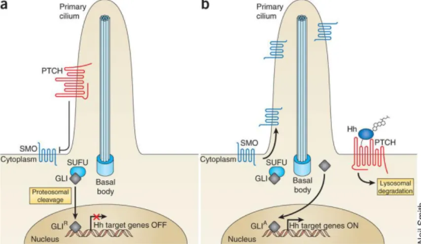
1.2.1. The Pathway
The precise mechanism of Hh signalling is not fully understood. Hh pathway activation begins when the ligand Hh is released from the cells through a transmembrane transporter Dispatched and binds the transmembrane receptor Patched1 (Ptch1). In basal conditions, Ptch1 is linked to the receptor Smoothened (Smo) and prevents location to the primary cilium (Figure 3a). In the presence of Hh, Ptch1 loses its inhibitory activity against Smo, allowing Smo activation. Localization of Smo in the primary cilium initiates a signalling cascade, which leadsto the activation of the glioma associated oncogene (Gli) family of zinc-finger transcription factors (Gli1, Gli2 and Gli3). Activated Glis, the final effectors of the pathway, translocate into the nucleus to induce the expression of various specific genes, such as those encoding the D-type cyclins, c-MYC (also called MYC), BCL2 and SNAIL (also called SNAI1), which regulate cellular differentiation, proliferation and survival.[21,22] Furthermore, Hh target genes include Gli1 itself, PTCH1
and the gene encoding for the Hh ligand.[23] The signalling of Gli is also
regulated by several protein mediators (PKA, GSK3-β, CK1-α), including suppressor of fused (SuFu). SuFu is a negative regulator of the Hh pathway because it is a sequester of Gli factors in the cytoplasm, thus repressing transcriptional activation (Figure 3b).[24,25]
Figure 3: Hh pathway activation. (a) Inactive Hh pathway. (b) Activated Hh pathway (Reproduced with permission).[26]
1.2.2. Alteration of the Hedgehog Pathway and
Cancer
In recent years, a multitude of studies have shown a correlation between the aberrant activation of the Hh pathway and cancer. In adults, Hh pathway mutation or dysregulation plays a crucial role in both proliferation and differentiation, leading to tumorigenesis or tumor growth acceleration. Basal cell carcinoma (BCC) and
medulloblastoma (MB) are two well-recognized cancers with mutations in components of the Hh pathway. Inappropriate activation of the Hh signaling pathway implies the development of several other types of cancer such as lung, prostate, breast, and pancreas cancer. Three basic models have been proposed for Hh pathway activity in cancer (Figure 4).[21,27]
1.2.2.1.
Type I: ligand-independentFirst the type I cancers were discovered. These feature harbouring Hh pathway-activating mutations which are independent of the Hh ligand, such as BCCs and MBs (Figure 4a). Most of these tumors either display inactivating mutations in Ptch1 (85%) or activating mutations in Smo (10%).[28] Furthermore, about one third of all medulloblastomas
and occasional rhabdomyosarcomas were shown to have inappropriate Hh pathway activation, often due to Ptch1 mutations or sometimes due to SuFu mutations.[29,30] Since these tumors are ligand independent, Hh
pathway inhibitors must act at or below the Smo level to be effective.
1.2.2.2. Type II: ligand-dependent autocrine mechanism
Autocrine activation of the Hh pathway in tumor cells through increased Hh ligand expression has been reported in a variety of tumors, such as lung, breast, stomach and prostate cancers (Figure 4b).[31] The relevance of this mechanism is not clearly understood. But
most of these tumors are dissimilar to BCC or MBs as they do not harbor any somatic mutations in the Hh signaling pathway. Instead, they demonstrate an autocrine, ligand-dependent, abnormal Hh
pathway activation. Most of these tumors have an elevated expression of the Hh ligand (Shh or Ihh) and/or ectopic Ptch1 and Gli expression within the epithelial compartment. This autocrine tumor growth can be effectively suppressed by various pathway inhibitors such as Hh neutralizing antibodies or Smo antagonists.
1.2.2.3. Type III: ligand-dependent paracrine mechanism
In contrast to the autocrine model, Bushman and colleagues were the first to propose that at least one model of prostate cancer signals in a paracrine manner to the stroma.[32] In paracrine signaling, Hh
produced by the tumor cells is received by the stroma, which signals back to the tumor to promote its growth or survival (Figure 4c). The precise mechanisms by which the Hh-stimulated stroma positively regulates tumor cell growth are not completely understood. However, it has been proposed that Hh regulates signaling mediators in the stroma, including insulin-like growth factor (IGF), Wnt, interleukin-6 (IL-6) and vascular endothelial growth factor (VEGF), which in turn promote tumor growth. Inhibition of this paracrine signaling in epithelial tumors may be of therapeutic value as specific inhibition of Hh signaling in the stroma did result in growth inhibition of tumor xenografts. Although the most effective way of treating these tumors relies possibly on a combination of a Hh pathway inhibitor targeting the stroma and other drugs targeting the tumor cells.
1.2.2.4. Type IIIb: reverse paracrine mechanism
Very recently, a “reverse paracrine” signalling model has also been introduced in which Hh is secreted from the stroma and is received by the tumor cells (Figure 4d). In this model, stromal Hh is thought to provide the appropriate microenvironment for potentiating tumor growth and would thus be a suitable therapeutic target as well.[31]
Figure 4: Different models of Hedgehog pathway activation in cancers. (a) Type I ligand-independent cancers. (b) Type II ligand-dependent autocrine cancers. (c) Type III ligand-dependent paracrine cancers. (d) Type IIIb reverse paracrine tumors (Reproduced with permission).[26]
1.3. Hedgehog Pathway and CSCs
Remarkably, Hh signaling is active in CSCs of various tumor types. It is responsible to sustainthe proliferation of these cells, which are responsible for tumor relapse and resistance to conventional anticancer therapies. Indeed, the Hh pathway controls the functional properties of CSCs, such as self-renewal, survival, metastatic spread, and neoangiogenesis by the regulation of stemness-determining genes such as Nanog, often overexpressed in cancer. Given the increasing evidences supporting the crucial role of the Hh pathway in cancer initiation, proliferation, metastasis, chemoresistance, and in the survival of CSCs, its components represent attractive druggable targets for anticancer therapy.[33,34]
1.3.1. Targeting the Hedgehog Pathway in
Cancer
Four major modes of Hh inhibition have been exploited therapeutically: 1) Smo inhibition; 2) receptor-ligand disruption; 3) inhibition of ligand processing; and 4) Gli inhibition (Figure 5).
Figure 5: Potential sites for blocking the Hh pathway with therapeutic agents (Reproduced with permission).[26]
In recent years, drug discovery efforts directed against the Hh pathway have focused predominantly on the development of Smo antagonists. As a consequencea remarkable number of small molecules of natural, semisynthetic or synthetic origin have been developed and reviewed extensively.[35] Despite the initial enthusiasm, clinical
development of Smo antagonists has ultimately proved disappointing, due to scarce pharmacokinetics, low selectivity on CSCs, severe side
effects, and the emergence of drug resistance.[35] Advanced tumors can
evolve resistance through pathway-dependent genetic mechanisms or through compensatory adaptation. Pathway-dependent genetic alterations discovered in resistant tumors from patients and animal models directly affect Hh pathway members. Vismodegib resistance is due to genetic alterations at the level of, or downstream from, Smo. Resistance can originate from Smo point mutations that ablate Smo– drug interaction while maintaining Hh pathway activation (Figure 6a).[36] These mutations occur in the ligand-binding pocket of Smo.
Other genetic alterations that lead to resistance come from gene duplications of Gli2 or Hh target gene cyclin D1 (Figure 6b) which bypass the requirement of Smo to inappropriately maintain or increase Gli target gene induction. These mutations promote high Hh pathway activation in the presence of Smo antagonists and mediate resistant tumor growth.
Compensatory alterations outside the Hh pathway have been found to mediate tumor resistance. In this case, Hh activation occurs in the absence of direct genetic mutation or copy number of variation of Hh members, and is epigenetic in nature. Developing Hh inhibitors that modulate targets acting downstream of Smo or independently from Smo, such as Gli, has recently emerged as a more promising therapeutic strategy for the treatment of Hh-dependent tumors. This approach could potentially overcomeanti-Smo resistance and adverse effects, which are responsible for more than 50% dropout’s rates in Smo antagonists clinical trials.
Figure 6: Genetic escape pathways evolving during Smo antagonist treatment and approaches to overcome resistance. (a) Smo point mutations. (b) Gli target gene amplification of Gli2. Compensatory escape pathways including (c) PI3K pathway up-regulation or (d) inappropriate activation of aPKC-/ (Reproduced with permission).[26]
1.3.1.1. Gli inhibitors
Gli transcription factors are the final effectors of the Hh pathway. Gli proteins have different functions in vivo: Gli1 acts as a transcriptional activator, whereas Gli2 and Gli3 can act both as activators and as repressors. It is important to consider that, whatever alteration leads to aberrant Hh pathway activation, all trigger the downstream effector Gli1. For this reason, in the last few years, Gli factors are emerging as the most attractive targets for the development of new anticancer drugs. However, not many Gli antagonists have been identified.
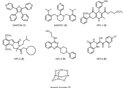
GANT58 (1) and GANT61 (2) are the first Gli antagonists that have been identified by Lauth et al in 2007 (Figure 7).[37] GANT61 (2) has
become the reference compound in many biological studies since it proved to inhibit the Hedgehog pathway in five different human colon carcinoma cell lines and in prostate cancer human xenografts. Recently, Chen and coworkers discovered a class of compounds named HPIs (Hedgehog Pathway Inhibitors) (3-6) by as Gli antagonist (Figure 7).[38]
The most promising compound, HPI-1 (3), was encapsulated in a polymeric nanoparticle. The formulation has undergone early preclinical testing showing a potent activity in HCC xenografts and inhibition of the proliferation and invasion of human HCC cell lines. Arsenic trioxide (ATO) (7), a drug approved by the FDA for the treatment of acute promyelocytic leukemia, was found to interfere directly with the Gli transcriptional factors, with efficacy both in vitro and in vivo (Figure 7).[39] Many other studies pointed out the efficacy of
trial study started to check the efficacy of ATO in treating patients with BCC.
Figure 7: Gli direct antagonists.
In conclusion, Gli transcriptional factors represent the most promising target for the development of new drugs targeting the Hh pathway in tumors.
1.4. Identification of the Active Site of Gli1
A previous study identified the active site of Gli1.[40] The starting
point of the investigation was the X-ray structure of cobalt ion-coordinated Gli1ZF in complex with DNA.[41] A representative Gli1ZF
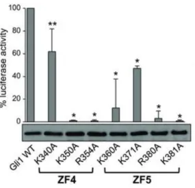
structure was extracted after molecular dynamics (MD) simulations (Figure 8A). Based on MD analysis and X-ray structure, a computational alanine scanning experiment was performed to evaluate which basic residues in ZF4 and ZF5 domains are relevant for the thermodynamic stability of the Gli1ZF/DNA adduct.[40] The delta
energy (G) of binding to DNA of Gli1 mutants (Figure 8B) and wild-type Gli1ZF (Gli1ZF-WT) (G) were calculated. Basic residues in ZF4 (K340, K350, R354) and ZF5 (K360, K371, R380 and K381) domains exhibit the strongest G contribution (Figure 8B), indicative of their role in the Gli1/DNA interaction.[40]
Figure 8: Structure-based analysis of Gli1/DNA complex. (A) Gli1ZF/DNA structure extrapolated from MD analysis. Gli1ZF (blue cartoon), residues involved in binding to DNA (magenta sticks), Zn ions (grey spheres) are shown. (B) Effect of Gli1ZF mutants on the binding affinity to DNA as predicted by in silico alanine scanning (Reproduced with permission).[40]
To correlate computational with experimental studies, a mutation experiment was performed on HEK293T cells. These cells have the peculiarity to transiently express ectopic Gli1 or different Gli1 mutants and a Gli-dependent luciferase reporter. The mutagenesis studies confirmed the role played by those residues in ZF4 and ZF5 domains in the transcriptional activity of Gli1 (Figure 9).[40] The representative
Gli1ZF structure extracted from MD trajectories served as a starting point for in silico ligand design.[40]
Figure 9: Luciferase assay on HEK293T cells. Effect of Gli1ZF mutants on Gli1-dependent transcriptional activity (Reproduced with permission).[40]
1.5. In Silico Screening
In Prof. Bruno Botta’s laboratory an in house library composed of more than 800 unique natural compounds belonging to different clusters is avalaible. Literature data[42] and results of the mutagenesis
studies were used as starting point for a docking of the library.[42] The
docking analysis was set so that at least one of the basic residues, highlighted by the previous studies, would be able to interact with small molecules.[40] The in silico study identified six molecules (three
vismiones, GlaB, the chalcone V94 and the opioid alkaloid narceine) (Figure 10) as potential Gli1 inhibitors.[40]
Figure 10: Structure of the six virtual hits.
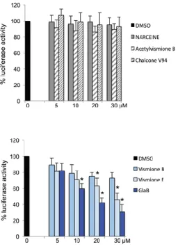
The biological activity of the selected hits as specific modulators of Gli1 was evaluated by a Gli-dependent luciferase reporter assay, using HEK293T cells (Figure 11).[40]
Figure 11: Gli1-induced transcription in HEK293T cells, treated with increasing concentrations of different compounds or DMSO as control (Reproduced with permission).[40]
The results of the biological tests showed that chalcone V94 (8), narceine (9) and acetylvismione B (10) were not active, yet vismione B (11) was partially active.[40] In contrast, vismione E (12) and
glabrescione B (GlaB) (13) strongly inhibited the luciferase activity (2) (Figure 11).[40] Since vismiones display chemical instability under
1.6. Aims and Scope
Aberrant Hh pathway activation is responsible for the tumorigenesis of several human cancers, including medulloblastoma (MB), rhabdomyosarcoma, melanoma, basal cell carcinoma (BCC), and breast, lung, liver, stomach, prostate, and pancreas tumors,[43–46]
through the aberrant regulation of the functional properties of CSCs, such as self-renewal, survival, metastatic spread, and neoangiogensis. Since much evidence supports the crucial role of the Hh pathway in cancer initiation, proliferation, metastasis, chemoresistance, and in the survival of CSCs,[45,47] its components represent attractive druggable
targets for anticancer therapy.
In this contest, targeting Gli transcription factors are extremely important to overcome the resistance to clinically available Smo antagonists. We previously identified GlaB (13) as the first Gli1 direct inhibitor. At the outset of this PhD thesis, no total synthesis of GlaB had been reported in literature, while the extremely limited supply of natural material had already been used for preliminary biological studies. In this context, it was a major goal of this PhD thesis to develop an efficient total synthesis of GlaB, in order to provide material for the assessment of its effects on human cancer cell growth. The chemistry developed in the course of this total synthesis was designed to also provide a basis for the synthesis of analogs to study theSAR and the underlying mechanism of action.
Total Synthesis, Biological
Evaluation, and SAR Studies of
GlaB
1
2.1. Chromones: A Privileged Structure in
Medicinal Chemistry
Chromones are benzoannulated γ-pyrone (4H-chromen-4-one, 4H-1-benzopyran-4-one) heterocycles (Figure 12). Since ancient times they have been used in traditional medicine[48] and are well-known for
their broad range of pharmacological properties, such as anti-allergic, anti-inflammatory, antidiabetic, antimicrobial, and antitumor.[49–51]
Extensive research on chromones and their derivatives have proven the pharmacological importance of their heterocyclic moiety. Also in medicial chemistry, chromones play an important role: their synthetic accessibility and structural diversity make them privileged structures for drug discovery.[52]
1This work of my PhD research was carried out in the laboratories of Prof. Bruno
The chromone ring system is the core fragment of several flavonoids, such as flavones and isoflavones (Figure 12).[49] In
particular, isoflavones are a class of natural compounds mainly occurring in plants of the Leguminosae family. The isoflavone nucleus consists of two phenyl rings linked by a propane bridge to form an oxygenated heterocyclic ring, resulting in the typical 15 carbon atoms (C6-C3-C6) skeleton with three rings, labelled A, B and C (Figure 12). Due to their interesting biological activity and benefits to human health, isoflavones have recently received attention.[53,54] Furthermore,
as underlined by genistein and its derivatives in differentiated pancreatic cancer cells and CSCs, isoflavones have displayed a noticeable pharmacophoric preference for Hh targets.[35]
Figure 12: Chromones, flavones and isoflavones skeleton.
Most of the isoflavones have been isolated from natural sources, but extraction methods provide only very limited amounts of pure samples. However, their simple structure features can be addressed conveniently by synthetic methods.
2.2. Synthetic Strategies for Isoflavones
Several synthetic procedures have been developed for the preparation of isoflavones.[55] Nevertheless, many of these new
synthetic approaches have not been demonstrated in the synthesis of polyhydroxylated isoflavones and isoflavones bearing other naturally-occurring substitution patterns. The most popular procedures are mainly based on two strategies, namely (i) the deoxybenzoin route (Scheme 1)[56] and (ii) the chalcone route (Scheme 2).[57]
Scheme 1: Deoxybenzoin route.
Scheme 2: Chalcone route.
Two other methods towards the synthesis of isoflavones include first the palladium catalyzed cross coupling reaction of a 3-alochromone (17) with an arylboronic acid/ester (18) (Scheme 3)[58,59],
and second the condensation of enamine (20) with salicylaldheyde (19).[60]
Scheme 3: Suzuki cross coupling route.
Scheme 4: Enamine-salicylaldheyde condensation.
Most of the available methods have major drawbacks, including the use of special and expensive reagents in large excess, long reaction times, vigorous conditions and very low yields. Hence, these approaches are overall unfeasible for the synthesis of GlaB, in particular for in vitro and in vivo studies requiring larger quantities. This prompted us to develop a mild and cost effective method, which also allows for scale up to gram quantities.
2.3. First Total Synthesis of GlaB
2.3.1. Retrosynthesis
The strategy to be followed for the synthesis of GlaB (13) entails a late stage functionalization (Scheme 5). The isoflavone 21, as the crucial precursor for the prenylation reaction, will be obtained by Suzuki coupling with protected boronic acid (23) and 3-iodochromone (22), respectively, followed by protecting group manipulation. 22 can be prepared from commercially available 2’,4’,6’-trimethoxyacetophenone (24) in a 3-step sequence (Scheme 5).
2.3.2. Synthesis
Our synthetic endeavours started from commercially available 2’,4’,6’-trimethoxyacetophenone (24) (Scheme 6).[61] Treatment of 24
with BBr3 led the selective ortho-O-demethylation to give
2’-hydroxy-4’,6’-dimethoxyacetophenone (25). 25 was first treated with N,N-dimethylformamide dimethyl acetal (DMF-DMA), which gave the enamino ketone 26. Stirring the latter with I2 excess has resulted in
tandem cyclization and iodination, to afford 3-iodo-chromone (22). Pd catalyzed Suzuki cross-coupling of 22 with 3,4-(methylenedioxy)-phenylboronic acid (23) using polyurea-encapsulated palladium (Pd EnCatTM 40)[62] gave the isoflavone 27. Methylendioxy group was then
removed in acidic conditions after the formation of the acetoxy acetal by treatment with Pb(OAc)4 allowing the formation of the desired
catechol 21. The prenylation reaction of the phenolic hydroxyl grops of 21 completed the synthesis of glabrescione B (13), with an overall yield of 7% (Scheme 6).[61] In total, the linear sequence from 24 to 13 comprises
Scheme 6: Forward synthesis of GlaB (13). i) BBr3, 98%; ii) DMF-DMA, quantitative crude; iii) I2, 33%; iv) 23, PdEnCatTM, Na2CO3, 57%; v) Pb(OAc)4; vi) AcOH, 42% over two
2.4. NMR Analysis of the Interaction GlaB/Gli1
2Virtual screening predicted GlaB (13) binding to Gli1 within ZF4 and ZF5 as well as interactions within K340 and K350 (Figure 13).
Figure 13: The predicted binding mode of GlaB (13) (blue sticks) to Gli1ZF (green cartoons). Residues K340 and K350 of the GlaB binding site are colored magenta.
NMR relaxation parameters, and in particular selective spin-lattice relaxation rates (R1SE), are highly sensitive indicators for binding
2 These experiments were carried out in collaboration with Prof. Gloria
Uccello-Barretta, University of Pisa, Italy. For detailed data about GlaB NMR conditions check the following article.[40]
processes between macromolecules and small molecules.[63,64] The
method relies on the change in molecular motion of the small molecule, i.e. its correlation time τc, upon interacting with a macromolecule. In its
free state the small molecule performs fast molecular motions (ω02 τc2
<<1, Figure 14) and enter the slow motional regime (ω02 τc2 >>1, Figure
14) only when bound to a macromolecule. R1SE increases strongly upon
passing ω02 τc2 = 1 as the slow motional regime is reached (Figure 14).
R1SEbased NMR relaxation studies rely on the favorable dependence of
R1SE on τc, while non-selective relaxation rates (R1NS) scale differently in
this motional regime (Figure 14).
Figure 14: Dependence of the specific and non-specific spin-lattice relaxation rates R1SE and R1NS, respectively, on ω0τc.
To demonstrate the predicted binding of GlaB (13) to Gli1 by means of NMR, a relaxation study was performed. For this mono-selective relaxation rates were determined for a chosen subgroup of GlaB (13) protons (Figure 15) by selectively inverting the corresponding proton signals.
Figure 15: Chosen subgroup of GlaB (13) protons for NMR based study of GlaB/Gli1 interaction, namely H-1 and H-3 and proton groups at 2-OCH3 and 4-OCH3 of methoxy
groups of ring A, H-11, H-12 and H-15 of ring B, and H-8 of ring C.
Mono-selective relaxation rates were obtained both for free GlaB (13) (Rf) and GlaB/Gli1 mixtures (Rms). The former allows normalization
of Rms, yielding ΔR/ Rf with ΔR = Rms-Rf (Table 1). In addition to the
GlaB/Gli mixtures, a GlaB/GST sample was included into the relaxation study and ensured that no specific contribution arises from a GlaB/GST interaction (Table 1).
Table 1: 1H-NMR mono-selective relaxation rate (Rf, s-1) of 0.412 mM free GlaB (13)
in DMSO-d6 measured at 25°C and 600 MHz for specified protons. Associated normalized
relaxation rates (ΔR/ Rf with ΔR=Rms-Rf) are listed for four different GlaB/protein
mixtures (Reproduced with permission).[40]
a According to the proton number scheme of GlaB (Figure 15). b Rms could not be determined due to overlapping water signal. c Rms could not be determined due to the large linewidth.
In order to demonstrate the predicted interaction of GlaB (13) to Gli1 within K340 and K350 (Figure 13), a mutagenesis NMR study was carried out. To this end, Rms values of GlaB in combination with the
single mutant K340A and the double mutant GST-Gli1ZF-K340A/K350A were determined. An overall smaller increase of the Rms
values upon addition of GST-Gli1ZF-K340A was observed, though preserving the stronger affinity towards ring B over ring A and C (Table 1; Figure 16). In case of the double mutant, change in Rms values for the
protein mixture is comparable to the GST sample (Table 1; Figure 16). These results suggest a comparable binding conformation of GlaB (13) upon interacting with GST-Gli1ZF-WT and the single mutant. Instead, double mutation to GST-Gli1ZF-K340A/K350A prohibits the specific binding via the GlaB ring B.
Figure 16: GlaB (13) is showed as green sticks, protons highlighted by the NMR study as magenta spheres and the radius is proportional to the normalize Rms value
(Reproduced with permission).[40]
In summary, results of NMR studies show that GlaB (13) interacts directly with Gli1 and further emphasize the role of K340 and K350. The ring B turns out to be the key determinants for GlaB (13) activity.
2.5. Biological Evaluation of GlaB
32.5.1. Influence of GlaB on Gli1/DNA Binding
and Transcriptional Activity
Biological tests validated the potency of GlaB (13) against Gli1. First, it was shown that GlaB reduces the luciferase activity arising from Gli-responsive element reporter in Smo-/- mouse embryonic fibroblasts
(MEFs) (Figure 17).[40] Second, 13 affects the transcriptional activity
specifically of a number of endogenous Gli target genes, while non-related genes were not influenced (Figure 18).[40]
Figure 17: Inhibition of Gli1-induced transcription in transfected Smo-/- MEF cells
(Reproduced with permission).[40]
3 These experiments were carried out in collaboration with Prof. Lucia Di
Marcotullio, Sapienza University of Rome, Italy. For detailed data about GlaB activity check the following article.[40]
Specifically, related experiments proofed that 13 suppresses Hh-gene expression in Ptch1-/- MEFs (Figure 18).[40] In the latter cells, the
Ptch1 receptor on Smo (Figure 18) is deleted along with its prohibiting effect, increasingly activating Gli transcription factors (Figure 18).[65] In
addition, the downstream activity of GlaB was demonstrated on SuFu -/- MEFs cells, which also feature high Gli1 reporter activity due to loss
of SuFu inhibition (Figure 18).[66] As for Ptch1-/- MEFs cells, 13 also
decreases the Hh target gene expression in SuFu-/- MEFs cells (Figure
18).[40]
Figure 18: Hh target genes expression levels in Ptch1-/- MEFs treated for 48h with
GlaB (13) or DMSO only (left). Representative model of Hh signaling hyperactivation: in Ptch1-/- MEFs and in SuFu-/- MEFs cells (center). SuFu-/- MEFs were treated for 48h with
GlaB (13) or DMSO as control (right) (Reproduced with permission).[40]
2.5.2. Effect of GlaB on Gli-dependent MB
Cells and Tumor SCs: Ex vivo Experiment
The potency of GlaB to prevent tumor growth was tested on primary MB cells, isolated from Ptch1+/- mice tumors. These MB cells
were treated with either GlaB (13), GANT61 (2) or DMSO and resulted in comparable inhibition of MB cell growth in case of GANT61 (2) and