Open Access
Research article
A 10 year study of the cause of death in children under 15 years in
Manhiça, Mozambique
Jahit Sacarlal*
1,2,3, Ariel Q Nhacolo
1, Betuel Sigaúque
1,5,
Delino A Nhalungo
1, Fatima Abacassamo
2, Charfudin N Sacoor
1,
Pedro Aide
1,5, Sonia Machevo
1,2, Tacilta Nhampossa
1,5, Eusébio V Macete
1,4,
Quique Bassat
1,3, Catarina David
5, Azucena Bardají
1,3, Emili Letang
1,3,
Francisco Saúte
4, John J Aponte
1,3, Ricardo Thompson
5and Pedro L Alonso
1,3Address: 1Centro de Investigação em Saúde da Manhiça (CISM), Mozambique, 2Faculdade de Medicina, Universidade Eduardo Mondlane,
Maputo, Mozambique, 3Barcelona Centre for International Health Research (CRESIB), Hospital Clínic/IDIBAPS, Universitat de Barcelona,
Barcelona, Spain, 4National Directorate of Health, Ministry of Health, Maputo, Mozambique and 5National Institute of Health, Ministry of Health,
Maputo, Mozambique
Email: Jahit Sacarlal* - [email protected]; Ariel Q Nhacolo - [email protected]; Betuel Sigaúque - [email protected]; Delino A Nhalungo - [email protected];
Fatima Abacassamo - [email protected]; Charfudin N Sacoor - [email protected]; Pedro Aide - [email protected]; Sonia Machevo - [email protected]; Tacilta Nhampossa - [email protected];
Eusébio V Macete - [email protected]; Quique Bassat - [email protected]; Catarina David - [email protected]; Azucena Bardají - [email protected]; Emili Letang - [email protected]; Francisco Saúte - [email protected];
John J Aponte - [email protected]; Ricardo Thompson - [email protected]; Pedro L Alonso - [email protected] * Corresponding author
Abstract
Background: Approximately 46 million of the estimated 60 million deaths that occur in the world
each year take place in developing countries. Further, this mortality is highest in Sub-Saharan Africa, although causes of mortality in this region are not well documented. The objective of this study is to describe the most frequent causes of mortality in children under 15 years of age in the demographic surveillance area of the Manhiça Health Research Centre, between 1997 and 2006, using the verbal autopsy tool.
Methods: Verbal autopsy interviews for causes of death in children began in 1997. Each
questionnaire was reviewed independently by three physicians with experience in tropical paediatrics, who assigned the cause of death according to the International Classification of Diseases (ICD-10). Each medical doctor attributed a minimum of one and a maximum of 2 causes. A final diagnosis is reached when at least two physicians agreed on the cause of death.
Results: From January 1997 to December 2006, 568499 person-year at risk (pyrs) and 10037
deaths were recorded in the Manhiça DSS. 3730 deaths with 246658 pyrs were recorded for children under 15 years of age. Verbal autopsy interviews were conducted on 3002 (80.4%) of these deaths. 73.6% of deaths were attributed to communicable diseases, non-communicable diseases accounted for 9.5% of the defined causes of death, and injuries for 3.9% of causes of deaths. Malaria
Published: 24 February 2009
BMC Public Health 2009, 9:67 doi:10.1186/1471-2458-9-67
Received: 26 March 2008 Accepted: 24 February 2009
This article is available from: http://www.biomedcentral.com/1471-2458/9/67 © 2009 Sacarlal et al; licensee BioMed Central Ltd.
This is an Open Access article distributed under the terms of the Creative Commons Attribution License (http://creativecommons.org/licenses/by/2.0), which permits unrestricted use, distribution, and reproduction in any medium, provided the original work is properly cited.
was the single largest cause, accounting for 21.8% of cases. Pneumonia with 9.8% was the second leading cause of death, followed by HIV/AIDS (8.3%) and diarrhoeal diseases with 8%.
Conclusion: The results of this study stand out the big challenges that lie ahead in the fight against
infectious diseases in the study area. The pattern of childhood mortality in Manhiça area is typical of developing countries where malaria, pneumonia and HIV/AIDS are important causes of death.
Background
One of the eight Millennium Development Goals (MDG), aims to reduce under five mortality rates by two-thirds, especially in African countries. To accomplish this goal, researchers, programme planners and policy-makers need information on the causes of death occurring in countries [1]. Approximately 46 million of the estimated 60 million deaths that occur in the world each year take place in developing countries [2]. Further, this mortality is highest in Sub-Saharan Africa, although causes of mortality in this region are not well documented [3]. In order to most appropriately allocate resources and to evaluate the impact of individual illnesses and its control, it is impor-tant to know the number and proportion of deaths in a community likely to be due to a given condition. In rural areas of developing African countries such as Mozam-bique, where many children are born and die without ever being registered, and where a significant proportion of all deaths take place outside health facilities, the only way of estimating the likely cause of death is through an inter-view of a witness of final illness. This is called a verbal autopsy (VA) [3-6].
Methods
Study areaManhiça district is located in southern Mozambique, in the Maputo Province, about 80 km north of Maputo City. The area has two distinct regions. The first is the fertile lowlands, comprising the Incomati River flood plain run-ning from the northern to the southern district boundary. This area is poorly inhabited and used mainly for sugar-cane and fruit plantations. The second area is an escarp-ment of moderate altitude bordering the west of the river, where the population inhabits an extensive plateau. There are two distinct seasons, a warm and rainy season between November and April and dry and cool season during rest of the year. A full description of the geographical and soci-odemographic characteristics of the study area has been presented elsewhere [7,8].
The Manhiça Demographic Surveillance System (DSS) in Manhiça District was established in 1996, and currently covers a 500 square kilometre area. An initial census was carried out in 1996, and vital events registration (births, deaths, pregnancy and, in/out-migration) were conducted on quarterly basis until the year 2000, when this was changed to twice yearly.
Verbal Autopsies (VA) data collection started in 1997 with the aim of generating cause-specific mortality data in the study area. Initially, VA were conducted only in January and July on deaths of children aged less than 15 years reported through the DSS in the previous 6 months. Since the introduction of new questionnaires in June 2002 through the MTIMBA (Malaria Transmission Intensity and Mortality Burden Across Africa) project from INDEPTH, VA interviews are carried out every day by a well-trained lay supervisor and field workers.
Description of the health delivery system
There are two referral health facilities in Manhiça district, the Manhiça District Hospital (MDH), with 110 beds, and the Xinavane Rural Hospital (XRH), with 59 beds. In addi-tion, 10 peripheral health facilities complete the official health facilities network. Most of the government medical services are provided free of charge except for drugs pre-scribed at the outpatient department that is available for purchase at subsidized prices. Adults pay a symbolic con-sultation fee of about USD 0.02.
Since 1996 the Manhiça Health Research Center (CISM) has been operating a round-the-clock, hospital-based morbidity surveillance system for children under 15 years of age attending the MDH and three other peripheral health facilities in the study area [8].
Voluntary counselling and testing to prevent mother to child transmission with Niverapina since 2003, and Highly Active Anti Retroviral Therapy (HAART) are avail-able since 2004 for all patients including pregnant women in MDH, according to national policies.
Obstetric services including obstetric emergency care, operation room and morbidity surveillance system were established at the MDH maternity clinic, as a passive case detection system, for all women (pregnant, puerperal and women with gynaecological complaints) attending this clinic with clinical complaints (i.e., not for those attend-ing the routine antenatal clinic).
Selected indicators for the DSS
Between 1997 and 2005, the number of inhabitants living in the study area increased from 32856 to 79783, due to population growth and the extension of the DSS area in August 2002. During these years, the total fertility rate
decreased from 5.2 to 4.8 children per woman. The infant mortality risk in 2005 was 77.5 per 1000 live births, the under five mortality (5q0) rate was 138.6 deaths per 1000 pyrs, and the life expectancy at birth was 40.2 years [9].
Data collection & processing
The methodology used in identifying vital events in the study area has been fully described elsewhere [7]. Infor-mation on deaths comes from one of several sources: (I) household visits twice a year that are conducted to record all deaths and other demographic events that have occurred since the previous visit, (II) daily visits to hospi-tal wards and maternity clinics by supervisors to gather information on all deaths and pregnancy related events that have taken place in the previous 24 hours and (III) weekly reports by local key informants on births, deaths and migrations that might be missed during household census visits by field workers and supervisors. Age is ascer-tained by direct questioning, referral to any existing per-sonal identification documents and, if necessary, an area-specific calendar of events is used. An identification card is issued to all children under 15 years of age to allow identification of patients in the morbidity surveillance system in the MDH.
Initially, eight medical students conducted VA interviews in the study area twice a year. The work was supplemented after June 2002 by a lay supervisor and field workers who interviewed key community informants and relatives of the deceased, daily. Between three and six months after a death, a field worker visited the family of the deceased to inquire whether they would accept to participate in a ver-bal autopsy. Upon acceptance, an oral consent was obtained from the interviewee and a date for the interview was agreed. On the day of the interview, a signed or finger-printed informed consent (IC) was sought before the VA took place. To ensure consent within the family, potential interviewers were given an information sheet with study objectives and procedures during the initial contact, and were encouraged to discuss with family members before proceeding. Interviewers who could not read were free to ask their relatives to read the document for them. The pri-mary informant was, whenever possible, the person who directly took care of the deceased child during the illness or condition that led to death. If the primary respondent was absent, information was sought from any other adult, including neighbours, who might have relevant informa-tion on the possible cause of death. In order to maintain confidentiality, only the coding physicians and the data entry clerks had access to the assigned causes of death.
A demographer was in charge of controlling the data qual-ity through an on-site review of questionnaires. After fieldwork, all questionnaires were checked for consistency and completeness. Questionnaires needing corrections
were returned to the field within two weeks of their receipt.
Nature of the VA tool
The study used a VA questionnaire standardized from INDEPTH [7] and adapted from the WHO model [10]. The standard questionnaire in Manhiça was written in Portuguese. However, the fieldworkers perform an on-site translation of the questions into the local language (Xan-gana). The questionnaire included questions on the iden-tification of the deceased and the respondent as well as the health seeking behaviour and use of health services by the deceased prior to the death. The questionnaire also had an open-ended section where circumstances sur-rounding the death of the child, as well as the signs and symptoms presented during the illness preceding death, are recorded. The final section had closed questions on signs and symptoms preceding the death that did not focus on any particular disease.
Assigning the cause of death
To assign the cause(s) of death, diagnoses are given using a standardised coding system. Three physicians with expe-rience in tropical diseases independently assigned the cause of death using the International Classification of Diseases (ICD-10) [11]. Each physician ascribes a mini-mum of one and a maximini-mum of 2 causes. Conditions should be additive and not alternative. For example, if more than one diagnosis was mentioned, it may be classi-fied as "malaria or pneumonia," but should be stated as "malaria and pneumonia". A final diagnosis was reached when at least two physicians agree on the cause of death. When at least two physicians assigned "unknown" as the cause of death, the final cause of death was considered undetermined. When the cause was different among the three reviewers, the final diagnosis was "not consensus", and these deaths were not redistributed to other diagnosis groups. When two final diagnoses were assigned for the same death, each of these was individually mapped onto ICD-10 for a calculated cause-specific rate.
To rank causes of death, we used the GBD tree structures [12]. The first level included three mortality groups: Group 1 consisted of deaths attributed to communicable diseases and to maternal, perinatal and nutritional condi-tions; Group 2 comprised deaths attributed to non-com-municable diseases and, Group 3 comprised deaths due to injuries. Each of the three groups was further divided into several major subcategories (second to fourth level). Third and fourth levels were used to classify specific causes of death.
Data Management & Analysis
Trained data entry clerks and a data manager ensured data entry into a network of computers under a Windows NT
environment. Double data entry was performed by two clerks using a modified version of The Household Regis-tration System (HRS) [13]. Inconsistencies, if any, were corrected after counter-checking with the original ques-tionnaires. Questionnaires with errors that could not be reconciled were returned to the field for correction. The database with the VA data was linked to other DSS data-bases. Data management, cleaning and statistical analysis were performed using STATA (Stata Corporation 2005, Stata Statistical Software: Release 9.2 College Station, TX: StataCorp LP, USA).
Calculation of mortality rates
Time at risk of disease was calculated for each individual registered in the demographic surveillance system, sub-tracting periods of absence due to migration. All-cause mortality rates were calculated by dividing the number of deaths in an age group by the time at risk, and expressed as deaths per 1000 person-years at risk. We calculated cause-specific death rates for each age group by multiply-ing the all-cause mortality rate by the proportion of deaths assigned to each cause.
Ethical considerations
The study falls within the national ethical clearance granted to the malaria epidemiological studies of the CISM (Ministry of Health/National Institute of Health of Mozambique, 1996). The participation of the respond-ents during the interview was voluntary and conducted only after the IC procedure described earlier. The inter-views were conducted at least one month after death, when the traditional grieving period was over.
Results
Population size and characteristics
From January 1997 to December 2006, 568499 person-year at risk (pyrs) and 10037 deaths were recorded in the Manhiça DSS. 3730 deaths with 246658 pyrs were recorded in children under 15 years old. Verbal autopsy interviews were conducted for 3002 (80.4%) of these deaths. Non-completion was due to family out-migration (9.8%), prolonged absence of the relatives of the deceased
(3.9%) or refusals (3.2%). Forty seven percent of the inter-views were conducted within a period of 6 months and 83.9% within 1 year from the time of death. The median time was 8 months. According to respondents, 54% of deaths occurred outside a health facility. However, medi-cal and other assisted care during the terminal illness was sought by 81.9% of those who died. Sources of care included: health centers and hospitals (67.8%), tradi-tional healers (8.3%), religious leaders (1.8%), friends and family (1.1%) and others (20.9%).
Age and sex distribution of deaths
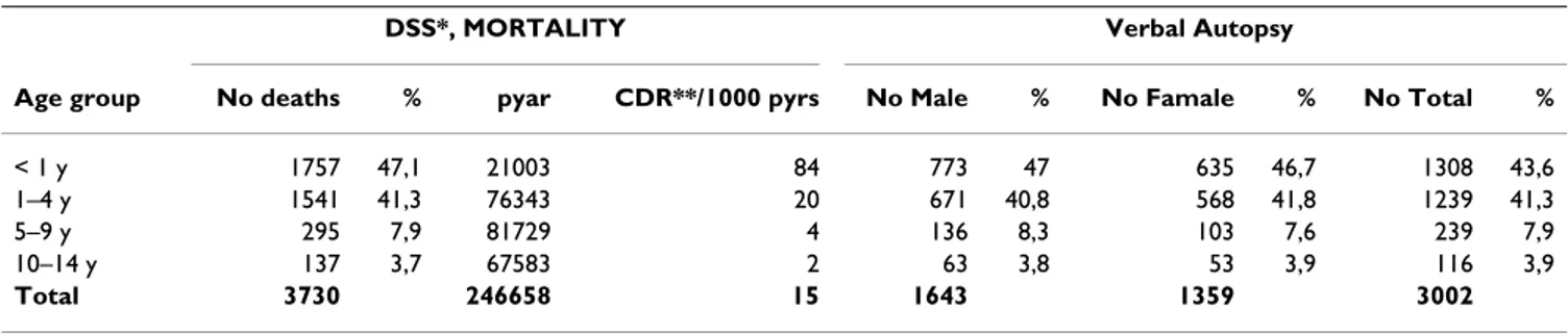
Most of the paediatric deaths occurred in children aged 1– 4 years (41.3%), followed by infants aged 29 days – 1 year (30.6%). Overall, males constituted 54.7% of the total children analyzed (Table 1).
Main causes and mortality rate of registered death
Table 2 summarizes the distribution of 3696 causes by group for the 3002 deaths. Communicable diseases were responsible for most deaths (73.6%), non-communicable diseases accounted for 9.5%, and injuries for 3.9%. Among communicable disease, the most frequent diagno-sis was infectious and parasitic diseases (60.0%), followed by perinatal disease (17.4%). Anaemia was a very com-mon diagnosis with 54.8% of total causes of death acom-mong non-communicable diseases. Injury, poisoning drowning and certain other consequences of external causes were also common in group 3.
The more frequent double death causes are malaria and anaemia with 13,5% (94/694) of cases followed by HIV/ AIDS and anaemia with 12,1% (84/694) of cases, and malaria and diarrhoea disease with 9,8% (68/694) of cases.
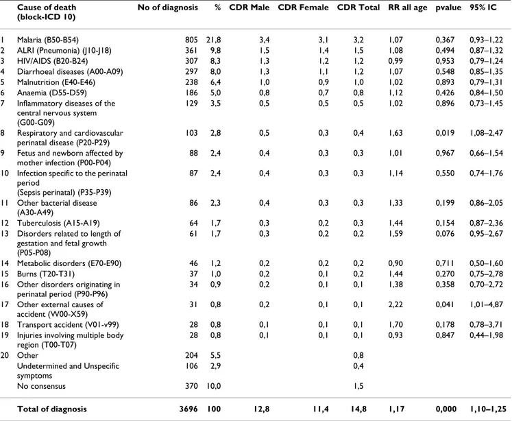
Table 3 shows the twenty leading causes of death and spe-cific crude death rate for different causes as reported from VA for children less than 15 years of age. Malaria was the leading cause, accounted for 21.8% of total diagnosis given physicians and for 3.2 deaths/1000 pyrs. Acute lower respiratory infection (ALRI) including pneumonia,
Table 1: Distribution of deaths and verbal autopsy by age and sex in Manhiça DSS, 1997–2006
DSS*, MORTALITY Verbal Autopsy
Age group No deaths % pyar CDR**/1000 pyrs No Male % No Famale % No Total %
< 1 y 1757 47,1 21003 84 773 47 635 46,7 1308 43,6
1–4 y 1541 41,3 76343 20 671 40,8 568 41,8 1239 41,3
5–9 y 295 7,9 81729 4 136 8,3 103 7,6 239 7,9
10–14 y 137 3,7 67583 2 63 3,8 53 3,9 116 3,9
Total 3730 246658 15 1643 1359 3002
*_DSS – Demographic Surveillance System **_CDR-Crude Death Rate
was the second leading cause of death with 9.8% of total diagnosis and 1.5 deaths/1000 pyrs, followed by HIV/ AIDS with 8.3% and 1.3 deaths/1000 pyrs. Diarrheal dis-eases with 8.0% and 1.2 deaths/1000 pyrs and malnutri-tion with 6.4% and 0.96 deaths/1000 pyrs were other important conditions.
Respiratory and cardiovascular disorders specific to the perinatal period accounted for 54.4 deaths/1000 pyrs and was the leading neonatal cause in children ≤ 28 days (data not shown). Foetus and newborn diseases affected by maternal related factors (complications of pregnancy, labour and delivery) were responsible for 51.4 deaths/ 1000 pyrs. Perinatal sepsis was the third principal cause of neonatal deaths with 47.1 deaths/1000 pyrs.
The mortality rate ratio for males compared to females, after controlling for age, during the study period was 1.17, (95% CI 1.10 – 1.25%; p < 0.001). This rate ratio was, in general, higher for males than for females in all diagnoses except HIV/AIDS, metabolic diseases and superficial trau-matic injuries (table 3). Deaths from external causes of
accident (poisoning, falls, animal bitten, drowning and suffocation) were frequent in children male compared to female with relative risk RR = 2.2 (95% CI 1.01–4.87; p = 0.041). The same observation occurs in perinatal deaths with respiratory and cardiovascular disease RR = 1.6 (95% CI 1.08–2.47; p = 0.019)
Table 4 presents mortality rates over the entire study period in children aged more than 29 days to 15 years of age. In the age group between 29 days to 1 year, malaria (12.5 deaths/1000 pyrs), ARLI (10.6 deaths/1000 pyrs) and HIV/AIDS (6.5 deaths/1000 pyrs) accounted for 37% of infant mortality.
Malaria was very common in children 1 to 4 years of age, with 34.4% of the total diagnoses and a mortality rate of 6.1 deaths/1000 pyrs. Malaria was followed by diarrhoea disease, HIV/AIDS and malnutrition with 2.3, 2.1 and 2.1 deaths/1000 pyrs, respectively.
Figure 1 presents the cause-specific death rates during the entire study. Overall mortality increased until 2001 in the
Table 2: Distribution of registered deaths by group and level-two cause in Manhiça DSS, Mozambique, 1997–2006.
Cause of death No of diagnosis %
I. Communicable Disease
Infectious and parasitic disease 1604 58,9
Perinatal disorders 473 17,4
Respiratory infections 361 13,3
Nutritional disorders 240 8,8
Others 43 1,6
Total 2721 73,6
II. Non-communicable Disease
Blood disease 193 54,8 Metabolic disorders 49 13,9 Congenital abnormalities 34 9,7 Neuropsychiatric disorders 28 8,0 Digestive disorders 12 3,4 Genitourinary disorders 11 3,1 Respiratory disorders 10 2,8 Cardiovascular disorders 9 2,6 Malignant disorders 3 0,9 Skin disorders 3 0,9 Total 352 9,5 III. Injuries
Injury, poisoning and certain other consequences of external causes 86 58,5
External causes of morbidity and mortality 61 41,5
Total 147 4,0
IV. Undetermined and badly defined symptoms 106 2,9
V. No consensus 370 10,0
study area and then decreased during the second period between 2001 and 2006. Malaria and pneumonia largely predominated among causes of death, accounting for 30% of the mortality during the study. The first period (1997 to 2001), was marked by an increase in death rates attributed to malaria, pneumonia, diarrhoeal disease and HIV/AIDS, but not malnutrition. Deaths attributed to malaria almost triplicate between 1997 and 2001, increas-ing from 2.2 deaths/1000 pyrs to 7.7 deaths/1000 pyrs, respectively, and then declined substantially between 2001 and 2006 to 2.8 deaths/1000 pyrs. During the sec-ond period all main mortality causes, declined in the study area.
Discussion
This study sought to identify the causes of death in our study area based on the verbal autopsy technique.
Varia-bility in recall period, a frequently cited limitation of VA is reported in our study. The median time was 8 months, about double that reported in other studies [14]. This is due to the data collection period used in our study that was only twice a year during the first six years of the study. However despite this limitation, interviews well recorded the signs and symptoms presented during the illness period preceding death. Other possible limitations included the relatively low specificity and sensitivity of the VA tool for detecting major causes of childhood death and the need to validate the diagnosis. In validation stud-ies conducted in Maputo in children under 5 years old, verbal autopsy was judged to be appropriate to detect measles (sensitivities 75%, specificity 98,6%), ALRI (sen-sitivities, 66,7%, specificity 85,4%) and malaria (sensitiv-ities 62,8%, specificity 90,3%) but it performed poorly for meningitis (sensitivities 33,3%, specificity 98,6%) and
Table 3: The 20 leading causes of verbal autopsy deaths and mortality rate by sex and relative risk in Manhiça DSS, Mozambique, 1997–2006
Cause of death (block-ICD 10)
No of diagnosis % CDR Male CDR Female CDR Total RR all age pvalue 95% IC
1 Malaria (B50-B54) 805 21,8 3,4 3,1 3,2 1,07 0,367 0,93–1,22
2 ALRI (Pneumonia) (J10-J18) 361 9,8 1,5 1,4 1,5 1,08 0,494 0,87–1,32
3 HIV/AIDS (B20-B24) 307 8,3 1,3 1,2 1,2 0,99 0,953 0,79–1,24
4 Diarrhoeal diseases (A00-A09) 297 8,0 1,3 1,1 1,2 1,07 0,548 0,85–1,35
5 Malnutrition (E40-E46) 238 6,4 1,0 0,9 1,0 1,02 0,893 0,79–1,31
6 Anaemia (D55-D59) 186 5,0 0,8 0,7 0,8 1,12 0,426 0,84–1,50
7 Inflammatory diseases of the central nervous system (G00-G09)
129 3,5 0,5 0,5 0,5 1,02 0,896 0,73–1,45
8 Respiratory and cardiovascular perinatal disease (P20-P29)
103 2,8 0,5 0,3 0,4 1,63 0,019 1,08–2,47
9 Fetus and newborn affected by mother infection (P00-P04)
88 2,4 0,4 0,3 0,3 1,01 0,967 0,66–1,54
10 Infection specific to the perinatal period
(Sepsis perinatal) (P35-P39)
87 2,4 0,4 0,3 0,3 1,14 0,550 0,74–1,76
11 Other bacterial disease (A30-A49)
86 2,3 0,4 0,3 0,3 1,33 0,199 0,86–2,05
12 Tuberculosis (A15-A19) 64 1,7 0,3 0,2 0,3 1,44 0,154 0,87–2,36
13 Disorders related to length of gestation and fetal growth (P05-P08)
61 1,7 0,3 0,2 0,2 1,59 0,076 0,95–2,67
14 Metabolic disorders (E70-E90) 46 1,2 0,2 0,2 0,2 0,90 0,711 0,50–1,60
15 Burns (T20-T31) 37 1,0 0,2 0,1 0,2 1,44 0,270 0,75–2,78
16 Other disorders originating in perinatal period (P90-P96)
34 0,9 0,2 0,1 0,1 1,38 0,358 0,70–2,72
17 Other external causes of accident (W00-X59)
31 0,8 0,2 0,1 0,1 2,22 0,041 1,01–4,87
18 Transport accident (V01-v99) 28 0,8 0,1 0,1 0,1 1,70 0,178 0,78–3,71
19 Injuries involving multiple body region (T00-T07)
28 0,8 0,1 0,1 0,1 0,93 0,847 0,44–1,98
20 Other 204 5,5 0,8
Undetermined and Unspecific symptoms
106 2,9 0,4
No consensus 370 10,0 1,5
anaemia (sensitivities 51,9%, specificity 84,9%)[15]. These results are comparable with the results of other val-idation studies made in Africa, including reported by Snow et al [3], Chandramohan et al [5], Kahn et al [16] and Philip WS et al [17].
Whether to use a single or a multiple diagnostic cause is arguable particularly in the regions where patients present more than one pathology [4], as it was observed in the current study. A single diagnosis of cause of death can be used, with the assumption that other causes are ignored in the analysis. An alternative type of analysis when more than one cause of death is considered has been described by Adjuik et al [18]. He has allocated percentages of a death to the codes assigned, in proportion to the number of coders who offered a diagnosis. This methodology may underestimate individual rates per cause. For us, many times it is difficult to assign a single cause of death because the process that leads to death is complex and several dis-eases are involved. We try to solve this question giving the same weight for each cause of death. However it is possi-ble that our methodology may overestimate the individ-ual rates per cause.
In our study about 54% of all deaths took place outside a health facility. This percentage is slightly lower than that found in another study carried out in Manhiça district between 1994–96 (59,9%) [19], and is a strong reminder that access to the health facilities is not just a matter of dis-tance, as other factors may be even more important in defining the pattern of health seeking behaviour in the area. Even when taking into account all the limitations
described above, the tool may be useful in monitoring changes in mortality patterns over time.
In children under 15 years of age, mortality decreased about 30% during the last 5 years in the Manhiça study area, but the same decrease has not been observed in adults (Nhacolo A, unpublished). A main factor not only in Mozambique but in other neighbouring countries is the rapid growth of AIDS cases in adults [9].
The crude mortality rates found in this study are similar to those reported by Adjuik M, for sites as Africa Center (ACDIS) in South Africa (16.5/1000 pyrs) and Navrongo in Ghana(15.6/1000 pyrs) [18] and by Korenromp EL, for sites in Hai district (16.6/1000 pyrs) and Dar es Salam (26/1000 pyr) in Tanzania [20]. The Manhiça study area has a similar mortality pattern and high rate of deaths due to infectious disease as other African countries. The pat-tern of child deaths found in Manhiça is typical of devel-oping countries [21,22].
Malaria due to Plasmodium falciparum was the main killer among children between 28 days to 4 years living in Man-hiça area. Overall, one in four deaths was due to malaria infection. The spread of resistance to antimalarial drugs, especially chloroquine, has probably contributed sub-stantially to this increase before 2001. After 2001, malaria cases and malaria mortality rapidly declined in the study area [23]. This may be due to several reasons such as use of more effective antimalarial drugs for treatment of non severe malaria cases such as sulfadoxine-pyrimethamine (SP) plus chloroquine initially in 2000 and SP plus
amo-Table 4: Mortality rate in children by age group in Manhiça DSS, Mozambique 1997–2006
Cause of death (block-ICD 10) CDR *
28-1 y CDR* 1–5 y CDR* 5–10 y CDR* 10–15 y 1 Malaria (B50-B54) 12,5 6,1 0,7 0,4 2 ALRI (Pneumonia) (J10-J18) 10,6 1,5 0,2 0,1 3 HIV/AIDS (B20-B24) 6,5 2,1 0,1 0,1
4 Diarrhoeal diseases (A00-A09) 4,9 2,3 0,2 0,1
5 Malnutrition (E40-E46) 3,4 2,1 0,1 0,0
6 Anaemia (D55-D59) 2,7 1,5 0,6 0,1
7 Inflamatory Central nervous System (G00-G09) 2,5 0,7 0,2 0,1
8 Other bacterial disease (A30-A49) 1,9 0,2 0,0 0,0
9 Tuberculosis (A15-A19) 1,1 0,3 0,1 0,1
10 Metabolic disorders (E70-E90) 0,5 0,4 0,0 0,0
11 Burns (T20-T31) 0,3 0,3 0,1 0,1
12 Other external causes of accident (W00-X59) 0,1 0,1 0,2 0,1
13 Transport accident (V01-v99) 0,0 0,1 0,2 0,1
14 Injuries involving multiple body region (T00-T07) 0,1 0,1 0,2 0,1
Total of diagnosis 46,9 17,7 2,8 1,4
diaquine that began in 2002. In addition, intervention studies including a malaria vaccine candidate trial for chil-dren aged 1 to 4 years, [24-26] or an the intermittent pre-ventive treatment trial in infants using SP [27] and the distribution bed nets to pregnant mother during the last 5 years may be contributed to drop in the malaria death rate.
Pneumonia was the second overall largest cause of death in children, and was the first cause in the neonatal group. This finding is not surprising [28] and highlights the importance of some pathogens, particularly the
Streptococ-cus pneumoniae and Haemophilus Influenzae, as major
health threats to African children. The observed age pat-tern of pneumonia deaths, whereby neonatal infants experience the highest burden, is confirmed by morbidity data from this area [8,29]. The specific death rates decreased after 2002, particularly in children less than 5 years, when surveillance of pneumococcal disease was established.
The high ranking of HIV/AIDS in children between 29 days and 1 year explained by the growing HIV epidemic in the study area. Many HIV/AIDS deaths were probably related to malnutrition, pneumonia, malaria and diar-rhoea. Given the 23.6% HIV maternal seroprevalence detected at the antenatal clinics between August 2003 and April 2005 (Menendez C – in press), we might have expected more deaths from AIDS than the reported 8.3%. It is probable that many deaths registered as diarrhoeal disease and malnutrition also had AIDS as the underlying cause, but was not reported as such. The crude death rate began decreasing later 2004, just after the implementation of antiretroviral treatment in MDH.
Diarrhoea related deaths accounted for 8% of all deaths and remains as another main contributor to child mortal-ity in Manhiça. A similar results has been reported by Dgedge et al in children from 0 to 14 years of age in Maputo City where up to 10% of paediatric deaths are attributed to diarrhoeal [21]. In sub-Saharan Africa,
hos-Cause-specific death rates per 1000 person-years in children under 15 years of age, Manhiça, Mozambique
Figure 1
pital-based mortality from acute diarrhoea varies from 1.9% of all deaths in The Gambia to 37% in Nigeria, with most of deaths occurring during the first year of life [30]. Even though morbidity caused by diarrhoea is still high, mortality has been decreasing worldwide, also in Man-hiça, mainly because of improved management and com-munity education [31-33].
Malnutrition constitutes an important cause of child death in Africa [34]. In Manhiça the specific rate decreased during the study period due to an effective malnutrition programme in MDH that included improved detection, treatment and community follow-up at home of children after discharge from hospital. However several other fac-tors such as poor socio-economic conditions, increasing prevalence of HIV/AIDS and tuberculosis, and the migra-tion of the adult male populamigra-tion to Maputo capital and South Africa [9] may have all contributed to maintaining a high prevalence of this disease in the study area.
In Manhiça the crude mortality rate for diarrhoeal dis-eases, decreased at the same pace as malaria and malnutri-tion deaths. These related patterns suggest the relationship and possible misclassification of cause of death among these three diseases.
Finally, these results confirm that most causes of death in children are preventable. Research and programs that ena-ble mothers to identify malaria, acute respiratory infec-tions (particularly pneumonia) and diarrhoea, and that encourage prompt care-seeking behaviour. Strengthening case management at the primary health care facilities are important priorities. Morbidity and mortality related to prenatal causes, including asphyxia, can be reduced if staff is well-trained. Mothers should be encouraged to seek early for antepartum and intrapartum care for adequate attendance. The quality of neonatal care, with a focus on preventing infection needs to be improved.
Conclusion
In conclusion results of this study highlight the big chal-lenge that lies ahead in the fight against preventable infec-tious diseases in developing countries.
Competing interests
The authors declare that they have no competing interests.
Authors' contributions
JS, AQN, DN and CNS were responsible for field data col-lection and quality control of questionnaires. JS with BS, FA, PA, SM, TN, EVM, QB, CD, AB, EL and FS, have assigned causes of death on VA. JS, JJA and PLA were involved in the data analysis and interpretation. RT, JS and PLA participated in the design of the study and the preparation of the manuscript. JS wrote the manuscript
with collaboration from all authors. All authors read and approved the final manuscript.
Acknowledgements
The authors would like to acknowledge the support of the staff of the Man-hiça Health Research Center during the period of data collection.
We are also very grateful to the community of the Manhiça District for allowing the DSS and verbal autopsies to take place. The Spanish Agency for International Cooperation (AECI) that funds the running costs of CISM and the Faculty of Medicine at Eduardo Mondlane University, for sending the students for the field work.
Finally Carolyn Daher, is acknowledged for her help in preparing the man-uscript.
References
1. World Health Organization: Millennium Development Goals & Health, Development and Poverty. New York 2006 [http:// mdgs.un.org/unsd/mdg/Resources/Static/Products/Progress2006/ MDGReport2006.pdf].
2. Work of the World Health Organization: The World health report: 2002: reducing risks, promoting healthy life: over-view. Edited by Geneva: World Health Organization 2; 2002:230. 3. Snow RW, Armstrong JR, Forster D, Winstanley MT, Marsh VM,
Newton CR, et al.: Childhood deaths in Africa: uses and limita-tions of verbal autopsies. Lancet 1992, 340:351-355.
4. Bang AT, Bang RA: Diagnosis of causes of childhood deaths in developing countries by verbal autopsy: suggested criteria. The SEARCH Team. Bull World Health Organ 1992, 70:499-507. 5. Chandramohan D, Maude GH, Rodrigues LC, Hayes RJ: Verbal
autopsies for adult deaths: their development and validation in a multicentre study. Trop Med Int Health 1998, 3(6):436-446. 6. Etard JF, Le Hesran JY, Diallo A, Diallo JP, Ndiaye JL, Delaunay V:
Childhood mortality and probable causes of death using ver-bal autopsy in Niakhar, Senegal, 1989–2000. Int J Epidemiol 2004, 33:1286-1292.
7. Alonso PL, Saúte F, Aponte JJ, Gómez-Olive FX, Nhacolo A, Thomp-son R, et al.: Manhiça Demographic surveillance System, Mozambique. Population, health, and survival at INDEPTH sites 2002, 1:295-308.
8. Loscertales MP, Roca A, Ventura PJ, Abacassamo F, Dos SF, Sitaube M, et al.: Epidemiology and clinical presentation of respiratory syncytial virus infection in a rural area of southern Mozam-bique. Pediatr Infect Dis J 2002, 21:148-155.
9. Nhacolo AQ, Nhalungo DA, Sacoor CN, Aponte JJ, Thompson R, Alonso P: Levels and trends of demographic indices in south-ern rural Mozambique: evidence from demographic surveil-lance in Manhica district. Bmc Public Health 2006, 6:.
10. Anker Martha, Black Robert E, Coldham Christopher, Kalter Henrry D, Quigley Maria A, Ross David, et al.: A Standard Verbal Autopsy Method for Investigating Causes of Death in Infants and Chil-dren. Edited by WHO/CDS/CSR/ISR/99.4. World Health Organiza-tion Department of Communicable Disease Surveillance and Response; The Johns Hopkins School of Hygiene and Public Health; The London School of Hygiene and Tropical Medicine; 1999. 11. WHO: International Statistical Classification of Diseases and Related
Health Problems, 10th Revision, Version 2006 [http://www.who.int/clas
sifications/apps/icd/icd10online2006/].
12. Murray CJ, Lopez AD: Mortality by cause for eight regions of the world: Global Burden of Disease Study. Lancet 1997, 349:1269-1276.
13. Phillips JF, Macleod Bruce B: The Household Registration Sys-tem: Computer Software for the Rapid Dissemination of Demographic Surveillance Systems. Demogr Res 2000, 2:40. 14. Garenne M, Fontaine O: Assesing probable causes of death
using standardized questionnaire: a study in rural Senegal. Edited by: Vallin Jacques, D'souza Stan, Palloni Alberto. Oxford, Clarendron Press: Editors; 1990.
15. Dgedge M, Farinha F: Diagnóstico Verbais baseados na história clínica fornecida pelos acompanhantes de crianças
severa-Publish with BioMed Central and every scientist can read your work free of charge "BioMed Central will be the most significant development for disseminating the results of biomedical researc h in our lifetime."
Sir Paul Nurse, Cancer Research UK Your research papers will be:
available free of charge to the entire biomedical community peer reviewed and published immediately upon acceptance cited in PubMed and archived on PubMed Central yours — you keep the copyright
Submit your manuscript here:
http://www.biomedcentral.com/info/publishing_adv.asp
BioMedcentral
mente doentes nos hospitais de Maputo. Revista Médica de
Moçambique 1994, 5:8-14.
16. Kahn K, Tollman SM, Garenne M, Gear JS: Validation and applica-tion of verbal autopsies in a rural area of South Africa. Trop
Med Int Health 2000, 5:824-831.
17. Setel PW, Whiting DR, Hemed Y, Chandramohan D, Wolfson LJ, Alberti KG, et al.: Validity of verbal autopsy procedures for determining cause of death in Tanzania. Trop Med Int Health 2006, 11:681-696.
18. Adjuik M, Smith T, Clark S, Todd J, Garrib A, Kinfu Y, et al.: Cause-specific mortality rates in sub-Saharan Africa and Bangla-desh. Bull World Health Organ 2006, 84:181-188.
19. Dgedge M, Simon F, Quinto L, Fontes F, Alonso PL: Principais cau-sas de morte em crianças com menos de 5 anos no distrito da Manhiça identificadas pela autópsia verbal 1994–1996.
Revista Médica de Moçambique 1997, 7:1-2.
20. Korenromp EL, Williams BG, Gouws E, Dye C, Snow RW: Measure-ment of trends in childhood malaria mortality in Africa: an assessment of progress toward targets based on verbal autopsy. Lancet Infect Dis 2003, 3:349-358.
21. Dgedge M, Novoa A, Macassa G, Sacarlal J, Black J, Michaud C, et al.: The burden of disease in Maputo City, Mozambique: regis-tered and autopsied deaths in 1994. Bulletin of the World Health
Organization 2001, 79:546-552.
22. WHO: The Word Health organization Report: Global Bur-den of Disease 2001, Geneva, WHO. 2007.
23. Bassat Q, Guinovart C, Sigauque B, Aide P, Sacarlal J, Nhampossa T,
et al.: Malaria in rural Mozambique. Part II: children admitted
to hospital. Malar J 2008, 7:37.
24. Alonso PL, Sacarlal J, Aponte JJ, Leach A, Macete E, Milman J, et al.: Efficacy of the RTS, S/AS02A vaccine against Plasmodium falciparum infection and disease in young African children: randomised controlled trial. Lancet 2004, 364:1411-1420. 25. Alonso PL, Sacarlal J, Aponte JJ, Leach A, Macete E, Aide P, et al.:
Duration of protection with RTS, S/AS02A malaria vaccine in prevention of Plasmodium falciparum disease in Mozam-bican children: single-blind extended follow-up of a ran-domised controlled trial. Lancet 2005, 366:2012-2018. 26. Sacarlal J, Aponte JJ, Aide P, Mandomando I, Bassat Q, Guinovart C,
et al.: Safety of the RTS, S/AS02A malaria vaccine in
Mozam-bican children during a Phase IIb trial. Vaccine 2008, 26:174-184.
27. Macete E, Aide P, Aponte JJ, Sanz S, Mandomando I, Espasa M, et al.: Intermittent preventive treatment for malaria control administered at the time of routine vaccinations in Mozam-bican infants: a randomized, placebo-controlled trial. J Infect
Dis 2006, 194:276-285.
28. Nykanen M, Tamaona W, Cullinan T, Van OV, Ashorn P: Verbal autopsy as a technique to establish causes of infant and child mortality. East Afr Med J 1995, 72:731-734.
29. Roca A, Sigauque B, Quinto L, Mandomando I, Valles X, Espasa M, et
al.: Invasive pneumococcal disease in children<5 years of age
in rural Mozambique. Trop Med Int Health 2006, 11:1422-1431. 30. Harner DH, Simon F, Thea D, Keush GT: Childhood diarrhea in
Sub-Saharan Africa. Child Health Research Project Special Report 2 1998:1-32.
31. Bern C, Martines J, Dezoysa I, Glass RI: The Magnitude of the Glo-bal Problem of Diarrheal Disease – A 10-Year Update. Bulletin
of the World Health Organization 1992, 70:705-714.
32. Kosek M, Bern C, Guerrant RL: The global burden of diarrhoeal disease, as estimated from studies published between 1992 and 2000. Bulletin of the World Health Organization 2003, 81:197-204. 33. Durley A, Shenoy A, Faruque ASG, Suskind R, Ahmed T: Impact of a standardized management protocol on mortality of chil-dren with diarrhoea: An update of risk factors for childhood death. Journal of Tropical Pediatrics 2004, 50:271-275.
34. Narman N, Norman R, Hendricks M, Dhansay MA, Bradshaw D: Esti-mating the burden of disease attributable to childhood and maternal undernutrition in South Africa in 2000. S Afr Med J 2007, 97(8 Pt 2):733-739.
Pre-publication history
The pre-publication history for this paper can be accessed here: